LightNeedle - RJ Laser
LightNeedle - RJ Laser
LightNeedle - RJ Laser

You also want an ePaper? Increase the reach of your titles
YUMPU automatically turns print PDFs into web optimized ePapers that Google loves.
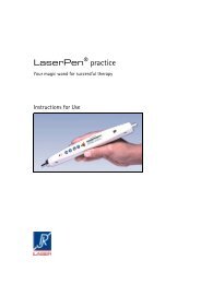
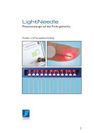
<strong>LightNeedle</strong><br />
Photon energy in the focus.<br />
Product and therapy description<br />
1
2<br />
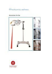
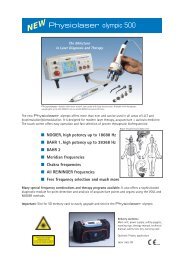
<strong>LightNeedle</strong> Concept
The <strong>LightNeedle</strong> device works in<br />
conjunction with the Physiolaser or<br />
Photonic and takes advantage of all<br />
therapy programs and settings. Simply attach<br />
it to the laser outlet of the control unit<br />
and start the therapy.<br />
The <strong>LightNeedle</strong> is a modular system<br />
and therefore offers economic treatment<br />
with lowest investment costs.<br />
The <strong>LightNeedle</strong> has 12 outlets (3x4)<br />
with 12 laser diodes (50 mW/655 nm).<br />
Extensive optional accessories:<br />
Single probes<br />
510A 785 nm/50 mW<br />
511A 810 nm/500 mW<br />
514A 638 nm/150 mW<br />
512A 670 nm/200 mW<br />
515A 904 nm/90 W<br />
Cluster probes<br />
516A Pulse laser<br />
5 x 904 nm/30 Watt<br />
517A Standard<br />
4 x 785 nm/55 mW + 4 x 655 nm/40 mW + 4 x 655/5 mW<br />
519 derma<br />
2 x 785 nm/55 mW + 6 x 655 nm/45 mW + 4 x 635/5 mW<br />
519A brush<br />
4 x 785 nm/55 mW + 4 x 655 nm/40 mW + 4 x 655/5 mW<br />
5090B Fiber optic (0 O )<br />
5090E Trolley (Physiolaser + <strong>LightNeedle</strong>)<br />
5090F Support for fiber optic<br />
Delivery contents: Main unit, 12 fiber<br />
optics, power supply, technical manual,<br />
holder, carrying case. <strong>Laser</strong> class 3B.<br />
Warning signs on the device:<br />
<strong>LightNeedle</strong> Concept<br />
3
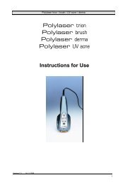
The innovative way for acupuncture<br />
and therapy of deep tissue layers<br />
The <strong>LightNeedle</strong> offers precise and<br />
strong laser stimulation of points and deep<br />
tissue layers and it can be widely used for<br />
many therapeutic applications.<br />
The operation of the <strong>LightNeedle</strong> is<br />
optimized for daily clinical use:<br />
- fast and easy operation<br />
- channel selection in groups<br />
- reliable and safe photon transfer<br />
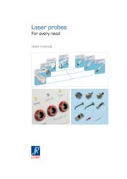
Patented tip<br />
The patented applicator offers for the first<br />
time a stable and secure treatment. Conventional<br />
systems are attached straight with<br />
special holders and are therefore faced with<br />
an unstable contact because the tip suffers<br />
under the weight of the fiber and tends to<br />
lose contact and its position.<br />
The patented tip of the <strong>LightNeedle</strong><br />
tip is placed flat onto the skin and has a<br />
beam outlet of 90 o . The tip can easily placed<br />
on any body part, may it be small or large,<br />
front and back at the same time, the patient<br />
can get a treatment in any position.<br />
Easy fixation of the tip with medical<br />
standard tape, no special tape is<br />
required!<br />
4<br />
<strong>LightNeedle</strong> Advantages<br />
The main advantages<br />
n modular system<br />
n 12 outlets<br />
n special fiber optic (stable)<br />
n standard tape<br />
n treat 2 patients simultaneously<br />
n all biofrequencies<br />
n compact and mobile
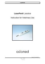
<strong>LightNeedle</strong> Therapy<br />
Choose the best therapy for your<br />
patients<br />
<strong>LightNeedle</strong> system offers a large<br />
variety of therapeutic applications.<br />
It is easy to use: Select the time and attach<br />
the patented tip of the fiber optic to the<br />
body or acupuncture points. It will shut off<br />
automatically once the treatment time is up.<br />
Acupuncture<br />
<strong>LightNeedle</strong> acupuncture is relaxing<br />
and painless it offers a long lasting stimulus<br />
and is widely accepted by the patients*.<br />
It seems to be even more effective than conventional<br />
needle acupuncture. Because once<br />
the fiber is placed no further manipulation is<br />
required.<br />
LLLT - wide range of applications<br />
The <strong>LightNeedle</strong> therapy can be used<br />
for any medical laser application except on<br />
open wounds (multi-cluster probe or Photonic<br />
is required). The tip can be positioned<br />
perfectly and allows that the beam easily<br />
penetrates deeply into the body.<br />
n Acupuncture<br />
n Pain management<br />
n Trigger points<br />
n Joints<br />
n Tendons (e.g. carpal tunnel)<br />
n Muscular skeletal system<br />
n Nerves<br />
* Patients’ sensation during and after laser needle versus metal<br />
needle treatment”. van Amerongen KS, et al<br />
RESULTS: The common metal needle technique was well known<br />
by the patients in comparison to the laser needle method<br />
(p
Place the fiber tip according to the guidelines<br />
of the classical acupuncture instead<br />
of metal needles directly on the point. The<br />
therapy duration is 10-15 minutes.<br />
If required further points can be treated<br />
additionally with the single probe (e.g. ear<br />
points).<br />
6<br />
<strong>LightNeedle</strong> Therapy
<strong>LightNeedle</strong> Therapy<br />
Trigger points, painfull spots<br />
Muscle pain, hardness, muscle stiffness<br />
Improvement of the regeneration<br />
after excercise<br />
Tendons (Tendinitis, Carpal tunnel syndr.)<br />
Distorsion, contusion, injuries<br />
Edema<br />
Spinal cord and intervertebral discs<br />
Lumbal pain<br />
Joints (arthrits, arthrosis)<br />
Shoulder/arm syndrome<br />
Pain in the neck area<br />
Muscle hardness<br />
Please refer to:<br />
Effect of 655-nm Low-Level <strong>Laser</strong> Therapy on Exercise-Induced<br />
Skeletal Muscle Fatigue in Humans<br />
Ernesto Cesar Pinto Leal Junior, M.Sc.,1,2,3 Rodrigo Álvaro<br />
Brandão Lopes-Martins, Ph.D.,4 Francis Dalan, P.T.,5 Maurício<br />
Ferrari, P.T.,5 Fernando Montanari Sbabo, P.T.,5 Rafael<br />
Abeche Generosi, P.E.,6 Bruno Manfredini Baroni, P.T.,5<br />
Sócrates Calvoso Penna, Ph.D.,4 Vegard V. Iversen, Ph.D.,8<br />
and Jan Magnus Bjordal, Ph.D.3,7<br />
7
For Ischalgia and lumbal pain place the fiber<br />
tips at and around the nerve outlet.<br />
It many cases it is recommended to place<br />
several tips along the nerve. Treatment<br />
duration 15-25 minutes.<br />
For the successful treatment of inner joint<br />
disorders as e.g. arthrosis, the correct position<br />
of the fiber tip is crucial. The beam must<br />
be directed into the joint and may not be<br />
absorbed by the bone.<br />
Please refer to:<br />
A systematic review of low level laser therapy with location-specific<br />
doses for pain from joint disorders. A systematic<br />
review of low level laser therapy with location-specific<br />
doses for pain from chronic joint disorders<br />
Jan M Bjordal1, Christian Couppé2, Roberta T Chow3,<br />
Jan Tunér4 and Elisabeth Anne Ljunggren1 1University of<br />
Bergen, Norway 2Lund University, Sweden 3Private Medical<br />
Practice, Sydney 4Private Dental Practice, Stockholm,<br />
Sweden<br />
Joint distorsion and contusion shall be<br />
treated by placing the fiber tips directly onto<br />
the painful area. Treatment duration 15-30<br />
minutes.<br />
8<br />
<strong>LightNeedle</strong> Therapy
<strong>LightNeedle</strong> Therapy<br />
Inner organs can be reached reflectory or<br />
locally. For reflectory treatment the Head<br />
zones (Henry Head) and the common reflex<br />
zones are most important.<br />
Place the fiber tips directly on the organ<br />
zones. Treatment duration 10 minutes.<br />
Head zones<br />
Reflex zones<br />
9
10<br />
<strong>LightNeedle</strong> Therapy<br />
Because the laser beam can penetrate deeply<br />
into the body, it can reach inner organs<br />
by local irradiation. The irradiation of the<br />
stomach (e.g. gastritis) and the pancreas (e.g.<br />
diabetis) can be a promissing indication.<br />
Good experience was made for kidney<br />
disease (Schmalix). Kidney insufficiency and<br />
nephro sclerosis showed good reaction.<br />
Treat neurological disturbance, nerve<br />
demage, rupture, injury etc. directly at the<br />
most painful and affected point.<br />
Please refer to:<br />
<strong>Laser</strong> Phototherapy, a New Modality in Treatment<br />
of Long-Term Incomplete Peripheral Nerve Injury:<br />
A Randomized Double-Blind Placebo-Controlled<br />
Study<br />
Shimon Rochkind et al.
Abstracts <strong>Laser</strong> Acupuncture<br />
Effects on Acupuncture, Acupressure and <strong>Laser</strong>needle acupuncture on EEG<br />
bispectral index and spectral edge frequency on healthy volunteers.<br />
G. Litscher<br />
University of Graz, Department of Biomedical Engineering and Researchin Anesthesia and Intensive<br />
Care, Graz, Austria<br />
Summary: Background and objective: The main purpose of this study was to investigate the effects<br />
of sensory (acupressure and acupuncture) and optical stimulation (<strong>Laser</strong>needle@ acupuncture)<br />
on electroencephalographic bispectral index, spectral edge frequency and a verbal sedation<br />
score.<br />
Methods: Twenty-five healthy volunteers (mean age :!: SD: 25.5 :!: 4.0yr) were investigated<br />
during the awake state. The acupuncture point Yintang and a placebo control point were stimulated.<br />
The study was performed as a randomized, controlled and partly blinded cross-over trial.<br />
Results: Bispectral index and spectral edge frequency values both decreased significantly (P <<br />
0.001) during acupressureonYintang tovalues of62.9 (minimum 35) :!: 13.9 bispectral indexand<br />
to 13.3 (minimum 2.9) :!: 8.1 Hz (spectral edge frequency right) and 13.8 (minimum 2.7) :!: 7.3<br />
Hz (spectral edge frequency left), respectively.<br />
Bispectral index was also significantly (P < 0.05) affected by <strong>Laser</strong>needle@ acupuncture and<br />
acupressure on the control point but the changes were not clinically relevant, 95.4 :!: 4 and 94.2<br />
:!: 4.8, respectively. All interventions significantly (Yintang: p < 0.001; control point: p < 0.012)<br />
reduced verbal sedation score.<br />
Conclusions: The study highlights the electroencephalographic similarities of acupressure induced<br />
sedation and general anaesthesia as assessed by bispectral index and spectral edge<br />
frequency.<br />
Biological Effects of Painless <strong>Laser</strong> Needle Acupuncture<br />
Gerhard Litscher, PhD Lu Wang, MD Detlef Schikora, PhD Dagmar Rachbauer, MSc Gerhard<br />
Schwarz, MD Andreas Sch�pfer, MD, Stefan Ropele, PhD Evamaria Huber<br />
Summary: <strong>Laser</strong> needle acupuncture is a new method to stimulate acupuncture points. We<br />
describe the technique, its first use, and its value in acupuncture research. <strong>Laser</strong> needle publications<br />
we included are based on 511 measurements in 231 healthy volunteers (129 female, 102<br />
male), with a mean (SD) age of 25 (3.5) years (range, 18-38 years). One pig experiment is also<br />
included.<br />
Results: The results of randomized, double-blind, controlled, crossover studies show that the<br />
methods of laser Doppler flowmetry, functional multidirectional transcranial Doppler sonography,<br />
functional magnetic resonance imaging, and near infrared spectroscopy are able to objectify<br />
and quantify peripheral and cerebral effects of laser needle acupuncture.<br />
Conclusion: For the first time, we were able to investigate scientifically the differences between<br />
needle acupuncture, which includes pain stimulation, and laser needle acupuncture, a continuous<br />
multichannel method of painless acupuncture stimulation. <strong>Laser</strong> needle acupuncture can<br />
induce specific, reproducible changes in the brain. These can be expressed by shifts in different<br />
parameters, such as cerebral blood flow velocity.<br />
11
12<br />
Abstracts <strong>Laser</strong> Acupuncture<br />
<strong>Laser</strong>needle Acupuncture: A Critical Review and Recent Results<br />
Detlef Schikora, PhD<br />
In the last 5 years, laserneedle acupuncture has become a new category in acupuncture, with its<br />
own scientific basics. It combines the tradition of Chinese acupuncture with the possibilities of<br />
modern technology. <strong>Laser</strong>needle acupuncture is in accordance with the aim of traditional medicine<br />
because it uses the most essential and most natural medium of our existence, the light,<br />
to heal illnesses. The painless laserneedle acupuncture is of proven medical effectiveness and<br />
particularly suited for the treatment of children and those patients who regard the metal needle<br />
insertion into the skin as unpleasant. In daily practical use, laserneedle acupuncture can<br />
be performed like any traditional needle acupuncture treatment.<br />
The diagnostic criteria of acupoint selection, the treatment duration, and treatment frequency<br />
are identical to the traditional Chinese acupuncture. To perform successful laserneedle acupuncture<br />
treatments, no additional qualification is required.<br />
Offering the painless laserneedle acupuncture to the patient means that the acupuncture needles<br />
are substituted and the risks of the metal needle are eliminated.<br />
Effect of New Non-invasive <strong>Laser</strong> Needles on Brain Function<br />
G. Litscher*, D. Schikora**<br />
* Department of Biomedical Eng. and Research in Anesthesia and Critical Care,<br />
University of Graz, Graz, Austria<br />
** Department of Physics and Optoelectronic, University of Paderborn, Paderborn, Germany<br />
This paper describes the first double-blind study in acupuncture research in 17 healthy<br />
volunteers using laserneedle acupuncture. Stimulation of vision related acupoints resulted<br />
in marked changes of mean blood flow velocity in the posterior cerebral artery measured by<br />
multidirectional transcranial Doppler sonography. Further studies using different laser stimulus<br />
intensities and wavelengths are in progress.<br />
Cerebral Vascular Effects of Non-invasive <strong>Laser</strong>needles Measured by Transorbital<br />
and Transtemporal Doppler Sonography<br />
G. Litscher1 and D. Schikora2<br />
1Department of Biomedical Engineering and Research in Anesthesia and Critical Care, University<br />
of Graz, Austria; 2Department of Physics and Optoelectronic, University of Paderborn, Germany<br />
<strong>Laser</strong>needles represent a new non-invasive optical stimulation method which is described for<br />
the first time in this paper. We investigated 27 healthy volunteers (mean ageSD: 25.154.12<br />
years; range: 21–38 years; 14 female, 13 male) in a randomised cross-over trial to study differences<br />
between laserneedle acupuncture and manual needle acupuncture in specific cerebral<br />
parameters. Mean blood flow velocity (vm) showed specific and significant increases in the<br />
ophthalmic artery during laserneedle stimulation (p=0.01) and during manual needle stimulation<br />
(p
Abstracts <strong>Laser</strong> Acupuncture<br />
Effects of laserneedle acupuncture on olfactory sensitivity of healthy human subjects:<br />
a placebo-controlled, double-blinded, randomized trial.<br />
Anzinger A, Albrecht J, Kopietz R, Kleemann AM, Schöpf V, Demmel M, Schreder T, Eichhorn I,<br />
Wiesmann M.<br />
Department of Neuroradiology, Ludwig-Maximilians-University Munich, Germany.<br />
The aims of the present study were to investigate the influence of laserneedle acupuncture on<br />
olfactory sensitivity and to examine whether the attitude towards laserneedle acupuncture affects<br />
the outcome. Olfaction was tested repeatedly on two days using the olfactory detection<br />
threshold subtest of the Sniffin’ Sticks test battery in sixty-four healthy subjects of which 32<br />
showed a positive attitude towards the effects of laserneedle acupuncture and 32 were sceptic<br />
about its effects.<br />
Testing was accomplished three times on day one (T1 = 0 min, T2 = 35 min, T3 = 105 min) without<br />
laserneedle acupuncture and on day two (T1* = 0 min, T2* = 35 min, T3* = 105 min) when<br />
the subjects were randomized in a non-stimulation (placebo) and a stimulation (laserneedle<br />
acupuncture) group. Stimulation or non-stimulation was conducted in a double-blinded design.<br />
Following laserneedle acupuncture a significant decrease in olfactory detection thresholds was<br />
observed at both, T2* and T3*, whereas no significant changes were found in the baseline or<br />
placebo group.<br />
Effects of laserneedle acupuncture on the olfactory detection threshold did not differ between<br />
sceptic and non-sceptic subjects. In conclusion, laserneedle acupuncture is an effective method<br />
to improve olfactory sensitivity after one session of stimulation for at least one hour, independently<br />
of the attitude of subjects towards the stimulation method.<br />
PMID: 19593972 [PubMed - indexed for MEDLINE<br />
Near-infrared spectroscopy for objectifying cerebral effects of needle and laserneedle<br />
acupuncture<br />
Gerhard Litschera* and Detlef Schikorab<br />
a Department of Biomedical Engineering and Research in Anesthesia and Critical<br />
Care, University of Graz, A-8036 Graz, Austria<br />
b Department of Physics and Optoelectronics, University of Paderborn, D-33095<br />
Paderborn, Germany<br />
Near infrared spectroscopy (NIRS) has been successfully used in this study to objectify cerebral<br />
alterations in oxyhemoglobin and desoxyhemoglobin, due to manual needle acupuncture and<br />
laserneedle acupuncture, in 88 healthy volunteers mean age 25.7 + 4.0 (x + SD) years (19 - 38<br />
years). Results from Traditional Chinese Acupuncture, Korean and Chinese hand acupuncture,<br />
ear acupuncture, combinations of the different acupuncture methods and placebo needling are<br />
presented. NIRS seems to be able to shed some light upon the functioning of the different acupuncture<br />
methods.<br />
13
14<br />
Abstracts <strong>Laser</strong> Acupuncture<br />
Patients’ sensation during and after laserneedle versus metal needle treatment.<br />
van Amerongen KS, Kuhn A, Mueller M.<br />
Department of Obstetrics and Gynaecology, Inselspital, Bern University Hospital, and University<br />
of Bern, Switzerland.<br />
OBJECTIVES: Aim of the study was to evaluate the patients’ sensations during and after laserneedle<br />
versus metal needle acupuncture.<br />
STUDY DESIGN: The prospective study was performed at the gynaecological outpatient department<br />
of a University Teaching Hospital of Bern, Switzerland. Thirty female patients per group<br />
were included in the study and randomized into laserneedle or metal needle group. All women<br />
visited the acupuncture out patient department because of gynaecological disorders.<br />
Age of the patients in the metal needle group was 38 years in median (range 18-73 years);<br />
mean age was 41+/-13.3. Age in the laserneedle group was 36 years in median (range 16-60<br />
years) and mean age was 39.1+/-12.2. I<br />
nterventions were laserneedle acupuncture and metal needle acupuncture. Patients answered<br />
a questionnaire before, after the first treatment and prior to the second treatment. The questionnaires<br />
asked about the patients’ knowledge of the various acupuncture methods and their<br />
health condition before treatment, their perception of pain, warmth, tiredness and relaxation<br />
during or after application of the needles or during or after the treatment.<br />
Statistics were performed by Graph Pad InStat 3 for windows.<br />
RESULTS: The common metal needle technique was well known by the patients in comparison<br />
to the laserneedle method (p
Abstracts <strong>Laser</strong> Acupuncture<br />
<strong>Laser</strong>-Needle Therapy for Spontaneous Osteonecrosis of the Knee<br />
Winfried Banzer, M.D., Ph.D.,1 Markus Hübscher, Ph.D.,1 and Detlef Schikora, Ph.D.2<br />
Objective: This case report describes the treatment of a 63-year-old patient with spontaneous<br />
osteonecrosis of the knee (SONK). Background Data: SONK usually appears in the elderly patient<br />
without the typical risk factors for osteonecrosis. It is characterized by acute and sudden pain,<br />
mostly occurring at the medial side of the knee joint. Symptoms usually worsen with physical<br />
activity and improve with rest. Besides physical therapy, limited weight-bearing and the use of<br />
analgesics and nonsteroidal anti-inflammatory drugs, we propose lowlevel laser therapy (LLLT)<br />
as a conservative treatment option.<br />
Methods: LLLT was carried out using laser needles emitting radiation with wavelengths of 685<br />
and 885 nm, and a power density of 17.8 W/cm2. Therapy sessions lasted 60 min and were<br />
performed daily over a period of 3 mo. The total irradiation dose emitted by 8 laser needles in 60<br />
min of treatment was 1008 J.<br />
Results: Magnetic resonance imaging revealed distinct restitution<br />
of the spongiosa edema 5 wk after treatment onset, and the final check-up at 35 wk demonstrated<br />
complete restoration of integrity.<br />
Conclusion: The present case report provides the first indication that laser-needle therapy may<br />
be a promising tool for complementary and alternative therapeutic intervention for those with<br />
SONK.<br />
Quantification of Gender Specific Thermal Sensory<br />
and Pain Threshold Before and After <strong>Laser</strong>needle Stimulation<br />
1Abteilung für Biomedizintechnische Forschung in Anästhesie und Intensivmedizin,<br />
Medizinische Universität Graz, Österreich<br />
2Fachbereich Physik – Optoelektronik, Universität Paderborn, Deutschland<br />
3Klinische Abteilung für Neuro- und Gesichtschirurgische Anästhesiologie und Intensivmedizin,<br />
Medizinische Universität Graz, Österreich<br />
Quantitative thermal sensory and pain threshold testing (QST) was performed in 29 adult<br />
healthy volunteers (mean age 24.2 ± 2.7 years; range: 18–29 years; 20 females, 9 males) using<br />
the Thermal Sensory Analyser TSA-II (Medoc Advanced Medical Systems, Ramat Yishai,<br />
Israel, and Minneapolis, Minnesota, USA) before and after laser needle acupuncture and placebo<br />
stimulation, respectively.<br />
Significant (p 0,001; t-test) gender-specific differences were seen on cold pain threshold<br />
analysis. No significant changes in parameters of thermal sensory and pain thresholds were<br />
found before and after laser needle or placebo stimulation at acupuncture points for acute pain.<br />
However, a trend towards change in the median value of cold pain sensation after laser needle<br />
stimulation (p = 0.479; paired t-test; n.s.) was seen within the group of healthy females.<br />
The influence of stimulation of acupuncture points for chronic pain on the various parameters<br />
needs to be clarified in future studies.<br />
Please contact <strong>RJ</strong>-LASER in order to get the complete paper:<br />
contact@rj-laser.com<br />
15
Original Article<br />
G. Litscher<br />
European Journal of Anaesthesiology 2004; 21: 13-19<br />
@ 2004 European Academy of Anaesrhesiology<br />
ISSN 0265-0215<br />
University of Graz, Department of Biomedical Engineering and Research in Anesthesia and Intensive Care, Graz, Austria<br />
Summary<br />
Background and objective: The main purpose of this study was to investigate the effects of sensory (acupressure<br />
and acupuncture) and optical stimulation (<strong>Laser</strong>needle@ acupuncture) on electroencephalographic bispectral<br />
index, spectral edge frequency and a verbal sedation score.<br />
Methods: Twenty-five healthy volunteers (mean age :!: SD: 25.5 :!: 4.0yr) were investigated during the<br />
awake state. The acupuncture point Yintang and a placebo control point were stimulated. The study was per-<br />
formed as a randomized, controlled and partly blinded cross-over trial.<br />
Results: Bispectral index and spectral edge frequency values both decreased significantly (P < 0.001) during acu-<br />
pressureonYintang tovalues of62.9 (minimum 35) :!: 13.9 bispectral indexand to 13.3 (minimum 2.9) :!: 8.1 Hz<br />
(spectral edge frequency right) and 13.8 (minimum 2.7) :!: 7.3 Hz (spectral edge frequency left), respectively.<br />
Bispectral index was also significantly (P < 0.05) affected by <strong>Laser</strong>needle@ acupuncture and acupressure on the<br />
control point but the changes were not clinically relevant, 95.4 :!: 4 and 94.2 :!: 4.8, respectively. All interventions<br />
significantly (Yintang: p < 0.001; control point: p < 0.012) reduced verbal sedation score.<br />
Conclusions: The study highlights the electroencephalographic similarities of acupressure induced sedation<br />
and general anaesthesia as assessed by bispectral index and spectral edge frequency.<br />
Keywords: ACUPRESSURE; ACUPUNCTURE; ELECTROENCEPHALOGRAPHY, bispectral index, spectral edge<br />
frequency.<br />
Noninvasive bioelectrical neuromonitoring is gaining<br />
more and more attention in anaesthesia and<br />
critical care [1,2]. The bispectral index (BIS) and the<br />
spectral edge frequency (SEF) are important numerical<br />
descriptors of the electroencephalogram (EEG)<br />
and both are mainly used for assessing depth of<br />
anaesthesia [3]. If anaesthetists relyon BIS and SEF<br />
to detect awareness, then it is very important to<br />
Correspondence ro. Gerhard Lirscher, Departmenr of Biomedical Engineering<br />
and Research in Anesthesia and Intensive Care, University of Graz,<br />
Auenbruggerplatz 29, A-8036 Graz, Austria. E-mail: gerhard.litscher@<br />
uni-grazat; Tel. +433163853907/83907; Fax. +433163853908<br />
Accepted for publication August 2003 EJA 1537<br />
exclude other influences that could give false readings.<br />
It is known that a number of environmental<br />
and physiological factors may affect BIS performance.<br />
Recently it has been reported that nonpharmacological<br />
interventions such as acupressure can also<br />
reduce BIS values significantly [4].<br />
This study is a randomized, controlled and partly<br />
blinded (<strong>Laser</strong>needle@ acupuncture; LASCO Int.<br />
Medical Mark. AG, Basel, Switzerland) cross-over<br />
trial intended to investigate the effects of three nonpharmacological<br />
interventions (acupressure, manual<br />
needle acupuncture and <strong>Laser</strong>needle@ acupuncture)<br />
on two processed EEG variables (BIS and SEF) and a<br />
verbal sedation score (VSS) in healthy volunteers.
14 G. Litscher<br />
Methods<br />
Subjects<br />
The study was approved by the Ethics Committee of<br />
the University of Graz (13-048 ex 02/03). Written<br />
informed consent was obtained from each subject.<br />
We studied 25 healthy volunteers (mean age :!: SD:<br />
25.5 :!: 4.0yr, range 21-39yr; 15 fernales, 10 males;<br />
height173.5 :!: 9.3cm;bodyweight69.1 :!: 16.1kg).<br />
None of the subjects had neurological or psychological<br />
disorders and they were not taking any medication.<br />
They were partly informed about the nature of<br />
the investigation and were paid for their participation.<br />
The investigators recording EEG and sedation<br />
data were blinded to the intervention applied to the<br />
volunteers. The subjects were not informed which of<br />
the four interventions was effectively a placebo control<br />
(acupressure on a control point).<br />
Procedure and study design<br />
The study was performed as a randomized, controlled<br />
cross-over trial. Four EEG electrodes (F7-Fpz,<br />
F8-Fpz' Fz = ground) and a noninvasive blood pres-<br />
sure cuff were attached to the volunteers after they<br />
arrived at the biomedical engineering laboratory.<br />
Two channels of spontaneous electrical activity<br />
were recorded from EEG electrodes (Zipprep@ selfprepping<br />
electrodes; Aspect Medical Systems Inc.,<br />
Natick, MA, USA). The skin-electrode impedance<br />
was
<strong>Laser</strong>needle@ acupuncture at the acupoint Yintang<br />
was performed using a new method for optical stimulation.<br />
This method was reported by our research<br />
group in the scientific literature in 2002 [6,7]. The<br />
<strong>Laser</strong>needle@ technique represents a new, noninvasive<br />
method for optical stimulation of acupuncture<br />
points. The laser used in this study emits red light<br />
in continuous-wave mode with an output power of<br />
30-40 rn W, which results in a radiant exposure energy<br />
of about 2.3 kJ cm-2 at the acupuncture point during<br />
a stimulation time of 10 min [6].<br />
Acupressure on the control point (location: 2 cm<br />
from lateral end of the left eyebrow; Fig. Id) was<br />
performed in similar manner as on the acupoint<br />
Yintang (duration 10 min).<br />
All subjects had four conditions applied (Fig. la-d).<br />
The persons were in a semi-lying position with<br />
closed eyes. The choice of the stimulation procedure<br />
was randomized within a subject and the interval<br />
between the different sessions was at least 20 min.<br />
Evaluation parameters<br />
The main evaluation parameters were BIS and SEF90<br />
during the different conditions (Fig. 1) and time<br />
intervals (Fig. 2). Measurements were made at time<br />
points a-g (Fig. 2). In any one condition we recorded<br />
BIS and SEF values continuously but sampled the<br />
data for subsequent analysis at seven points. A single<br />
reading was taken at each point. The stimulation<br />
was not stopped at the time of reading. The whole<br />
study session lasted 2-3 h. BIS and SEF90 represent<br />
single numbers, which should decrease continuously<br />
with decreasing level of consciousness (hypnosis).<br />
There are several review articles for methodological<br />
details of signal processing of BIS and SEF [3].<br />
After 5 min of stimulation (Fig. 2d) the subjects<br />
were asked to move their right hand to clarify that<br />
they were awake and not asleep. In addition, before<br />
and after each stimulation mode the persons were<br />
asked to score their stress and tension based on a VSS<br />
from 0 (no stress) to 10 (maximum stress) [4]. Heart<br />
rate (HR) and noninvasive blood pressure (BP) were<br />
also recorded before and after acupressure stimulation<br />
Figure 2.<br />
Stimulation procedure and different measuring points before<br />
(a) during (b-f) and after (g) stimulation.<br />
Nonpharmacological influences on BIS and SEF 15<br />
at Yintang (measurement points:<br />
and 1 min after 'g' (cf Fig. 2».<br />
Statistical analysis<br />
min before<br />
The BIS and SEF data were tested with analysis of<br />
variance (one-way repeated measures ANOVA;<br />
similar data were found to be normally distributed<br />
in previous investigations) using SigmaStat@ Oandel<br />
Scientific Corp., Erkrath, Germany). Dunnett's<br />
method was used for post hoc analysis; VSS data were<br />
compared using paired t-test. The results were<br />
graphically presented as box plots (BIS and SEF) and<br />
as scatter plot (VSS). Changes were considered<br />
significant at p < 0.05.<br />
Results<br />
All subjects completed the study. Figure 3 shows the<br />
decreases of BIS values during acupressure applied to<br />
the acupoint Yintang in all 25 healthy volunteers.<br />
1 : r- ST'J ~<br />
60[<br />
4Oi<br />
100~S 5<br />
i -<br />
80' T\J<br />
6oi \Ji<br />
40~<br />
100~S --<br />
:r fJ:iijJ<br />
4of<br />
9<br />
100~S<br />
r .-<br />
13<br />
8° 1<br />
60,<br />
:rrrrrr<br />
I U r<br />
401<br />
100r.ߧ<br />
801 r<br />
60[<br />
40,<br />
~<br />
1COrJ?!J§- 21<br />
:~\ ~<br />
40[<br />
100;=-~J§ 25<br />
:~I ~<br />
4oi<br />
2<br />
rw<br />
6<br />
f"'ir<br />
17 18<br />
~<br />
~<br />
3<br />
7<br />
~<br />
4<br />
"'W<br />
14 15 16<br />
vii:"<br />
~<br />
~<br />
~<br />
'V<br />
,-,<br />
22<br />
10 min<br />
~<br />
19 20<br />
-<br />
23<br />
:r--r<br />
f\;Mr<br />
"*\\I<br />
Y intang n= 25<br />
Figure 3.<br />
The trend of BIS values of 25 healthy volunteers { 1-25) before,<br />
during and after acupressure performed on the acupoint Yintang.<br />
All subjects were awake. Note the significant decrease {minimum<br />
BIS = 35; no.14) due to acupressure.<br />
24
16<br />
100<br />
80<br />
~ 60<br />
m<br />
N<br />
I<br />
N<br />
I<br />
40<br />
20<br />
O<br />
35<br />
G. Litscher<br />
30 .<br />
25<br />
20<br />
10<br />
Figure 4.<br />
.<br />
.<br />
.<br />
.<br />
.<br />
SEFr<br />
Box plots of alterations of BIS and SEF values ( r: right; I: left) in<br />
25 healthy volunteers before (a), during (b-f) and after (g) acu-<br />
pressure (cf. Fig. 2) on the acupoint Yintang. The ends of the boxes<br />
define the 25%0 and 75%0, with a line at the median and error<br />
bars defining the 10%0 and 90%0.<br />
Before the subjects were stimulated, their mean<br />
BIS values (:tSD) were 97.4 (98-95) :t 1.0 andtheir<br />
mean SEF values (:tSD) were 23.9 :t 4.1 (right) and<br />
23.5 :t 4.9Hz (left). The BIS and SEF values both<br />
decreased significantly (P < 0.001) after starting acupressure.<br />
After 5 min acupressure at the acupoint<br />
Yintang, the mean BIS values were 62.9 (minimum<br />
35; see no.14 in Fig. 3) :t 13.9, and the mean SEF<br />
values were 13.3 (minimum 2.9) :t 8.1 (right) and<br />
13.8 (minimum 2.7) :!: 7.3 Hz (left). The release of<br />
acupressure caused an increase in BIS and SEF back<br />
to the baseline values before stimulation (cf Fig. 4).<br />
Figure 5 summarizes the BIS and SEF results<br />
obtained during manual needle acupuncture,<br />
<strong>Laser</strong>needle @ acupuncture and acupressure on the<br />
control point. Significant (P < 0.05) changes were<br />
found in BIS values during <strong>Laser</strong>needle@ acupuncture<br />
(measuring points d and e; cf Figs. 2 and 5) and<br />
during acupuncture on the control point (measuring<br />
points d-f). After 7.5 min <strong>Laser</strong>needle@ acupuncture at<br />
acupoint Yintang, the mean BIS values (:!:SD) were<br />
95.4 (minimum 81; see Fig. 5, middle, upper panel)<br />
:!: 4.1. After 5 min acupressure at the control point,<br />
the mean BIS values (:!:SD) were 94.2 (minimum 77;<br />
see Fig. 5, right, upper panel) :!: 4.8. SEF did not show<br />
any significant alteration.<br />
The results of the analysis of the VSS are demonstrated<br />
in Figure 6. The VSS values were significantly<br />
(P < 0.001) reduced after pressure application on<br />
Yintang, needle acupuncture and <strong>Laser</strong>needle@<br />
acupuncture but also after pressure application on<br />
the control point (P = 0.012). Mean baseline VSS<br />
values were insignificantly lower in <strong>Laser</strong>needle@<br />
and control conditions.<br />
HR and BP values (mean :!: SD) before and after<br />
acupressure at Yintang were calculated to be 73.2 :!:<br />
12.4beatsmin-l, 109.8:!: 14.0mmHg (systolic) and<br />
69.3 :!: 10.6 mmHg (diastolic), respectively. After<br />
stimulation the values decreased to 63.7 :!:<br />
11.9beatsmin-l, 107.7:!: 8.7mmHg(systolic)and<br />
66.8 :!: 8.6 mmHg (diastolic), respectively.<br />
Discussion<br />
The BIS and the SEF are mainly used intraoperatively<br />
to monitor the hypnotic effect of anaesthetic drugs.<br />
There are several studies reported in the literature<br />
proposing target values for EEG parameters to guide<br />
the depth of anaesthesia. A number of authors have<br />
reported a low probability of recall and a high probability<br />
of unresponsiveness during surgery at a level<br />
of 60 for BIS [8,9]. BIS values
(8) (b) (c)<br />
120<br />
100 ~ ...T T T T T<br />
80<br />
(I)<br />
tC 60<br />
40<br />
20<br />
0<br />
30<br />
25 ,<br />
n.s.<br />
.~ ."'(7i'ti!! ~ "<br />
BIS<br />
N 20<br />
:I:<br />
15<br />
.<br />
10<br />
5 n.s.<br />
0 I. ... ~<br />
Nonpharmacological influences on BIS and SEF 17<br />
b c d e 9<br />
Figure 5.<br />
Box plots of changes of BIS and SEF values ( r: right; I: left) during ( a) manual needle acupuncture, ( b) <strong>Laser</strong>needle@ acupuncture and<br />
( c) acupressure at the control point. Further explanations see Figure 4.<br />
Figure 6.<br />
Mean (::t:SD) values ofthe vss of25 healthy volunteers before<br />
(a) and after (b) different modalities of nonpharmacological stimu-<br />
lation (0: no stress; 10: maximum stress).<br />
Acupuncture has been shown to reduce medication<br />
use in a number of trials [12]. Acupressure has been<br />
studied and offered in scientific literature as a valuable<br />
treatment in improving the quality of sleep [13].<br />
In previous studies, it has also been shown that pressure<br />
on acupoints can decrease postoperative pain [14] and<br />
that Korean hand acupressure reduces postoperative<br />
nausea and vomiting after gynaecologicallaparoscopic<br />
N<br />
I<br />
surgery [15]. Acupressure has also been used in some<br />
other studies for prevention of emesis [16]. There are<br />
a number of theories as to how acupressure or acupuncture<br />
works. All these hypotheses show that the brain<br />
plays a key role in acupuncture and acupressure research<br />
[17-20]. Modulation of subcortical structures may be<br />
an important mechanism by which acupuncture and<br />
acupressure exerts its complex multisystem effects<br />
[20]. Demonstration of regionally specific, quantifiable<br />
acupuncture and acupressure effects on relevant<br />
structures of the human brain would facilitate acceptance<br />
and integration of these therapeutic modalities<br />
into the practice of modern medicine [17-20].<br />
It has been shown in several publications that different<br />
narcotics have different influence on BIS and<br />
SEF [8-11,21-26]. However, nonpharmacological<br />
influences such as electromyographic activity may<br />
contribute to the low specificity of the absolute values<br />
of the electrophysiological measurement data [21].<br />
In the majority of the cases, the BIS is falsely elevated<br />
[21]. Our results appear to confirm the results<br />
of the study ofFassoulaki and colleagues [4] who also<br />
found that acupressure on Yintang resulted in a<br />
significant and clinically relevant reduction on BIS<br />
values and they concluded that BIS is therefore<br />
of limited clinical relevance for monitoring depth<br />
of anaesthesia [22-26]. However, Fassoulaki and
18<br />
G. Litscher<br />
colleagues [4] did consider the SEF, nor did they<br />
investigate the effects of manual needle acupuncture<br />
and the effects of<strong>Laser</strong>needle@ acupuncture.<br />
We have shown in this study that awake volunteers<br />
subjected to acupressure at Yintang can have<br />
similar BIS and SEF values to anaesthetized patients.<br />
While it is unlikely that a patient will receive acupressure<br />
or acupuncture during surgery, the question<br />
as to what causes BIS readings below 50 in awake<br />
subjects remains. It is unlikely to be a placebo effect<br />
as we have shown in several test measurements usin~<br />
placebo points that BIS is not affected by <strong>Laser</strong>needle<br />
stimulation per se. In the present study there were<br />
small statistically significant but not clinically important<br />
changes with needle acupuncture, <strong>Laser</strong>needle@<br />
acupuncture and acupressure at control point. These<br />
findings also help confirm that the BIS and SEF<br />
reductions induced by acupressure at Yintang are<br />
not a placebo effect. Reduced electromyographic<br />
levels could be partially responsible [21]. At the<br />
moment it is unclear to what degree system algorithms<br />
contribute to such findings. BIS is certainly<br />
affected by electrical activity nearby, especially<br />
diathermy. Therefore, there could also be a possibility<br />
that local movement in the region of the recording<br />
electrode might be responsible for the EEG effects<br />
observed. These are apparently less during control<br />
point acupuncture than during Yintang acupressure,<br />
where pressure is applied to a point immediately<br />
adjacent to the Zipprep@ electrode. Further investigations<br />
are necessary to clarify these questions.<br />
In conclusion, we found in healthy awake volunteers<br />
that acupressure at Yintang results in statistically<br />
significant and clinically relevant reductions in<br />
BIS and SEF while needle acupuncture, <strong>Laser</strong>needle@<br />
acupuncture and acupressure at a control point result<br />
in statistically significant but clinically unimportant<br />
reductions. Although the validity of BIS in anaesthesia<br />
is higher than that of SEF, BIS too has to be<br />
interpreted very carefully as our results show. Our<br />
results also highlight the EEG similarities of acupressure<br />
induced sedation and anaesthesia.<br />
Acknowledgements<br />
The author would like to express his thanks to Dr Lu<br />
Wang, Mag. Petra Petz and Evamaria Huber (Department<br />
of Biomedical Engineering and Research in<br />
Anesthesia and Intensive Care, University of Graz)<br />
for their valuable help.<br />
References<br />
Litscher G, Schwarz G. Noninvasive bioelectrical neuromonitoring<br />
in anaesthesia and critical care. Bur J Anaesthesiol<br />
2001; 18: 785-788.<br />
2. Litschet G. Editotial. The future of neuromonitoring.<br />
Internet J Neuromonitoring 2000; 1(1): http:/ /www.ispub.com/<br />
ostia/ index. php ?xmlF ilePath = journals/ i j nm/voll n 1/<br />
editorial2.xml<br />
3. Rampil IJ. A primer for EEG signal processing in anesthesia.<br />
Anesthesiology 1998; 89: 980-1002.<br />
4. Fassoulaki A, Paraskeva A, Patris K, Pourgiezi T,<br />
Kostopanagiotou G. Pressure applied on the extra 1<br />
acupuncture point reduces bispectral index values and<br />
stress in volunteers. Anesth Analg 2003; 96: 885-889.<br />
5. Stux G, Pomeranz B. Basics of Acupuncture. Berlin, Germany:<br />
Springer, 1998.<br />
6. Litscher G, Schikora D. Cerebral vascular effects of noninvasive<br />
laserneedles measured by transorbital and transtemporal<br />
Doppler sonography. <strong>Laser</strong> Med Sci 2002; 17:<br />
289-295.<br />
7. Litscher G, Schikora D. Near-infrared spectroscopy for<br />
objectifying cerebral effects of needle and laserneedle<br />
acupuncture. Spectroscopy 2002; 16: 335-342.<br />
8. Sebel PS, Lang E, Rampil IJ, et al. A multicenter study of<br />
bispectral electroencephalogram analysis for monitoring<br />
anesthetic effect. Anesth Analg 1997; 84: 891-899.<br />
9. LiuJ, Singh H, White PF. Electroencephalographic bispectral<br />
index correlates with intraoperative recall and depth of<br />
propofol-induced sedation. Anesth Analg 1997; 84: 185-189.<br />
10. HeckM, Kurnie B, BoldtJ, LangJ, Lehmann A, Saggau W.<br />
Electroencephalogram bispectral index predicts hemodynamic<br />
and arousal reactions during induction of anesthesia<br />
in patients undergoing cardiac surgery.J Cardiothorac Vasc<br />
Anesth 2000; 14: 693-697.<br />
11. Chan MTV, Gin T. What does the bispectral EEG index<br />
monitor? Eur J Anaesthesiol2000; 17: 146-148.<br />
12. Greif R, Laciny S, Mokhtarani M, et al. Transcutaneous<br />
electrical stimulation of an auricular acupuncture point<br />
decreases anesthetic requirement. Anesthesiology 2002; 96:<br />
306-312.<br />
13. Tsay SL, Chen ML. Acupressure and quality of sleep in<br />
patients with end-stage renal disease -a randomized control<br />
trial. Int J Nurs Stud 2003; 40: 1-7.<br />
14. Felhendler D, Lisander B. Pressure on acupoints decreases<br />
postoperative pain. ClinJ Pain 1996; 12: 326-329.<br />
15. Boehler M, Mitterschiffthaler G, Schlager A. Korean hand<br />
acupressure reduces postoperative nausea and vomiting after<br />
gynecologicallaparoscopic surgery. Anesth Analg 2002; 94:<br />
872-875.<br />
16. Eizember FL, Tomaszewski CA, Kerns WP. Acupressure<br />
for prevention of emesis in patients receiving activated<br />
charcoal.J Toxicol-Clin Toxicol2002; 40: 775-780.<br />
17. Cho ZH, Wong EK, Fallon JH. Neuro-Acupuncture. Los<br />
Angeles, USA: Q-Puncture, 2001.<br />
18. Litscher G, Cho ZH, eds. Computer Controlled Acupuncture@.<br />
Berlin, Germany: Pabst Science Publishers, 2000.<br />
19. Litscher G. High-Tech Akupunktur@. Berlin, Germany: Pabst<br />
Science Publishers, 2001.<br />
20. Hui KKS, Liu J, Makris N, et al. Acupuncture modulates<br />
the limbic system and subcortical gray structures of the<br />
human brain: evidence from fMRI studies in normal subjects.<br />
Hum Brain Mapp 2000; 9: 13-25.<br />
21. Bruhn J, Bouillon TW, Shafer SL. Electromyographic<br />
activiry falsely elevates the bispectral index. Anesthesiology<br />
2000; 92: 1485-1487.
MEDICAL ACUPUNCTURE<br />
Volume 20, Number 1, 2008<br />
© Mary Ann Liebert, Inc.<br />
DOI: 10.1089/acu.2007.0606<br />
Original Paper<br />
<strong>Laser</strong>needle Acupuncture: A Critical Review and Recent Results<br />
Detlef Schikora, PhD<br />
ABSTRACT<br />
In the last 5 years, laserneedle acupuncture has become a new category in acupuncture, with its own scientific<br />
basics. It combines the tradition of Chinese acupuncture with the possibilities of modern technology. <strong>Laser</strong>needle<br />
acupuncture is in accordance with the aim of traditional medicine because it uses the most essential and<br />
most natural medium of our existence, the light, to heal illnesses. The painless laserneedle acupuncture is of<br />
proven medical effectiveness and particularly suited for the treatment of children and those patients who regard<br />
the metal needle insertion into the skin as unpleasant. In daily practical use, laserneedle acupuncture can<br />
be performed like any traditional needle acupuncture treatment. The diagnostic criteria of acupoint selection,<br />
the treatment duration, and treatment frequency are identical to the traditional Chinese acupuncture. To perform<br />
successful laserneedle acupuncture treatments, no additional qualification is required. Offering the painless<br />
laserneedle acupuncture to the patient means that the acupuncture needles are substituted and the risks of<br />
the metal needle are eliminated.<br />
Key Words: <strong>Laser</strong>needle, Acupuncture, <strong>Laser</strong><br />
INTRODUCTION<br />
THE BASIC IDEA FOR THE DEVELOPMENT of “laserneedles”<br />
for acupuncture in the Biophotonic Research Group at<br />
Paderborn University (in Germany) originated from an<br />
acupuncture analysis in Europe 10 years ago. At that time,<br />
the first hand-held devices/”laser pens” arrived on the market,<br />
which were recommended as instruments to perform<br />
painless laser acupuncture treatments. It is obvious, however,<br />
that acupuncture treatments using such devices are not in accordance<br />
with the long tradition of Chinese acupuncture<br />
which is based on a simultaneous stimulation of a selected<br />
acupuncture point combination. Hand-held acupuncture laser<br />
devices allow just a serial stimulation of acupuncture points,<br />
i.e., 1 point after the other. The question arises: is it acupuncture<br />
if one sticks a needle in the first acupuncture point, takes<br />
University of Paderborn, Paderborn, Germany.<br />
37<br />
it out after 2 minutes, punctures the second point for 2 minutes,<br />
takes it out again, and stimulates the third point, and so<br />
forth. Every experienced acupuncturist would perhaps answer,<br />
“No, that is not acupuncture. I would never do that.<br />
The needles must remain for at least 20 minutes in the selected<br />
acupuncture points.” That is, the points have to be<br />
stimulated simultaneously. Our analysis came to the same<br />
conclusion; furthermore, we could not find proof in the literature<br />
of a serial point stimulation approach, neither for<br />
metal needles nor for laser pens.<br />
With the development of laserneedles, we have tried to<br />
preserve the methodical rules of the classic Chinese<br />
acupuncture. Several fundamental scientific and medical<br />
problems had to be resolved and investigated:<br />
1) How can visible laser light stimulate acupuncture<br />
points? It is known from daily experience that visible light
38<br />
that is shining on the skin does not create any acupuncturelike<br />
reaction nor does it interact with peripheral sensory<br />
nerves. The pleasant warmth that we feel on the skin during<br />
a summer day does not come from the visible light of<br />
the sun. How can laser light stimulate acupoints? If one<br />
pricks a metal needle in the skin, the patient often feels pain.<br />
Is that physical stimulation an essential requirement for efficient<br />
acupuncture?<br />
2) Which parameter and properties are important in laser<br />
acupuncture that determine the therapeutic efficacy?<br />
3) Is it dangerous to stimulate acupuncture points by laser<br />
irradiation? Are there any risks or side effects?<br />
4) Is laser acupuncture comparable to the traditional metal<br />
needle acupuncture regarding its therapeutic efficacy?<br />
5) What are the limits and challenges of laserneedle stimulation.<br />
These 5 topics are addressed in this paper. One of the<br />
practical outcomes of our previous research work was a new<br />
medical instrument, the “laserneedle.” 1 We are aware that<br />
the term laserneedle is somewhat misleading; it suggests that<br />
these instruments hurt the skin. This is not true; laserneedles<br />
are non-invasive instruments that do not puncture the<br />
skin. They are brought in contact with the skin and can be<br />
fixed on the skin, but do not penetrate the skin. Therefore,<br />
acupuncture treatments with laserneedles are of non-invasive<br />
character and are free of the unpleasant metal needle<br />
pain sensations.<br />
HOW DOES LASERNEEDLE<br />
ACUPUNCTURE WORK?<br />
The mechanism of acupuncture analgesia has been studied<br />
extensively in the past 2 decades in Western countries. 2<br />
Studies using biomedical instruments have demonstrated the<br />
key role of the brain in acupuncture. 3–11 It was also found<br />
that the insertion of a metal needle into an acupuncture point<br />
leads to a release of different chemical substances like histamine,<br />
bradykinin, substance P, and ATP in the tissue at the<br />
acupuncture point. Due to the increased concentration of<br />
these substances, the peripheral nociceptors, which exist in<br />
great numbers at acupoints, seem to become depolarized. As<br />
SCHIKORA<br />
a consequence, rhythmic discharges occur in nociceptors and<br />
a cascade of electrical signals (action potentials) is generated<br />
and transmitted via afferent nerve fibres to the brain. Specific<br />
cortical areas like the pain-related cores of the hypothalamus<br />
and areas of the limbic system become activated. 3,12<br />
The following effect transduction from the central nervous<br />
system to the periphery, accompanied by a release of �-endorphins<br />
and other opiogen or non-opiogen neurotransmitters,<br />
uses efferent signal paths. This rather short description of the<br />
basic mechanism of acupuncture analgesia shall illustrate the<br />
essential point. That means that acupuncture effects are based<br />
on rhythmic discharges of nociceptors and are not based on<br />
the needle pain. As a consequence, painless acupuncture<br />
should be possible, provided that the rhythmic discharges of<br />
nociceptors can be induced in a non-invasive, non-traumatic<br />
way. In this context, the question arises: can we use laser irradiation<br />
for the induction of discharges in peripheral nociceptors?<br />
The answer is not as simple as it seems. Visible light<br />
does not interact with peripheral nerves; we do not feel pain<br />
during light illumination of the skin. How can nociceptors<br />
discharges be generated by light if there is no interaction? To<br />
investigate this problem, we did cell research studies, using<br />
mast cells selected from human connective tissue. 13 Single<br />
mast cells were isolated by a patch-clamp technique and illuminated<br />
with the red radiation of a laserneedle. Figure 1<br />
demonstrates the effect of the laserneedle-radiation.<br />
A few minutes after laserneedle illumination, the mast<br />
cell degranulates, releasing histamine. Conversly, a mast cell<br />
that is not illuminated does not show any effects under the<br />
same experimental conditions. This suggests that the irradiation<br />
of red laser light, emitted by a laserneedle, leads to a<br />
release of histamine in the connective tissue at the acupoint.<br />
When the histamine concentration increases, the nociceptors<br />
again become depolarized and rhythmic discharges may initiate.<br />
This may be the basic mechanism of laser acupuncture<br />
and it suggests the important role of connective tissue<br />
in acupuncture. The stimulation is of indirect character; the<br />
light does not directly influence the peripheral nociceptors,<br />
but probably influences indirectly the alteration of the histamine<br />
concentration in the surrounding connective tissue.<br />
We have found that the release of histamine from the connective<br />
tissue mast cells occurs only when a critical value<br />
FIG. 1. Degranulation of a single human mast cell of connective tissue, irradiated 60 s by a laserneedle, in-vitro isolated by a patchclamp<br />
technique (a: before irradiation; b: after 10 minutes; c: after 25 minutes).
LASERNEEDLE ACUPUNCTURE 39<br />
Transmitted light intensity, � W<br />
10 5<br />
10 4<br />
1000<br />
100<br />
10<br />
0<br />
Skin fold<br />
5 10 15 20 25<br />
<strong>Laser</strong>needle<br />
Photodetector<br />
Tissue penetration depth, mm<br />
30 35 40<br />
FIG. 2. Experimentally-determined tissue penetration depth of<br />
laserneedle radiation of 685 nm wavelength. The intensity of the<br />
transmitted laser radiation is reduced by 1 magnitude of order after<br />
penetration of a 35 mm skin fold. The effective penetration<br />
depth of 685 nm laserneedle radiation in human tissue, therefore,<br />
is in the order of 35 mm.<br />
of laser irradiation (light power per area) is exceeded. The<br />
laserneedles had an irradiance of 20 W/cm 2 , a value able to<br />
induce needle-equivalent acupuncture effects. 4–11 The conclusion<br />
may be that there is basically no difference in the<br />
stimulation of an acupoint by insertion of a metal needle<br />
compared to the stimulation or laser radiation. Both approaches<br />
generate the same rhythmic discharges and action<br />
potentials in peripheral nociceptors and activate analogous<br />
afferent and efferent signal transduction paths and therefore,<br />
similar acupuncture effects. Invasive needle acupuncture<br />
and non-invasive laserneedle acupuncture probably only differ<br />
in the specific way of inducing changes in the chemical<br />
composition of the connective tissue around the acupointnociceptors.<br />
What Determines the Therapeutic Efficacy of<br />
<strong>Laser</strong> Acupuncture?<br />
The most important laser acupuncture parameter is power<br />
per area of the laser beam. This parameter was optimized<br />
by our mast cell experiments. The degranulation of connective<br />
tissue mast cells requires an irradiation of about<br />
20 W/cm 2 . For laserneedles, which emit 40 mW at their distal<br />
output, this critical value is exceeded. Only laserneedles<br />
of 35–40 mW distal light power induce the histamine release<br />
and, therefore, the acupuncture effects. When the distal<br />
light power is not sufficient, either none or weak,<br />
acupuncture effects are not generated.<br />
The second important laser acupuncture parameter is the<br />
wavelength of the laser light. The laser wavelength determines<br />
the absorption of the photons in the tissue and therefore,<br />
the “penetration depth” of the light. For needle<br />
acupuncture treatments, the insertion depth varies because<br />
traditional Chinese acupuncture assumes that acupuncture<br />
points are located in different depths in the tissue. Photon<br />
penetration into tissue is inversely proportional to its absorption.<br />
To achieve a substantial penetration depth, we have<br />
to use laser wavelength which exhibits the lowest absorption<br />
in human skin, muscle, and fat tissue. It is believed that<br />
infrared light penetrates deeper in human tissue than red<br />
light, but is controversial. It has been recently demonstrated<br />
in experimental measurements 14 that the dispersion of the<br />
absorption coefficients in complex human tissue shows 2<br />
distinct absorption minima: the lowest absorption exists at<br />
about 700 nm, i.e., red light of 700 nm exhibits the deepest<br />
penetration in human tissue. Therefore, the best choice for<br />
the wavelength of laser acupuncture devices is red light near<br />
700 nm. There exists a second absorption minimum at<br />
820 nm (infrared radiation) which is the second best choice.<br />
A bichromatic combination or mixture of red and infrared<br />
radiation would probably be the optimum for an efficient<br />
stimulation of acupuncture points. In conclusion, the most<br />
important laserneedle parameters that directly influence the<br />
therapeutic efficacy are laser irradiance (laser power per<br />
FIG. 3. <strong>Laser</strong>needle treatment of a 6-year-old asthma patient<br />
(courtesy of R. Klowersa, MD, Berlin, Germany).
40<br />
FIG. 4. <strong>Laser</strong>needle treatment of cervical syndrome. Acupuncture<br />
point combination: SI 3 (Hou Xi), BL 62 (Shen Mai), BL 60<br />
(Kun Lun), LI 4 (He Gu), LR 3 (Tai Chong). Treatment duration,<br />
20 minutes; treatment frequency, 8 treatments, 3 times a week.<br />
area) and absorption. In this context, the laser irradiance is<br />
physiologically equivalent to the stimulation strength of the<br />
laserneedle at the acupoint; the wavelength is equivalent to<br />
the “penetration depth” of a laserneedle.<br />
At 40 mW distal laser power, which corresponds to an<br />
irradiation of 20 W/cm 2 and an emission wavelength of<br />
about 685–690 nm, we found that the stimulation effects at<br />
the acupoints are comparable to the stimulation effects of<br />
metal needles (Figure 2), although the patients did not feel<br />
any pain from the activated laserneedles. Further proof for<br />
that observation could be the fact that De Qi sensations are<br />
sometimes felt and reported by the patients during laserneedle<br />
acupuncture analgesia treatments.<br />
In comparison to the large stimulation strength (large irradiance)<br />
which is necessary to induce the specific acupuncture<br />
mechanism, the energy that is transferred into the body<br />
during a laserneedle acupuncture treatment is rather moderate.<br />
Assuming a treatment duration of 1000 s (�17 min) using<br />
10 laserneedles of 40 mJ distal energy, the total energy<br />
that is transferred into the tissue during the treatment is about<br />
4 J. For better understanding and simplification and expressed<br />
in more familiar quantities, 4 J correspond to an energy<br />
transfer into the body of about 17 cal during a normal<br />
laserneedle treatment. This is much less than a teaspoon of<br />
yogurt. Due to the moderate dosage and its painless stimulation<br />
character, the laserneedle acupuncture is particularly<br />
suited for acupuncture treatments of children (Figure 3).<br />
Is It Dangerous to Stimulate Acupuncture Points<br />
by <strong>Laser</strong> Light?<br />
We have studied this important question carefully in the<br />
past. To determine the temperature effects of activated<br />
laserneedles, we have performed animal experiments as well<br />
as experiments with healthy volunteers. In this context, we<br />
SCHIKORA<br />
used different biomedical methods: laser Doppler flowmetry<br />
and laser Doppler imaging, for registration of changes<br />
in microcirculation and different temperature measurement<br />
equipments. 15 The main result of these studies was that the<br />
temperature increased about 1°C in the tissue by laserneedle<br />
activation. This increase is negligible and of no critical<br />
relevance. The temperature-increase of about 1°C is accompanied<br />
by an increase of the peripheral microcirculation<br />
in the acupoint area during laserneedle stimulation. 14<br />
To investigate micromorphological changes in the skin<br />
during and after laserneedle stimulation, we performed animal<br />
experimental studies. 16 We studied in particular the<br />
possible influence of laserneedle radiation on a necrosis of<br />
the epidermis, alterations of endothelia cells, blood vessels,<br />
and occurrence of microthrombosis using histological preparations<br />
of the skin. In all these investigations, we could not<br />
detect any micromorphological alterations of the animal (sus<br />
scrofa domesticus) skin.<br />
From the results of these experimental studies, we can,<br />
therefore, conclude that laserneedle stimulation using these<br />
specific technical parameters does not induce measurable<br />
micromorphological changes in the illuminated skin. 15,16<br />
Is <strong>Laser</strong>needle Acupuncture Therapeutically<br />
Equivalent to Traditional Metal<br />
Needle Acupuncture?<br />
This question must be discussed under 2 different aspects.<br />
The first aspect regards the physiological equivalency that can<br />
be determined exactly by modern spectroscopic methods. The<br />
second aspect regards the clinical equivalency, which can only<br />
be assessed by a statistically significant number of therapeutic<br />
reports and clinical studies. Regarding the physiological<br />
equivalency, a larger number of scientific studies exist for the<br />
peripheral physiological effects as well as for the central physiological<br />
effects in the brain (summarized in reference 15).<br />
FIG. 5. <strong>Laser</strong>needle regeneration therapy of a retropatellar chondropathy.<br />
Treatment time, 30 minutes; treatment frequency, 3 times<br />
a week; duration, 8 weeks.
LASERNEEDLE ACUPUNCTURE 41<br />
An important result was obtained by studying the alterations<br />
of the blood flow velocity in the ophthalmic artery during<br />
acupuncture of a visual acupuncture scheme. Combining 3<br />
different acupuncture microsystems: the Traditional Chinese<br />
Medicine body acupuncture (acupoints Zanzhu/ BL 2 and<br />
Yuyao/Ex. 3), ear acupuncture (points Eye and Liver), and<br />
Korean Hand Acupuncture (point E 2). Eighty-eight healthy<br />
volunteers were investigated in the study using metal needles<br />
and laserneedles. This scheme is known as particularly successful<br />
for the treatment of eye diseases. For the stimulation<br />
with metal needles, a significant increase (factor: 1.9) of the<br />
cerebral blood flow velocity in the ophthalmic artery was detected.<br />
The increase was observed only in the ophthalmic<br />
artery. In comparison, stimulation of the same acupuncture<br />
points using laserneedles resulted in an increase (factor: 1.6)<br />
of the blood flow velocity in the ophthalmic artery. Also, the<br />
oxygen metabolism in the brain, measured by near-infrared<br />
spectroscopic parameters, 17 was increased during the stimulation<br />
by a factor of 1.6 for metal needles and a factor of 1.8<br />
for laserneedle stimulation. These results demonstrate that<br />
laserneedle stimulation may be nearly equivalent to the traditional<br />
metal needle stimulation.<br />
The clinical equivalency between traditional metal needle<br />
acupuncture and laserneedle acupuncture can be assessed<br />
on the basis of 1.4 million laserneedle treatments worldwide.<br />
No side effects of laserneedle treatments have been reported.<br />
The clinical reports and studies confirmed that laserneedle<br />
acupuncture is comparable to traditional needle acupuncture<br />
also from the clinical point of view. <strong>Laser</strong>needle<br />
acupuncture has been employed to treat allergic diseases like<br />
rhinitis allergica, asthma bronchiale, neurodermatitis; neurological<br />
diseases like migraine, trigeminusneuralgia, herpes<br />
zoster neuralgia, hemiparese, phantom pain, paresis after<br />
stroke; orthopedic diseases like cervical syndromes<br />
(Figure 4), gonarthritis, rhizarthritis, epicondylitis, tendonitis,<br />
fibromyalgia, polyarthritis, spine syndromes; and<br />
pediatric diseases like bronchitis, asthma bronchiale, otitis<br />
media, bladder inflammations, enuresis, etc. Practitioners<br />
report better clinical efficacy of laserneedle treatments compared<br />
to metal needle treatments. We explain that by the<br />
“double effect” of laser needles: they stimulate specifically<br />
the acupuncture points as metal needles do, and, in addition,<br />
they stimulate the surrounding tissue by laser light, resulting<br />
in typical laser therapy effects like enhanced microcirculation,<br />
increased ATP-synthesis in the mitochondria, and<br />
improved anti-inflammatory effects. <strong>Laser</strong>needles, combine,<br />
always and during each treatment, the laser acupuncture with<br />
the laser therapy. This is, perhaps, a remarkable difference<br />
to metal needles.<br />
New Therapeutic Possibilities of<br />
<strong>Laser</strong>needle Stimulation<br />
Recent cell research studies with laserneedles demonstrate<br />
other therapeutic possibilities. 18 It was found that in-<br />
vitro studies of human osteoblast cells metabolism could be<br />
increased by a factor of 9.1 by laserneedle irradiation. A<br />
shift of the osteoblast-osteoclast-activity equilibrium to the<br />
bone regeneration side can be induced and maintained by<br />
the laserneedle therapy. Successful clinical treatments of osteoarthritic<br />
illnesses (Figure 5) like chondropathy and osteonecrotic<br />
illnesses, like morbus Ahlbäck, morbus Osgood-<br />
Schlatter, and morbus Perthes have been reported. In all<br />
these reports, the regeneration effects were achieved without<br />
any accompanying medication.<br />
ACKNOWLEDGEMENTS<br />
The author thanks Professor Ch. Kasperk, Dr V. Haxsen<br />
(University Hospital. Heidelberg, Germany), and Professor<br />
W. Schwarz and Ms Zhang Di (Max Planck Institute for<br />
Biophysics, Frankfurt/Main, Germany) for the cell research<br />
collaboration.<br />
REFERENCES<br />
1. Schikora D. European Patent EP 1 298:337.<br />
2. Irnich D, Beyer A. Neurobiologic mechanisms of acupuncture<br />
analgesia. Schmerz. 2002;16:93–102.<br />
3. Cho ZH, Wong EK, Fallon J. Neuro-Acupuncture. Los Angeles,<br />
CA: Q-Puncture Inc; 2001.<br />
4. Litscher G. Bioengineering assessment of acupuncture. part 1:<br />
thermography. Crit Rev Biomed Eng. 2006;34(1):1–22.<br />
5. Litscher G. Bioengineering assessment of acupuncture. part 2:<br />
monitoring of microcirculation. Crit Rev Biomed Eng.<br />
2006;34(4):273–294.<br />
6. Litscher G. Bioengineering assessment of acupuncture. part 3:<br />
ultrasound. Crit Rev Biomed Eng. 2006;34(4):295–326.<br />
7. Litscher G. Bioengineering assessment of acupuncture. part 4:<br />
functional magnetic resonance imaging. Crit Rev Biomed Eng.<br />
2006;34(4):327–345.<br />
8. Litscher G. Bioengineering assessment of acupuncture. part 5:<br />
cerebral near-infrared spectroscopy. Crit Rev Biomed Eng.<br />
2006;34(6):439–457.<br />
9. Litscher G. Bioengineering assessment of acupuncture. part 6:<br />
monitoring—neurophysiology. Crit Rev Biomed Eng. 2007;<br />
35(1):1–38.<br />
10. Litscher G. Bioengineering assessment of acupuncture. part 7:<br />
heart rate variability. Crit Rev Biomed Eng. In press.<br />
11. Litscher G, Wang L, Schikora D, et al. Biological effects of<br />
painless laserneedle acupuncture. Medical Acupuncture.<br />
2004;16(1):24–29.<br />
12. Siedentopf C, Haala I, Koppelstätter F, et al. Placebo laser controlled,<br />
computer controlled double blind study—a new attempt<br />
at basic research. D Zeit Akupunktur. 2005;48(1):18–23.<br />
13. Zhang D, Schwarz W, Schikora D. Proceedings of the 2 nd International<br />
Workshop on TCM, held at University Hospital<br />
Heidelberg. October 14, 2007.<br />
14. Walter H. Photobiological basics of low level laser irradiation.<br />
Helbo-Medizintechnik GmbH. 2001.<br />
15. Litscher G, Schikora D. <strong>Laser</strong>needle Acupuncture. Science and<br />
Practice. Lengerich, Pabst Science Publishers; 2004.
42<br />
16. Litscher G, Nemetz W, Smolle J, Schwarz G, Schikora D,<br />
Uranüs S. Histological investigation of the micromorphological<br />
effects of the application of a laserneedle—results of an<br />
animal experiment. Biomed Tech. 2004;49(1–2):2–5.<br />
17. Litscher G, Schikora D. Near-infrared spectroscopy for objectifying<br />
cerebral effects of needle and laserneedle acupuncture.<br />
Spectroscopy. 2002;16:335–342.<br />
18. Haxsen V, Schikora D, Sommer U, Remppis A, Greten J,<br />
Kasperk C. Relevance of laser irradiance threshold in the induction<br />
of alkaline phophatase of human osteoblast cultures.<br />
<strong>Laser</strong>s Med Sci. In press.<br />
SCHIKORA<br />
Address correspondence to:<br />
Detlef Schikora, PhD<br />
University of Paderborn<br />
Faculty of Science<br />
Warburger Street 100<br />
33098 Paderborn, Germany<br />
E-mail: detlefschikora@tiscali.de
EFFECTS OF NEW NONINVASIVE LASERNEEDLES<br />
ON BRAIN FUNCTION<br />
G. Litscher*, D. Schikora**<br />
* Department of Biomedical Engineering and Research in Anesthesia and Critical Care,<br />
University of Graz, Graz, Austria<br />
** Department of Physics and Optoelectronic, University of Paderborn, Paderborn, Germany<br />
Abstract: This paper describes the first double-blind<br />
study in acupuncture research in 17 healthy volunteers<br />
using laserneedle acupuncture. Stimulation of<br />
vision related acupoints resulted in marked changes<br />
of mean blood flow velocity in the posterior cerebral<br />
artery measured by multidirectional transcranial<br />
Doppler sonography. Further studies using different<br />
laser stimulus intensities and wavelengths are in<br />
progress.<br />
Keywords: laser, laserneedle, acupuncture, brain<br />
function, blood flow velocity<br />
Introduction<br />
A new noninvasive laserneedle system has been developed<br />
and used for the first time in acupuncture research<br />
[1,2]. This new optical stimulation technique has<br />
the advantage that the stimulation cannot be felt by the<br />
patient. The operator may also be unaware of whether<br />
the laserneedle system is active and therefore for the<br />
first time true double blind studies in acupuncture research<br />
can be performed.<br />
Materials and Methods<br />
The laser radiation is coupled into eight optical fibres<br />
and the laserneedles are arranged at the distal ends<br />
of the optical fibres. Due to the direct contact of the<br />
laserneedles and the skin, no loss of intensity occurs and<br />
the laser power, which affects the acupoints, can be<br />
exactly determined [1,2].<br />
Simultaneous and continuous transtemporal Doppler<br />
sonographic examinations of the posterior cerebral artery<br />
(PCA) and the middle cerebral artery (MCA) were<br />
performed to objectify alterations of cerebral blood flow<br />
velocity. Seventeen healthy volunteers with a mean age<br />
of 25.1 + 4.1 years (⎺x + SD) were examined. Eight<br />
vision related distal acupoints (Hegu, Zusanli, Kunlun,<br />
Zhiyin) on both sides and 8 placebo points were tested<br />
using two schemes, each in one session in a randomized<br />
double-blind cross-over study design. The data before<br />
(a), during (b - d), and after (e) laserneedle acupuncture<br />
(Fig.1) were tested with Kruskal-Wallis one way<br />
ANOVA on ranks (SigmaStat, Jandel Scientific Corp.,<br />
Erkrath, Germany).<br />
Fig. 1: Measurement profile.<br />
Results<br />
The results of the alterations of mean blood flow velocities<br />
in the PCA and MCA before (a), during (b - d),<br />
and after (e) laserneedle and placebo acupuncture are<br />
summarized in Table 1.<br />
Tab. 1: Changes in mean blood flow velocity (vm;<br />
⎺x + SE) in 17 healthy volunteers during laserneedle<br />
(A) and placebo (B) acupuncture. Note the<br />
increase of vm at the conditions b - d (* n.s.) during<br />
A.<br />
v m (cm/s) A: PCA B: PCA A: MCA B: MCA<br />
a 42.2 ± 2.5 42.9 ± 2.6 48.0 ± 3.1 47.5 ± 3.2<br />
b 44.7 ± 2.7 * 41.6 ± 2.7 48.0 ± 3.2 45.9 ± 3.1<br />
c 43.9 ± 2.6 41.6 ± 2.7 47.6 ± 3.2 45.7 ± 3.1<br />
d 44.1 ± 2.6 41.9 ± 2.4 48.6 ± 3.1 45.9 ± 3.0<br />
e 42.3 ± 2.4 42.1 ± 2.8 46.8 ± 3.2 46.2 ± 3.1<br />
Discussion<br />
Recent scientific and technological progress has<br />
truly revolutionized acupuncture. The usage of advanced<br />
exploratory tools, such as laser Doppler flowmetry,<br />
laser Doppler imaging, ultrasound or magnetic<br />
resonance imaging, provides revealing insights and<br />
attempt to shine scientific light upon the most spectacular<br />
of the eastern medical procedures [3-9].<br />
Similar like in animal studies we have found recently<br />
that the brain is the key to acupuncture’s and<br />
laserpuncture’s effects. New experimental constructions<br />
to measure ultrasound, light and bioelectrical processes<br />
can reproducibly demonstrate effects of stimulation of<br />
acupoints in the brain [1,3-9].<br />
Streitberger and Kleinhenz [10] have reported that<br />
the stimulus strength at the acupuncture points are of
decisive importance for the therapeutic efficiency of<br />
acupuncture treatments. Using placebo-needles in<br />
comparison with metal needles, it was found that the<br />
efficiency of acupuncture treatments decreases<br />
significantly, if placebo needles were used.<br />
Our studies show that the new high optical<br />
stimulation with laserneedles can elicit reproducible<br />
cerebral effects which are in the same order (half<br />
dimension) with respect to the maximum amplitude of<br />
the mean blood flow velocity as compared to needle<br />
acupuncture [1].<br />
Conclusions<br />
The new laserneedle system is able to induce specific,<br />
marked alterations in cerebral blood flow velocity<br />
of the PCA after stimulation of vision-related acupoints<br />
on the foot and hand. At the same time blood flow velocity<br />
in the MCA did show minor changes. Further<br />
studies using different laser stimulus intensities and<br />
wavelengths are in progress.<br />
Acknowledgements<br />
We are especially indebted to Dr. Lu Wang,<br />
Evamaria Huber and Mag. Petra Petz (all Department of<br />
Biomedical Engineering and Research in Anesthesia<br />
and Critical Care, University of Graz, Austria) for their<br />
support to this study. We thank Petra Thöne, Tanja<br />
Prohaska, Marianne Hubbert and Jörg Reitemeyer from<br />
<strong>Laser</strong>needle ® Systems GmbH (Germany) for technical<br />
support.<br />
The present report is a pilot study to the presumtive<br />
FWF project P16020.<br />
REFERENCES<br />
[1] G. Litscher and D. Schikora, “Cerebral vascular<br />
effects of non invasive laserneedles measured by<br />
transorbital and transtemporal Doppler sonography”,<br />
<strong>Laser</strong>s Med. Sci., vol. 17, 2002, in press.<br />
[2] D. Schikora, European Patent Nr. PCT/EP<br />
01/08504.<br />
[3] Litscher G and Z.H. Cho, Eds., “Computer-<br />
Controlled Acupuncture ® ”, Lengerich Berlin Rom<br />
Riga Wien Zagreb: Pabst Science Publishers,<br />
2000.<br />
[4] G Litscher, “Computer-based quantification of<br />
traditional Chinese-, ear- and Korean hand acupuncture:<br />
Needle-induced changes of regional<br />
cerebral blood flow velocity”, Neurol. Res., vol 24,<br />
2002, in press.<br />
[5] G. Litscher, L. Wang and M. Wiesner-<br />
Zechmeister, “Specific effects of laserpuncture on<br />
the cerebral circulation”, <strong>Laser</strong>s Med. Sci., vol 15,<br />
pp 57-62, 2000.<br />
[6] G. Litscher, “High-Tech Akupunktur ® ”, Lengerich<br />
Berlin Rom Riga Wien Zagreb: Pabst Science Publishers,<br />
2001.<br />
[7] Z.H. Cho, E.K. Wong and J. Fallon, Eds., “Neuro-<br />
Acupuncture I. Neuroscience Basics”, Los Angeles:<br />
Q-Puncture Inc, 2001.<br />
[8] G. Litscher, L. Wang, N.H. Yang NH and G.<br />
Schwarz, “Computer-controlled acupuncture.<br />
Quantification and separation of specific effects”,<br />
Neurol. Res., vol. 21(6), pp 530-534, 1999.<br />
[9] G. Litscher, L. Wang, N.H. Yang NH and G.<br />
Schwarz, “Ultrasound-monitored effects of acupuncture<br />
on brain and eye”, Neurol. Res., vol<br />
21(4), pp 373-377, 1999.<br />
[10] K. Streitberger and J. Kleinhenz, “Introducing a<br />
placebo needle into acupuncture research, Lancet,<br />
vol. 352, pp 364-365, 1998.<br />
Information<br />
gerhard.litscher@uni-graz.at<br />
www.litscher.info
Biological Effects Of Painless <strong>Laser</strong> Needle Acupuncture<br />
Gerhard Litscher, PhD<br />
Lu Wang, MD<br />
Detlef Schikora, PhD<br />
Dagmar Rachbauer, MSc<br />
Gerhard Schwarz, MD<br />
Andreas Sch�pfer, MD<br />
Stefan Ropele, PhD<br />
Evamaria Huber<br />
ABSTRACT<br />
<strong>Laser</strong> needle acupuncture is a new method to stimulate acupuncture points. We describe the<br />
technique, its first use, and its value in acupuncture research. <strong>Laser</strong> needle publications we included<br />
are based on 511 measurements in 231 healthy volunteers (129 female, 102 male), with a mean (SD)<br />
age of 25 (3.5) years (range, 18-38 years). One pig experiment is also included.<br />
The results of randomized, double-blind, controlled, crossover studies show that the methods of laser<br />
Doppler flowmetry, functional multidirectional transcranial Doppler sonography, functional magnetic<br />
resonance imaging, and near infrared spectroscopy are able to objectify and quantify peripheral and<br />
cerebral effects of laser needle acupuncture.<br />
For the first time, we were able to investigate scientifically the differences between needle<br />
acupuncture, which includes pain stimulation, and laser needle acupuncture, a continuous<br />
multichannel method of painless acupuncture stimulation. <strong>Laser</strong> needle acupuncture can induce<br />
specific, reproducible changes in the brain. These can be expressed by shifts in different parameters,<br />
such as cerebral blood flow velocity.<br />
KEY WORDS<br />
<strong>Laser</strong> Needle Acupuncture, Acupuncture, <strong>Laser</strong>, <strong>Laser</strong> Doppler Flowmetry, Transcranial Doppler<br />
Sonography, Functional Magnetic Resonance Imaging (fMRI), Near Infrared Spectroscopy<br />
INTRODUCTION<br />
The stimulation of acupuncture points with laser light can evoke specific effects in the periphery and in<br />
the brain. These effects can be objectified and quantified using modern biomedical engineering<br />
techniques. <strong>Laser</strong> needle acupuncture represents a new, painless method for primary optical<br />
stimulation of acupuncture points.1-14 <strong>Laser</strong> needles are not inserted in the skin; they are simply<br />
applied to the acupuncture point. This method allows the simultaneous stimulation of individually<br />
combined points.<br />
This study gives a current summary regarding scientific proof and innovative aspects of laser needle<br />
acupuncture. We discuss studies of the peripheral effects using registration of temperature and laser<br />
Doppler flowmetry8,9,13 as well as publications regarding the objectification of cerebral effects of laser<br />
needle acupuncture aided by functional multidirectional transcranial Doppler sonography,1-3,8,9,11,12<br />
functional magnetic resonance imaging (fMRI),11,12 and near infrared spectroscopy.2,4-6,8,9<br />
METHODS<br />
Temperature and Microcirculatory Monitoring<br />
The surface temperature of the skin and the measurement parameter Flux (= product of concentration<br />
and velocity of erythrocytes) were measured with the <strong>Laser</strong>-Doppler-Flowmetry Monitor DRT 4 (Moor<br />
Instruments, Millway, Axminster, England). A DPIT-probe (diameter, 8 mm; length, 7 mm) with a<br />
power of 1 mW was used. The edge frequencies were 20 Hz and 22.5 kHz.8,9,13<br />
Functional Multidirectional Transcranial Doppler Sonography<br />
The Multi-Dop T System (DWL Electronic Systems GmbH, Sipplin-gen, Germany) was used to<br />
measure the mean blood flow velocity in different cerebral arteries. A 4-MHz (ophthalmic artery), as<br />
well as 2-MHz probes (posterior cerebral artery, anterior cerebral artery , middle cerebral artery) were<br />
applied with a specially developed ultrasound probe-holding construction.<br />
Functional Magnetic Resonance Imaging (fMRI) The fMRI investigations were performed using a 1.5-T<br />
total body system (Intera, Philips Medical Systems, Best, Netherlands). The blood oxygen leveldependent<br />
contrast sensitive images were acquired with a T2-weighted gradient echo sequence<br />
(single shot planar readout, flip angle 90�, TE 50 ms, FOV 250 mm, matrix 96 x 96 interpolated at<br />
128 x 128, layer number 30, layer thickness 4 mm). A total of 144 volume images were registered<br />
continuously in succession, with a repetition time of 5 seconds.
The fMRI-study was based on a block design with alternating resting conditions for 1 minute and 1<br />
minute of laser needle activation. A total of 6 resting and 6 activation intervals were registered. Each<br />
fMRI data registration required 12 minutes.11,12<br />
Near Infrared Spectroscopy<br />
Near infrared spectroscopic investigations for monitoring laser needle acupuncture were done with the<br />
NIRO 300 Monitor (Hamamatsu Photonics, Japan). Measurement values and changes such as in<br />
oxyhemoglobin and desoxyhemoglobin were determined using the Lambert-Beer principle. Alterations<br />
in parameters could be measured absolutely with this system, but not the level (absolute<br />
concentration) at which these changes (in a positive or negative direction) occur. As long as no<br />
change in concentration was given, the measurement value was zero. The fixating of the sensor<br />
(emitter and near infrared detectors) on the head was done with a silicone holder.<br />
<strong>Laser</strong> Needle Stimulation<br />
As mentioned, laser needle acupuncture (D. Schikora: European Patent PCT/EP 01/08504) allows the<br />
simultaneous stimulation of individual point combinations. Variation and combination of acupuncture<br />
points on the body are possible according to Traditional Chinese Medicine (TCM), or at the ear and<br />
hand using Korean (KHA) or Chinese(CHA) Hand Acupuncture. The laser needle method is based on<br />
a multichannel system with 8 separate semiconductor laser diodes and emission wavelengths of 685<br />
and 785 nm. The system consists of flexible optical light fibers, which conduct the laser light without<br />
loss to the laser needle. Thus, a high optical density at the distal end of the laser needle is achievable.<br />
The intensity of the laser needles is optimized in such a way so that the patient does not feel the<br />
activation of the needle (30-40 mW per needle; diameter 500 mm; duration 10 min; power density 2.3<br />
kJ/cm2 per acupuncture point). More details regarding this method are described in previous<br />
studies.1,6<br />
Volunteers, Animal Experiments, and Procedures<br />
This summarizing study presents a total of 511 measurements in 231 healthy volunteers (129 females,<br />
102 males) with a mean (SD) age of 25 (3.5) years (range, 18-38 years). Protocols were approved by<br />
the local ethics committee, Medical University of Graz, and all volunteers gave their written consent.<br />
None of the volunteers had obvious visual, neurological, olfactory, or mental deficits, or were under the<br />
influence of drugs acting on the central nervous system. A maximum of 7 acupuncture points were<br />
investigated simultaneously in different measurement<br />
series.<br />
Figure 1. Pig (top) and human<br />
experimental (bottom) studies using laser<br />
needle stimulation. Flux (product of<br />
concentration and velocity of<br />
erythrocytes), surface skin temperature,<br />
and room (R) temperature before (a),<br />
during (b-d), and after (e) laser needle<br />
activation.<br />
Animal Experiment (sus scrofa<br />
domesticus), n=1<br />
Modified from: Biomed. Technik,<br />
2004;49:2-5[13]
In addition, an animal study (pig) was included in this<br />
report. The pigs were put under general anesthesia in the<br />
animal surgical suite of the Department of Surgical<br />
Research at the Medical University of Graz. This study was<br />
performed in accordance with the rules defined by the<br />
ethics committee (animal study approval number GZ<br />
66.010/10-BRGT/2003).<br />
Statistical Analysis<br />
Data were analyzed with 1-way repeated measure analysis<br />
of variance using the computer program SigmaStat (Jandel<br />
Scientific Corp, Erkrath, Germany). The tests described in<br />
single publications were used for post hoc analyses. The<br />
level of significance was defined as P
Figures 2 and 3 document specific changes in cerebral blood flow velocities in different arteries. Using<br />
the laser acupuncture scheme (TCM: Zanzhu and Yuyao; ear: eye and liver; KHA: E2; CHA: Yan<br />
Dian), the blood flow velocity in the ophthalmic artery using a wavelength of 685 nm increased<br />
significantly (P = .01). However, a 30% increase in stimulation intensity only increased vm in the<br />
ophthalmic artery to a mean value of 11%. Simultaneously, no significant changes in vm occurred in<br />
the middle cerebral artery. Using laser needle acupuncture with a wavelength of 785 nm, a marked but<br />
nonsignificant (P = .55) increase in vm in the ophthalmic artery during stimulus application occurred.<br />
Brief stimulation (20 seconds each) of the single points with a hand-held low-level laser (19 mW) did<br />
not reveal any significant (P = .94) differences in vm in the ophthalmic artery concerning the conditions<br />
before and after stimulation.<br />
Figure 3<br />
Figure 3 shows the changes in vm in the anterior and posterior cerebral arteries when applying<br />
different laser acupuncture schemes. When using laser acupuncture scheme A, vm increased during<br />
stimulation significantly in the anterior cerebral artery (P
The first fMRI results using laser needle acupuncture are summarized in Figure 4. Significant (P
Figure 6 (at the left) shows the hypothetically assumed course of stimulus intensity, in random units of<br />
a metal and laser needle, as a function of time. At the right, real time signals registered with near<br />
infrared spectroscopy and bioelectric methods are illustrated.<br />
DISCUSSION<br />
Innovation and laser are nearly synonymous. In 1917, Albert Einstein already formulated the physical<br />
foundation for so-called light intensification with stimulated emission. In the field of medicine, laser not<br />
only allows careful treatment for patients but also, a manifold of selective therapies in nearly all special<br />
fields. <strong>Laser</strong> has become an important instrument in acupuncture for the treatment of small children or<br />
patients with needlephobia.<br />
Our goal was to give a summary about previous clinical experimental studies dealing with this new<br />
method of optical acupuncture stimulation. Since the volunteer or patient does not feel the<br />
intervention, the different acupuncture points can be stimulated continuously and simultaneously. The<br />
double-blind, randomized, controlled, crossover studies indicated that cerebral effects of this manner<br />
of stimulation are nearly equivalent to that of needles. In addition to complex multidirectional<br />
sonography, it was also possible to provide proof regarding cerebral functional changes after laser<br />
needle stimulation using fMRI for the first time. At the same time, points "near the head" could be<br />
stimulated during fMRI examination, which has not been possible with acupuncture needles and handheld<br />
laser instruments.<br />
These findings may be of great importance, not only for the field of laser medicine but also for<br />
acupuncture research in general.<br />
CONCLUSION<br />
We report that laser needle acupuncture allows simultaneous optical stimulation of individual<br />
acupuncture point combinations. Variations in acupuncture on the body, ear, or hand, as performed<br />
and described in these studies, are also possible. The studies were able to objectify and specify the<br />
cerebral effects of laser needle stimulation for the first time. The cerebral effects triggered by this new,<br />
painless laser needle technique were of similar dimension to those evoked by manual needle<br />
acupuncture.<br />
Painless laser needle acupuncture can induce specific, reproducible changes in the brain. These can<br />
be expressed by shifts in different parameters, such as cerebral blood flow velocity.<br />
ACKNOWLEDGEMENT<br />
We would like to thank Ingrid Gaischek, MS (Biomedical Research in Anesthesia and Critical Care,<br />
Medical University Graz) for her valuable support in this study.
REFERENCES<br />
1. Litscher G, Schikora D. Cerebral effects of non-invasive laserneedles measured by<br />
transorbital and transtemporal Doppler sonography. <strong>Laser</strong>s Med Sci. 2002;17:289-295.<br />
2. Litscher G, Schikora D. Neue Konzepte in der experimentellen Akupunkturforschung �<br />
Computerkontrollierte <strong>Laser</strong>punktur (CCL) mit der <strong>Laser</strong>needle Technik. Der Akupunkturarzt<br />
Aurikulotherapeut. 2002;28:18-28.<br />
3. Litscher G, Schikora D. Effects of New Non-invasive <strong>Laser</strong>needles on Brain Function. EMBEC<br />
2002. Proceedings of the 2nd European Medical & Biological Engineering Conference.<br />
December 4-8, 2002; Vienna, Austria. Graz, Austria: Verlag der Technischen Universit�t<br />
Graz; 996-997.<br />
4. Litscher G, Schikora D. Nahinfrarot-spektroskopische Untersuchungen zur Nadel und<br />
<strong>Laser</strong>akupunktur. AKU. 2002;30:140-146.<br />
5. Litscher G, Schikora D. Near-infrared spectroscopy for objectifying cerebral effects of needle<br />
and laserneedle acupuncture. Internet J Neuromonitoring. 2003;3(2). Link:<br />
http://www.ispub.com/ostia/index.php?xmlFilePath=journals/ijnm/vol3n1/nirs.xml. Accessibility<br />
verified June 27, 2004.<br />
6. Litscher G, Schikora D. Near-infrared spectroscopy for objectifying cerebral effects of needle<br />
and laserneedle acupuncture. Spectroscopy. 2002;16: 335-342.<br />
7. Litscher G. <strong>Laser</strong>nadelakupunktur Eine neue nicht-invasive optische<br />
Akupunkturstimulationsmethode. Dtsch Z Akupunktur. 2003;1:News.<br />
8. Litscher G. <strong>Laser</strong>needle-Akupunktur auf dem Pr�fstand der Wissenschaft. Schweizerische Z<br />
Ganzheitsmedizin. 2003;15:253-259.<br />
9. Litscher G. Cerebral and peripheral effects of laserneedle stimulation. Neurol Res.<br />
2003;25:722-728.<br />
10. Litscher G. Effects of acupressure, manual acupuncture and <strong>Laser</strong>needle acupuncture on<br />
EEG bispectral index (BIS) and spectral edge frequency (SEF) in healthy volunteers. Eur J<br />
Anaesthesiol. 2004;21:13-19.<br />
11. Litscher G, Rachbauer D, Ropele S, et al. Acupuncture using laserneedles modulates brain<br />
function: first evidence from functional transcranial Doppler sonography (fTCD) and functional<br />
magnetic resonance imaging (fMRI). <strong>Laser</strong>s Med Sci. 2004;19(1):6-11.<br />
12. Litscher G, Rachbauer D, Ropele S, Wang L, Schikora D. Die schmerzfreie<br />
<strong>Laser</strong>nadelakupunktur moduliert die Gehirnaktivit�t: Erste Nachweise mit funktioneller<br />
transkranieller Dopplersonographie (fTCD) und funktionellem Magnetresonanzimaging (fMRI).<br />
Schmerz Akupunktur. 2004; 1:4-11.<br />
13. Litscher G, Nemetz W, Smolle J, Schwarz G, Schikora D, Uran�s S. Histologische<br />
Untersuchungen zu mikromorphologischen Einfl�ssen von <strong>Laser</strong>nadelstrahlung. Ergebnisse<br />
einer tierexperimentellen Untersuchung. Biomed Technik. 2004;49:2-5.<br />
14. Litscher G, Schikora D. <strong>Laser</strong>needle-Akupunktur: Wissenschaft und Praxis. Berlin, Germany:<br />
Pabst Science Publishers; 2004.<br />
AUTHORS' INFORMATION<br />
Dr Gerhard Litscher is Doctor of Technical Sciences and Doctor of Medical Sciences, and is Head of<br />
the Department of Biomedical Engineering and Research in Anesthesia and Intensive Care at the<br />
Medical University of Graz, Austria. Dr Litscher's special interests are Neuromonitoring and<br />
Acupuncture Research.<br />
Gerhard Litscher, MSc, PhD, MDsc*<br />
Dept of Biomedical Engineering and Research in Anesthesia and Intensive Care, Medical University of<br />
Graz<br />
Auenbruggerplatz 29<br />
A-8036 Graz/Austria<br />
Phone: ++43 316 385-3907, -83907 � Fax: ++43 316 385-3908<br />
E-mail: gerhard.litscher@meduni-graz.at
Dr Lu Wang is a Doctor of Western and Traditional Chinese Medicine, and is a Research Assistant in<br />
the Department of Biomedical Engineering and Research in Anesthesia and Intensive Care at the<br />
Medical University of Graz, Austria.<br />
Lu Wang, MD<br />
Biomedical Engineering and Research in Anesthesia and Intensive Care<br />
Medical University of Graz<br />
Auenbruggerplatz 29<br />
A-8036 Graz/Austria<br />
Phone: ++43 316 385-3907 � Fax: ++43 316 385-3908<br />
E-mail: probanden@gmx.at<br />
Detlef Schikora, PhD, is Associate Professor of Physics and Head of the Bio-photonics Group at the<br />
University of Paderborn in Paderborn, Germany.<br />
Detlef Schikora, PhD<br />
Faculty of Science<br />
University of Paderborn<br />
Warburger Strasse 100<br />
D-33098 Paderborn/Germany<br />
Phone: ++49 5251 60-3566 � Fax: ++49 5251 60-3490<br />
E-mail: schikora@upb.de<br />
Dagmar Rachbauer, MSc, is a Research Associate and PhD student (2004) at the Department of<br />
Neurology, Medical University of Graz, Austria. She is working with functional Magnetic Resonance<br />
Imaging (fMRI) and multiple sclerosis patients, and is interested in acupuncture and Chinese herbal<br />
therapy from a scientific point of view.<br />
Dagmar Rachbeuer, MSc<br />
Dept of Neurology<br />
Medical University of Graz<br />
Auenbruggerplatz 22<br />
A-8036 Graz/Austria<br />
Phone: ++43 316 385-3691 � Fax: ++43 316 385-6808<br />
E-mail: dagmar.rachbauer@meduni-graz.at; drachbauer@gmx.at<br />
Dr Gerhard Schwarz is Head of the Department of Anesthesiology for Neurosurgical and Craniofacial<br />
Surgery and Intensive Care at the Medical University of Graz, Austria. Dr Schwarz's specialties are<br />
Neuroanesthesia and Neurointensive Care.<br />
Gerhard Schwarz, MD Prof<br />
Dept of Anesthesiology for Neurosurgical and Craniofacial Surgery and Intensive Care<br />
Medical University of Graz<br />
Auenbruggerplatz 29<br />
A-8036 Graz<br />
Phone: ++43 316 385-3911 � Fax: ++43 316 385-3491<br />
E-mail: gerhard.schwarz@meduni-graz.at<br />
Dr Andreas Schoepfer is a Neuroanesthesiologist in the Department of Anesthesiology for<br />
Neurosurgical and Craniofacial Surgery and Intensive Care at Medical University of Graz, Austria. Dr<br />
Scheopfer's specialties are Neuroanesthesia and Pain Therapy.<br />
Andreas Schoepfer, MD<br />
Dept of Anesthesiology for Neurosurgical and Craniofacial Surgery and Intensive Care<br />
Medical University of Graz<br />
Auenbruggerplatz 29<br />
A-8036 Graz<br />
Phone: ++43 316 385-80436 � Fax: ++43 316 385-3491<br />
E-mail: andreas.schoepfer@meduni-graz.at
Stefan Ropele, PhD, is a senior MR-Physicist, Department of Neurology and MR Research Unit,<br />
Medical University of Graz, Austria.<br />
Stefan, Ropele, PhD Prof<br />
MR Research Unit<br />
Medical University of Graz<br />
Auenbruggerplatz 9<br />
8036 Graz/Austria<br />
Phone: ++43 316 385-3529 � Fax: ++43 316 385-3164<br />
E-mail: stefan.ropele@meduni-graz.at<br />
Evamaria Huber is a student of Medicine at Medical University of Graz, and a Research Assistant at<br />
the Department of Biomedical Engineering and Research in Anesthesia and Intensive Care.<br />
Evamaria Huber<br />
Biomedical Engineering and Research in Anesthesia and Intensive Care<br />
Medical University of Graz<br />
Auenbruggerplatz 29<br />
A-8036 Graz/Austria<br />
Phone: ++43 316 385-3907 � Fax: ++ 316 385-3908<br />
E-mail: evamaria.huber@stud.meduni-graz.at<br />
*Correspondence and reprint requests
Gerhard Litscher<br />
Department 0( Biomedical Engineering and Research in Anesthesia and Critical Care, University 0( Graz, Austria<br />
This study comprises scientific-theoretic fundamental investi~ations of laserneedle@ technology, a new<br />
and painless method of acupuncture stimulation. <strong>Laser</strong>needlesfß> are not inserted in the skin, but are merely<br />
placed on the surface of the acupuncture point. The study documents the significant changes in peripheral<br />
microcirculation (p = 0.005) and surface temperature of the skin (p = 0.02) induced by laser, in 22 healthy<br />
volunteers (mean age 24.4:1: 2.6 years). In addition, a randomised cross-over study to characterise the<br />
specific changes in cerebral blood flow velocity with laserneedleil;) acupuncture (p < 0.001) is presented.<br />
These results provide important information for characterising the effects of laserneedleil;) acupuncture.<br />
[Neurol Res 2003; 25: 000-000] 2003, in press<br />
Keywords: <strong>Laser</strong>needlelgl acupuncture; High-Tech Acupuncturelgl; computer-controlled laserpuncture;<br />
transcranial Doppler sonography; near-infrared spectroscopy; laser Doppler flowmetry<br />
INTRODUCTION<br />
By stimulating acupuncture points with laserbeam and/<br />
or needles, specific effects can be achieved at the<br />
periphery and in the brain. For the first time, these effects<br />
can be characterised and quantified with modern<br />
biomedical equipment and cerebral monitoring<br />
methods 1-37.<br />
<strong>Laser</strong>needle@-acupuncture represents a new, painless<br />
method for primary optic stimulation of acupuncture<br />
points, whose effects were first described in international<br />
scientific literature in the year 20023,31-34,36,37. The goal<br />
of this stud~ is to convey current knowledge regarding<br />
laserneedle acupuncture from a theoretic-experimental<br />
point of view, based on the scientific studies<br />
mentioned above.<br />
METHODS<br />
The multi-parameter monitoring methods described in<br />
our study comprise investigations with laserneedle@<br />
acupuncture in regard to peripheral effects (temperature<br />
and microcirculation), as weil as central effects (blood<br />
flow velocity and regional cerebral oxygenation).<br />
<strong>Laser</strong>needle@.acupuncture<br />
Für the first time, laserneedle@ acupuncture allows<br />
the simultaneaus stimulation of individual acupuncture<br />
point combinations. Thus, variations and combinations<br />
of acupuncture points are possible on the body, ear or<br />
hand (Figure 1). More details regarding this method and<br />
its technical realisation can be found in previously<br />
publ ished stud iesJl-J4,J6,J7 .<br />
--<br />
Correspondence and reprint requests to: Prof. Dr G. Litscher,<br />
Departrnent of Biomedical Engineering and Research in Anesthesia<br />
and Critical Care, University of Graz, Auenbruggerplatz 29, A-8036<br />
Graz, Austria. [gerhard.litscher@uni-graz.at] Accepted for publication<br />
June2003.<br />
(C) 2003 Forefront Publishing Group<br />
0161-6412!03/00000{}-ü7<br />
Thermography<br />
The thermography system used in our study to<br />
determine surface temperature of the hand includes a<br />
Therma CAMTM P60 infrared camera (Flir Systems Inc.,<br />
Portland, OR, USA) in connection with a computeraided<br />
warmth visualisation system for real time video<br />
recording and analysis (Tracer Plus, Flir Systems Inc.).<br />
The infrared camera (distance to the measuring point<br />
on the hand 0.5 rn) was connected to a notebook via<br />
interface. For these investigations, a total of 20 min per<br />
volunteer with a resolution time of 3 min were registered<br />
and analysed. The registered temperatures were shown<br />
color-coded on a display which yielded color pictures of<br />
the color distribution.<br />
<strong>Laser</strong> Doppler flowmetry (LDF)<br />
A laser Doppler monitor DRT4 (Moor Instruments,<br />
Millwey, Axminster, England) was used to determine<br />
flux (product of concentration and velocity of erythrocytes)<br />
and temperature (selective measurement).<br />
Specified performance of the probe was 1 mW. Cutoff<br />
frequencies were 20 Hz and 22.5 kHz. The temperature<br />
measurement unit (5°C to sa°C) had a resolution of<br />
0.2°C. Several measurement probes are available for the<br />
DRT4 monitor. Derivatives documented in this study<br />
were performed with a DPIT probe (diameter 8 mm,<br />
length 7 mm).<br />
Mu'tidirectional transcrania' Doppler-sonography<br />
(TCD)<br />
In this study, Doppler sonographic signals were<br />
simultaneously registered in the left anterior cerebral<br />
artery (ACA) and the right posterior cerebral artery (PCA)<br />
with a Multi-Dop T System (DWL Elektronische Systeme<br />
GmbH, Sipplingen, Germany). Two 2-MHz probes were<br />
applied with a multi-functional construction2,11. Seg-<br />
Neurological Research, 2003, Volume 25,
ment A 1 of the ACA was exposed to ultrasonic waves at<br />
a depth of 58 to 88 mm. The flow direction in the ACA<br />
was opposite from the ipsilaterally applied probe. The<br />
PCA was exposed to ultrasonic waves between 60 and<br />
78 mm and the flow direction in segment Pl was<br />
directed towards the probe.<br />
Near-infrared spectroscopy (NIRS)<br />
The NIRS method allows the monitaring of changes in<br />
cerebral oxygenation through the intact skull, and is<br />
gaining importance in acupuncture research because of<br />
its non i nvasiveness2,6, 17,28=-30,34,36,37.<br />
One equipment für this fjeld of research is the NIRO<br />
300 Monitor (Hamamatsu Photonics, Japan). Measurement<br />
values such as changes in oxyhemoglobin<br />
(AO2Hb) and desoxyhemoglobin (AHHb) are determined<br />
by Lambert-Beer's Principle3,37. With this system,<br />
changes in parameters can be measured absolutely<br />
(Jlmolar), however, the niveau (absolute concentration)<br />
at which these changes (in positive ar negative direction)<br />
occur cannot be determined. As lang as no change in<br />
concentration takes place, the measurement value is<br />
zero. Using a silicone holder, the application of the<br />
probe (emitter and near-infrared detectors) on the head<br />
is simple. Data reproduction of AO2Hb and AHHb was<br />
düne with a color LCD-display and color printer.<br />
2 Neurological Research, 2003, Volume 25,<br />
Volunteers and procedure<br />
Twenty-two healthy volunteers, mean age 24.4 :f: 2.6<br />
years (21-29 years) were examined with LDF and<br />
temperature monitoring. At the time of measurement,<br />
the volunteers were informed about the procedure, were<br />
not under influence of medication and gave their written<br />
consent. In addition, thermography imaging was performed<br />
on two volunteers. This study was approved by<br />
the Ethics Committee of the University of Graz, Austria<br />
(<strong>Laser</strong>needle@-Stimulation; 13-048 ex 02/03).<br />
Continuous measurement of microcirculation parameters<br />
and temperature at the right hand were initiated,<br />
after a resting period of 10 min. When the 'steady-state'<br />
condition was reached, acupuncture point Hegu (Li.4)<br />
on the right hand was stimulated with a laserneedle@.<br />
After cleaning the skin with alcohol, the laserneedle@<br />
was fixed at the acupuncture point with special adhesive<br />
tape. A semi-conductor with an emitting wavelength of<br />
680 nm was used as light source. <strong>Laser</strong> intensity was<br />
rv60 mW.<br />
The LDF-tem~erature probe was applied 1 cm from<br />
the laserneedle .This distance was selected in the<br />
infrared range of 1 cm, based on the given geometric<br />
dimensions of the probe holder and the supposed optic<br />
penetration depth. In addition, temperature at the<br />
measurement point as weil as room temperature were<br />
determined for comparison.
a<br />
+-+<br />
2 min<br />
b c<br />
miR<br />
10 min<br />
20 min<br />
lasemeedle@-stimulation activ<br />
Effects of laserneedle@-stimulation: Gerhard Litscher<br />
d<br />
'..e<br />
I min<br />
e<br />
-.-+ 2 min<br />
Figure 2: Measurement profile and measuring times (a-e) before, during and after laserneedle\n1-acupuncture<br />
Figure 2a-e shows the different measurement times<br />
before, during and after laserneedle@ stimulation.<br />
In a further study series, flow profiles from the ACA<br />
and P CA, before, during and after acupuncture were<br />
registered continuously in the same 22 adult volunteers3.<br />
Two laserpuncture schemes were tested during two<br />
different measurements on each test person. The first<br />
scheme (Yingxiang, Hegu and Pianli, stimulated<br />
bilaterally) was used to influence the olfactory system<br />
according to the expectations of traditional Chinese<br />
medicine, the second scheme (Guangming, Taichong<br />
and Zhiyin, bilateral stimulation of all three points) to<br />
activate the optic system3, has already been documented<br />
in preliminary studies 1-3.<br />
Statistical analysis<br />
LDF-data were analysed with 'Friedman repeated<br />
measures ANOV A on ranks' using SigmaStat Oandel<br />
Scientific Corp., Erkrath, Germany). The Tukey test was<br />
used for post hoc analysis. Mean values before (a),<br />
during (b-d) and after (e) laserneedle@ acupuncture are<br />
shown graphically. Statistical significance was estab-<br />
lished at p< 0.05.<br />
Mean blood flow velocity (vm) in the left ACA as weil<br />
as Vm in the PCA were referred to as evaluative<br />
parameters for TCD-measurements. This data was<br />
analysed with 'one-way repeated measure ANOVA'.<br />
RESULTS<br />
Figure 3 shows the summary of results from the three<br />
parameters flux, as weil as hand and room temperature<br />
at different measurement times.<br />
Note the significant changes in flux (p= 0.005) as weil<br />
as the increase in temperature at the measuring point<br />
(p= 0.02). Even 2 min after discontinuing laser stimulation<br />
(measurement time e), the flux was no longer<br />
significant, however markedly higher compared to the<br />
initial value.<br />
Temperature at the stimulation point (distance "",1 cm)<br />
showed an obvious increase, which reached its maximum<br />
at measurement time d. The nearly constant room<br />
temperature was registered simultaneously as a com-<br />
parative parameter.<br />
Figure 4 shows a typical example of Vm jncrease in the<br />
ACA during laserneedle@-stjmulation.<br />
Figure 5 (middle and lower sectjons) summarizes the<br />
TCD-results from all 22 volunteers and both laserpuncture<br />
schemes. When usjng laserpuncture scheme<br />
A, Vm increases significantly (p < 0.001) during stimulatjon<br />
(b-d) in the ACA and at the end of the test (e) is stjll<br />
higher than before laserneedle@-acupuncture (a). At the<br />
same time, no significant changes jn Vm in the PCA<br />
occurred. However, durjng optjc stjmulation of the<br />
acupuncture points jn scheme B, a signjfjcant<br />
(p< 0.002) increase in Vm in the PCA without signjficant<br />
simultaneous changes jn the ACA, took place.<br />
A frontal derivation of near-jnfrared spectroscopic<br />
parameters js shown jn Figure 6.<br />
DISCUSSION<br />
<strong>Laser</strong> Doppler technology js a suitable method for<br />
determinjng concentratjon and velocjty of moving blood<br />
cells in surface vessels. Penetratjon depth is Ijmjted to<br />
about 1 mm. <strong>Laser</strong> Ijght js usually transferred to the<br />
derivation point via optical fibres. Due to the Doppler<br />
effect, a frequency change in scattered Ijght occurs,<br />
whjch enables determination of e.g. flow velocjty.<br />
Currently, the possible advantages of this method jn<br />
acupuncture research are being investjgated2.3,21-23,26.<br />
Based on the results in this study, we see that the<br />
energy dose emjtted from a laserneedle@ jn 20 min at a<br />
distance of 1 cm, was able to warm local skin and<br />
subcutaneous tissues about 0.7°C (p= 0.02). Thus,<br />
stimulation wjth laserneedles@ is not only optical, but<br />
also thermic.<br />
It is interestjng that flux is signjficantly ~= 0.005)<br />
increased, 2 mjn after begjnnjng laserneedle stimulation.<br />
<strong>Laser</strong>needle@ acupuncture leads to an jmprovement<br />
in local, peripheral microcirculation at the area<br />
where stjmulus applicatjon was done. This fact could be<br />
useful in case of dermatologic indications (p=0.005).<br />
The additional use of TCD enables continuous,<br />
noninvasjve monitorjng of blood flow in different<br />
cerebral vessels. The results presented in this study<br />
document a further important factor for characterising<br />
cerebral effects of laserneedle@ acupuncture. laserneedle@<br />
stimulatjon of particular acupuncture points<br />
Neurological Research, 2003, Volume 2
Effects of laserneedle@-stimulation: Gerhard Litscher<br />
c.e Bi;-<br />
-T~~"S.-<br />
~;~,L;J<br />
I" ..,5<br />
- 07 ~ "<br />
",<br />
;.<br />
~~~.?~.~<br />
:~<br />
-.<br />
B '! B ,; ' b c""<br />
, " " .<br />
-0 "": c 9 . :<br />
" """ 5 min<br />
+<br />
.i4,~4.J<br />
SJ.. 28),', f, 11..02..03<br />
Figure 6: Near infrared spectroscopy parameters from a 28-year-old healthy male volunteer during laserneedlelß)stimulation<br />
with scheme A in Figure 5. Observe the relative increase ( + 1.5 p.mol) in oxyhemoglobin (O2Hb) and the<br />
simultaneous decrease (-0.9 p.mol) in desoxyhemoglobin (HHb) correlated to the initial value<br />
showed specific changes in blood flow velocity and<br />
regional cerebral oxygenation in our 22 healthy<br />
volunteers.<br />
<strong>Laser</strong>needle@ stimulation of points Yingxiang, Hegu<br />
and Pianli, which are connected to the olfactory system<br />
according to traditional Chinese medicine, lead to a<br />
significant change in blood flow velocity in the ACA.<br />
The ACA supplies major regions of the frontal and<br />
median area of the brain, including the olfactory cortex.<br />
At the same time, blood flow in the PCA remained<br />
nearly unchanged. In comparison, laserneedle@ stimulation<br />
of points Guangming, Taichong and Zhiyin,<br />
which are supposed to be connected to the optic<br />
system, showed a significant increase in blood flow<br />
velocity in the PCA, whereas flow in the ACA only<br />
changed irrelevantly3.<br />
Modern biomedical techniques such as multidirectional<br />
transcranial Doppler sonography, near-infrared<br />
spectroscopy or laser Doppler flowmetry combined with<br />
thermography are able to quantify the effects of<br />
laserneedle@ acupuncture objectively.<br />
ACKNOWlEDGEMENTS<br />
The author thanks the members of the Department of Biomedical<br />
Engineering and Research in Anesthesia and Critical Care, Lu Wang,<br />
MD, for performing laserneedleal> acupuncture, Evamaria Huber for her<br />
support in data recording and Mag. Petra Petz for her help in data<br />
analysis. We also thank PD Dr Detief Schikora (University Paderborn,<br />
Department of Physics-OptoelectronicsJ for his valuable help a:ld<br />
technical support in connection with the laserneedleal>-system. These<br />
investigations were partially supported by LASCO Int. Medical<br />
Marketing AG, Basel, Switzerland.<br />
Neuroloj!ical Research, 2003, Volume 25<br />
-"ii;; ;; "<br />
REFERENCES<br />
1 Litscher G, Cho ZH, eds. Computer-Control'ed AcupuncturelK><br />
(CCA), Lengerich: Pabst Science Publishers, 2000<br />
2 Litscher G. High-Tech Akupunktu~, Lengerich: Pabst Science<br />
Publishers, 2001<br />
3 Litscher G, Schikora D (Hrsg.). LASERneedlelK>-Akupunktur. Wissenschaft<br />
und Praxis, Lengerich: Pabst Science Publishers, 2003, in<br />
press<br />
4 Litscher G, Schwarz G, Sandner-Kiesling A, Hadolt I. Transkranielle<br />
Doppler-Sonographie -Robotergesteuerte Sonden zur Quantifizierung<br />
des Einflusses der Akupunktur. Biomed Technik 1997; 42:<br />
116-122<br />
5 Litscher G, Schwarz G, Sandner-Kiesling A, Hadolt I, Eger E,<br />
Lenhard H. Transcranial near infrared spectroscopy and transcranial<br />
Doppler sonography during acupuncture. In: Litscher G,<br />
Schwarz G, eds. Transcranial Cerebral Oximetry, Lengerich: Pabst<br />
Science Publishers, 1997: pp. 184-198<br />
6 Litscher G, Schwarz G, Sandner-Kiesling A, Hadolt I, Eger E. Effects<br />
of acupuncture on the oxygenation of cerebral tissue. Neurol Res<br />
1998; 20: 28-32<br />
7 Litscher G, Schwarz G, Sandner-Kiesling A, Hadolt I. Robotic<br />
transcranial Doppler sonography probes and acupuncture. Int J<br />
Neurosci 1998; 95: 1-15<br />
8 Litscher G, Schwarz G, Sandner-Kiesling A. Computerkontrollierte<br />
AkupunkturlK> .Akupunktur Theorie und Praxis 1998; 26: 133-142<br />
9 Litscher G, Yang NH, Wang L. Ultrasound-controlled acupuncture.<br />
Internet J Anesthesiology 1998; 2/4: http://www.ispub.com/<br />
jou rnals/IJA/V 012 N4/acu .htm<br />
10 Litscher G, Yang NH, Wang L, Schwarz G. Quantitative Separation<br />
spezifischer Akupunktureffekte von Gehim und Auge mittels<br />
bidirektionaler Ultraschallmeßkonstruktion. AKU 1998i 26:<br />
212-217<br />
11 Litscher G, Yang NH, Schwarz G, Wang L. Computerkontrollierte<br />
AkupunkturlB>: Eine neue Konstruktion zur simultanen und kontinuierlichen<br />
Erfassung der Blutflußgeschwindigkeit in der A.<br />
supratrochlearis und A. cerebri media. Biomed Technik 1999;<br />
44: 58-63<br />
12 Litscher G, Wang L, Yang NH, Schwarz G. Ultrasound-monitored<br />
effects of acupuncture on brain and eye. Neurol Res 1999; 21 :<br />
373-377
13 Litscher G, Wang L. Visualisierung von peripheren Durchblutungsänderungen<br />
während der Akupunktur mittels Thermographic.<br />
Biomed Technik 1999; 44: 129-134<br />
14 Litscher G, Wang L, Yang NH, Schwarz G. Computer-controlled<br />
acupuncture(ß). Quantification and separation of specific effects.<br />
Neurol Res 1999; 21: 530-534<br />
15 Litscher G, Wang L, Wiesner-Zechmeister M. Specific effects of<br />
laserpuncture on the cerebral circulation. <strong>Laser</strong>s Med Sci 2000; 15:<br />
57-62<br />
16 Litscher G, Wang L, Lehmann R. Computerkontrollierte Akupunktur(ß)<br />
-Einfluß der Körperakupunktur auf die Blutflußgeschwindigkeit<br />
von intra- und extrakraniellen Arterian. Europ Z Akup<br />
Der Akupunkturarzt/Aurikulotherapeut 2000; 1: 4-19<br />
17 Litscher G. Wang L. Zerebrale Nahinfrarot-Spektroskopie und<br />
Akupunktur- Ergebnisse einer Pilotstudie. Biomed Technik 2000;<br />
45: 215-218<br />
18 Litscher G, Wang L, Niederwieser G. Computer-aided<br />
neuromonitoring techniques to objectify the effects of<br />
acupuncture in the treatment of migraine. Intemet J Neuro-<br />
monitoring 2000; 1/1: http://www.ispub.com/joumals/IJNM/<br />
Voll N1/compaide.html<br />
19 Litscher G, Wang L, Yang N, Hanselmayer R. Transorbital Doppler<br />
assessment of blood flow velocity during stimulation of acupoints<br />
Yuyao and Zanzhu. Internet J Neuromonitoring 2000; 1/1 : http://<br />
www.ispub.com/journals/IJNM/VoI1 N1/transorbit.html<br />
20 Litscher G, Wenzel G, Niederwieser G, Schwarz G. Effects of Qi<br />
Gong on brain function. Neurol Res 2001; 23: 501-505<br />
21 Litscher G, Wang L, Huber E, Nilsson G. <strong>Laser</strong> Doppler Imaging -<br />
Objektivierung und Quantifizierung primärer peripherer Mikrozirkulationsänderungen<br />
durch Akupunktur. Europ Z Akup Der<br />
Akupunkturarzt/Aurikulotherapeut 2001; 3: 7-17<br />
22 Litscher G, Wang L, Nilsson G. <strong>Laser</strong> Doppler Imaging und<br />
Kryoglobulinämie. Biomed Technik 2001; 46: 154-157<br />
23 Sandner-Kiesling A, Litscher G, Voit-Augustin H, James RL,<br />
Schwarz G. <strong>Laser</strong> Doppler flowmetry in combined needle<br />
acupuncture and moxibustion: A pilot study in healthy adults.<br />
<strong>Laser</strong>s Med Sci 2001; 16: 184-191<br />
24 Litscher G, Wang L, Huber E. High-frequency brain oscillations<br />
(600 Hz) in human somatosensory evoked potentials -quantification<br />
of effects after needling and stimulating large intestine 4 using<br />
new EEG-electrodes. Internet J Neuromonitoring 2001; 1: http://<br />
www.ispub.com/journals/IJNM/VoI1 N2/sep.html<br />
25 Litscher G. Visual evoked potentials and acupuncture. Intemet J<br />
Neuromonitoring 2001; 2: http://www.ispub.com/journals/ijnrn/<br />
voI2n2/visual.html<br />
Effects of laserneedlelß)-stimulation: Gerhard Litschel<br />
26 Litscher G, Wang L, Huber E, Nilsson G. Changed skin blood<br />
perfusion in the fingertip following acupuncture needle introduction<br />
as evaluated by laser Doppler imaging. <strong>Laser</strong>s Med Sci 2002;<br />
17: 19-25<br />
27 Litscher G. Computer-based objectivation of traditional Chinese-,<br />
ear-, and Korean hand acupuncture: Needle-induced changes of<br />
regional cerebra! blood flow velocity. Neurol Res 2002; 24:<br />
377-380<br />
28 Litscher G, Wang L. Computergestützte Objektivierung der<br />
Grenzen der Akupunktur. AKU Akupunktur Theorie und Praxis<br />
2002; 1: 13-19<br />
29 Litscher G, Wang L, Huber E. Veränderungen zerebraler nahinfrarot-spektroskopischer<br />
Parameter während manueller Akupunkturnadelstimulation.<br />
Biorned Technik 2002; 47: 76-79<br />
30 Litscher G. Quantifizierung zerebraler Effekte der Ohrakupunktur<br />
durch innovative computergestützte Verfahren -Vergleich mit der<br />
Traditionellen Chinesischen Körperakupunktur und der Koreanischen<br />
Handakupunktur. Europ Z Akup Der Akupunkturarzt/<br />
Aurikulotherapeut 2002; 4: 4-13<br />
31 Litscher G, Schikora D. Cerebral effects of noninvasive laserneedles<br />
measured by transorbital and transtemporal Doppler<br />
sonography. <strong>Laser</strong>s Med Sci 2002; 17: 289-295<br />
32 Litscher G, Schikora D. Neue Konzepte in der Experimentellen<br />
Akupunkturforschunl! -Computerkontrollierte <strong>Laser</strong>punktur (C CL)<br />
mit der <strong>Laser</strong>needle Technik. Der Akupunkturarzt/Aurikulotherapeut<br />
2002; 28: 18-28<br />
33 Litscher G, 5chikora D. Effects of new noninvasive !aserneedles on<br />
brain function. EMBEC 2002. 2nd European Medical & Biological<br />
Engineering Conference, Vienna, Decernber 2002. Proc Int Fed<br />
Med Biol Engng 2002; 996-997<br />
34 Litscher G, Schikora D. Near-infrared spectroscopy for<br />
objectifying cerebral effects of needle and laserneedle<br />
acupuncture. Internet J Neurornonitoring 2003; 3: http://<br />
www .ispub.com/ostia/i ndex.pholxml Fi lePath=journal s/i j nm/<br />
voI3nl/nirs.xml<br />
35 Litscher G. High-Tech Acupuncture(ß) -A scientific interdisciplinary<br />
website. Internet J Alternative Medicine 2002; 1 :<br />
httpj /www .i spub.com/osti a/i ndex.phplxm I F i lePath=jou rna Is/i jam/<br />
voll nl/acuweb.xml<br />
36 Litscher G, Schikora D. Nahinfrarot-spektroskopische Untersuchungen<br />
zur Nadel- und <strong>Laser</strong>nadelakupunktur. AKU 2002;<br />
30: 136-141<br />
37 Litscher G, Schikora D. Near-infrared spectroscopy for objectifying<br />
cerebral effects of needle and laserneedle acupuncture. Spectro-<br />
SCOpy 2002; 16: 335-342<br />
Neurological Research, 2003, Volume 25,
Effects of laserneedle acupuncture on olfactory sensitivity of healthy human subjects: a<br />
placebo-controlled, double-blinded, randomized trial.<br />
Anzinger A, Albrecht J, Kopietz R, Kleemann AM, Schöpf V, Demmel M, Schreder<br />
T, Eichhorn I, Wiesmann M.<br />
Department of Neuroradiology, Ludwig-Maximilians-University Munich, Germany.<br />
The aims of the present study were to investigate the influence of laserneedle<br />
acupuncture on olfactory sensitivity and to examine whether the attitude towards<br />
laserneedle acupuncture affects the outcome. Olfaction was tested repeatedly on two<br />
days using the olfactory detection threshold subtest of the Sniffin' Sticks test battery in<br />
sixty-four healthy subjects of which 32 showed a positive attitude towards the effects of<br />
laserneedle acupuncture and 32 were sceptic about its effects. Testing was accomplished<br />
three times on day one (T1 = 0 min, T2 = 35 min, T3 = 105 min) without laserneedle<br />
acupuncture and on day two (T1* = 0 min, T2* = 35 min, T3* = 105 min) when the<br />
subjects were randomized in a non-stimulation (placebo) and a stimulation (laserneedle<br />
acupuncture) group. Stimulation or non-stimulation was conducted in a double-blinded<br />
design. Following laserneedle acupuncture a significant decrease in olfactory detection<br />
thresholds was observed at both, T2* and T3*, whereas no significant changes were<br />
found in the baseline or placebo group. Effects of laserneedle acupuncture on the<br />
olfactory detection threshold did not differ between sceptic and non-sceptic subjects. In<br />
conclusion, laserneedle acupuncture is an effective method to improve olfactory<br />
sensitivity after one session of stimulation for at least one hour, independently of the<br />
attitude of subjects towards the stimulation method.<br />
PMID: 19593972 [PubMed - indexed for MEDLINE<br />
Patients' sensation during and after laserneedle versus metal needle treatment.<br />
van Amerongen KS, Kuhn A, Mueller M.<br />
Department of Obstetrics and Gynaecology, Inselspital, Bern University Hospital, and University of<br />
Bern, Switzerland. k.staehler@spin.ch<br />
OBJECTIVES: Aim of the study was to evaluate the patients' sensations during and after<br />
laserneedle versus metal needle acupuncture. STUDY DESIGN: The prospective study<br />
was performed at the gynaecological outpatient department of a University Teaching<br />
Hospital of Bern, Switzerland. Thirty female patients per group were included in the<br />
study and randomized into laserneedle or metal needle group. All women visited the<br />
acupuncture out patient department because of gynaecological disorders. Age of the<br />
patients in the metal needle group was 38 years in median (range 18-73 years); mean<br />
age was 41+/-13.3. Age in the laserneedle group was 36 years in median (range 16-60<br />
years) and mean age was 39.1+/-12.2. Interventions were laserneedle acupuncture and<br />
metal needle acupuncture. Patients answered a questionnaire before, after the first<br />
treatment and prior to the second treatment. The questionnaires asked about the<br />
patients' knowledge of the various acupuncture methods and their health condition<br />
before treatment, their perception of pain, warmth, tiredness and relaxation during or
after application of the needles or during or after the treatment. Statistics were<br />
performed by Graph Pad InStat 3 for windows. RESULTS: The common metal needle<br />
technique was well known by the patients in comparison to the laserneedle method<br />
(p
Cerebral and peripheral effects of laser needle-stimulation.<br />
Litscher G.<br />
Department of Biomedical Engineering and Research in Anesthesia and Critical Care, University of<br />
Graz, Auenbruggerplatz 29, A-8036 Graz, Austria. gerhard.litscher@uni-graz.at<br />
This study comprises scientific-theoretic fundamental investigations of laserneedle<br />
technology, a new and painless method of acupuncture stimulation. <strong>Laser</strong>needles are not<br />
inserted in the skin, but are merely placed on the surface of the acupuncture point. The<br />
study documents the significant changes in peripheral microcirculation (p = 0.005) and<br />
surface temperature of the skin (p = 0.02) induced by laser, in 22 healthy volunteers<br />
(mean age 24.4 +/- 2.6 years). In addition, a randomised cross-over study to<br />
characterise the specific changes in cerebral blood flow velocity with laserneedle<br />
acupuncture (p < 0.001) is presented. These results provide important information for<br />
characterising the effects of laserneedle acupuncture.<br />
PMID: 14579790 [PubMed - indexed for MEDLINE]
EFFECTS OF NEW NONINVASIVE LASERNEEDLES<br />
ON BRAIN FUNCTION<br />
G. Litscher*, D. Schikora**<br />
* Department of Biomedical Engineering and Research in Anesthesia and Critical Care,<br />
University of Graz, Graz, Austria<br />
** Department of Physics and Optoelectronic, University of Paderborn, Paderborn, Germany<br />
Abstract: This paper describes the first double-blind<br />
study in acupuncture research in 17 healthy volunteers<br />
using laserneedle acupuncture. Stimulation of<br />
vision related acupoints resulted in marked changes<br />
of mean blood flow velocity in the posterior cerebral<br />
artery measured by multidirectional transcranial<br />
Doppler sonography. Further studies using different<br />
laser stimulus intensities and wavelengths are in<br />
progress.<br />
Keywords: laser, laserneedle, acupuncture, brain<br />
function, blood flow velocity<br />
Introduction<br />
A new noninvasive laserneedle system has been developed<br />
and used for the first time in acupuncture research<br />
[1,2]. This new optical stimulation technique has<br />
the advantage that the stimulation cannot be felt by the<br />
patient. The operator may also be unaware of whether<br />
the laserneedle system is active and therefore for the<br />
first time true double blind studies in acupuncture research<br />
can be performed.<br />
Materials and Methods<br />
The laser radiation is coupled into eight optical fibres<br />
and the laserneedles are arranged at the distal ends<br />
of the optical fibres. Due to the direct contact of the<br />
laserneedles and the skin, no loss of intensity occurs and<br />
the laser power, which affects the acupoints, can be<br />
exactly determined [1,2].<br />
Simultaneous and continuous transtemporal Doppler<br />
sonographic examinations of the posterior cerebral artery<br />
(PCA) and the middle cerebral artery (MCA) were<br />
performed to objectify alterations of cerebral blood flow<br />
velocity. Seventeen healthy volunteers with a mean age<br />
of 25.1 + 4.1 years (⎺x + SD) were examined. Eight<br />
vision related distal acupoints (Hegu, Zusanli, Kunlun,<br />
Zhiyin) on both sides and 8 placebo points were tested<br />
using two schemes, each in one session in a randomized<br />
double-blind cross-over study design. The data before<br />
(a), during (b - d), and after (e) laserneedle acupuncture<br />
(Fig.1) were tested with Kruskal-Wallis one way<br />
ANOVA on ranks (SigmaStat, Jandel Scientific Corp.,<br />
Erkrath, Germany).<br />
Fig. 1: Measurement profile.<br />
Results<br />
The results of the alterations of mean blood flow velocities<br />
in the PCA and MCA before (a), during (b - d),<br />
and after (e) laserneedle and placebo acupuncture are<br />
summarized in Table 1.<br />
Tab. 1: Changes in mean blood flow velocity (vm;<br />
⎺x + SE) in 17 healthy volunteers during laserneedle<br />
(A) and placebo (B) acupuncture. Note the<br />
increase of vm at the conditions b - d (* n.s.) during<br />
A.<br />
v m (cm/s) A: PCA B: PCA A: MCA B: MCA<br />
a 42.2 ± 2.5 42.9 ± 2.6 48.0 ± 3.1 47.5 ± 3.2<br />
b 44.7 ± 2.7 * 41.6 ± 2.7 48.0 ± 3.2 45.9 ± 3.1<br />
c 43.9 ± 2.6 41.6 ± 2.7 47.6 ± 3.2 45.7 ± 3.1<br />
d 44.1 ± 2.6 41.9 ± 2.4 48.6 ± 3.1 45.9 ± 3.0<br />
e 42.3 ± 2.4 42.1 ± 2.8 46.8 ± 3.2 46.2 ± 3.1<br />
Discussion<br />
Recent scientific and technological progress has<br />
truly revolutionized acupuncture. The usage of advanced<br />
exploratory tools, such as laser Doppler flowmetry,<br />
laser Doppler imaging, ultrasound or magnetic<br />
resonance imaging, provides revealing insights and<br />
attempt to shine scientific light upon the most spectacular<br />
of the eastern medical procedures [3-9].<br />
Similar like in animal studies we have found recently<br />
that the brain is the key to acupuncture’s and<br />
laserpuncture’s effects. New experimental constructions<br />
to measure ultrasound, light and bioelectrical processes<br />
can reproducibly demonstrate effects of stimulation of<br />
acupoints in the brain [1,3-9].<br />
Streitberger and Kleinhenz [10] have reported that<br />
the stimulus strength at the acupuncture points are of
decisive importance for the therapeutic efficiency of<br />
acupuncture treatments. Using placebo-needles in<br />
comparison with metal needles, it was found that the<br />
efficiency of acupuncture treatments decreases<br />
significantly, if placebo needles were used.<br />
Our studies show that the new high optical<br />
stimulation with laserneedles can elicit reproducible<br />
cerebral effects which are in the same order (half<br />
dimension) with respect to the maximum amplitude of<br />
the mean blood flow velocity as compared to needle<br />
acupuncture [1].<br />
Conclusions<br />
The new laserneedle system is able to induce specific,<br />
marked alterations in cerebral blood flow velocity<br />
of the PCA after stimulation of vision-related acupoints<br />
on the foot and hand. At the same time blood flow velocity<br />
in the MCA did show minor changes. Further<br />
studies using different laser stimulus intensities and<br />
wavelengths are in progress.<br />
Acknowledgements<br />
We are especially indebted to Dr. Lu Wang,<br />
Evamaria Huber and Mag. Petra Petz (all Department of<br />
Biomedical Engineering and Research in Anesthesia<br />
and Critical Care, University of Graz, Austria) for their<br />
support to this study. We thank Petra Thöne, Tanja<br />
Prohaska, Marianne Hubbert and Jörg Reitemeyer from<br />
<strong>Laser</strong>needle ® Systems GmbH (Germany) for technical<br />
support.<br />
The present report is a pilot study to the presumtive<br />
FWF project P16020.<br />
REFERENCES<br />
[1] G. Litscher and D. Schikora, “Cerebral vascular<br />
effects of non invasive laserneedles measured by<br />
transorbital and transtemporal Doppler sonography”,<br />
<strong>Laser</strong>s Med. Sci., vol. 17, 2002, in press.<br />
[2] D. Schikora, European Patent Nr. PCT/EP<br />
01/08504.<br />
[3] Litscher G and Z.H. Cho, Eds., “Computer-<br />
Controlled Acupuncture ® ”, Lengerich Berlin Rom<br />
Riga Wien Zagreb: Pabst Science Publishers,<br />
2000.<br />
[4] G Litscher, “Computer-based quantification of<br />
traditional Chinese-, ear- and Korean hand acupuncture:<br />
Needle-induced changes of regional<br />
cerebral blood flow velocity”, Neurol. Res., vol 24,<br />
2002, in press.<br />
[5] G. Litscher, L. Wang and M. Wiesner-<br />
Zechmeister, “Specific effects of laserpuncture on<br />
the cerebral circulation”, <strong>Laser</strong>s Med. Sci., vol 15,<br />
pp 57-62, 2000.<br />
[6] G. Litscher, “High-Tech Akupunktur ® ”, Lengerich<br />
Berlin Rom Riga Wien Zagreb: Pabst Science Publishers,<br />
2001.<br />
[7] Z.H. Cho, E.K. Wong and J. Fallon, Eds., “Neuro-<br />
Acupuncture I. Neuroscience Basics”, Los Angeles:<br />
Q-Puncture Inc, 2001.<br />
[8] G. Litscher, L. Wang, N.H. Yang NH and G.<br />
Schwarz, “Computer-controlled acupuncture.<br />
Quantification and separation of specific effects”,<br />
Neurol. Res., vol. 21(6), pp 530-534, 1999.<br />
[9] G. Litscher, L. Wang, N.H. Yang NH and G.<br />
Schwarz, “Ultrasound-monitored effects of acupuncture<br />
on brain and eye”, Neurol. Res., vol<br />
21(4), pp 373-377, 1999.<br />
[10] K. Streitberger and J. Kleinhenz, “Introducing a<br />
placebo needle into acupuncture research, Lancet,<br />
vol. 352, pp 364-365, 1998.<br />
Information<br />
gerhard.litscher@uni-graz.at<br />
www.litscher.info
Spectroscopy 16 (2002) 335-342<br />
lOS Press<br />
Gerhard Litscher a,* and DetIef Schikora b<br />
Reprinted from<br />
An International Journal<br />
a Department of Biomedical Engineering and Research in Anesthesia and Critical Care, University of<br />
Graz, Auenbruggerplatz 29, A-8036 Graz, Austria<br />
b Department of Physics and Optoelectronics, University of Paderborn, D-33095 Paderborn, Germany<br />
lOS Press<br />
Amsterdam, Washington DC, Tokyo
SPECTRQSCOPY: An International Journal<br />
Editors-in-Chief J.H. Banoub<br />
R.G.E. Moraleg<br />
Fisheries & Oceans Canada<br />
St. John's, NF, Canada<br />
University at Chile, Santiaga, Chile<br />
Esstern Hemisphere:<br />
Professor V. Asakawa<br />
Faculty of Pharmaceutical<br />
Sciences<br />
Tokushima Bunri University<br />
Yamashiro-cho<br />
Tokushima 770. Japan<br />
Tel.: +81 886229611<br />
Fax: +81 886553051<br />
E-mail: asakawatlph.bunri-u.ac.jp<br />
Western Hemisphere:<br />
Professor C.K. Jankowski<br />
Department of Chemistry<br />
University of Moncton<br />
Moncton, NB<br />
Canada E1A 3E9<br />
Tel.: + 1 506 8584331<br />
Fax: +15068584541<br />
E-mail: jankowc@umoncton.ca<br />
Europe:<br />
Dr. P.A. Leclercqt<br />
Consultlng Editor:<br />
Professor J.R.J. Pare<br />
Environment Canada, ETC<br />
Ottawa, ON, Canada K1A OH3<br />
Te/.:+16139909122<br />
Fax: + 1 6139902855<br />
E-mail: pare.jrj@etc.ec.gc.ca<br />
Editorial Board<br />
F. Andre<br />
DBCM- Centre d'Etudes de Saclay<br />
Gif.sur- Yvette, Fmnce<br />
Atta-ur-Rahman<br />
Universily of Kamchi, Pakistan<br />
J.M.R. Belanger<br />
Environment Canada, Ottawa, ON<br />
Canada<br />
J.H. Bowie<br />
University of Adelaide<br />
SA, Australia<br />
H. Budzlklewicz<br />
Universität Köln, Germany<br />
P. Cozzone<br />
CRMBM, Marsei"e, France<br />
E. Dlaz Torres<br />
Univ. Aut6noma de Mexico, Mexico<br />
J. Elguero<br />
Inst. de Quimica Medica<br />
Madrid, Spain<br />
P. Joseph-Nathan<br />
Centro de Investigaci6n y Estudios<br />
Avanzados delIPN, Mexico<br />
M. Kainosho<br />
Tokyo Metropolitan University<br />
Japan<br />
s. Kaplan<br />
Xerox Corp. Webster Research<br />
Webster, N~ USA<br />
A.M. Lawson<br />
Clinjcal Mass Spectrometry<br />
Harrow Middlesex, UK<br />
G. Lhoest<br />
UCL. Ecole de Pharmacie, Brussels<br />
Belgium<br />
H.H. Mantsch<br />
Natl. Research Council Canada<br />
Winnipeg, MB, Canada<br />
A.J. Marsaioli<br />
Universidade Estadual de Campinas<br />
SP; Brazil<br />
A. Nikiforov<br />
Universität Wien, Austria<br />
J.-C. Prome<br />
Centre de Biochimie, Toulouse, France<br />
J.D. Roberts<br />
California Institute of Technology<br />
Pasadena, CA, USA<br />
J. Roboz<br />
Mount Sinai Medical Center, New York<br />
USA<br />
Co Sandcrfy<br />
University cf Mcntreal, Canada<br />
J.-C. Tabel<br />
Univ. P. et M. Curie, Paris, France<br />
M. Tori<br />
Tokushima Bunri University, Japan<br />
A. Trka<br />
Czech Academy of Sciences. Prague<br />
Czech Republic<br />
B.J. van der Veken<br />
Rijksuniv. Ctr. Antwerpen. Antwerp<br />
Belgium<br />
L. Vereczkey<br />
Institute tor Drug Research Berlini<br />
Budapest, Hungary<br />
F. van de Voort<br />
McGi11 University, Bellevue, ac, Canada<br />
M. Witanowski<br />
Polish Academy of Sciences, Warszaw<br />
Poland<br />
Aims and scope<br />
Sp9ctroscopy: An Int9rnational Journal publishes papers in the English language on all aspects of spectroscopy in coordination<br />
with chemistry. organic chemistry. environmental chemistry. pharmaceuticar chemistry, biochemistry. molecular biology. medical<br />
chemistry. biophysics and related fields. In addition to the regular sections dealing with molecular spectroscopy. MS. NMR. IR/UV,<br />
Raman and ESR. a special section is devoted to structure elucidation work. where applications of artificial intelligence for chemical<br />
interference are welcome. Papers dealing with methods and developments (e.g.. the description of a new interface of LC/MS or<br />
GC/IR) will also be considered.<br />
~~ ~- ---~<br />
(92001 lOS Press. All rights reserved<br />
No pan 01 this publication may be reproduced, stored in a retriwal system or transmitted in any form or by any means, eleclronic, mechanical, ptIotocopying,<br />
recording or othelWise, without the prior permission 01 the publisher, lOS Press, Nieuwe Hemweg 6B, 1013 BG Amsterdam, The Nethertands. No responsibility<br />
is asstnled by the Publisher tor any injury and/or damage to persons or property as a matter of products liability, negligence or otherwise, or from any use or<br />
operation 01 any methods, instructions or ideas contained in the material herein. AJthough all advertising material is expected to conform to ethical standards,<br />
incJusion in this publication does not constitute a guarantee or endorsement of the quality or value of such produc t or 01 the claims made of il by its manufaC\urer.<br />
Speo:aJ regulations tor readers in Ihe USA. This journal has been registered with lt18 Copyright Clearance Center, Inc. Consenl is given tor copying of articles for<br />
personal or intemal use, or tor the personal use ot specific ctients. This consent is given on the condition that the copier pays through the Center the per-copy<br />
fee stated in the code on the lirst page of each anicte br copying beyond that permitted by Seclions t07 or 108 of the U.S. Copyright Law. Th8 appropriate fee<br />
should be torwarded with a copy 01 the lirst page of the article to the Copyright Clearance Center, Inc., 222 Rosewood Drive, Danvers, MA 01923, USA. If no<br />
code appears in an article, the author has not given broad consent to copy and permission to copy must be obtained direC\ly from the aulhor. This consent does<br />
not exI8nd 10 oIher kinds 01 copying, such as br general dislriblAioo, resale, advertising and promotion purposes, or tor creating new coUeclive wor1
Spectroscopy 16 (2002) 335-342<br />
lOS Press<br />
Gerhard Litscher a.* and Detlef Schikora b<br />
a Department of Biomedical Engineering and Research in Anesthesia and Critical Care, Universit.v (~f<br />
Graz, Auenbruggerplatz 29, A-8036 Graz, Austri(/<br />
b Department of Physics and Optoelectronics, University of Paderborn, D-33095 Paderborn, Germ(/ny<br />
Abstract. Near infrared spectroscopy (NIRS) has been successfully used in this study to objectify cerebral alterations in oxYhemoglobin<br />
and desoxyhemoglobin, due to manual needle acupuncture and laserneedle acupuncture. in 88 healthy volunteers<br />
mean age 25.7 ::I: 4.0 (x ::I: SD) yeal.s (19-38 years). Results from Traditional Chinese Acupuncture, Korean and Chinese hand<br />
acupuncture, ear acupuncture, combinations of the different acupuncture methods and placebo needling are prcsented. NIRS<br />
seems to bc able to shed somc light upon the functioning of the ditIcrcnt acupuncturc methods.<br />
Keywords; Near-infrarcd spectl.oscopy, necdle acupuncturc, lasernecdlc@ acupuncture, High-Tech Acupul1cture
336 G. Lit.\'cher l/lld f). Schikur rvellr-itifrared spectrO.\'L'opy lmd UL'UpU/Ic(1<br />
Fig. 1. Test person during laserneedle slimulation and simultaneous registration of NIRS paramereT~. Righr botrom single active<br />
laserneedle and application device.<br />
The NIRO 300 Monitor (Hamamatsu Photonics, Japan) is a new instrument in this field 01: research.<br />
Parameters such as changes in oxyhemoglobin (1102Hb) and desoxyhemoglobin (I1HHb) are determined<br />
by Lambert-Beer's principle [4]. The system can measure the absolute value (/tmol) of changes ill parameters,<br />
but not the level (absolute concentration) at which these changes occur (in positive or Ilegative<br />
direction). The measurement value is zero, as long as no change ill concentration occurs. Placement of the<br />
sensor (emitter and near-int'rared detectors) on the head witll a silicone holder is easy and rcproducibll'<br />
Data output of 1102Hb and L\HHb were presented on a color LCD-display and color printer.<br />
In addition to the spectroscopic method, non-invasive, standard monitoring parameters such as blood<br />
pressure (Cardiocap@ CC-]04, Datex Medical Electronics, Hoevelaken, The Netherlands) were determined<br />
before, during and after dift.erent manners of stimulation.<br />
2.2. <strong>Laser</strong>needle@ ~1imulation<br />
The Lasemeedle@ -technique represents a new, non-invasive method für üptic stimulation üf acupuncture<br />
points and was first dcscribed in literature in 2002 [9,10]. <strong>Laser</strong>needle@ (Schikora D.: EurOpeUlJ<br />
Patent Nr. PCT/EP 01/08504) acupuncture allüws the simultaneous stimulation of individual a
E2<br />
G. Litscher und D. Schikoru / Neu r.infrured spectroscopy und ucupuncture<br />
Fig. 2. Acupuncture schemes used in this study.<br />
committee of the University of Graz (11-017) and all test persons gave their written consent. None of<br />
the volunteers had visual, neurologjcal or psychological deficits or were under the influence of central<br />
nervous system effective drugs.<br />
A maxjmum of 7 acupuncture points jn difterent measurement series (needle acupuncture and<br />
<strong>Laser</strong>needle@ acupuncture) were investigated. The acupuncture scheme included two acupuncture points<br />
from TCM: Zanzhu (localization: at the medjal end of the eyebrow, perpendicular to and above the inner<br />
corner of the eye, at the foramina of the supraorbjtal nerve; need]ing: perpendjcu]ar 0.5-0.8 cun) and<br />
Yuyao (localization: at the middle of the eyebrow, perpendicular .and above the pupil; need]ing: inclined<br />
0.3-0.5 cun). In additjon, 2 ear acupuncture points (eye and liver: localization see Fig. 2; need]ing: in-<br />
clined 0.3 cun) and 2 eye acupuncture points from Korean hand acupuncture (E2: loca]ization see Fig. 2;<br />
needling: perpendjcu]ar 0.1-0.2 cun) and one acupuncture point from Chinese hand acupuncture (Yan<br />
Dian: localization: on the ulnar side of the mjddle phalanx of the thumb; needling: inclined 0.2 cun) were<br />
included in the study [9-11] .<br />
In addjtion, possible responses in NIRS parameters after needling and stjmulating of a placebo point<br />
(localizatjon: lateral from the radjus 6 cun above the horizontal fo]d of the wrist exactly on the radial<br />
ledge, lateral from the pulmonary meridian) were tested.<br />
The different acupuncture schemes were applied alone and in combination, since preliminary stud-<br />
ies indicated that the selection of different combinations also result in different effects in the cerebral<br />
parameters to be measured (e.g., bloodflow velocity in the ophthalmic artery) [9-11] (Fig. 2).<br />
Acupuncture pojnts were needled wjth single-use needles after local desinfection of the skin. We used<br />
three different types of needles (body: 0.25 x 25 mm, Huan Qiu, Suzhou, China; ear: 0.2 x 13 mm.<br />
337
338 G. Lit.l'cher and D. Schikora / Near-infrared spectroscopy and acupuncture<br />
0 2 4 6 8 10<br />
a<br />
1 .OzHb<br />
~ ~ ~V.,j} /~<br />
b ",,".. ~ ..WI ,... #<br />
10 min<br />
';;.:.: Illlb<br />
B.J., 22y, f, 2002<br />
Fig. 3. Left: Stimulus intensity (SI f(t» as a function of time (hypothesis). Right: Real measured cerebral responses o<br />
NIRS-parameters 02Hb (oxyhernoglobin) and HHb (desoxyhernoglobin) on manual, brief (20 seconds) acupuncture needl,<br />
stimulation (a) and lasemeedle stimulation (b) in 22-year-old fernale test person. The arrows indicate the beginning of stimula<br />
tion.<br />
European Marco Polo Comp., Albi, France; hand: 0.1 x 8 mm, Sooji-Chim, Korea). Stimulation was<br />
performed with simultaneous rotating, pulling and thrusting movements of medium intensity.<br />
In the case of lasemeedle acupuncture, the skin at the acupuncture point was cleaned with alcohol, the<br />
lasemeedle was positioned at the surface of the skin and then fixated with special adhesive tape. We used<br />
the same acupuncture schemes as in the combined measurements using needle acupuncture.<br />
During the experimental phase, the test persons were positioned in a relaxed manner on a lounge.<br />
After applying the near-infrared spectroscopic sensors in the frontal area of the skull, a 10 minute resting<br />
period was observed. Then, either lasemeedle stimulation was activated or the acupuncture needles were<br />
inserted and stimulated for 10 seconds. Thereafter, the laser was activated for 10 minutes or the needles<br />
were left alone. The maximum amplitude of ~O2Hb and ~HHb (phase during acupuncture) was analyzed<br />
during this period of time. Randornized selection of which technique should be started with, as well as<br />
selection of sequence of the particular type of stimu.!~tion (body, ear, hand, combination) was done. The<br />
resting period between each investigation was at least 30 minutes.<br />
2.4. Statistical analysis<br />
Data was analyzed with the computer program SigmaStat (Jandel Scientific Corp., Erkrath, Germany).<br />
Results from the phases before (= zeropoint calibration), during and 5 minutes after needle acllpllnctllrc<br />
or of laserneedle acupuncture are shown in the diagrams as mean values, respectively.<br />
3. Results<br />
At the left side of Fig. 3, the hypothetical functional curve of stimulus intensity dependent upon thc<br />
treatment time is shown. This diagram gains in importance due to the actually measured, specific cerebral
L\ ~mol<br />
G. Lit.l'cher and D. Schikoru / Near-itifrured spectroscopy und ucupuncture<br />
NIRS-t1°2Hb<br />
Fig. 4. Changes in Jl,mol of oxyhemoglobin (O2Hb) during needling of a placebo point, 3 hand acupuncture points, 2 ear<br />
acupuncture points, 2 acupuncture points from TCM, a combination of hand, ear and body acupuncture as weil as lasemeedle<br />
stimulation and an intensity-increased ( +30% ) lasemeedle acupuncture (from left to right) during and 5 minutes after acupul1cture.<br />
data in regard to changes in 02Hb and HHb shown at the right. During manual, metal needle stimulation a<br />
nearly exponential maximum increase in 02Hb and a exponential decrease to a higher level than initially,<br />
occurred, whereas the trend of 02Hb during lasemeedle NIRS r8sponse remains plateau-like.<br />
Figures 4 and 5 show the mean values of maximum ch{{nge in 02Hb (Fig. 4) and HHb (Fig. 5) para-<br />
meters during and 5 minutes after manual needle acupuncture or lasemeedle acupuncture.<br />
It is obvious that needling and stimulation of the placebo point does not lead to marked changes in<br />
cerebral NIRS parameters during and 5 minutes after acupuncture. Manual needling and lasemeedle<br />
stimulation leads to a marked increase in 02Hb (compare Fig. 4) and simultaneous decrease in HHb<br />
(compare Fig. 5) when using the combined Korean hand acupuncture (E2) and Chinese hand acupuncture<br />
(Yan Dian), as well as TCM-body (Zhanzu and Yuyao) acupuncture, as well as combined body, ear,<br />
and hand acupuncture. This effect is still present 5 minutes after removing the needles or deactivating<br />
lasemeedle stimulation. An almost negligible, but contrary behavior of 02Hb and HHb occurs when both<br />
ear points (eye and liver) are needled or stimulated with laser.<br />
None of the acupuncture stimuiation methods or combinations resulted in significant changes in stan-<br />
dard monitoring parameters (blood pressure).<br />
339
340 G. Litscher and D. Schikora / Near-infrared spectroscopy and acupuncture<br />
il .umol<br />
NIRS -L\HHb<br />
Fig. 5. Cerebral changes (generally decreases) in desüxyhemüglübin (HHb). Für further descriptiüns see Fig. 4.<br />
4. Discussion<br />
One of the main advantages of the Lasemeedle@-technique is its non-invasiveness. It is possible to<br />
apply the laser in such a manner, that the test person cannot feel optical stimulation of the acupuncture<br />
point. In addition, the acupuncturer does not need to know if the system is activated or deactivated.<br />
Thus, double-blind studies using this new method are possible in acupuncture research for the first time.<br />
This method of study was already performed by our research group [12] and included simultaneous and<br />
continuous monitoring of blood flow velocity in the P9,sterior cerebral artery and the middle cerebral<br />
artery in 17 healthy volunteers. This study showed that Lasemeedle@ stimulation of distant acupuncture<br />
points at hands and feet (Hegu, Zusanli, Kunlun, Zhiyin) is able to achieve marked and specific changes<br />
in cerebral blood flow velocity [10,12].<br />
Even though laser puncture using low-Ievel-laser stimulation devices is an established method, measurable<br />
cerebral effects lie far below conventional needle acupuncture [13]. The results from the first<br />
studies [9,10,12] using the Lasemeedle@ system revealed significant changes in cerebral parameters<br />
(blood flow velocities), which were otherwise only achieved by manual needle acupuncture. The propor-<br />
tion of maximum change in blood flow velocity (needle/lasemeedle) is approximately factor 2.<br />
Since Chinese medicine and acupuncture are considered an integrative part of TCM based on energetic<br />
processes, the registration of changes in the cerebral metabolism could express energetic processes in the<br />
brain and obviously plays a key role in investigating the effects of acupuncture. To date, it has not been<br />
possible to obtain non-invasive and continuous results regarding regional cerebral oxygenation. Nearinfrared<br />
spectroscopy can register changes in oxygenation in the cerebral vascular region very sensitively.
G. Litscher and D. .S.chiktJra / Near-infrared spectroscopy and acupuncture<br />
The advantages of transcranial oximetry are its non-invasiveness, low risks and continuity, as weIl as its<br />
easy and time-saving application. A wide range of indications are the result for the potential use of this<br />
spectroscopic method [4].<br />
A number of factors which can influence adequate interpretation of data must be considered. Contam-<br />
ination with surrounding light, mechanical irritations, intracerebral hematoma, misplacement of optodes<br />
or other user errors are just some possibilities which should be noted [4] .<br />
A number of studies which deal with NIRS conclude that NIRS can exactly determine extremely small<br />
changes in cerebral hemodynamics, as a response to different functional stimulations.<br />
In this study, 328 systematic NIRS registrations on healthy volunteers during manual and laserneedie<br />
acupuncture stimulation were performed for the first time. The results trom two preceding publications<br />
[6,7] were the reference points for this study.<br />
The first study regarding acupuncture and NIRS [6] indicated that the changes in the occipital region<br />
after acupuncture stimulation in 3 healthy volunteers, was measurable and reproducible in each of the<br />
test persons. In the second study [7], NIRS-changes were measurable and reproducible at the central<br />
region after acupuncture stimulation at the Hegu point. This study showed, that reproducible changes in<br />
frontally monitored NIRS parameters could be determined, after stimulation of specific eye acupuncture<br />
points.<br />
In general, changes in NIRS parameters are unspecific and we do not know if an isolated decrease in<br />
saturation is caused by an increase in cerebral oxygenation consumption or results from a decrease in<br />
cerebral blood flow. Therefore, not only the extent of oxygenation is shown, but the interaction between<br />
oxygenation and desoxygenation is reflected. This is possible since the measurement zone is mainly<br />
dominated by the venous part of the cerebral vascular bed ( "" 75% ). The arterial part ( ",,20% ) or the<br />
capillary (",,5%) flow region is respectively smaller [4].<br />
For these reasons, we were able to determine changes, which for example, occur due to an increase<br />
in oxygenation. Which ruling mechanisms are present is still unclear. Increased desoxygenation by<br />
stimulus-induced neuronal activation, i.e., caused by changes in membrane potentials or release of neu-<br />
rotransmitters could be possibilities [14]. For whatever reason, acupuncture obviously inftuences the<br />
oxygen metabolism of the brain in healthy test persons.<br />
Similar to this study using ear acupuncture, a paradox contra-directional change in blood flow velocity<br />
(increase) and regional cerebral 02-saturation (decrease) occurred in a vascular based case of dementia,<br />
when an individually adapted acupuncture scheme was used [14]. The described case report showed that<br />
acupuncture could improve the clinical status of vascular dementia:Using NIRS and transcranial Doppler<br />
sonography, we were able to register the effects on cerebralblood ftow velocity and the 02-metabolism.<br />
In combination with clinical findings, an inverse decrease in regional cerebral 02-saturation during si-<br />
multaneous increase in cerebral blood flow velocity during acupuncture could be interpreted, as a sign of<br />
increased cerebral oxygenation. A decrease in regional cerebral 02-saturation does not necessarily indicate<br />
a poor condition of the 02-metabolism in the sense of reduced oxygen supply, however could also<br />
document the beneficial effects of regionally increased oxygenation, activated by acupuncture [ 14]. In<br />
a similar manner, the minor contradirectional regional changes in NIRS parameters using ear acupunc-<br />
ture could be interpreted, since the monitoring method conveys the balance between oxygenation and<br />
desoxygenation.<br />
Further studies are necessary to investigate the importance of these phenomena on acupuncture, since<br />
not only the influence in general and in detail of laser acupuncture, but also the inftuence of combined<br />
ear and body acupuncture, are still discussed controversially. Spectroscopic methods probably are useful<br />
tools for this investigations.
342 G. Lit.5cher and D. Schikora / Near-infrared spectroscopy and acupunctul'('<br />
Acknowledgements<br />
The authors thank Dr. med. Lu Wang for performing the acupuncture, Ms. Evamaria Huber for help in<br />
data recording and Mag. Petra Petz for her valuable support in data analysis (all Department of Biomed-<br />
ical Engineering and Research in Anesthesia and Critical Care Graz).<br />
The present report is a pilot study to the presumptive FWF project P16020.<br />
Rererences<br />
[1] G. Litscher, High-Tech Akupunktur@, Pabst Science Publishers, Lengerich, Berlin, Düsseldorf, 2001.<br />
[2] G. Litscher and Z.H. Cho, eds, Computer-Contralled Acupuncture@ , Pabst Science Publishers, Lengerich, Berlin, Düsseidorf,<br />
Riga, Scottsdale, Wien, Zagreb, 2000.<br />
[3] Z.H. Cho, E.K. Wong and I. Fallon, Neura-Acupuncture, Q-puncture, Los Angeles, 2001.<br />
[4] G. Litscher and G. Schwarz, eds, Transcranial Cerebral Oximetry, Pabst Science Publishers, Lengerich, Berlin, Düsseldorf,<br />
Riga, Scottsdale, Wien, Zagreb, 1997.<br />
[5] G. Litscher, G. Schwarz, A. Sandner-Kiesling, I. Hadolt and E. Eger, Effects of acupuncture on the oxygenation of cerebral<br />
tissue, Neurol. Res. 20(Sl) (1998), 28-32,<br />
[6] G. Litscher and L. Wang, Zerebrale Nahinfrarot-Spektroskopie und Akupunktur -Ergebnisse einer Pilotstudie, Biomed.<br />
Technik 45 (2000), 215-218.<br />
[7] G. Litscher, L. Wang and E. Huber, Veränderungen zerebraler nahinfrarot-spektroskopischer Parameter währl:nd<br />
manueller Akupunkturnadelstimulation, Biamed. Technik. 47 (2002),76-79.<br />
[8] G. Litscher and L. Wang, Computergestützte Objektivierung der Grenzen der Akupunktur, AKU Akupunktur Theorie ulld<br />
Pra.\"is 30(1) (2002), 13-19.<br />
[9] G. Litscher and D. Schikora, Cerebral effects of noninvasive laserneedles measured by transorbital alld trallstemporal<br />
Doppler sonography, <strong>Laser</strong>s Med. Sci. 17 (2002), in press.<br />
[10] G. Litscher and D. Schikora, Neue Konzepte in der experimentellen Akupunkturforschung -Computerkontrollierte <strong>Laser</strong>punktur<br />
(C CL) mit der <strong>Laser</strong>needle@ Technik, Der Akupunkturarzt/Aurikulatherapeut 3 (2002), 18-28.<br />
[11] Litscher G., Computer-based objectivation of traditional Chinese-, ear- and Korean hand acupuncture, needle-induced<br />
changes of regional cerebral blood fiow velocity, Neural. Res. 24 (2002), 377-380.<br />
[12] G. Litscher and D. Schikora, Effects of new noninvasive laserneedles on brain function, EMBEC 2002, 2nd EurOpelll1<br />
Medical & Bialagical Engineering Canference, Vienna, December 2002, in press.<br />
[13] G. Litscher, L. Wang and M. Wiesner-Zechmeister, Specific effects of laserpuncture on the cerl:bral circulation, La.I.er.v<br />
Med. Sci. 15 (2000), 57-62.<br />
[14] G. Litscher, G. Schwarz, L. Wang and A. Sandner-Kiesling, Akupunktur bei vaskulär bedingtem dementiellen Abbau.<br />
Iahrestagung der Österreichischen Alzheimer-Gesellschaft. 14. Klagenfurter Arbeitstagung für Neurologie, 24-25 Mai<br />
2002, K1a~enfurt, Austria.
SPECTROSCOPY: An International Journal<br />
Subscription information<br />
Spectroscopy: An International Journal (ISSN 0712-<br />
4813) is published in volumes of four issues. The subscription<br />
price for 2001 (Volume 15) is EUR 230 + EUR<br />
23 p.h. = EUR 253 (US$ 242). The Euro price is defini.<br />
tive. The US dollar price is subject to exchange-rate<br />
fluctuations and is given only as a guide. 6% VAT is<br />
applicable for certain customers in the EC Countries.<br />
Subscriptions are accepted on a prepaid basis only,<br />
unless different terms have been previously agreed<br />
upon. Personal subscription rates and conditions, if<br />
applicable, are available upon request from the Publisher.<br />
Subscription orders can be entered only by calendar<br />
year (Jan.-Dec.) and should be sent to the Subscription<br />
Department of lOS Press, or to your usual<br />
subscription agent. Postage and handling charges in.<br />
clude printed airmail delivery to countries outside Europe.<br />
Claims for missing issues must be made within<br />
six months of our publication (mailing) date, otherwise<br />
such claims cannot be honoured free of charge.<br />
Instructions to authors<br />
For detailed instructions pIease refer to the Information<br />
for authors published in Spectroscopy: An International<br />
JournaI13(2) (1997), 175-180. You may also refer to<br />
our website on the Internet, www.iospress.nl.<br />
Submission of manuscripts: Authors are requested to<br />
submit 3 copies of their manuscript as weIl as a floppy<br />
disk containing the electronic files of the paper ta one<br />
of the Editors-in-Chief. It 15 important that the file on<br />
5. Figure captions should be provided all together on<br />
a separate sheet.<br />
6. Each illustration should be provided on a separate<br />
sheet. Illustrations should not be included in the<br />
text. The original drawings (no photocopies) are required.<br />
Electronic files of illustrations should preferab<br />
I y be formatted in Encapsulated PostScript Format.<br />
7. For nomenclature, abbreviations, and symbols, in<br />
general, the recommendations of the International<br />
Union of Pure and Applied Chemistry (IUPAC)<br />
should be followed.<br />
8. Footnotes should be kept to a minimum, and they<br />
should be provided all together on a separate sheet.<br />
9. Rererences should be listed alphabetically in the<br />
following style:<br />
[1) S. Hofmann, in: WiJson and Wilson's Comprehensive<br />
AnaJytical Chemistry, G. Svehla, ed.,<br />
Vol. 9, Elsevier, Amsterdam, 1979, p. 89.<br />
[2) M. McDaniel, A.D. Shendrikar, K.D. Reizneir<br />
and P.W. West, Anal. Chem. 48 (1976), 2240.<br />
Proors: The corresponding author is asked to check<br />
the galley proofs. Corrections other than printer's errors,<br />
however, should be avoided. Costs arising from<br />
such corrections will be charged to the authors.<br />
Offprints: For each contribution Ihe corresponding author<br />
will receive one copy of the issue free of charge.<br />
An order form for offprints, additional issues, or a pdf<br />
file will be provided along with the galley proofs.<br />
di5k and the prlntout are identical. Copyright: Authors submitting a manuscript do so on<br />
Preparafion of manuscripfs:<br />
1. Manuscripts must be written in English. Authors<br />
whose native language is not English are recommended<br />
to seek the advice of a native English<br />
speaker, if possible, before submitting their<br />
manuscripts.<br />
2. Manuscripts should be typed on one side of the<br />
paper only, with wide margins and double spacing<br />
throughout. For the electronic file of the text you<br />
may use any standard word processor. Do not use<br />
page layout software and do not send PostScript<br />
files of the text.<br />
3. The title page should contain (i) a title, (ii) author's<br />
name(s), (iii) affiliation(s), (iv) an abstract,<br />
(v) a complete correspondence address, including<br />
a telephone number, a fax number, and an e-mail<br />
address.<br />
4. Each fable should be provided on a separate sheet.<br />
Tables should not be included in the text.<br />
the understanding that if their paper is accepted for<br />
publication, copyright in the article, including the right<br />
to reproduce the article in all forms and media, shall<br />
be assigned exclusively to the Publisher.<br />
Publisher<br />
lOS Press<br />
Nieuwe Hemweg 68<br />
1013 8G Amsterdam<br />
The Netherlands<br />
Tel.: +31 206883355<br />
Fax: +31 2062034 19<br />
E-mail:<br />
Subscription Department: order@ iospress.nl<br />
Advertising Department: market@iospress.nl<br />
Desk Editorial Department: editorial@iospress.nl<br />
Internet:<br />
www.iospress.nl
Internet Journal of Neuromonitoring, 2002, in press<br />
Near-infrared spectroscopy for objectifying cerebral effects<br />
of needle and laserneedle acupuncture<br />
Gerhard Litscher a* and Detlef Schikora b<br />
a Department of Biomedical Engineering and Research in Anesthesia and Critical<br />
Care, University of Graz, A-8036 Graz, Austria<br />
b Department of Physics and Optoelectronics, University of Paderborn, D-33095<br />
Paderborn, Germany<br />
Correspondence to:<br />
Prof. G. Litscher, Ph.D.<br />
Department of Biomedical Engineering and Research in Anesthesia and Critical Care Graz<br />
University of Graz<br />
Auenbruggerplatz 29<br />
A-8036 Graz<br />
Austria<br />
Tel.: +43 316 385-3907; -83907<br />
Fax: +43 316 385-3908<br />
E-mail: gerhard.litscher@uni-graz.at<br />
www.litscher.info<br />
www.litscher.at<br />
www.neuromonitoring.org<br />
Abstract<br />
Near infrared spectroscopy (NIRS) has been successfully used in this study to<br />
objectify cerebral alterations in oxyhemoglobin and desoxyhemoglobin, due to<br />
manual needle acupuncture and laserneedle acupuncture, in 88 healthy volunteers<br />
mean age 25.7 + 4.0 (⎺x + SD) years (19 - 38 years). Results from Traditional<br />
Chinese Acupuncture, Korean and Chinese hand acupuncture, ear acupuncture,<br />
combinations of the different acupuncture methods and placebo needling are
presented. NIRS seems to be able to shed some light upon the functioning of the<br />
different acupuncture methods.<br />
Key words: near-infrared spectroscopy, needle acupuncture, laserneedle ®<br />
acupuncture, High-Tech Acupuncture ® , brain function, Computer-Controlled<br />
Acupuncture ® (CCA ® ), Computer-Controlled <strong>Laser</strong>puncture (CCL)<br />
Introduction<br />
The connection between puncturing the body with a needle and the reaction at<br />
another area of the body is still unclear. However, it has been proven that when<br />
particular acupuncture points are stimulated with needles or laser light, specific<br />
effects in the brain can be objectivized and quantified with modern cerebral<br />
monitoring methods [1-3].<br />
In this present study, we objectivized the systematic changes of oxygenation in the<br />
brain [4] using cerebral near-infrared spectroscopy (NIRS), after stimulating<br />
acupuncture points according to Traditional Chinese Medicine (TCM), Korean and<br />
Chinese hand acupuncture, ear acupuncture and combinations of these different<br />
methods.<br />
We analyzed a total of 328 recordings after manual needle and laser needle<br />
stimulation from 88 healthy volunteers.<br />
2
Methods<br />
Near-infrared spectroscopy<br />
The NIRS method allows the evaluation of changes in cerebral oxygenation through<br />
the intact skull and is also gaining importance in acupuncture research because of<br />
its non-invasive approach [4-8].<br />
The NIRO 300 Monitor (Hamamatsu Photonics, Japan) is a new instrument in this<br />
field of research. Parameters such as changes in oxyhemoglobin ( O 2Hb) and<br />
desoxyhemoglobin ( HHb) are determined by Lambert-Beer´s principle [4]. The<br />
system can measure the absolute value (µmol) of changes in parameters, but not<br />
the level (absolute concentration) at which these changes occur (in positive or<br />
negative direction). The measurement value is zero, as long as no change in<br />
concentration occurs. Placement of the sensor (emitter and near-infrared detectors)<br />
on the head with a silicone holder is easy and reproducible. Data output of O 2Hb<br />
and HHb were presented on a color LCD-Display and color printer.<br />
In addition to the spectroscopic method, non-invasive, standard monitoring<br />
parameters such as blood pressure (Cardiocap ® CC-104, Datex Medical Electronics,<br />
Hoevelaken, Netherlands) were determined before, during and after different<br />
manners of stimulation.<br />
<strong>Laser</strong>needle ® stimulation<br />
The <strong>Laser</strong>needle ® -technique represents a new, non-invasive method for optic<br />
stimulation of acupuncture points and was first described in literature in 2002 [9,10].<br />
<strong>Laser</strong>needle ® (Schikora D.: European Patent Nr. PCT/EP 01/08504) acupuncture<br />
allows the simultaneous stimulation of individual acupuncture point combinations<br />
[9,10]. Variations and combinations of acupuncture at different areas of the body,<br />
ear or hand, as performed particularly in this study are possible (compare Fig. 1).<br />
Details regarding this method can be found in preceding studies [9,10].<br />
3
Fig. 1: Test person during laserneedle stimulation and simultaneous registration of<br />
NIRS parameters. Right bottom: single active laserneedle and application device.<br />
Changes in near-infrared spectroscopic parameters in the frontal region of the brain<br />
were continuously registered and analyzed.<br />
Healthy volunteers, acupuncture, measurement procedure<br />
In this study, a total of 328 measurements on 88 healthy volunteers (50 female, 38<br />
male) mean age 25.7 + 4.0 (⎺x + SD) years (19 - 38 years) were performed. The<br />
study protocol was approved by the ethics committee of the University of Graz (11-<br />
017) and all test persons gave their written consent. None of the volunteers had<br />
4
visual, neurological or psychological deficits or were under the influence of central<br />
nervous system effective drugs.<br />
A maximum of 7 acupuncture points in different measurement series (needle<br />
acupuncture and <strong>Laser</strong>needle ® acupuncture) were investigated. The acupuncture<br />
scheme included two acupuncture points from TCM: Zanzhu (localization: at the<br />
medial end of the eyebrow, perpendicular to and above the inner corner of the eye,<br />
at the foramina of the supraorbital nerve; needling: perpendicular 0.5 - 0.8 cun) and<br />
Yuyao (localization: at the middle of the eyebrow, perpendicular and above the<br />
pupil; needling: inclined 0.3 - 0.5 cun). In addition, 2 ear acupuncture points (eye<br />
and liver: localization see Fig. 2; needling: inclined 0.3 cun) and 2 eye acupuncture<br />
points from Korean hand acupuncture (E2: localization see Fig. 2; needling:<br />
perpendicular 0.1 – 0.2 cun) and one acupuncture point from Chinese hand<br />
acupuncture (Yan Dian: localization: on the ulnar side of the middle phalanx of the<br />
thumb; needling: inclined 0.2 cun) were included in the study [9-11].<br />
In addition, possible responses in NIRS parameters after needling and stimulating of<br />
a placebo point (localization: lateral from the radius 6 cun above the horizontal fold<br />
of the wrist exactly on the radial ledge, lateral from the pulmonary meridian) were<br />
tested.<br />
The different acupuncture schemes were applied alone and in combination, since<br />
preliminary studies indicated that the selection of different combinations also result<br />
in different effects in the cerebral parameters to be measured (e.g. bloodflow velocity<br />
in the ophthalmic artery) [9-11] (Fig. 2).<br />
Fig. 2: Acupuncture schemes used in this study.<br />
5
Acupuncture points were needled with single-use needles after local desinfection of<br />
the skin. We used three different types of needles (body: 0.25 x 25 mm, Huan Qiu,<br />
Suzhou, China; ear: 0.2 x 13 mm, European Marco Polo Comp., Albi, France; hand:<br />
0.1 x 8 mm, Sooji-Chim, Korea). Stimulation was performed with simultaneous<br />
rotating, pulling and thrusting movements of medium intensity.<br />
In the case of laserneedle acupuncture, the skin at the acupuncture point was<br />
cleaned with alcohol, the laserneedle was positioned at the surface of the skin and<br />
then fixated with special adhesive tape. We used the same acupuncture schemes as<br />
in the combined measurements using needle acupuncture.<br />
During the experimental phase, the test persons were positioned in a relaxed<br />
manner on a lounge. After applying the near-infrared spectroscopic sensors in the<br />
frontal area of the skull, a 10 minute resting period was observed. Then, either<br />
laserneedle stimulation was activated or the acupuncture needles were inserted and<br />
6
stimulated for 10 seconds. Thereafter, the laser was activated for 10 minutes or the<br />
needles were left alone. The maximum amplitude of O 2Hb and HHb (phase during<br />
acupuncture) was analyzed during this period of time. Randomized selection of<br />
which technique should be started with, as well as selection of sequence of the<br />
particular type of stimulation (body, ear, hand, combination) was done. The resting<br />
period between each investigation was at least 30 minutes.<br />
Statistical analysis<br />
Data was analyzed with the computer program SigmaStat (Jandel Scientific Corp.,<br />
Erkrath, Germany). Results from the phases before (=zeropoint calibration), during<br />
and 5 minutes after needle acupuncture or of laserneedle acupuncture are shown in<br />
the diagrams as mean values, respectively.<br />
Results<br />
At the left side of Figure 3, the hypothetical functional curve of stimulus intensity<br />
dependent upon the treatment time is shown. This diagram gains in importance due<br />
to the actually measured, specific cerebral data in regard to changes in O 2Hb and<br />
HHb shown at the right. During manual, metal needle stimulation a nearly<br />
exponential maximum increase in O 2Hb and a exponential decrease to a higher<br />
level than initially, occurred, whereas the trend of O 2Hb during laserneedle NIRS<br />
response remains plateau-like.<br />
Fig. 3: Left: Stimulus intensity (SI f(t)) as a function of time (hypothesis). Right: Real<br />
measured cerebral responses of NIRS-parameters O2Hb (oxyhemoglobin) and HHb<br />
(desoxyhemoglobin) on manual, brief (20 seconds) acupuncture needle stimulation<br />
(a) and laserneedle stimulation (b) in 22-year-old female test person. The arrows<br />
indicate the beginning of stimulation.<br />
7
Figures 4 and 5 show the mean values of maximum change in O 2Hb (Fig. 4) and<br />
HHb (Fig. 5) parameters during and 5 minutes after manual needle acupuncture or<br />
laserneedle acupuncture.<br />
8
Fig. 4: Changes in µmol of oxyhemoglobin (O2Hb) during needling of a placebo<br />
point, 3 hand acupuncture points, 2 ear acupuncture points, 2 acupuncture points<br />
from TCM, a combination of hand, ear and body acupuncture as well as laserneedle<br />
stimulation and an intensity-increased (+ 30 %) laserneedle acupuncture (from left<br />
to right) during and 5 minutes after acupuncture.<br />
9
Fig. 5: Cerebral changes (generally decreases) in desoxyhemoglobin (HHb). For<br />
further descriptions see Fig. 4.<br />
It is obvious that needling and stimulation of the placebo point does not lead to<br />
marked changes in cerebral NIRS parameters during and 5 minutes after<br />
acupuncture. Manual needling and laserneedle stimulation leads to a marked<br />
increase in O 2Hb (compare Fig. 4) and simultaneous decrease in HHb (compare<br />
Fig. 5) when using the combined Korean hand acupuncture (E2) and Chinese hand<br />
acupuncture (Yan Dian), as well as TCM-body (Zhanzu and Yuyao) acupuncture, as<br />
well as combined body, ear, and hand acupuncture. This effect is still present 5<br />
minutes after removing the needles or deactivating laserneedle stimulation. An<br />
almost negligible, but contrary behavior of O 2Hb and HHb occurs when both ear<br />
points (eye and liver) are needled or stimulated with laser.<br />
None of the acupuncture stimulation methods or combinations resulted in significant<br />
changes in standard monitoring parameters (blood pressure).<br />
10
Discussion<br />
One of the main advantages of the <strong>Laser</strong>needle ® -technique is its non-invasiveness. It<br />
is possible to apply the laser in such a manner, that the test person cannot feel<br />
optical stimulation of the acupuncture point. In addition, the acupuncturer does not<br />
need to know if the system is activated or deactivated. Thus, double-blind studies<br />
using this new method are possible in acupuncture research for the first time. This<br />
method of study was already performed by our research group [12] and included<br />
simultaneous and continuous monitoring of blood flow velocity in the posterior<br />
cerebral artery and the middle cerebral artery in 17 healthy volunteers. This study<br />
showed that <strong>Laser</strong>needle ® stimulation of distant acupuncture points at hands and<br />
feet (Hegu, Zusanli, Kunlun, Zhiyin) is able to achieve marked and specific changes<br />
in cerebral blood flow velocity [10,12].<br />
Even though laser puncture using Low-Level-<strong>Laser</strong> stimulation devices is an<br />
established method, measurable cerebral effects lie far below conventional needle<br />
acupuncture [13]. The results from the first studies [9,10,12] using the <strong>Laser</strong>needle ®<br />
system revealed significant changes in cerebral parameters (blood flow velocities),<br />
which were otherwise only achieved by manual needle acupuncture. The proportion<br />
of maximum change in blood flow velocity. (needle/laserneedle) is approximately<br />
factor 2.<br />
Since Chinese medicine and acupuncture are considered an integrative part of TCM<br />
based on energetic processes, the registration of changes in the cerebral<br />
metabolism could express energetic processes in the brain and obviously plays a<br />
key role in investigating the effects of acupuncture. To date, it has not been possible<br />
to obtain non-invasive and continuous results regarding regional cerebral<br />
oxygenation. Near-infrared spectroscopy can register changes in oxygenation in the<br />
cerebral vascular region very sensitively. The advantages of transcranial oximetry<br />
are its non-invasiveness, low risks and continuity, as well as its easy and time-saving<br />
application. A wide range of indications are the result for the potential use of this<br />
spectroscopic method [4].<br />
11
A number of factors which can influence adequate interpretation of data must be<br />
considered. Contamination with surrounding light, mechanical irritations,<br />
intracerebral hematoma, misplacement of optodes or other user errors are just some<br />
possibilities which should be noted [4].<br />
A number of studies which deal with NIRS conclude that NIRS can exactly determine<br />
extremely small changes in cerebral hemodynamics, as a response to different<br />
functional stimulations.<br />
In this study, 328 systematic NIRS registrations on healthy volunteers during manual<br />
and laserneedle acupuncture stimulation were performed for the first time. The<br />
results from two preceding publications [6,7] were the reference points for this<br />
study.<br />
The first study regarding acupuncture and NIRS [6] indicated that the changes in the<br />
occipital region after acupuncture stimulation in 3 healthy volunteers, was<br />
measurable and reproducible in each of the test persons. In the second study [7],<br />
NIRS-changes were measurable and reproducible at the central region after<br />
acupuncture stimulation at the Hegu point. This study showed, that reproducible<br />
changes in frontally monitored NIRS parameters could be determined, after<br />
stimulation of specific eye acupuncture points.<br />
In general, changes in NIRS parameters are unspecific and we do not know if an<br />
isolated decrease in saturation is caused by an increase in cerebral oxygenation<br />
consumption or results from a decrease in cerebral blood flow. Therefore, not only<br />
the extent of oxygenation is shown, but the interaction between oxygenation and<br />
desoxygenation is reflected. This is possible since the measurement zone is mainly<br />
dominated by the venous part of the cerebral vascular bed (~ 75 %). The arterial<br />
part (~20 %) or the capillary (~5 %) flow region is respectively smaller [4].<br />
For these reasons, we were able to determine changes, which for example, occur<br />
due to an increase in oxygenation. Which ruling mechanisms are present is still<br />
12
unclear. Increased desoxygenation by stimulus-induced neuronal activation, i.e.<br />
caused by changes in membrane potentials or release of neurotransmitters could be<br />
possibilities [14]. For whatever reason, acupuncture obviously influences the oxygen<br />
metabolism of the brain in healthy test persons.<br />
Similar to this study using ear acupuncture, a paradox contra-directional change in<br />
blood flow velocity (increase) and regional cerebral O 2-saturation (decrease)<br />
occurred in a vascular based case of dementia, when an individually adapted<br />
acupuncture scheme was used [14]. The described case report showed that<br />
acupuncture could improve the clinical status of vascular dementia. Using NIRS and<br />
transcranial Doppler sonography, we were able to register the effects on cerebral<br />
blood flow velocity and the O 2-metabolism. In combination with clinical findings, an<br />
inverse decrease in regional cerebral O 2-saturation during simultaneous increase in<br />
cerebral blood flow velocity during acupuncture could be interpreted, as a sign of<br />
increased cerebral oxygenation. A decrease in regional cerebral O 2-saturation does<br />
not necessarily indicate a poor condition of the O 2-metabolism in the sense of<br />
reduced oxygen supply, however could also document the beneficial effects of<br />
regionally increased oxygenation, activated by acupuncture [14]. In a similar<br />
manner, the minor contradirectional regional changes in NIRS parameters using ear<br />
acupuncture could be interpreted, since the monitoring method conveys the balance<br />
between oxygenation and desoxygenation.<br />
Further studies are necessary to investigate the importance of these phenomena on<br />
acupuncture, since not only the influence in general and in detail of laser<br />
acupuncture, but also the influence of combined ear and body acupuncture, are still<br />
discussed controversially. Spectroscopic methods probably are useful tools for this<br />
investigations.<br />
Acknowledgements<br />
The authors thank Dr. med. Lu Wang for performing the acupuncture, Ms. Evamaria<br />
Huber for help in data recording and Mag. Petra Petz for her valuable support in<br />
13
data analysis (all Department of Biomedical Engineering and Research in<br />
Anesthesia and Critical Care Graz).<br />
The present report is a pilot study to the presumptive FWF project P16020.<br />
References<br />
[1] G. Litscher, High-Tech Akupunktur ® , Pabst Science Publishers, Lengerich Berlin<br />
Düsseldorf, 2001.<br />
[2] G. Litscher and Z.H. Cho, eds., Computer-Controlled Acupuncture ® , Pabst<br />
Science Publishers, Lengerich Berlin Düsseldorf Riga Scottsdale Wien Zagreb,<br />
2000.<br />
[3] Z.H. Cho, E.K. Wong and J. Fallon, Neuro-Acupuncture, Q-puncture, Los<br />
Angeles, 2001.<br />
[4] G. Litscher and G. Schwarz, eds., Transcranial cerebral oximetry, Pabst Science<br />
Publishers, Lengerich Berlin Düsseldorf Riga Scottsdale Wien Zagreb, 1997.<br />
[5] G. Litscher, G. Schwarz, A. Sandner-Kiesling, I. Hadolt and E. Eger, Effects of<br />
acupuncture on the oxygenation of cerebral tissue, Neurol. Res. 20/S1 (1998),<br />
28-32.<br />
[6] G. Litscher and L. Wang, Zerebrale Nahinfrarot-Spektroskopie und Akupunktur –<br />
Ergebnisse einer Pilotstudie, Biomed. Technik 45 (2000), 215-218.<br />
[7] G. Litscher, L. Wang and E. Huber, Veränderungen zerebraler nahinfrarot-<br />
spektroskopischer Parameter während manueller Akupunkturnadelstimulation,<br />
Biomed. Technik. 47 (2002), 76-79.<br />
[8] G. Litscher and L. Wang, Computergestützte Objektivierung der Grenzen der<br />
Akupunktur, AKU Akupunktur Theorie und Praxis 30/1 (2002), 13-19.<br />
[9] G. Litscher and D. Schikora, Cerebral effects of noninvasive laserneedles<br />
measured by transorbital and transtemporal Doppler sonography, <strong>Laser</strong>s Med.<br />
Sci. 17 (2002), in press.<br />
[10] G. Litscher, D. Schikora, Neue Konzepte in der experimentellen<br />
Akupunkturforschung - Computerkontrollierte <strong>Laser</strong>punktur (CCL) mit der<br />
<strong>Laser</strong>needle ® Technik, Der Akupunkturarzt / Aurikulotherapeut (2002), in press.<br />
14
[11] Litscher G., Computer-based objectivation of traditional Chinese-, ear- and<br />
Korean hand acupuncture, Needle-induced changes of regional cerebral blood<br />
flow velocity. Neurol. Res. 24 (2002), 377-380.<br />
[12] G. Litscher and D. Schikora, Effects of new noninvasive laserneedles on brain<br />
function. EMBEC 2002. 2 nd European Medical & Biological Engineering<br />
Conference, Vienna, December 2002, in press.<br />
[13] G. Litscher, L. Wang and M. Wiesner-Zechmeister, Specific effects of<br />
laserpuncture on the cerebral circulation, <strong>Laser</strong>s Med. Sci. 15 (2000), 57-62.<br />
[14] G. Litscher, G. Schwarz, L. Wang and A. Sandner-Kiesling, Akupunktur bei<br />
vaskulär bedingtem dementiellem Abbau. Jahrestagung der Österreichischen<br />
Alzheimer-Gesellschaft. 14. Klagenfurter Arbeitstagung für Neurologie. 24. - 25.<br />
Mai 2002, Klagenfurt / Austria.<br />
15
Photomedicine and <strong>Laser</strong> Surgery<br />
Volume 26, Number 4, 2008<br />
© Mary Ann Liebert, Inc.<br />
Pp. 301–306<br />
DOI: 10.1089/pho.2007.2188<br />
Abstract<br />
<strong>Laser</strong>-Needle Therapy<br />
for Spontaneous Osteonecrosis of the Knee<br />
Winfried Banzer, M.D., Ph.D., 1 Markus Hübscher, Ph.D., 1 and Detlef Schikora, Ph.D. 2<br />
Objective: This case report describes the treatment of a 63-year-old patient with spontaneous osteonecrosis of<br />
the knee (SONK). Background Data: SONK usually appears in the elderly patient without the typical risk factors<br />
for osteonecrosis. It is characterized by acute and sudden pain, mostly occurring at the medial side of the<br />
knee joint. Symptoms usually worsen with physical activity and improve with rest. Besides physical therapy,<br />
limited weight-bearing and the use of analgesics and nonsteroidal anti-inflammatory drugs, we propose lowlevel<br />
laser therapy (LLLT) as a conservative treatment option. Methods: LLLT was carried out using laser needles<br />
emitting radiation with wavelengths of 685 and 885 nm, and a power density of 17.8 W/cm 2 . Therapy sessions<br />
lasted 60 min and were performed daily over a period of 3 mo. The total irradiation dose emitted by 8<br />
laser needles in 60 min of treatment was 1008 J. Results: Magnetic resonance imaging revealed distinct restitution<br />
of the spongiosa edema 5 wk after treatment onset, and the final check-up at 35 wk demonstrated complete<br />
restoration of integrity. Conclusion: The present case report provides the first indication that laser-needle<br />
therapy may be a promising tool for complementary and alternative therapeutic intervention for those with<br />
SONK.<br />
Introduction<br />
STEONECROSIS OF THE KNEE was first described by Ahlbäck<br />
Oand<br />
colleagues in 1968, and has been classified into two<br />
distinct types: (1) spontaneous or idiopathic osteonecrosis,<br />
and (2) secondary osteonecrosis associated with various risk<br />
factors such as steroid therapy, renal transplantation, systemic<br />
lupus erythematosus (SLE), alcohol abuse, caisson decompression<br />
sickness, Gaucher’s disease, and hemoglobinopathies.<br />
1,2 Spontaneous osteonecrosis of the knee<br />
(SONK) usually appears in the elderly patient over 55 years<br />
of age without the typical risk factors for osteonecrosis, with<br />
an age-related prevalence between 3.4% and 9.4%. 3 Women<br />
are three times more often affected than men. SONK is characterized<br />
by acute and sudden pain, mostly occurring at the<br />
medial side of the knee joint. Symptoms usually worsen with<br />
physical activity and improve with rest. Also, nocturnal pain<br />
is frequently observed, and clinical examination shows local<br />
hypersensitivity to pressure. 4 Even though the precise etiology<br />
still remains unclear, two major theories have been proposed.<br />
5 The traumatic theory suggests that repeated microtraumata<br />
in porotic bone cause stress fractures and<br />
301<br />
successive necrosis. 6 According to the vascular theory, occlusion<br />
of the blood supply at the arterial and venous side<br />
may lead to decreased bone microcirculation with subsequent<br />
edema formation. Edema increases bone marrow pressure,<br />
further diminishing the blood supply and resulting in<br />
osseus ischemia and necrosis. 4 Furthermore, elevated bone<br />
marrow pressure due to increased fat cell size and fat microemboli<br />
has been suggested to impair intraosseus microcirculation.<br />
7 Established treatment options comprise physical<br />
therapy, limited weight-bearing, and the use of analgesics<br />
and nonsteroidal anti-inflammatory drugs. These conservative<br />
approaches are recommended, usually in the early<br />
stages of disease. But even in such cases, progression can not<br />
always be successfully hindered, and patients with severe<br />
necrotic changes may require surgical intervention (e.g., high<br />
tibial osteotomy or total knee replacement). 2,8–10 In this context<br />
and in consideration of the basic research on the biostimulatory<br />
effects of low-level laser therapy (LLLT) on microcirculation<br />
and vascularization as well as on osteogenesis,<br />
and its clinical effectiveness in bone and joint diseases such<br />
as osteoarthritis and rheumatoid arthritis, we propose LLLT<br />
as a promising therapeutic option for patients with<br />
1 Department of Sports Medicine, Goethe-University Frankfurt/Main, and 2 Department of Physics and Optoelectronic, University of<br />
Paderborn, Germany.
302<br />
SONK. 11–16 However, clinical data on its effectiveness are<br />
currently lacking. For the first time, we describe the treatment<br />
of a 63-year-old patient with SONK using low-level<br />
laser irradiation.<br />
Case Report<br />
A 63-year-old man presented to sports medicine consultation<br />
with pain in the right medial femur radiating to the<br />
medial joint cavity. The complaints had first developed a<br />
year before, were aggravated by exercise, ceased spontaneously,<br />
then recurred during exercise on the treadmill 3 wk<br />
earlier. Up to that point the patient had worked out 1–2 h<br />
daily. In recent months, however, exercising was possible<br />
only with limitations. This otherwise healthy patient denied<br />
preceding trauma and had no history of diabetes mellitus or<br />
other metabolic disorders.<br />
On clinical examination the medial distal femur of the<br />
knee joint was sensitive to pressure. The circumference measurement<br />
at the joint cavity revealed a left-to-right proportion<br />
of 36.5 cm to 35 cm. No other side discrepancies,<br />
swelling, or hyperthermia were apparent. All relevant functional<br />
tests of the knee were normal. With the exception of<br />
increased homocysteine values, all relevant laboratory values,<br />
including the rheumatoid factors, were unremarkable.<br />
Furthermore, predisposing factors associated with secondary<br />
osteonecrosis, such as long-term glucocorticoid therapy,<br />
renal transplantation, SLE, alcohol abuse, caisson<br />
decompression sickness, Gaucher’s disease, and hemoglobinopathies<br />
could be excluded.<br />
The magnetic resonance imaging (MRI) examination of the<br />
right knee joint performed 2 d later (on March 16, 2005) revealed<br />
Morbus Ahlbäck (spontaneous osteonecrosis of the<br />
knee, stage III) at the coronary fat-suppressed PD TSE se-<br />
A<br />
BANZER ET AL.<br />
quence (Fig. 1). There was a linearly demarcated subcortical<br />
focus at the medial femur condyle with adjacent spongiosa<br />
edema (necrotic zone) reaching deep into the bone marrow.<br />
There was no osteochondritis dissecans. Furthermore, chondral<br />
irregularities were present at the medial femur condyle,<br />
with a lesion consisting of less than 50% of the normal cartilage<br />
thickness, in accordance with grade II chondropathy.<br />
The frontal view showed centrally and along the medial<br />
condyle a retropatellar cartilage lesion also in accordance<br />
with grade III chondropathy. Irritation at the lower portion<br />
of the medial retinaculum was also revealed.<br />
We explained to our patient the various therapeutic alternatives,<br />
in particular the conservative options, and recommended<br />
no or only very minor surgical intervention. The patient<br />
decided on a conservative course of laser therapy. The<br />
therapy was conducted with the commercially available<br />
<strong>Laser</strong>needle ® System (Germany).<br />
The device consists of eight laser needles, each attached<br />
to the end of an optical fiber. <strong>Laser</strong> diodes were used for the<br />
light source, and they emit red light at a wavelength of 685<br />
nm and infrared light at a wavelength of 885 nm (bichromatic<br />
emission) in continuous-wave mode with an output<br />
power of 35 mW per laser needle. The fiber core diameter<br />
was 0.5 mm, resulting in a power density of 17.8 W/cm 2 per<br />
laser needle. The use of two wavelengths with different scattering<br />
properties has the advantage that the tissue light absorbance<br />
is more homogeneous, which is critical to achieve<br />
the optimal therapeutic effect. The laser needles were not inserted<br />
into the skin, but were taped to the skin along the distal<br />
part of the femur in the region of the medial condyle and<br />
joint cavity with the patient lying relaxed on his back. Therapy<br />
sessions lasted 60 min and were performed daily over a<br />
period of 3 mo. The total irradiation dose emitted by the eight<br />
laser needles in each 60-min treatment session was 1008 J.<br />
FIG. 1. These MRI images, made March 16, 2005, are a coronary fat-suppressed PD TSE sequence. (A) Axial and (B) frontal<br />
images, showing a linearly subcortical focus at the medial femur condyle with adjacent spongiosa edema (necrotic zone)<br />
reaching deep into the bone marrow.<br />
B
LASER-NEEDLE THERAPY IN SONK 303<br />
The treatment parameters were deduced from our experimental<br />
data on human osteoblast cultures, which demonstrated<br />
a significant increase in osteoanabolic activity. 17 The<br />
treatment duration and frequency of sessions were chosen<br />
and adapted according to the patient’s functional status and<br />
level of pain.<br />
No additional treatment was administered during the<br />
LLLT therapy period. The patient quit jogging and kept fit<br />
only by weight training once a week, daily exercising on a<br />
cycling ergometer for 30 min, and playing golf occasionally.<br />
We agreed with our patient to assess therapeutic progress<br />
regularly with MRI. The first check-up on April 25, 2005, revealed<br />
distinct regression of the spongiosa edema at the medial<br />
femur condyle, as well as a decrease in size of the subcortical<br />
focus (Fig. 2). The cartilage lesion grade II at the<br />
medial inner femur condyle did not at that point show any<br />
change. The signal intensity of the linearly demarcated subcortical<br />
focus lay under that of the joint cavity, implying no<br />
communication between the lesion and the joint cavity. The<br />
check-up revealed as secondary findings small synovial cysts<br />
in Hoffa’s fat pad. Clinically marked pain reduction and virtually<br />
complete resolution of the patient’s complaints had<br />
also taken place. The patient experienced pain only when<br />
briskly walking. Since he was satisfied with therapy results<br />
up to that point, we agreed to continue it.<br />
The next check-up on June 16, 2005, 3 mo after treatment<br />
onset, revealed nearly complete restitution of the spongiosa<br />
edema (Fig. 3). A faintly visible subcortical demarcation continued<br />
to show on the images. The depth of the cartilage ulceration<br />
at the femoral condyle had receded to less than 50%<br />
of the total cartilage thickness, in accordance with grade II<br />
A<br />
chondropathy. Our patient was clinically entirely pain-free,<br />
even when training vigorously on the bicycle.<br />
During the follow-up period, the patient also did not take<br />
any medication. The final check-up on December 5, 2005,<br />
confirmed the findings of June 16: full recovery had taken<br />
place (Fig. 4). The initially diagnosed Morbus Ahlbäck could<br />
no longer be seen, and 35 wk after treatment onset the chondropathy<br />
at the medial femur condyle showed complete<br />
restitution. Any remaining pathology was at this point minor,<br />
in accordance with grade I chondropathy. The cartilage<br />
at the femoral condyle also showed marked improvement.<br />
There were slight alterations seen in the chondral surface,<br />
consisting only of a solitary, flat lesion, in accordance with<br />
grade I–II chondropathy.<br />
Discussion<br />
MRI has proven to be the most sensitive method to detect<br />
SONK at an early stage, 18 to enhance visualization of bone<br />
marrow, and to distinguish necrotic tissue from viable tissue<br />
with a high level of specificity. 2 This method is currently<br />
considered the gold standard and was therefore used for diagnosis<br />
and therapeutic evaluation in the present case.<br />
Based on MRI findings, SONK can be categorized into four<br />
stages. 19 In stage I, bone marrow edema is present in the<br />
load-bearing zone of the femoral condyle. This initial stage<br />
is reversible. In stage II, early subchondral fracturing with<br />
flattening of the affected weight-bearing portion of the<br />
femoral condyle is observed. Further progression leads to osteochondral<br />
fracturing in stage III, and consequently to secondary<br />
osteoarthritis in stage IV.<br />
FIG. 2. These MRI images, made on April 25, 2005, are a coronary fat-suppressed PD TSE sequence. (A) Axial and (B)<br />
frontal images, demonstrating distinct regression of the spongiosa edema at the medial femur, as well as a decrease in size<br />
of the subcortical focus.<br />
B
304<br />
A<br />
Derived from the MRI data showing SONK stage III and<br />
in accordance with our patient’s request, we decided to<br />
treat nonsurgically. With regard to the basic evidence of<br />
laser-induced biostimulation, 11–14 clinical research demonstrates<br />
the potential positive effects of LLLT in osteoarthritis<br />
and rheumatoid arthritis, 15,16 and with our own clinical<br />
experience we considered this treatment option to be appropriate<br />
in the present case. <strong>Laser</strong>-needle stimulation using<br />
the parameters described above demonstrated succes-<br />
BANZER ET AL.<br />
FIG. 3. These MRI images, made on June 16, 2005, are a coronary fat-suppressed PD TSE sequence. (A) Axial and (B)<br />
frontal images, showing almost complete restitution of the spongiosa edema.<br />
A<br />
sive restitution over the course of the therapy. Distinct restitution<br />
of the spongiosa edema took place 5 wk after treatment<br />
onset, and the final check-up of December 5, 2005 revealed<br />
complete restoration of joint integrity. These findings<br />
may be explained by the above-mentioned evidence suggesting<br />
the biostimulatory effects of LLLT on biological<br />
processes. <strong>Laser</strong>-induced vascular relaxation and increased<br />
microcirculation have been demonstrated in animals 20,21<br />
and in humans. 11,22 Data from the study of Bayat et al. in-<br />
FIG. 4. These MRI images, made on December 5, 2005, are a coronary fat-suppressed PD TSE sequence. (A) Axial and (B)<br />
frontal images, demonstrating complete restoration of joint integrity.<br />
B<br />
B
LASER-NEEDLE THERAPY IN SONK 305<br />
dicated that laser irradiation of random skin flaps without<br />
recognizable blood vessels in rats reduced vasospasm, produced<br />
vasodilation, and significantly reduced necrosis. 23 In<br />
addition, recent research has identified laser-induced regenerative<br />
processes in surgically-damaged rat tibia. Garavello<br />
et al. demonstrated that laser therapy applied transcutaneously<br />
accelerated the deposition of bone matrix, and<br />
histological characteristics showing active recovery of the<br />
injured tissue. 12 Using biochemical and radioactive labeling<br />
methods, Yaakobi et al. found a significant increase in<br />
osteoblastic activity at the injured site, as reflected by increased<br />
alkaline phosphatase activity. 13 The laser treatment<br />
also evoked a twofold increase in the rate of bone repair,<br />
as evidenced by the rate and extent of calcification. Likewise,<br />
morphometrical and histological analyses by Garavello-Freitas<br />
et al. showed that daily laser irradiation stimulated<br />
the growth of the trabecular area, and improved<br />
parallel organization of bone matrix collagen fibers within<br />
the first week, probably related to the activation of osteoblasts<br />
to produce bone matrix. 14 In the second week, the<br />
effects of laser irradiation changed from stimulatory action<br />
on bone growth to an inhibitory action, indicating increased<br />
activity of osteoclasts to promote bone resorption and remodeling.<br />
The authors therefore concluded that a two-stage<br />
mechanism might be involved in the interaction of the laser<br />
and the bone repair process. Since an adequate blood supply<br />
or neovascularization is crucial for regenerative and<br />
proliferative processes, it was suggested that laser irradiation<br />
also promotes angioneogenesis. This assumption may<br />
be verified by light microscopic examination of histological<br />
sections that reveal new formation of blood vessels over<br />
the course of low-level laser stimulation. 12<br />
Conclusion<br />
The present case report provides the first indication that<br />
laser-needle stimulation with the parameters described here<br />
may be a promising tool for complementary and alternative<br />
therapeutic intervention for spontaneous osteonecrosis of the<br />
knee. It is likely that laser-needle therapy stimulated neovascularization<br />
and osteogenesis. Further research in the form of<br />
randomized, controlled trials should be done to assess the clinical<br />
effectiveness of laser-needle therapy and compare it to current<br />
standard treatments for SONK.<br />
Acknowledgments<br />
The authors acknowledge the help of Dr. Dominik Weber<br />
for providing the MRT recordings and assisting on the<br />
diagnositc process. They also acknowledge Eszter Füzeki<br />
for help with correcting syntax.<br />
References<br />
1. Ahlbäck, S., Bauer, G.C., and Bohne, W.H. (1968). Spontaneous<br />
osteonecrosis of the knee. Arthritis Rheum. 11,<br />
705–733.<br />
2. Patel, D.V., Breazeale, N.M., Behr, C.T., Warren, R.F., Wickiewicz,<br />
T.L., and O’Brien S.J. (1998). Osteonecrosis of the<br />
knee: current clinical concepts. Knee Surg. Sports Traumatol.<br />
Arthrosc. 6, 2–11.<br />
3. Pape, D., Seil, R., Fritsch, E., Rupp, S., and Kohn, D. (2002).<br />
Prevalence of spontaneous osteonecrosis of the medial<br />
femoral condyle in elderly patients. Knee Surg. Sports Traumatol.<br />
Arthrosc. 10, 233–240.<br />
4. Lotke, P.A., and Ecker, M.L. (1988). Osteonecrosis of the<br />
knee. J. Bone Joint Surg. Am. 70, 470–473.<br />
5. Narvaéz, J., Narvaez, J.A., Rodriguez-Moreno, J., and Roig-<br />
Escofet, D. (2000). Osteonecrosis of the knee: differences<br />
among idiopathic and secondary types. Rheumatology. 39,<br />
982–989.<br />
6. Pruès-Latour, V., Bonvin, J.C., and Fritschy, D. (1998). Nine<br />
cases of osteonecrosis in elderly patients following arthroscopic<br />
meniscectomy. Knee Surg. Sports Traumatol. Arthrosc.<br />
6, 142–147.<br />
7. Assouline-Dayan, Y., Chang, C., Greenspan, A., Shoenfeld,<br />
Y., and Gershwin, M.E. (2002). Pathogenesis and natural<br />
history of osteonecrosis. Semin. Arthritis Rheum. 32,<br />
94–124.<br />
8. al-Rowaih, A., Lindstrand, A., Björkengren, A., Wingstrand,<br />
H., and Thorngren, K.G. (1991). Osteonecrosis of the knee.<br />
Diagnosis and outcome in 40 patients. Acta. Orthop. Scand.<br />
62, 19–23.<br />
9. Forst, J., Forst, R., Heller, K.-D., and Adam, G. (1998). Spontaneous<br />
osteonecrosis of the femoral condyle: causal treatment<br />
by early core decompression. Arch. Orthop. Trauma<br />
Surg. 117, 18–22.<br />
10. Ragland, P.S., Dolphin, M., Etienne, G., and Mont, M.A.<br />
(2004). Treatment of osteonecrosis of the knee. Tech. Knee<br />
Surg. 3, 163–169.<br />
11. Banzer, W., Hübscher, M., Seib, M., and Vogt, L. (2006).<br />
Short-time effects of laser needle stimulation on the peripheral<br />
microcirculation assessed by laser Doppler spectroscopy<br />
and near-infrared spectroscopy. Photomed. <strong>Laser</strong><br />
Surg. 24, 575–580.<br />
12. Garavello, I., Baranauskas, V., and da Cruz-Hofling, M.A.<br />
(2004). The effects of low laser irradiation on angiogenesis<br />
in injured rat tibiae. Histol. Histopathol. 19, 43–48.<br />
13. Yaakobi, T., Maltz, L., and Oron, U. (1996). Promotion of<br />
bone repair in the cortical bone of the tibia in rats by low<br />
energy laser (He-Ne) irradiation. Calcif. Tissue Int. 59,<br />
297–300.<br />
14. Garavello-Freitas, I., Baranauskas, V., Joazeiro, P.P., Padovani,<br />
C.R., Dal Pai-Silva, M., and da Cruz-Hofling, M.A. (2003). Lowpower<br />
laser irradiation improves histomorphometrical parameters<br />
and bone matrix organization during tibia wound<br />
healing in rats. J. Photochem. Photobiol. B. 70, 81–89.<br />
15. Brosseau, L., Welch, V., Wells, G., et al. (2000). Low level<br />
laser therapy for osteoarthritis and rheumatoid arthritis: a<br />
metaanalysis. J. Rheumatol. 27, 1961–1969.<br />
16. Gur, A., Cosut, A., Sarac, A.J., Cevik, R., Nas, K., and<br />
Uyar, A. (2003). Efficacy of different therapy regimes of<br />
low-power laser in painful osteoarthritis of the knee: a<br />
double-blind and randomized trial. <strong>Laser</strong>s Surg. Med. 33,<br />
330–338.<br />
17. Haxsen, V., Schikora, D., Sommer, U., Remppis, A., Greten,<br />
J., and Kasperk, C. Relevance of laser irradiance threshold<br />
in the induction of alkaline phosphatase of human osteoblast<br />
cultures. <strong>Laser</strong>s Med. Sci. doi: 10.1007/s/0103-007-<br />
0517-5.<br />
18. Mitchell, D.G., Rao, V.M., Dalinka, M.K., et al. (1987).<br />
Femoral head avascular necrosis: correlation of MR imaging,<br />
radiographic staging, radionuclide imaging, and clinical<br />
findings. Radiology. 162, 709–715.<br />
19. Hofmann, S., Kramer, J., Vakil-Adli, A., Aigner, N., and Breitenseher,<br />
M. (2004). Painful bone marrow edema of the<br />
knee: differential diagnosis and therapeutic concepts. Orthop.<br />
Clin. North Am. 35, 321–333.
306<br />
20. Karlsson, J.O., Axelsson, K.L., and Andersson, R.G. (1984).<br />
Effects of ultraviolet radiation on the tension and the cyclic<br />
GMP level of bovine mesenteric arteries. Life Sci. 34,<br />
1555–1563.<br />
21. Maegawa, Y., Itoh, T., Hosokawa, T., Yaegashi, K., and<br />
Nishi, M. (2000). Effects of near-infrared low-level laser irradiation<br />
on microcirculation. <strong>Laser</strong>s Surg. Med. 27,<br />
427–437.<br />
22. Schindl, A., Schindl, M., Schon, H., Knobler, R., Havelec, L.,<br />
and Schindl, L. (1998). Low-intensity laser irradiation improves<br />
skin circulation in patients with diabetic microangiopathy.<br />
Diabetes Care. 21, 580–584.<br />
BANZER ET AL.<br />
23. Bayat, M., Tabatabai, H., and Shemshadi, H. (2004). Effects<br />
of low-power laser irradiation on survival of random skin<br />
flap in rats. Eur. J. Plast. Surg. 27, 178–181.<br />
Address reprint requests to:<br />
Prof. Winfried Banzer, M.D., Ph.D.<br />
Department of Sports Medicine<br />
Goethe-University Frankfurt/Main<br />
Ginnheimer Landstrasse 39<br />
60487 Frankfurt am Main<br />
Frankfurt/Main, Germany<br />
E-mail: banzer@sport.uni-frankfurt.de
16<br />
Service & Support<br />
REIMERS & JANSSEN GmbH<br />
Medical — <strong>Laser</strong> — Technology<br />
Frohnacker 8 79297 Winden, Germany<br />
Tel. +49-7682-6558 Fax +49-7682-6640<br />
service@rj-laser.com www.rj-laser.com<br />
Quality Management EN ISO 13485: 2003<br />
Distributor<br />
<strong>RJ</strong>-LASER GmbH develops and markets innovative medical laser devices for non-invasive<br />
diagnostic and therapeutical purposes.<br />
<strong>RJ</strong>-<strong>Laser</strong> devices stand out due to their many features, ergonomic design, and high level of<br />
efficiency.<br />
V. 1.6, 20.04.2010