Diagnostic Ultrasound - Abdomen and Pelvis
Tubal Ectopic Pregnancy (Left) Sagittal transvaginal ultrasound of the uterus shows fluid in the endometrial cavity with low-level echoes st, compatible with a pseudosac. (Right) Sagittal oblique transvaginal ultrasound of the left adnexa shows an echogenic ring-like mass within a thickened tube ſt containing echogenic fluid st. Diagnoses: Female Pelvis (Left) Transverse transvaginal ultrasound of the right adnexa shows a hyperechoic tubular mass adjacent to the right ovary ſt. Note the small amount of adjacent echogenic free fluid st. (Right) Transverse color Doppler ultrasound in the same patient shows a ring of vascular flow st in the tubal mass . (Left) Sagittal transvaginal ultrasound shows pelvic free fluid with low-level echoes ſt that are compatible with moderate hemoperitoneum, which could be due to bleeding from the tube or rupture. (Right) Longitudinal oblique transabdominal ultrasound of the pelvis was repeated after transvaginal imaging showed no evidence of IUP st in a patient with hCG > 2000mIU/mL. A small echogenic ring could only be seen with transabdominal imaging above the uterus and separate from the left ovary ſt. 779
Unusual Ectopic Pregnancies Diagnoses: Female Pelvis TERMINOLOGY • Interstitial ectopic pregnancy (EP): Pregnancy occurring in intramural portion of fallopian tube • Cornual EP: Pregnancy in cornua of an anomalous uterus • Uterine scar EP: Implantation of pregnancy at site of previous uterine surgery • Cervical EP: Gestational sac (GS) in wall of the cervix • Ovarian EP: GS implanted on ovary • Abdominal EP: Implantation in peritoneal cavity • Heterotopic pregnancy: Concurrent intrauterine and ectopic pregnancies IMAGING • Best diagnostic clues on ultrasound (US) ○ Interstitial: Interstitial line sign = echogenic line from endometrium to ectopic sac ○ Cornual: Occurs with uterine mullerian duct anomaly ○ Scar EP: Eccentrically located GS at site of C-section scar ○ Cervical EP: GS eccentric to the endocervical canal KEY FACTS • 3D ultrasound: Improves visualization of gestational sac in relation to the intrauterine cavity • MR: Reserved for problem solving when US findings are equivocal or preoperative planning for large ectopics TOP DIFFERENTIAL DIAGNOSES • Intrauterine pregnancy vs. interstitial or scar EP • Tubal ectopic vs. interstitial EP • Septate uterus vs. interstitial EP • Abortion in progress vs. cervical EP • Corpus luteum vs. ovarian EP CLINICAL ISSUES • Overall, increased morbidity and mortality compared to usual tubal EP ○ Improved blood supply in setting of unusual EP often allows GS to grow larger and present later • Diagnosis of unusual ectopic pregnancy can be difficult; must have a high degree of suspicion, especially in a highrisk patient (Left) Transverse transvaginal US of an interstitial EP shows an eccentrically located GS with a fetal pole protruding from the lateral aspect of the uterus, separate from the empty endometrial cavity ſt. Note thinned overlying myometrium st. (Right) Longitudinal transvaginal ultrasound of a cervical EP shows an empty uterus ſt with a round, eccentrically located gestational sac in the anterior wall of the cervix. Note thin central curved line of the endocervical canal st and fetal pole . (Left) Longitudinal transvaginal US of a cesarean section scar ectopic, shows an eccentrically located gestation sac within the lower anterior uterine myometrium at the site of a cesarean section scar ſt. Note the yolk sac and fetal pole and empty endometrial cavity st. (Right) Longitudinal transvaginal US shows an abortion in progress with a centrally located, tear-drop shaped gestational sac spanning the lower uterine cavity and endocervical canal. Note wide open internal os st and fetal pole ſt. 780
- Page 750 and 751: Nabothian Cyst (Left) Longitudinal
- Page 752 and 753: Cervical Carcinoma IMAGING General
- Page 754 and 755: Cervical Carcinoma (Left) Longitudi
- Page 756 and 757: Cervical Carcinoma (Left) Longitudi
- Page 758 and 759: Adenomyosis TERMINOLOGY Synonyms
- Page 760 and 761: Adenomyosis (Left) Longitudinal tra
- Page 762 and 763: Leiomyoma TERMINOLOGY Abbreviations
- Page 764 and 765: Leiomyoma (Left) Transvaginal ultra
- Page 766 and 767: Leiomyoma (Left) Transverse transab
- Page 768 and 769: Uterine Anomalies TERMINOLOGY Abbre
- Page 770 and 771: Uterine Anomalies (Left) 3D ultraso
- Page 772 and 773: Uterine Anomalies (Left) Graphic of
- Page 774 and 775: Hematometrocolpos TERMINOLOGY Abbre
- Page 776 and 777: Hematometrocolpos (Left) Transverse
- Page 778 and 779: Endometrial Polyp TERMINOLOGY Abbre
- Page 780 and 781: Endometrial Polyp (Left) Longitudin
- Page 782 and 783: Endometrial Polyp (Left) Transabdom
- Page 784 and 785: Endometrial Carcinoma TERMINOLOGY A
- Page 786 and 787: Endometrial Carcinoma (Left) Longit
- Page 788 and 789: Endometritis TERMINOLOGY Synonyms
- Page 790 and 791: Endometritis (Left) Longitudinal tr
- Page 792 and 793: Intrauterine Device TERMINOLOGY Abb
- Page 794 and 795: Intrauterine Device (Left) Longitud
- Page 796 and 797: Tubal Ectopic Pregnancy TERMINOLOGY
- Page 798 and 799: Tubal Ectopic Pregnancy (Left) Tran
- Page 802 and 803: Unusual Ectopic Pregnancies TERMINO
- Page 804 and 805: Unusual Ectopic Pregnancies (Left)
- Page 806 and 807: Unusual Ectopic Pregnancies (Left)
- Page 808 and 809: Failed First Trimester Pregnancy TE
- Page 810 and 811: Failed First Trimester Pregnancy (L
- Page 812 and 813: Failed First Trimester Pregnancy (L
- Page 814 and 815: Retained Products of Conception TER
- Page 816 and 817: Retained Products of Conception (Le
- Page 818 and 819: Gestational Trophoblastic Disease T
- Page 820 and 821: Gestational Trophoblastic Disease (
- Page 822 and 823: Functional Ovarian Cyst TERMINOLOGY
- Page 824 and 825: Functional Ovarian Cyst (Left) Typi
- Page 826 and 827: Hemorrhagic Cyst TERMINOLOGY Abbrev
- Page 828 and 829: Hemorrhagic Cyst (Left) Using color
- Page 830 and 831: Ovarian Hyperstimulation Syndrome T
- Page 832 and 833: Ovarian Hyperstimulation Syndrome (
- Page 834 and 835: Serous Ovarian Cystadenoma/Carcinom
- Page 836 and 837: Serous Ovarian Cystadenoma/Carcinom
- Page 838 and 839: Mucinous Ovarian Cystadenoma/Carcin
- Page 840 and 841: Mucinous Ovarian Cystadenoma/Carcin
- Page 842 and 843: Ovarian Teratoma TERMINOLOGY Synony
- Page 844 and 845: Ovarian Teratoma (Left) Ultrasound
- Page 846 and 847: Polycystic Ovarian Syndrome TERMINO
- Page 848 and 849: Endometrioma TERMINOLOGY Synonyms
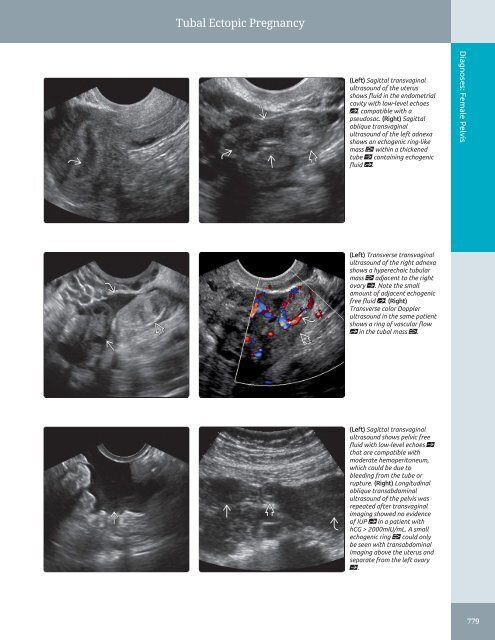
Tubal Ectopic Pregnancy<br />
(Left) Sagittal transvaginal<br />
ultrasound of the uterus<br />
shows fluid in the endometrial<br />
cavity with low-level echoes<br />
st, compatible with a<br />
pseudosac. (Right) Sagittal<br />
oblique transvaginal<br />
ultrasound of the left adnexa<br />
shows an echogenic ring-like<br />
mass within a thickened<br />
tube ſt containing echogenic<br />
fluid st.<br />
Diagnoses: Female <strong>Pelvis</strong><br />
(Left) Transverse transvaginal<br />
ultrasound of the right adnexa<br />
shows a hyperechoic tubular<br />
mass adjacent to the right<br />
ovary ſt. Note the small<br />
amount of adjacent echogenic<br />
free fluid st. (Right)<br />
Transverse color Doppler<br />
ultrasound in the same patient<br />
shows a ring of vascular flow<br />
st in the tubal mass .<br />
(Left) Sagittal transvaginal<br />
ultrasound shows pelvic free<br />
fluid with low-level echoes ſt<br />
that are compatible with<br />
moderate hemoperitoneum,<br />
which could be due to<br />
bleeding from the tube or<br />
rupture. (Right) Longitudinal<br />
oblique transabdominal<br />
ultrasound of the pelvis was<br />
repeated after transvaginal<br />
imaging showed no evidence<br />
of IUP st in a patient with<br />
hCG > 2000mIU/mL. A small<br />
echogenic ring could only<br />
be seen with transabdominal<br />
imaging above the uterus <strong>and</strong><br />
separate from the left ovary<br />
ſt.<br />
779


