Diagnostic Ultrasound - Abdomen and Pelvis
Endometrial Polyp (Left) Longitudinal transvaginal ultrasound shows focal nonspecific thickening of the endometrium ſt in a patient with dysfunctional uterine bleeding. The majority of the endometrium is atrophic, with a thin trilaminar appearance . (Right) Longitudinal transvaginal ultrasound during saline infusion in the same patient reveals an elongated pedunculated polyp ſt with a small stalk corresponding to the focal area of endometrial thickening. Diagnoses: Female Pelvis (Left) Longitudinal transvaginal ultrasound of the cervix shows a large polyp ſt prolapsing through the external os . Small internal cystic spaces are visible near the internal os. (Right) Sagittal T2-weighted MR in the same patient shows the polyp protruding through the cervical canal ſt. The polyp is hypointense relative to the normal endometrium , with the exception of internal cystic spaces . (Left) Longitudinal transvaginal ultrasound shows nonspecific focal thickening ſt in the upper endometrium, which could be secondary to hyperplasia, a polyp, or carcinoma. (Right) Longitudinal transvaginal ultrasound in the same patient following saline infusion reveals 2 sessile polyps ſt corresponding to the region of focal endometrial thickening. 759
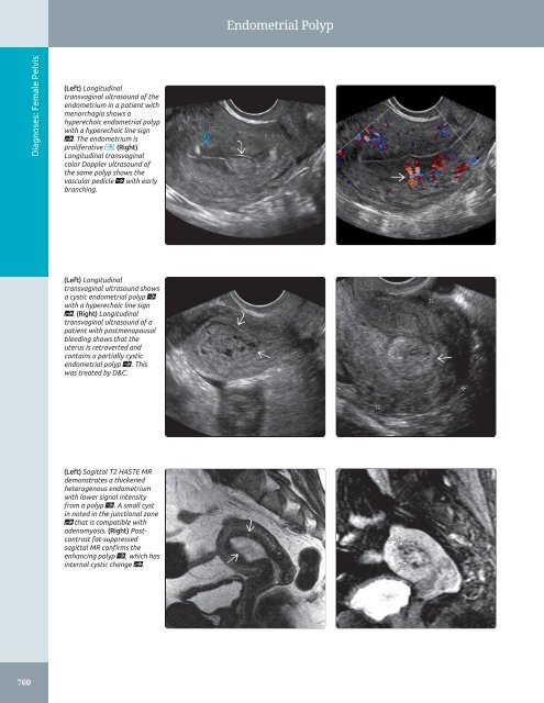
Endometrial Polyp Diagnoses: Female Pelvis (Left) Longitudinal transvaginal ultrasound of the endometrium in a patient with menorrhagia shows a hyperechoic endometrial polyp with a hyperechoic line sign st. The endometrium is proliferative . (Right) Longitudinal transvaginal color Doppler ultrasound of the same polyp shows the vascular pedicle ſt with early branching. (Left) Longitudinal transvaginal ultrasound shows a cystic endometrial polyp ſt with a hyperechoic line sign st. (Right) Longitudinal transvaginal ultrasound of a patient with postmenopausal bleeding shows that the uterus is retroverted and contains a partially cystic endometrial polyp ſt. This was treated by D&C. (Left) Sagittal T2 HASTE MR demonstrates a thickened heterogenous endometrium with lower signal intensity from a polyp ſt. A small cyst in noted in the junctional zone st that is compatible with adenomyosis. (Right) Postcontrast fat-suppressed sagittal MR confirms the enhancing polyp ſt, which has internal cystic change st. 760
- Page 730 and 731: Epididymitis/Orchitis (Left) Sagitt
- Page 732 and 733: Scrotal Trauma TERMINOLOGY Definiti
- Page 734 and 735: Scrotal Trauma (Left) Transverse gr
- Page 736 and 737: Hydrocele TERMINOLOGY Definitions
- Page 738 and 739: Spermatocele/Epididymal Cyst TERMIN
- Page 740 and 741: Adenomatoid Tumor TERMINOLOGY Defin
- Page 742 and 743: Varicocele TERMINOLOGY Definitions
- Page 744 and 745: Non-Ovarian Cystic Masses Hydrosalp
- Page 746 and 747: Approach to Sonography of the Femal
- Page 748 and 749: Nabothian Cyst TERMINOLOGY Synonyms
- Page 750 and 751: Nabothian Cyst (Left) Longitudinal
- Page 752 and 753: Cervical Carcinoma IMAGING General
- Page 754 and 755: Cervical Carcinoma (Left) Longitudi
- Page 756 and 757: Cervical Carcinoma (Left) Longitudi
- Page 758 and 759: Adenomyosis TERMINOLOGY Synonyms
- Page 760 and 761: Adenomyosis (Left) Longitudinal tra
- Page 762 and 763: Leiomyoma TERMINOLOGY Abbreviations
- Page 764 and 765: Leiomyoma (Left) Transvaginal ultra
- Page 766 and 767: Leiomyoma (Left) Transverse transab
- Page 768 and 769: Uterine Anomalies TERMINOLOGY Abbre
- Page 770 and 771: Uterine Anomalies (Left) 3D ultraso
- Page 772 and 773: Uterine Anomalies (Left) Graphic of
- Page 774 and 775: Hematometrocolpos TERMINOLOGY Abbre
- Page 776 and 777: Hematometrocolpos (Left) Transverse
- Page 778 and 779: Endometrial Polyp TERMINOLOGY Abbre
- Page 782 and 783: Endometrial Polyp (Left) Transabdom
- Page 784 and 785: Endometrial Carcinoma TERMINOLOGY A
- Page 786 and 787: Endometrial Carcinoma (Left) Longit
- Page 788 and 789: Endometritis TERMINOLOGY Synonyms
- Page 790 and 791: Endometritis (Left) Longitudinal tr
- Page 792 and 793: Intrauterine Device TERMINOLOGY Abb
- Page 794 and 795: Intrauterine Device (Left) Longitud
- Page 796 and 797: Tubal Ectopic Pregnancy TERMINOLOGY
- Page 798 and 799: Tubal Ectopic Pregnancy (Left) Tran
- Page 800 and 801: Tubal Ectopic Pregnancy (Left) Sagi
- Page 802 and 803: Unusual Ectopic Pregnancies TERMINO
- Page 804 and 805: Unusual Ectopic Pregnancies (Left)
- Page 806 and 807: Unusual Ectopic Pregnancies (Left)
- Page 808 and 809: Failed First Trimester Pregnancy TE
- Page 810 and 811: Failed First Trimester Pregnancy (L
- Page 812 and 813: Failed First Trimester Pregnancy (L
- Page 814 and 815: Retained Products of Conception TER
- Page 816 and 817: Retained Products of Conception (Le
- Page 818 and 819: Gestational Trophoblastic Disease T
- Page 820 and 821: Gestational Trophoblastic Disease (
- Page 822 and 823: Functional Ovarian Cyst TERMINOLOGY
- Page 824 and 825: Functional Ovarian Cyst (Left) Typi
- Page 826 and 827: Hemorrhagic Cyst TERMINOLOGY Abbrev
- Page 828 and 829: Hemorrhagic Cyst (Left) Using color
Endometrial Polyp<br />
Diagnoses: Female <strong>Pelvis</strong><br />
(Left) Longitudinal<br />
transvaginal ultrasound of the<br />
endometrium in a patient with<br />
menorrhagia shows a<br />
hyperechoic endometrial polyp<br />
with a hyperechoic line sign<br />
st. The endometrium is<br />
proliferative . (Right)<br />
Longitudinal transvaginal<br />
color Doppler ultrasound of<br />
the same polyp shows the<br />
vascular pedicle ſt with early<br />
branching.<br />
(Left) Longitudinal<br />
transvaginal ultrasound shows<br />
a cystic endometrial polyp ſt<br />
with a hyperechoic line sign<br />
st. (Right) Longitudinal<br />
transvaginal ultrasound of a<br />
patient with postmenopausal<br />
bleeding shows that the<br />
uterus is retroverted <strong>and</strong><br />
contains a partially cystic<br />
endometrial polyp ſt. This<br />
was treated by D&C.<br />
(Left) Sagittal T2 HASTE MR<br />
demonstrates a thickened<br />
heterogenous endometrium<br />
with lower signal intensity<br />
from a polyp ſt. A small cyst<br />
in noted in the junctional zone<br />
st that is compatible with<br />
adenomyosis. (Right) Postcontrast<br />
fat-suppressed<br />
sagittal MR confirms the<br />
enhancing polyp ſt, which has<br />
internal cystic change st.<br />
760


