Diagnostic Ultrasound - Abdomen and Pelvis
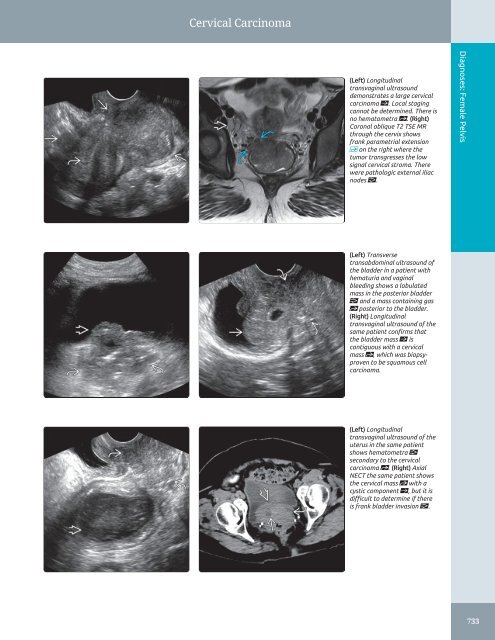
Cervical Carcinoma (Left) Longitudinal transvaginal ultrasound demonstrates a large cervical carcinoma ſt. Local staging cannot be determined. There is no hematometra st. (Right) Coronal oblique T2 TSE MR through the cervix shows frank parametrial extension on the right where the tumor transgresses the low signal cervical stroma. There were pathologic external iliac nodes . Diagnoses: Female Pelvis (Left) Transverse transabdominal ultrasound of the bladder in a patient with hematuria and vaginal bleeding shows a lobulated mass in the posterior bladder and a mass containing gas st posterior to the bladder. (Right) Longitudinal transvaginal ultrasound of the same patient confirms that the bladder mass ſt is contiguous with a cervical mass st, which was biopsyproven to be squamous cell carcinoma. (Left) Longitudinal transvaginal ultrasound of the uterus in the same patient shows hematometra secondary to the cervical carcinoma st. (Right) Axial NECT the same patient shows the cervical mass st with a cystic component ſt, but it is difficult to determine if there is frank bladder invasion . 733
Cervical Carcinoma Diagnoses: Female Pelvis (Left) Longitudinal transabdominal ultrasound shows an enlarged cervix ſt relative to the uterus st in a patient with profuse vaginal bleeding. (Right) Sagittal transvaginal ultrasound of the same patient shows the cervix ſt to be enlarged and hyperechoic to the myometrium st. The endometrium is thin. (Left) Coronal oblique transvaginal ultrasound of the same patient shows that the mass ſt infiltrates the entire cervix. Small cystic foci are present st. (Right) Coronal T2 FSE MR parallel to the endometrium in a patient with HIV and CIN 3 shows the normal cervical high signal mucosa ſt and intact low signal intensity stroma st. (Left) Coronal oblique TSE perpendicular to the cervix ("doughnut" view) shows a mass with intermediate T2 signal intensity st that extends through the cervical stroma ſt on the right and into the parametrium. (Right) Axial FDG PET/CT of the same patient shows increased metabolic activity in the primary tumor ſt (SUV 15.6) posterior to the bladder st. 734
- Page 704 and 705: Testicular Germ Cell Tumors TERMINO
- Page 706 and 707: Testicular Germ Cell Tumors (Left)
- Page 708 and 709: Gonadal Stromal Tumors, Testis TERM
- Page 710 and 711: Gonadal Stromal Tumors, Testis (Lef
- Page 712 and 713: Testicular Lymphoma/Leukemia TERMIN
- Page 714 and 715: Epidermoid Cyst TERMINOLOGY Synonym
- Page 716 and 717: Tubular Ectasia of Rete Testis TERM
- Page 718 and 719: Testicular Microlithiasis TERMINOLO
- Page 720 and 721: Testicular Microlithiasis (Left) Tr
- Page 722 and 723: Testicular Torsion/Infarction TERMI
- Page 724 and 725: Testicular Torsion/Infarction (Left
- Page 726 and 727: Undescended Testis TERMINOLOGY Syno
- Page 728 and 729: Epididymitis/Orchitis TERMINOLOGY S
- Page 730 and 731: Epididymitis/Orchitis (Left) Sagitt
- Page 732 and 733: Scrotal Trauma TERMINOLOGY Definiti
- Page 734 and 735: Scrotal Trauma (Left) Transverse gr
- Page 736 and 737: Hydrocele TERMINOLOGY Definitions
- Page 738 and 739: Spermatocele/Epididymal Cyst TERMIN
- Page 740 and 741: Adenomatoid Tumor TERMINOLOGY Defin
- Page 742 and 743: Varicocele TERMINOLOGY Definitions
- Page 744 and 745: Non-Ovarian Cystic Masses Hydrosalp
- Page 746 and 747: Approach to Sonography of the Femal
- Page 748 and 749: Nabothian Cyst TERMINOLOGY Synonyms
- Page 750 and 751: Nabothian Cyst (Left) Longitudinal
- Page 752 and 753: Cervical Carcinoma IMAGING General
- Page 756 and 757: Cervical Carcinoma (Left) Longitudi
- Page 758 and 759: Adenomyosis TERMINOLOGY Synonyms
- Page 760 and 761: Adenomyosis (Left) Longitudinal tra
- Page 762 and 763: Leiomyoma TERMINOLOGY Abbreviations
- Page 764 and 765: Leiomyoma (Left) Transvaginal ultra
- Page 766 and 767: Leiomyoma (Left) Transverse transab
- Page 768 and 769: Uterine Anomalies TERMINOLOGY Abbre
- Page 770 and 771: Uterine Anomalies (Left) 3D ultraso
- Page 772 and 773: Uterine Anomalies (Left) Graphic of
- Page 774 and 775: Hematometrocolpos TERMINOLOGY Abbre
- Page 776 and 777: Hematometrocolpos (Left) Transverse
- Page 778 and 779: Endometrial Polyp TERMINOLOGY Abbre
- Page 780 and 781: Endometrial Polyp (Left) Longitudin
- Page 782 and 783: Endometrial Polyp (Left) Transabdom
- Page 784 and 785: Endometrial Carcinoma TERMINOLOGY A
- Page 786 and 787: Endometrial Carcinoma (Left) Longit
- Page 788 and 789: Endometritis TERMINOLOGY Synonyms
- Page 790 and 791: Endometritis (Left) Longitudinal tr
- Page 792 and 793: Intrauterine Device TERMINOLOGY Abb
- Page 794 and 795: Intrauterine Device (Left) Longitud
- Page 796 and 797: Tubal Ectopic Pregnancy TERMINOLOGY
- Page 798 and 799: Tubal Ectopic Pregnancy (Left) Tran
- Page 800 and 801: Tubal Ectopic Pregnancy (Left) Sagi
- Page 802 and 803: Unusual Ectopic Pregnancies TERMINO
Cervical Carcinoma<br />
(Left) Longitudinal<br />
transvaginal ultrasound<br />
demonstrates a large cervical<br />
carcinoma ſt. Local staging<br />
cannot be determined. There is<br />
no hematometra st. (Right)<br />
Coronal oblique T2 TSE MR<br />
through the cervix shows<br />
frank parametrial extension<br />
on the right where the<br />
tumor transgresses the low<br />
signal cervical stroma. There<br />
were pathologic external iliac<br />
nodes .<br />
Diagnoses: Female <strong>Pelvis</strong><br />
(Left) Transverse<br />
transabdominal ultrasound of<br />
the bladder in a patient with<br />
hematuria <strong>and</strong> vaginal<br />
bleeding shows a lobulated<br />
mass in the posterior bladder<br />
<strong>and</strong> a mass containing gas<br />
st posterior to the bladder.<br />
(Right) Longitudinal<br />
transvaginal ultrasound of the<br />
same patient confirms that<br />
the bladder mass ſt is<br />
contiguous with a cervical<br />
mass st, which was biopsyproven<br />
to be squamous cell<br />
carcinoma.<br />
(Left) Longitudinal<br />
transvaginal ultrasound of the<br />
uterus in the same patient<br />
shows hematometra <br />
secondary to the cervical<br />
carcinoma st. (Right) Axial<br />
NECT the same patient shows<br />
the cervical mass st with a<br />
cystic component ſt, but it is<br />
difficult to determine if there<br />
is frank bladder invasion .<br />
733


