Diagnostic Ultrasound - Abdomen and Pelvis
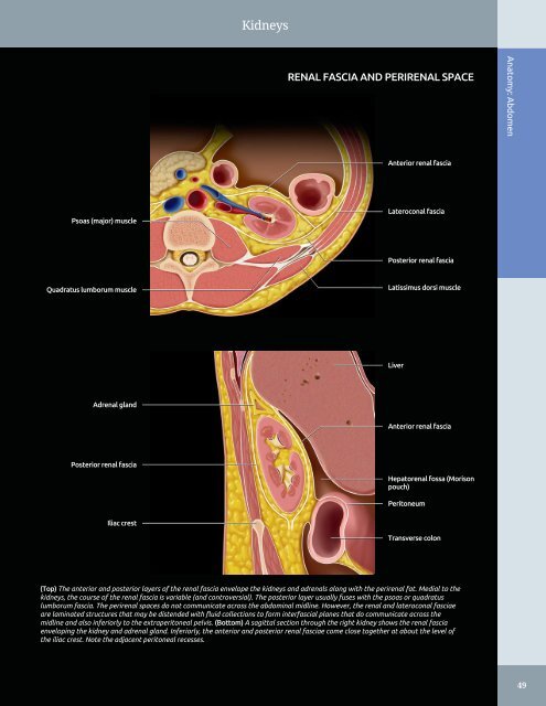
Kidneys RENAL FASCIA AND PERIRENAL SPACE Anatomy: Abdomen Anterior renal fascia Psoas (major) muscle Lateroconal fascia Posterior renal fascia Quadratus lumborum muscle Latissimus dorsi muscle Liver Adrenal gland Anterior renal fascia Posterior renal fascia Hepatorenal fossa (Morison pouch) Peritoneum Iliac crest Transverse colon (Top) The anterior and posterior layers of the renal fascia envelope the kidneys and adrenals along with the perirenal fat. Medial to the kidneys, the course of the renal fascia is variable (and controversial). The posterior layer usually fuses with the psoas or quadratus lumborum fascia. The perirenal spaces do not communicate across the abdominal midline. However, the renal and lateroconal fasciae are laminated structures that may be distended with fluid collections to form interfascial planes that do communicate across the midline and also inferiorly to the extraperitoneal pelvis. (Bottom) A sagittal section through the right kidney shows the renal fascia enveloping the kidney and adrenal gland. Inferiorly, the anterior and posterior renal fasciae come close together at about the level of the iliac crest. Note the adjacent peritoneal recesses. 49
Kidneys Anatomy: Abdomen RIGHT KIDNEY, ANTERIOR ABDOMEN SCAN Oblique muscles Right lobe of liver Renal sinus Medullary pyramid Psoas muscle Right lobe of liver Right main renal vein Psoas muscle Vertebral bodies Shadowing from rib Right lobe of liver Right psoas muscle (Top) Longitudinal grayscale ultrasound of the right kidney using the liver as an acoustic window is shown. This approach usually provides excellent visualization of the right kidney and is useful for measuring bipolar renal length. (Middle) Longitudinal oblique grayscale ultrasound of the right kidney using the liver as an acoustic window with the view obtained with a bit more medial angulation (when compared with the previous image). (Bottom) Longitudinal oblique grayscale ultrasound of the right kidney using the liver as an acoustic window with the view obtained with more lateral angulation (when compared with the previous 2 images) cuts through the renal parenchyma on the lateral aspect of the right kidney. Note that the echogenic sinus is not demonstrated and that there is shadowing from ribs. 50
- Page 20 and 21: TABLE OF CONTENTS 906 Hyperechoic G
- Page 22 and 23: Diagnostic Ultrasound
- Page 24 and 25: PART I SECTION 1 Abdomen Liver 4 Bi
- Page 26 and 27: Liver ○ Appear as echolucent defe
- Page 28 and 29: Liver Coronary ligament HEPATIC ATT
- Page 30 and 31: Liver Segment 8 HEPATIC SEGMENTS Se
- Page 32 and 33: Liver Rectus abdominis muscle LEFT
- Page 34 and 35: Liver Abdominal muscle LEFT LOBE OF
- Page 36 and 37: Liver Anterior right portal vein RI
- Page 38 and 39: Liver PORTA HEPATIS Anatomy: Abdome
- Page 40 and 41: Liver Inferior liver margin OTHER V
- Page 42 and 43: Biliary System • Harmonic imaging
- Page 44 and 45: Biliary System Left hepatic duct Ri
- Page 46 and 47: Biliary System Right rectus muscle
- Page 48 and 49: Biliary System COMMON BILE DUCT Ana
- Page 50 and 51: Biliary System LEFT INTRAHEPATIC DU
- Page 52 and 53: Spleen SPLEEN ANATOMY AND HISTOLOGY
- Page 54 and 55: Spleen Fat in splenic hilum Left he
- Page 56 and 57: Spleen SPLENIC VESSELS Anatomy: Abd
- Page 58 and 59: Spleen Splenosis ANATOMICAL VARIANT
- Page 60 and 61: Pancreas PANCREAS IN SITU Anatomy:
- Page 62 and 63: Pancreas PANCREAS, TRANSVERSE VIEW
- Page 64 and 65: Pancreas Left lobe of liver PANCREA
- Page 66 and 67: Kidneys - Normal peak systolic velo
- Page 68 and 69: Kidneys KIDNEY ARTERIES AND INTERIO
- Page 72 and 73: Kidneys Right hemidiaphragm RIGHT K
- Page 74 and 75: Kidneys RIGHT KIDNEY, CT CORRELATIO
- Page 76 and 77: Kidneys Right erector spinae muscle
- Page 78 and 79: Kidneys RIGHT INTRARENAL ARTERY AND
- Page 80 and 81: Kidneys LEFT KIDNEY, CT CORRELATION
- Page 82 and 83: Kidneys LEFT KIDNEY, CT CORRELATION
- Page 84 and 85: Kidneys Subcutaneous fat Left latis
- Page 86 and 87: Kidneys LEFT MAIN RENAL ARTERY AND
- Page 88 and 89: Kidneys Right lobe of liver MULTIPL
- Page 90 and 91: Bowel - Forms an incomplete ring in
- Page 92 and 93: Bowel Falciform ligament STOMACH AN
- Page 94 and 95: Bowel SMALL INTESTINE Anatomy: Abdo
- Page 96 and 97: Bowel Abdominal wall STOMACH Anatom
- Page 98 and 99: Bowel Rectus muscle SMALL BOWEL Ana
- Page 100 and 101: Bowel Abdominal wall musculature Ce
- Page 102 and 103: Bowel LARGE BOWEL Abdominal wall mu
- Page 104 and 105: Bowel Urinary bladder RECTOSIGMOID
- Page 106 and 107: Abdominal Lymph Nodes RETROPERITONE
- Page 108 and 109: Abdominal Lymph Nodes LYMPHANGIOGRA
- Page 110 and 111: Peritoneal Spaces and Structures PE
- Page 112 and 113: Peritoneal Spaces and Structures PE
- Page 114 and 115: Peritoneal Spaces and Structures Li
- Page 116 and 117: Peritoneal Spaces and Structures IN
- Page 118 and 119: Abdominal Wall ANTERIOR ABDOMINAL W
Kidneys<br />
RENAL FASCIA AND PERIRENAL SPACE<br />
Anatomy: <strong>Abdomen</strong><br />
Anterior renal fascia<br />
Psoas (major) muscle<br />
Lateroconal fascia<br />
Posterior renal fascia<br />
Quadratus lumborum muscle<br />
Latissimus dorsi muscle<br />
Liver<br />
Adrenal gl<strong>and</strong><br />
Anterior renal fascia<br />
Posterior renal fascia<br />
Hepatorenal fossa (Morison<br />
pouch)<br />
Peritoneum<br />
Iliac crest<br />
Transverse colon<br />
(Top) The anterior <strong>and</strong> posterior layers of the renal fascia envelope the kidneys <strong>and</strong> adrenals along with the perirenal fat. Medial to the<br />
kidneys, the course of the renal fascia is variable (<strong>and</strong> controversial). The posterior layer usually fuses with the psoas or quadratus<br />
lumborum fascia. The perirenal spaces do not communicate across the abdominal midline. However, the renal <strong>and</strong> lateroconal fasciae<br />
are laminated structures that may be distended with fluid collections to form interfascial planes that do communicate across the<br />
midline <strong>and</strong> also inferiorly to the extraperitoneal pelvis. (Bottom) A sagittal section through the right kidney shows the renal fascia<br />
enveloping the kidney <strong>and</strong> adrenal gl<strong>and</strong>. Inferiorly, the anterior <strong>and</strong> posterior renal fasciae come close together at about the level of<br />
the iliac crest. Note the adjacent peritoneal recesses.<br />
49


