- Page 2 and 3:
Diagnostic Ultrasound
- Page 4 and 5:
Diagnostic Ultrasound Aya Kamaya, M
- Page 6 and 7:
Dedications To my sweet and support
- Page 8 and 9:
Asef Khwaja, MD Assistant Professor
- Page 10 and 11:
Preface
- Page 12 and 13:
Acknowledgements Text Editors Nina
- Page 14 and 15:
Sections PART I - Anatomy SECTION 1
- Page 16 and 17:
TABLE OF CONTENTS VASCULAR CONDITIO
- Page 18 and 19:
TABLE OF CONTENTS 562 Perigraft Flu
- Page 20 and 21:
TABLE OF CONTENTS 906 Hyperechoic G
- Page 22 and 23:
Diagnostic Ultrasound
- Page 24 and 25:
PART I SECTION 1 Abdomen Liver 4 Bi
- Page 26 and 27:
Liver ○ Appear as echolucent defe
- Page 28 and 29:
Liver Coronary ligament HEPATIC ATT
- Page 30 and 31:
Liver Segment 8 HEPATIC SEGMENTS Se
- Page 32 and 33:
Liver Rectus abdominis muscle LEFT
- Page 34 and 35:
Liver Abdominal muscle LEFT LOBE OF
- Page 36 and 37:
Liver Anterior right portal vein RI
- Page 38 and 39:
Liver PORTA HEPATIS Anatomy: Abdome
- Page 40 and 41:
Liver Inferior liver margin OTHER V
- Page 42 and 43:
Biliary System • Harmonic imaging
- Page 44 and 45:
Biliary System Left hepatic duct Ri
- Page 46 and 47:
Biliary System Right rectus muscle
- Page 48 and 49:
Biliary System COMMON BILE DUCT Ana
- Page 50 and 51:
Biliary System LEFT INTRAHEPATIC DU
- Page 52 and 53:
Spleen SPLEEN ANATOMY AND HISTOLOGY
- Page 54 and 55:
Spleen Fat in splenic hilum Left he
- Page 56 and 57:
Spleen SPLENIC VESSELS Anatomy: Abd
- Page 58 and 59:
Spleen Splenosis ANATOMICAL VARIANT
- Page 60 and 61:
Pancreas PANCREAS IN SITU Anatomy:
- Page 62 and 63:
Pancreas PANCREAS, TRANSVERSE VIEW
- Page 64 and 65:
Pancreas Left lobe of liver PANCREA
- Page 66 and 67:
Kidneys - Normal peak systolic velo
- Page 68 and 69:
Kidneys KIDNEY ARTERIES AND INTERIO
- Page 70 and 71:
Kidneys RENAL FASCIA AND PERIRENAL
- Page 72 and 73:
Kidneys Right hemidiaphragm RIGHT K
- Page 74 and 75:
Kidneys RIGHT KIDNEY, CT CORRELATIO
- Page 76 and 77:
Kidneys Right erector spinae muscle
- Page 78 and 79:
Kidneys RIGHT INTRARENAL ARTERY AND
- Page 80 and 81:
Kidneys LEFT KIDNEY, CT CORRELATION
- Page 82 and 83:
Kidneys LEFT KIDNEY, CT CORRELATION
- Page 84 and 85:
Kidneys Subcutaneous fat Left latis
- Page 86 and 87:
Kidneys LEFT MAIN RENAL ARTERY AND
- Page 88 and 89:
Kidneys Right lobe of liver MULTIPL
- Page 90 and 91:
Bowel - Forms an incomplete ring in
- Page 92 and 93:
Bowel Falciform ligament STOMACH AN
- Page 94 and 95:
Bowel SMALL INTESTINE Anatomy: Abdo
- Page 96 and 97:
Bowel Abdominal wall STOMACH Anatom
- Page 98 and 99:
Bowel Rectus muscle SMALL BOWEL Ana
- Page 100 and 101:
Bowel Abdominal wall musculature Ce
- Page 102 and 103:
Bowel LARGE BOWEL Abdominal wall mu
- Page 104 and 105:
Bowel Urinary bladder RECTOSIGMOID
- Page 106 and 107:
Abdominal Lymph Nodes RETROPERITONE
- Page 108 and 109:
Abdominal Lymph Nodes LYMPHANGIOGRA
- Page 110 and 111:
Peritoneal Spaces and Structures PE
- Page 112 and 113:
Peritoneal Spaces and Structures PE
- Page 114 and 115:
Peritoneal Spaces and Structures Li
- Page 116 and 117:
Peritoneal Spaces and Structures IN
- Page 118 and 119:
Abdominal Wall ANTERIOR ABDOMINAL W
- Page 120 and 121:
Abdominal Wall MUSCLES OF BACK IN S
- Page 122 and 123:
Abdominal Wall Subcutaneous fat Rig
- Page 124 and 125:
Abdominal Wall Right lobe of liver
- Page 126 and 127:
Abdominal Wall Right rectus abdomin
- Page 128 and 129:
Abdominal Wall Subcutaneous fat Rig
- Page 130 and 131:
PART I SECTION 2 Pelvis Ureters and
- Page 132 and 133:
Ureters and Bladder - Distended bla
- Page 134 and 135:
Ureters and Bladder URINARY BLADDER
- Page 136 and 137:
Ureters and Bladder CT UROGRAM CORR
- Page 138 and 139:
Ureters and Bladder Liver URETER An
- Page 140 and 141:
Ureters and Bladder WEIGERT-MEYER L
- Page 142 and 143:
Prostate ○ Sac-like structures su
- Page 144 and 145:
Prostate ZONAL ANATOMY OF THE PROST
- Page 146 and 147:
Prostate SEMINAL VESICLES AND VAS D
- Page 148 and 149:
Prostate PROSTATE ANATOMY Anatomy:
- Page 150 and 151:
Testes ○ Internal oblique muscle
- Page 152 and 153:
Testes EPIDIDYMIS AND SCROTAL WALL
- Page 154 and 155:
Testes TESTIS, SAGITTAL VIEW Anatom
- Page 156 and 157:
Testes Scrotal wall EPIDIDYMIS, HEA
- Page 158 and 159:
Testes TESTICULAR AND EPIDIDYMAL AP
- Page 160 and 161:
Testes ARTERIAL AND VENOUS SUPPLY A
- Page 162 and 163:
Uterus ARTERIES OF UTERUS AND ADJAC
- Page 164 and 165:
Uterus NORMAL VARIATIONS, UTERINE P
- Page 166 and 167:
Uterus UTERINE VARIATIONS WITH AGE
- Page 168 and 169:
Uterus CYCLIC CHANGES OF ENDOMETRIU
- Page 170 and 171:
Uterus FALLOPIAN TUBE Anatomy: Pelv
- Page 172 and 173:
Cervix GRAPHICS OF CERVIX ANATOMY A
- Page 174 and 175:
Cervix TRANSVAGINAL ULTRASOUND OF C
- Page 176 and 177:
Cervix CHANGES OF CERVIX DURING PRE
- Page 178 and 179:
Vagina GRAPHICS OF NORMAL VAGINAL A
- Page 180 and 181:
Vagina Urinary bladder TRANSVERSE U
- Page 182 and 183:
Vagina SPECTRAL WAVEFORM OF VAGINAL
- Page 184 and 185:
Ovaries Mesosalpinx LIGAMENTOUS SUP
- Page 186 and 187:
Ovaries Transvaginal transducer NOR
- Page 188 and 189:
Ovaries Ovarian artery SPECTRAL WAV
- Page 190 and 191:
Ovaries CYCLIC CHANGES OF OVARY Ana
- Page 192 and 193:
Ovaries CYCLIC CHANGES OF INTRAOVAR
- Page 194 and 195:
Liver Transplant Hepatic Artery Ste
- Page 196 and 197:
Approach to Hepatic Sonography (Lef
- Page 198 and 199:
Approach to Hepatic Sonography (Lef
- Page 200 and 201:
Acute Hepatitis TERMINOLOGY Definit
- Page 202 and 203:
Acute Hepatitis (Left) Transverse g
- Page 204 and 205:
Hepatic Cirrhosis TERMINOLOGY Defin
- Page 206 and 207:
Hepatic Cirrhosis (Left) Longitudin
- Page 208 and 209:
Hepatic Steatosis TERMINOLOGY Synon
- Page 210 and 211:
Hepatic Steatosis (Left) Transverse
- Page 212 and 213:
Hepatic Schistosomiasis TERMINOLOGY
- Page 214 and 215:
Venoocclusive Disease TERMINOLOGY A
- Page 216 and 217:
Venoocclusive Disease (Left) Graysc
- Page 218 and 219:
Hepatic Cyst TERMINOLOGY Synonyms
- Page 220 and 221:
Hepatic Cyst (Left) Transverse and
- Page 222 and 223:
Biliary Hamartoma TERMINOLOGY Synon
- Page 224 and 225:
Biliary Hamartoma (Left) Ultrasound
- Page 226 and 227:
Caroli Disease TERMINOLOGY Synonyms
- Page 228 and 229:
Caroli Disease (Left) Oblique abdom
- Page 230 and 231:
Biloma TERMINOLOGY Definitions •
- Page 232 and 233:
Biliary Cystadenoma/Carcinoma TERMI
- Page 234 and 235:
Biliary Cystadenoma/Carcinoma (Left
- Page 236 and 237:
Pyogenic Hepatic Abscess TERMINOLOG
- Page 238 and 239:
Pyogenic Hepatic Abscess (Left) Obl
- Page 240 and 241:
Amebic Hepatic Abscess TERMINOLOGY
- Page 242 and 243:
Amebic Hepatic Abscess (Left) Longi
- Page 244 and 245:
Hepatic Echinococcus Cyst TERMINOLO
- Page 246 and 247:
Hepatic Echinococcus Cyst (Left) Ob
- Page 248 and 249:
Hepatic Diffuse Microabscesses TERM
- Page 250 and 251:
Peribiliary Cyst TERMINOLOGY Synony
- Page 252 and 253:
Ciliated Hepatic Foregut Cyst TERMI
- Page 254 and 255:
Hepatic Cavernous Hemangioma TERMIN
- Page 256 and 257:
Hepatic Cavernous Hemangioma (Left)
- Page 258 and 259:
Hepatic Cavernous Hemangioma (Left)
- Page 260 and 261:
Focal Nodular Hyperplasia TERMINOLO
- Page 262 and 263:
Focal Nodular Hyperplasia (Left) Tr
- Page 264 and 265:
Hepatic Adenoma TERMINOLOGY Synonym
- Page 266 and 267:
Hepatic Adenoma (Left) Transverse t
- Page 268 and 269:
Hepatocellular Carcinoma TERMINOLOG
- Page 270 and 271:
Hepatocellular Carcinoma (Left) Tra
- Page 272 and 273:
Hepatocellular Carcinoma (Left) Tra
- Page 274 and 275:
Hepatic Metastases TERMINOLOGY Defi
- Page 276 and 277:
Hepatic Metastases (Left) Transvers
- Page 278 and 279:
Hepatic Lymphoma TERMINOLOGY Defini
- Page 280 and 281:
Hepatic Lymphoma (Left) Transverse
- Page 282 and 283:
Transjugular Intrahepatic Portosyst
- Page 284 and 285:
Transjugular Intrahepatic Portosyst
- Page 286 and 287:
Portal Vein Occlusion TERMINOLOGY A
- Page 288 and 289:
Portal Vein Occlusion (Left) Color
- Page 290 and 291:
Budd-Chiari Syndrome TERMINOLOGY Ab
- Page 292 and 293:
Budd-Chiari Syndrome (Left) Color D
- Page 294 and 295:
Portal Vein Gas TERMINOLOGY Abbrevi
- Page 296 and 297:
Liver Transplant Portal Vein Stenos
- Page 298 and 299:
Liver Transplant Biliary Stricture
- Page 300 and 301:
PART II SECTION 2 Biliary System In
- Page 302 and 303:
Approach to Biliary Sonography hosp
- Page 304 and 305:
Approach to Biliary Sonography (Lef
- Page 306 and 307:
Approach to Biliary Sonography (Lef
- Page 308 and 309:
Cholelithiasis TERMINOLOGY Synonyms
- Page 310 and 311:
Cholelithiasis (Left) Longitudinal
- Page 312 and 313:
Cholelithiasis (Left) Abdominal rad
- Page 314 and 315:
Echogenic Bile TERMINOLOGY Synonyms
- Page 316 and 317:
Echogenic Bile (Left) Transverse ul
- Page 318 and 319:
Gallbladder Cholesterol Polyp TERMI
- Page 320 and 321:
Gallbladder Cholesterol Polyp (Left
- Page 322 and 323:
Acute Calculous Cholecystitis TERMI
- Page 324 and 325:
Acute Calculous Cholecystitis (Left
- Page 326 and 327:
Acute Acalculous Cholecystitis TERM
- Page 328 and 329:
Acute Acalculous Cholecystitis (Lef
- Page 330 and 331:
Chronic Cholecystitis TERMINOLOGY D
- Page 332 and 333:
Xanthogranulomatous Cholecystitis T
- Page 334 and 335:
Porcelain Gallbladder TERMINOLOGY A
- Page 336 and 337:
Hyperplastic Cholecystosis (Adenomy
- Page 338 and 339:
Hyperplastic Cholecystosis (Adenomy
- Page 340 and 341:
Gallbladder Carcinoma TERMINOLOGY A
- Page 342 and 343:
Gallbladder Carcinoma (Left) Sagitt
- Page 344 and 345:
Biliary Ductal Dilatation IMAGING G
- Page 346 and 347:
Choledochal Cyst TERMINOLOGY Synony
- Page 348 and 349:
Choledochal Cyst (Left) Longitudina
- Page 350 and 351:
Choledocholithiasis TERMINOLOGY Abb
- Page 352 and 353:
Choledocholithiasis (Left) A single
- Page 354 and 355:
Biliary Ductal Gas TERMINOLOGY Syno
- Page 356 and 357:
Cholangiocarcinoma TERMINOLOGY Syno
- Page 358 and 359:
Cholangiocarcinoma (Left) Ultrasoun
- Page 360 and 361:
Ascending Cholangitis TERMINOLOGY S
- Page 362 and 363:
Ascending Cholangitis (Left) Longit
- Page 364 and 365:
Recurrent Pyogenic Cholangitis TERM
- Page 366 and 367:
Recurrent Pyogenic Cholangitis (Lef
- Page 368 and 369:
AIDS-Related Cholangiopathy TERMINO
- Page 370 and 371:
PART II SECTION 3 Pancreas Introduc
- Page 372 and 373:
Approach to Pancreatic Sonography C
- Page 374 and 375:
Approach to Pancreatic Sonography (
- Page 376 and 377:
Acute Pancreatitis TERMINOLOGY Abbr
- Page 378 and 379:
Acute Pancreatitis (Left) Transvers
- Page 380 and 381:
Pancreatic Pseudocyst TERMINOLOGY D
- Page 382 and 383:
Pancreatic Pseudocyst (Left) Transv
- Page 384 and 385:
Chronic Pancreatitis TERMINOLOGY Ab
- Page 386 and 387:
Chronic Pancreatitis (Left) Transve
- Page 388 and 389:
Mucinous Cystic Pancreatic Tumor TE
- Page 390 and 391:
Mucinous Cystic Pancreatic Tumor (L
- Page 392 and 393:
Serous Cystadenoma of Pancreas TERM
- Page 394 and 395:
Serous Cystadenoma of Pancreas (Lef
- Page 396 and 397:
Intraductal Papillary Mucinous Neop
- Page 398 and 399:
Intraductal Papillary Mucinous Neop
- Page 400 and 401:
Intraductal Papillary Mucinous Neop
- Page 402 and 403:
Pancreatic Ductal Carcinoma TERMINO
- Page 404 and 405:
Pancreatic Ductal Carcinoma (Left)
- Page 406 and 407:
Pancreatic Neuroendocrine Tumor TER
- Page 408 and 409:
Pancreatic Neuroendocrine Tumor (Le
- Page 410 and 411:
Solid Pseudopapillary Neoplasm TERM
- Page 412 and 413:
Solid Pseudopapillary Neoplasm (Lef
- Page 414 and 415:
PART II SECTION 4 Spleen Introducti
- Page 416 and 417:
Approach to Splenic Sonography sple
- Page 418 and 419:
Approach to Splenic Sonography (Lef
- Page 420 and 421:
Approach to Splenic Sonography (Lef
- Page 422 and 423:
Splenomegaly TERMINOLOGY Abbreviati
- Page 424 and 425:
Splenomegaly (Left) US in a 92-year
- Page 426 and 427:
Splenic Cyst TERMINOLOGY Definition
- Page 428 and 429:
Splenic Cyst (Left) Grayscale US of
- Page 430 and 431:
Splenic Tumors TERMINOLOGY Definiti
- Page 432 and 433:
Splenic Tumors (Left) Longitudinal
- Page 434 and 435:
Splenic Tumors (Left) A solid, hypo
- Page 436 and 437:
Splenic Infarct TERMINOLOGY Abbrevi
- Page 438 and 439:
Splenic Infarct (Left) Power Dopple
- Page 440 and 441:
Vascular Conditions Renal Artery St
- Page 442 and 443:
Approach to Urinary Tract Sonograph
- Page 444 and 445:
Approach to Urinary Tract Sonograph
- Page 446 and 447:
Column of Bertin, Kidney TERMINOLOG
- Page 448 and 449:
Renal Junction Line TERMINOLOGY Syn
- Page 450 and 451: Renal Ectopia TERMINOLOGY Abbreviat
- Page 452 and 453: Renal Ectopia (Left) Grayscale ultr
- Page 454 and 455: Horseshoe Kidney TERMINOLOGY Defini
- Page 456 and 457: Horseshoe Kidney (Left) Longitudina
- Page 458 and 459: Ureteral Duplication TERMINOLOGY Sy
- Page 460 and 461: Ureteral Duplication (Left) Longitu
- Page 462 and 463: Ureteral Ectopia TERMINOLOGY Abbrev
- Page 464 and 465: Ureteral Ectopia (Left) Coronal T2
- Page 466 and 467: Ureteropelvic Junction Obstruction
- Page 468 and 469: Ureteropelvic Junction Obstruction
- Page 470 and 471: Urolithiasis TERMINOLOGY Abbreviati
- Page 472 and 473: Urolithiasis (Left) Longitudinal US
- Page 474 and 475: Urolithiasis (Left) Intravenous pye
- Page 476 and 477: Nephrocalcinosis TERMINOLOGY Abbrev
- Page 478 and 479: Nephrocalcinosis (Left) Coronal MIP
- Page 480 and 481: Hydronephrosis TERMINOLOGY Synonyms
- Page 482 and 483: Hydronephrosis (Left) Longitudinal
- Page 484 and 485: Simple Renal Cyst TERMINOLOGY Defin
- Page 486 and 487: Simple Renal Cyst (Left) Longitudin
- Page 488 and 489: Complex Renal Cyst TERMINOLOGY Defi
- Page 490 and 491: Complex Renal Cyst (Left) Transvers
- Page 492 and 493: Cystic Disease of Dialysis TERMINOL
- Page 494 and 495: Cystic Disease of Dialysis (Left) L
- Page 496 and 497: Multilocular Cystic Nephroma TERMIN
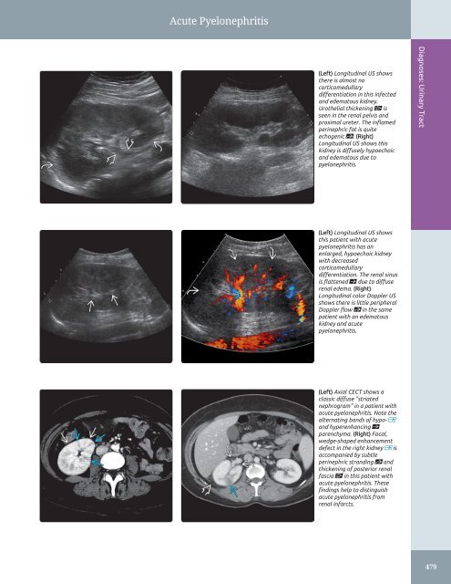
- Page 498 and 499: Acute Pyelonephritis TERMINOLOGY Ab
- Page 502 and 503: Renal Abscess TERMINOLOGY Definitio
- Page 504 and 505: Emphysematous Pyelonephritis TERMIN
- Page 506 and 507: Emphysematous Pyelonephritis (Left)
- Page 508 and 509: Pyonephrosis TERMINOLOGY Definition
- Page 510 and 511: Xanthogranulomatous Pyelonephritis
- Page 512 and 513: Tuberculosis, Urinary Tract TERMINO
- Page 514 and 515: Tuberculosis, Urinary Tract (Left)
- Page 516 and 517: Renal Cell Carcinoma TERMINOLOGY Ab
- Page 518 and 519: Renal Cell Carcinoma (Left) Longitu
- Page 520 and 521: Renal Metastases IMAGING General Fe
- Page 522 and 523: Renal Angiomyolipoma TERMINOLOGY Ab
- Page 524 and 525: Renal Angiomyolipoma (Left) Longitu
- Page 526 and 527: Upper Tract Urothelial Carcinoma TE
- Page 528 and 529: Upper Tract Urothelial Carcinoma (L
- Page 530 and 531: Renal Lymphoma TERMINOLOGY Abbrevia
- Page 532 and 533: Renal Lymphoma (Left) Longitudinal
- Page 534 and 535: Renal Artery Stenosis TERMINOLOGY A
- Page 536 and 537: Renal Artery Stenosis (Left) Obliqu
- Page 538 and 539: Renal Vein Thrombosis TERMINOLOGY A
- Page 540 and 541: Renal Vein Thrombosis (Left) Longit
- Page 542 and 543: Renal Infarct TERMINOLOGY Definitio
- Page 544 and 545: Perinephric Hematoma TERMINOLOGY De
- Page 546 and 547: Prostatic Hyperplasia TERMINOLOGY A
- Page 548 and 549: Prostatic Hyperplasia (Left) Axial
- Page 550 and 551:
Prostatic Carcinoma TERMINOLOGY Abb
- Page 552 and 553:
Prostatic Carcinoma (Left) Transver
- Page 554 and 555:
Prostatic Carcinoma (Left) Transver
- Page 556 and 557:
Bladder Carcinoma TERMINOLOGY Defin
- Page 558 and 559:
Bladder Carcinoma (Left) Transverse
- Page 560 and 561:
Ureterocele TERMINOLOGY Definitions
- Page 562 and 563:
Ureterocele (Left) Transabdominal l
- Page 564 and 565:
Bladder Diverticulum TERMINOLOGY Ab
- Page 566 and 567:
Bladder Diverticulum (Left) Transab
- Page 568 and 569:
Bladder Calculi TERMINOLOGY Synonym
- Page 570 and 571:
Schistosomiasis, Bladder TERMINOLOG
- Page 572 and 573:
PART II SECTION 6 Kidney Transplant
- Page 574 and 575:
Approach to Sonography of Renal All
- Page 576 and 577:
Approach to Sonography of Renal All
- Page 578 and 579:
Approach to Sonography of Renal All
- Page 580 and 581:
Allograft Hydronephrosis TERMINOLOG
- Page 582 and 583:
Allograft Hydronephrosis (Left) Lon
- Page 584 and 585:
Perigraft Fluid Collections TERMINO
- Page 586 and 587:
Perigraft Fluid Collections (Left)
- Page 588 and 589:
Transplant Renal Artery Stenosis TE
- Page 590 and 591:
Transplant Renal Artery Stenosis (L
- Page 592 and 593:
Transplant Renal Artery Thrombosis
- Page 594 and 595:
Transplant Renal Vein Thrombosis TE
- Page 596 and 597:
Renal Transplant Arteriovenous (AV)
- Page 598 and 599:
Renal Transplant Pseudoaneurysm TER
- Page 600 and 601:
Renal Transplant Rejection IMAGING
- Page 602 and 603:
Delayed Renal Graft Function TERMIN
- Page 604 and 605:
PART II SECTION 7 Adrenal Gland Adr
- Page 606 and 607:
Adrenal Hemorrhage TERMINOLOGY Abbr
- Page 608 and 609:
Adrenal Hemorrhage (Left) Longitudi
- Page 610 and 611:
Myelolipoma TERMINOLOGY Definitions
- Page 612 and 613:
Myelolipoma (Left) Longitudinal US
- Page 614 and 615:
Adrenal Adenoma TERMINOLOGY Synonym
- Page 616 and 617:
Adrenal Adenoma (Left) Longitudinal
- Page 618 and 619:
Adrenal Cyst TERMINOLOGY Definition
- Page 620 and 621:
Pheochromocytoma TERMINOLOGY Defini
- Page 622 and 623:
Pheochromocytoma (Left) Longitudina
- Page 624 and 625:
Adrenal Carcinoma TERMINOLOGY Synon
- Page 626 and 627:
Adrenal Carcinoma (Left) Transverse
- Page 628 and 629:
PART II SECTION 8 Abdominal Wall/Pe
- Page 630 and 631:
Approach to Sonography of Abdominal
- Page 632 and 633:
Approach to Sonography of Abdominal
- Page 634 and 635:
Abdominal Wall Hernia TERMINOLOGY D
- Page 636 and 637:
Abdominal Wall Hernia (Left) Transv
- Page 638 and 639:
Abdominal Wall Hernia (Left) Transv
- Page 640 and 641:
Groin Hernia TERMINOLOGY Definition
- Page 642 and 643:
Groin Hernia (Left) Graphic shows a
- Page 644 and 645:
Groin Hernia (Left) Longitudinal ul
- Page 646 and 647:
Ascites TERMINOLOGY Definitions •
- Page 648 and 649:
Ascites (Left) Transverse ultrasoun
- Page 650 and 651:
Peritoneal Carcinomatosis TERMINOLO
- Page 652 and 653:
Peritoneal Carcinomatosis (Left) Lo
- Page 654 and 655:
Peritoneal Carcinomatosis (Left) Tr
- Page 656 and 657:
Peritoneal Space Abscess TERMINOLOG
- Page 658 and 659:
Peritoneal Space Abscess (Left) Tra
- Page 660 and 661:
Segmental Omental Infarction TERMIN
- Page 662 and 663:
PART II SECTION 9 Bowel Approach to
- Page 664 and 665:
Approach to Bowel Sonography Given
- Page 666 and 667:
Approach to Bowel Sonography (Left)
- Page 668 and 669:
Appendicitis TERMINOLOGY Definition
- Page 670 and 671:
Appendicitis (Left) Axial ultrasoun
- Page 672 and 673:
Appendicitis (Left) Axial ultrasoun
- Page 674 and 675:
Appendiceal Mucocele TERMINOLOGY De
- Page 676 and 677:
Appendiceal Mucocele (Left) Sagitta
- Page 678 and 679:
Intussusception TERMINOLOGY Definit
- Page 680 and 681:
Intussusception (Left) Transverse t
- Page 682 and 683:
Epiploic Appendagitis TERMINOLOGY A
- Page 684 and 685:
Epiploic Appendagitis (Left) Graysc
- Page 686 and 687:
Diverticulitis TERMINOLOGY Definiti
- Page 688 and 689:
Diverticulitis (Left) Acute diverti
- Page 690 and 691:
Crohn Disease TERMINOLOGY Synonyms
- Page 692 and 693:
Crohn Disease (Left) Long-axis ultr
- Page 694 and 695:
Crohn Disease (Left) Long-axis ultr
- Page 696 and 697:
Large Bowel Malignancy TERMINOLOGY
- Page 698 and 699:
Large Bowel Malignancy (Left) Hepat
- Page 700 and 701:
PART II SECTION 10 Scrotum Introduc
- Page 702 and 703:
Approach to Scrotal Sonography (Lef
- Page 704 and 705:
Testicular Germ Cell Tumors TERMINO
- Page 706 and 707:
Testicular Germ Cell Tumors (Left)
- Page 708 and 709:
Gonadal Stromal Tumors, Testis TERM
- Page 710 and 711:
Gonadal Stromal Tumors, Testis (Lef
- Page 712 and 713:
Testicular Lymphoma/Leukemia TERMIN
- Page 714 and 715:
Epidermoid Cyst TERMINOLOGY Synonym
- Page 716 and 717:
Tubular Ectasia of Rete Testis TERM
- Page 718 and 719:
Testicular Microlithiasis TERMINOLO
- Page 720 and 721:
Testicular Microlithiasis (Left) Tr
- Page 722 and 723:
Testicular Torsion/Infarction TERMI
- Page 724 and 725:
Testicular Torsion/Infarction (Left
- Page 726 and 727:
Undescended Testis TERMINOLOGY Syno
- Page 728 and 729:
Epididymitis/Orchitis TERMINOLOGY S
- Page 730 and 731:
Epididymitis/Orchitis (Left) Sagitt
- Page 732 and 733:
Scrotal Trauma TERMINOLOGY Definiti
- Page 734 and 735:
Scrotal Trauma (Left) Transverse gr
- Page 736 and 737:
Hydrocele TERMINOLOGY Definitions
- Page 738 and 739:
Spermatocele/Epididymal Cyst TERMIN
- Page 740 and 741:
Adenomatoid Tumor TERMINOLOGY Defin
- Page 742 and 743:
Varicocele TERMINOLOGY Definitions
- Page 744 and 745:
Non-Ovarian Cystic Masses Hydrosalp
- Page 746 and 747:
Approach to Sonography of the Femal
- Page 748 and 749:
Nabothian Cyst TERMINOLOGY Synonyms
- Page 750 and 751:
Nabothian Cyst (Left) Longitudinal
- Page 752 and 753:
Cervical Carcinoma IMAGING General
- Page 754 and 755:
Cervical Carcinoma (Left) Longitudi
- Page 756 and 757:
Cervical Carcinoma (Left) Longitudi
- Page 758 and 759:
Adenomyosis TERMINOLOGY Synonyms
- Page 760 and 761:
Adenomyosis (Left) Longitudinal tra
- Page 762 and 763:
Leiomyoma TERMINOLOGY Abbreviations
- Page 764 and 765:
Leiomyoma (Left) Transvaginal ultra
- Page 766 and 767:
Leiomyoma (Left) Transverse transab
- Page 768 and 769:
Uterine Anomalies TERMINOLOGY Abbre
- Page 770 and 771:
Uterine Anomalies (Left) 3D ultraso
- Page 772 and 773:
Uterine Anomalies (Left) Graphic of
- Page 774 and 775:
Hematometrocolpos TERMINOLOGY Abbre
- Page 776 and 777:
Hematometrocolpos (Left) Transverse
- Page 778 and 779:
Endometrial Polyp TERMINOLOGY Abbre
- Page 780 and 781:
Endometrial Polyp (Left) Longitudin
- Page 782 and 783:
Endometrial Polyp (Left) Transabdom
- Page 784 and 785:
Endometrial Carcinoma TERMINOLOGY A
- Page 786 and 787:
Endometrial Carcinoma (Left) Longit
- Page 788 and 789:
Endometritis TERMINOLOGY Synonyms
- Page 790 and 791:
Endometritis (Left) Longitudinal tr
- Page 792 and 793:
Intrauterine Device TERMINOLOGY Abb
- Page 794 and 795:
Intrauterine Device (Left) Longitud
- Page 796 and 797:
Tubal Ectopic Pregnancy TERMINOLOGY
- Page 798 and 799:
Tubal Ectopic Pregnancy (Left) Tran
- Page 800 and 801:
Tubal Ectopic Pregnancy (Left) Sagi
- Page 802 and 803:
Unusual Ectopic Pregnancies TERMINO
- Page 804 and 805:
Unusual Ectopic Pregnancies (Left)
- Page 806 and 807:
Unusual Ectopic Pregnancies (Left)
- Page 808 and 809:
Failed First Trimester Pregnancy TE
- Page 810 and 811:
Failed First Trimester Pregnancy (L
- Page 812 and 813:
Failed First Trimester Pregnancy (L
- Page 814 and 815:
Retained Products of Conception TER
- Page 816 and 817:
Retained Products of Conception (Le
- Page 818 and 819:
Gestational Trophoblastic Disease T
- Page 820 and 821:
Gestational Trophoblastic Disease (
- Page 822 and 823:
Functional Ovarian Cyst TERMINOLOGY
- Page 824 and 825:
Functional Ovarian Cyst (Left) Typi
- Page 826 and 827:
Hemorrhagic Cyst TERMINOLOGY Abbrev
- Page 828 and 829:
Hemorrhagic Cyst (Left) Using color
- Page 830 and 831:
Ovarian Hyperstimulation Syndrome T
- Page 832 and 833:
Ovarian Hyperstimulation Syndrome (
- Page 834 and 835:
Serous Ovarian Cystadenoma/Carcinom
- Page 836 and 837:
Serous Ovarian Cystadenoma/Carcinom
- Page 838 and 839:
Mucinous Ovarian Cystadenoma/Carcin
- Page 840 and 841:
Mucinous Ovarian Cystadenoma/Carcin
- Page 842 and 843:
Ovarian Teratoma TERMINOLOGY Synony
- Page 844 and 845:
Ovarian Teratoma (Left) Ultrasound
- Page 846 and 847:
Polycystic Ovarian Syndrome TERMINO
- Page 848 and 849:
Endometrioma TERMINOLOGY Synonyms
- Page 850 and 851:
Endometrioma (Left) Longitudinal en
- Page 852 and 853:
Hydrosalpinx TERMINOLOGY Definition
- Page 854 and 855:
Hydrosalpinx (Left) Longitudinal tr
- Page 856 and 857:
Tubo-Ovarian Abscess TERMINOLOGY De
- Page 858 and 859:
Tubo-Ovarian Abscess (Left) Longitu
- Page 860 and 861:
Parovarian Cyst TERMINOLOGY Abbrevi
- Page 862 and 863:
Peritoneal Inclusion Cyst TERMINOLO
- Page 864 and 865:
Peritoneal Inclusion Cyst (Left) Sa
- Page 866 and 867:
Bartholin Cyst TERMINOLOGY Definiti
- Page 868 and 869:
Gartner Duct Cyst TERMINOLOGY Abbre
- Page 870 and 871:
Gartner Duct Cyst (Left) Longitudin
- Page 872 and 873:
Sex Cord-Stromal Tumor TERMINOLOGY
- Page 874 and 875:
Sex Cord-Stromal Tumor (Left) Trans
- Page 876 and 877:
Sex Cord-Stromal Tumor (Left) Sagit
- Page 878 and 879:
Adnexal/Ovarian Torsion TERMINOLOGY
- Page 880 and 881:
Adnexal/Ovarian Torsion (Left) Long
- Page 882 and 883:
Ovarian Metastases Including Kruken
- Page 884 and 885:
Ovarian Metastases Including Kruken
- Page 886 and 887:
PART III SECTION 1 Liver Hepatomega
- Page 888 and 889:
Hepatomegaly - Firm consistency (du
- Page 890 and 891:
Hepatomegaly Lymphoma Lymphoma (Lef
- Page 892 and 893:
Diffuse Liver Disease Acute/Chronic
- Page 894 and 895:
Cystic Liver Lesion ○ May be soli
- Page 896 and 897:
Cystic Liver Lesion Peribiliary Cys
- Page 898 and 899:
Hypoechoic Liver Mass - Adjacent he
- Page 900 and 901:
Hypoechoic Liver Mass Infected Bilo
- Page 902 and 903:
Echogenic Liver Mass • Fibrolamel
- Page 904 and 905:
Echogenic Liver Mass Hepatic Ligame
- Page 906 and 907:
Target Lesions in Liver Hepatic Met
- Page 908 and 909:
Multiple Hepatic Masses ○ Cluster
- Page 910 and 911:
Multiple Hepatic Masses Cirrhosis W
- Page 912 and 913:
Hepatic Mass With Central Scar Foca
- Page 914 and 915:
Periportal Lesion Helpful Clues for
- Page 916 and 917:
Periportal Lesion Peribiliary Cyst
- Page 918 and 919:
Irregular Hepatic Surface Subcapsul
- Page 920 and 921:
Portal Vein Abnormality Bland Porta
- Page 922 and 923:
PART III SECTION 2 Biliary System
- Page 924 and 925:
Diffuse Gallbladder Wall Thickening
- Page 926 and 927:
Diffuse Gallbladder Wall Thickening
- Page 928 and 929:
Hyperechoic Gallbladder Wall Porcel
- Page 930 and 931:
Focal Gallbladder Wall Thickening/M
- Page 932 and 933:
Echogenic Material in Gallbladder S
- Page 934 and 935:
Dilated Gallbladder ○ Distended n
- Page 936 and 937:
Dilated Gallbladder Mucocele/Hydrop
- Page 938 and 939:
Intrahepatic and Extrahepatic Duct
- Page 940 and 941:
PART III SECTION 3 Pancreas Cystic
- Page 942 and 943:
Cystic Pancreatic Lesion Helpful Cl
- Page 944 and 945:
Cystic Pancreatic Lesion Mucinous C
- Page 946 and 947:
Solid Pancreatic Lesion ○ Usually
- Page 948 and 949:
Solid Pancreatic Lesion Serous Cyst
- Page 950 and 951:
Pancreatic Duct Dilatation Chronic
- Page 952 and 953:
PART III SECTION 4 Spleen Focal Spl
- Page 954 and 955:
Focal Splenic Lesion - Typically mu
- Page 956 and 957:
Focal Splenic Lesion Pyogenic Absce
- Page 958 and 959:
Focal Splenic Lesion Splenic Infarc
- Page 960 and 961:
PART III SECTION 5 Urinary Tract 9
- Page 962 and 963:
Intraluminal Bladder Mass Bladder C
- Page 964 and 965:
Abnormal Bladder Wall □ Uterine c
- Page 966 and 967:
Abnormal Bladder Wall Invasion by P
- Page 968 and 969:
PART III SECTION 6 Kidney Enlarged
- Page 970 and 971:
Enlarged Kidney - Nonneoplastic cau
- Page 972 and 973:
Enlarged Kidney Perinephric Fluid C
- Page 974 and 975:
Small Kidney ○ Pseudotumors from
- Page 976 and 977:
Small Kidney Postobstructive Atroph
- Page 978 and 979:
Hypoechoic Kidney • Multiple Myel
- Page 980 and 981:
Hypoechoic Kidney Acute Renal Arter
- Page 982 and 983:
Hyperechoic Kidney ○ Echogenic co
- Page 984 and 985:
Hyperechoic Kidney Chronic Glomerul
- Page 986 and 987:
Cystic Renal Mass ○ Associated wi
- Page 988 and 989:
Cystic Renal Mass Multicystic Dyspl
- Page 990 and 991:
Solid Renal Mass • Horseshoe Kidn
- Page 992 and 993:
Solid Renal Mass Renal Lymphoma Ren
- Page 994 and 995:
Renal Pseudotumor Column of Bertin
- Page 996 and 997:
Dilated Renal Pelvis • Intrarenal
- Page 998 and 999:
Dilated Renal Pelvis Pyonephrosis P
- Page 1000 and 1001:
PART III SECTION 7 Abdominal Wall/P
- Page 1002 and 1003:
Diffuse Peritoneal Fluid Hemoperito
- Page 1004 and 1005:
Solid Peritoneal Mass - Higher dens
- Page 1006 and 1007:
Solid Peritoneal Mass Mimics Benign
- Page 1008 and 1009:
Cystic Peritoneal Mass ○ Women of
- Page 1010 and 1011:
Cystic Peritoneal Mass Pseudomyxoma
- Page 1012 and 1013:
PART III SECTION 8 Prostate Enlarge
- Page 1014 and 1015:
Enlarged Prostate Benign Prostatic
- Page 1016 and 1017:
Focal Lesion in Prostate ○ Variab
- Page 1018 and 1019:
Focal Lesion in Prostate Müllerian
- Page 1020 and 1021:
PART III SECTION 9 Bowel Bowel Wall
- Page 1022 and 1023:
Bowel Wall Thickening - Distal ileu
- Page 1024 and 1025:
Bowel Wall Thickening Crohn Disease
- Page 1026 and 1027:
Bowel Wall Thickening Clostridium D
- Page 1028 and 1029:
PART III SECTION 10 Scrotum 1008
- Page 1030 and 1031:
Diffuse Testicular Enlargement Test
- Page 1032 and 1033:
Decreased Testicular Size Testicula
- Page 1034 and 1035:
Testicular Calcifications Sertoli C
- Page 1036 and 1037:
Focal Testicular Mass - Most common
- Page 1038 and 1039:
Focal Testicular Mass Testicular Ly
- Page 1040 and 1041:
Focal Extratesticular Mass - 3-50 m
- Page 1042 and 1043:
Focal Extratesticular Mass Inguinal
- Page 1044 and 1045:
Focal Extratesticular Mass Liposarc
- Page 1046 and 1047:
Extratesticular Cystic Mass Varicoc
- Page 1048 and 1049:
PART III SECTION 11 Female Pelvis
- Page 1050 and 1051:
Cystic Adnexal Mass □ Hemorrhagic
- Page 1052 and 1053:
Cystic Adnexal Mass Dermoid (Mature
- Page 1054 and 1055:
Solid Adnexal Mass - May masquerade
- Page 1056 and 1057:
Solid Adnexal Mass Fibrothecoma Hem
- Page 1058 and 1059:
Extraovarian Adnexal Mass Helpful C
- Page 1060 and 1061:
Extraovarian Adnexal Mass Paraovari
- Page 1062 and 1063:
Extraovarian Adnexal Mass Lymph Nod
- Page 1064 and 1065:
Enlarged Ovary - Often bilateral (5
- Page 1066 and 1067:
Enlarged Ovary Theca Lutein Cysts T
- Page 1068 and 1069:
Enlarged Uterus Leiomyoma Adenomyos
- Page 1070 and 1071:
Abnormal Endometrium ○ Multiple e
- Page 1072 and 1073:
Abnormal Endometrium Pregnancy and
- Page 1074 and 1075:
Abnormal Endometrium Tamoxifen-Indu
- Page 1076 and 1077:
INDEX A Abdominal aorta, 34, 40, 42
- Page 1078 and 1079:
INDEX - myelolipoma vs., 590 - stag
- Page 1080 and 1081:
INDEX Biliary cyst. See Choledochal
- Page 1082 and 1083:
INDEX Caroli disease, 204-207 - bil
- Page 1084 and 1085:
INDEX - solid renal mass vs., 968 -
- Page 1086 and 1087:
INDEX diagnostic checklist, 839 dif
- Page 1088 and 1089:
INDEX Efferent ductules, 130 Ejacul
- Page 1090 and 1091:
INDEX Focal myometrial contraction
- Page 1092 and 1093:
INDEX - hydrocele vs., 715 - sperma
- Page 1094 and 1095:
INDEX Hepatocellular carcinoma (HCC
- Page 1096 and 1097:
INDEX Inflammatory pseudotumor, sol
- Page 1098 and 1099:
INDEX - focal extratesticular mass
- Page 1100 and 1101:
INDEX irregular hepatic surface vs.
- Page 1102 and 1103:
INDEX Normal postpartum, enlarged u
- Page 1104 and 1105:
INDEX Pararenal fat, posterior, 64
- Page 1106 and 1107:
INDEX - inflammatory, gallbladder c
- Page 1108 and 1109:
INDEX Pyelogenic cyst - dilated ren
- Page 1110 and 1111:
INDEX Renal infection - renal lymph
- Page 1112 and 1113:
INDEX - macrocystic variant, mucino
- Page 1114 and 1115:
INDEX - right, 92 lesser sac, 93 Su
- Page 1116 and 1117:
INDEX Tunica albuginea cyst - calci
- Page 1118:
INDEX X Xanthogranulomatous cholecy