Diagnostic Ultrasound - Abdomen and Pelvis
Hydronephrosis (Left) Longitudinal transabdominal ultrasound of the kidney shows significant dilation of the renal collecting system ſt. The degree of marked parenchymal thinning indicates this is a longstanding process. (Right) Color Doppler ultrasound in the same patient shows that the RI = 0.7 is in the normal range, consistent with the chronic nature of this patient's significant hydronephrosis ſt. Diagnoses: Urinary Tract (Left) Transverse transabdominal color Doppler ultrasound of the urinary bladder shows an obstructing stone at right UVJ. Twinkling artifact is seen distal to the stone, which can be useful to identify stones in the urinary system. The ureter proximal to the obstructing stone is moderately dilated st. A normal left ureteral jet ſt is seen on the contralateral side. (Right) Coronal antenatal fetal US shows a bilateral dilated renal collecting system ſt (pelvis = 8 mm, normally measured in the AP dimension on a transverse image). (Left) Longitudinal ultrasound of a kidney with moderate hydronephrosis shows increased echogenicity of the medulla ſt, which is a finding consistent with medullary nephrocalcinosis, a condition that can predispose patients to development of renal calculi. (Right) Coronal reformat NECT of the abdomen in the same patient confirms nephrocalcinosis with bilateral multiple stones and right moderate hydronephrosis ſt, caused by an obstructing right ureteral stone . 461
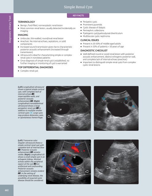
Simple Renal Cyst Diagnoses: Urinary Tract TERMINOLOGY • Benign, fluid-filled, nonneoplastic renal lesion • Most common renal lesion, usually detected incidentally on imaging IMAGING • Unilocular, thin-walled, round/oval renal lesion • Anechoic: No internal echoes, septations, or solid components • Increased sound transmission gives rise to characteristic posterior acoustic enhancement (increased through transmission) • Ultrasound is ideal for characterizing simple or complex renal cysts in nonobese patients • Once diagnosis of simple renal cyst is established, no further imaging or monitoring of cyst is warranted TOP DIFFERENTIAL DIAGNOSES • Complex renal cyst KEY FACTS • Peripelvic cysts • Prominent pyramids • Cystic disease of dialysis • Perinephric collections • Pyelogenic cyst/pyelocalyceal diverticulum • Multilocular cystic nephroma CLINICAL ISSUES • Present in 20-30% of middle-aged adults • Present in 50% of patients > 50 years of age DIAGNOSTIC CHECKLIST • Well-defined round or ovoid renal lesion with posterior acoustic enhancement, distinct echogenic posterior wall, and complete lack of internal echoes (anechoic) • Important to distinguish simple renal cysts from complex cystic renal lesions (Left) Longitudinal ultrasound shows a typical simple cortical cyst, with complete lack of internal echoes ſt, imperceptible walls, and posterior acoustic enhancement . (Right) Longitudinal transabdominal ultrasound shows a large parapelvic renal cyst ſt in addition to multiple smaller cortical cysts . Large cysts may produce distension, pain, or spontaneous hemorrhage. (Left) Transverse color Doppler ultrasound shows a simple cortical renal cyst with complete lack of internal flow ſt. Posterior acoustic enhancement remains visible. (Right) Longitudinal US shows a small simple cyst in an echogenic kidney. Minimal echoes within the peripheral aspect of the cyst st are artifactual and confounding. Posterior acoustic enhancement remains evident . In the setting of underlying renal disease, the possibility of acquired cystic disease should be considered. 462
- Page 432 and 433: Splenic Tumors (Left) Longitudinal
- Page 434 and 435: Splenic Tumors (Left) A solid, hypo
- Page 436 and 437: Splenic Infarct TERMINOLOGY Abbrevi
- Page 438 and 439: Splenic Infarct (Left) Power Dopple
- Page 440 and 441: Vascular Conditions Renal Artery St
- Page 442 and 443: Approach to Urinary Tract Sonograph
- Page 444 and 445: Approach to Urinary Tract Sonograph
- Page 446 and 447: Column of Bertin, Kidney TERMINOLOG
- Page 448 and 449: Renal Junction Line TERMINOLOGY Syn
- Page 450 and 451: Renal Ectopia TERMINOLOGY Abbreviat
- Page 452 and 453: Renal Ectopia (Left) Grayscale ultr
- Page 454 and 455: Horseshoe Kidney TERMINOLOGY Defini
- Page 456 and 457: Horseshoe Kidney (Left) Longitudina
- Page 458 and 459: Ureteral Duplication TERMINOLOGY Sy
- Page 460 and 461: Ureteral Duplication (Left) Longitu
- Page 462 and 463: Ureteral Ectopia TERMINOLOGY Abbrev
- Page 464 and 465: Ureteral Ectopia (Left) Coronal T2
- Page 466 and 467: Ureteropelvic Junction Obstruction
- Page 468 and 469: Ureteropelvic Junction Obstruction
- Page 470 and 471: Urolithiasis TERMINOLOGY Abbreviati
- Page 472 and 473: Urolithiasis (Left) Longitudinal US
- Page 474 and 475: Urolithiasis (Left) Intravenous pye
- Page 476 and 477: Nephrocalcinosis TERMINOLOGY Abbrev
- Page 478 and 479: Nephrocalcinosis (Left) Coronal MIP
- Page 480 and 481: Hydronephrosis TERMINOLOGY Synonyms
- Page 484 and 485: Simple Renal Cyst TERMINOLOGY Defin
- Page 486 and 487: Simple Renal Cyst (Left) Longitudin
- Page 488 and 489: Complex Renal Cyst TERMINOLOGY Defi
- Page 490 and 491: Complex Renal Cyst (Left) Transvers
- Page 492 and 493: Cystic Disease of Dialysis TERMINOL
- Page 494 and 495: Cystic Disease of Dialysis (Left) L
- Page 496 and 497: Multilocular Cystic Nephroma TERMIN
- Page 498 and 499: Acute Pyelonephritis TERMINOLOGY Ab
- Page 500 and 501: Acute Pyelonephritis (Left) Longitu
- Page 502 and 503: Renal Abscess TERMINOLOGY Definitio
- Page 504 and 505: Emphysematous Pyelonephritis TERMIN
- Page 506 and 507: Emphysematous Pyelonephritis (Left)
- Page 508 and 509: Pyonephrosis TERMINOLOGY Definition
- Page 510 and 511: Xanthogranulomatous Pyelonephritis
- Page 512 and 513: Tuberculosis, Urinary Tract TERMINO
- Page 514 and 515: Tuberculosis, Urinary Tract (Left)
- Page 516 and 517: Renal Cell Carcinoma TERMINOLOGY Ab
- Page 518 and 519: Renal Cell Carcinoma (Left) Longitu
- Page 520 and 521: Renal Metastases IMAGING General Fe
- Page 522 and 523: Renal Angiomyolipoma TERMINOLOGY Ab
- Page 524 and 525: Renal Angiomyolipoma (Left) Longitu
- Page 526 and 527: Upper Tract Urothelial Carcinoma TE
- Page 528 and 529: Upper Tract Urothelial Carcinoma (L
- Page 530 and 531: Renal Lymphoma TERMINOLOGY Abbrevia
Simple Renal Cyst<br />
Diagnoses: Urinary Tract<br />
TERMINOLOGY<br />
• Benign, fluid-filled, nonneoplastic renal lesion<br />
• Most common renal lesion, usually detected incidentally on<br />
imaging<br />
IMAGING<br />
• Unilocular, thin-walled, round/oval renal lesion<br />
• Anechoic: No internal echoes, septations, or solid<br />
components<br />
• Increased sound transmission gives rise to characteristic<br />
posterior acoustic enhancement (increased through<br />
transmission)<br />
• <strong>Ultrasound</strong> is ideal for characterizing simple or complex<br />
renal cysts in nonobese patients<br />
• Once diagnosis of simple renal cyst is established, no<br />
further imaging or monitoring of cyst is warranted<br />
TOP DIFFERENTIAL DIAGNOSES<br />
• Complex renal cyst<br />
KEY FACTS<br />
• Peripelvic cysts<br />
• Prominent pyramids<br />
• Cystic disease of dialysis<br />
• Perinephric collections<br />
• Pyelogenic cyst/pyelocalyceal diverticulum<br />
• Multilocular cystic nephroma<br />
CLINICAL ISSUES<br />
• Present in 20-30% of middle-aged adults<br />
• Present in 50% of patients > 50 years of age<br />
DIAGNOSTIC CHECKLIST<br />
• Well-defined round or ovoid renal lesion with posterior<br />
acoustic enhancement, distinct echogenic posterior wall,<br />
<strong>and</strong> complete lack of internal echoes (anechoic)<br />
• Important to distinguish simple renal cysts from complex<br />
cystic renal lesions<br />
(Left) Longitudinal ultrasound<br />
shows a typical simple cortical<br />
cyst, with complete lack of<br />
internal echoes ſt,<br />
imperceptible walls, <strong>and</strong><br />
posterior acoustic<br />
enhancement . (Right)<br />
Longitudinal transabdominal<br />
ultrasound shows a large<br />
parapelvic renal cyst ſt in<br />
addition to multiple smaller<br />
cortical cysts . Large cysts<br />
may produce distension, pain,<br />
or spontaneous hemorrhage.<br />
(Left) Transverse color<br />
Doppler ultrasound shows a<br />
simple cortical renal cyst with<br />
complete lack of internal flow<br />
ſt. Posterior acoustic<br />
enhancement remains<br />
visible. (Right) Longitudinal US<br />
shows a small simple cyst in an<br />
echogenic kidney. Minimal<br />
echoes within the peripheral<br />
aspect of the cyst st are<br />
artifactual <strong>and</strong> confounding.<br />
Posterior acoustic<br />
enhancement remains evident<br />
. In the setting of<br />
underlying renal disease, the<br />
possibility of acquired cystic<br />
disease should be considered.<br />
462


