Diagnostic Ultrasound - Abdomen and Pelvis
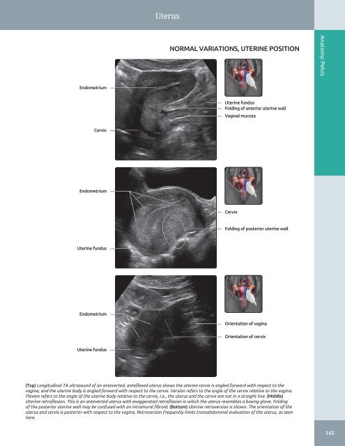
Uterus NORMAL VARIATIONS, UTERINE POSITION Anatomy: Pelvis Endometrium Uterine fundus Folding of anterior uterine wall Vaginal mucosa Cervix Endometrium Cervix Folding of posterior uterine wall Uterine fundus Endometrium Orientation of vagina Orientation of cervix Uterine fundus (Top) Longitudinal TA ultrasound of an anteverted, anteflexed uterus shows the uterine cervix is angled forward with respect to the vagina, and the uterine body is angled forward with respect to the cervix. Version refers to the angle of the cervix relative to the vagina. Flexion refers to the angle of the uterine body relative to the cervix, i.e., the uterus and the cervix are not in a straight line. (Middle) Uterine retroflexion. This is an anteverted uterus with exaggerated retroflexion in which the uterus resembles a boxing glove. Folding of the posterior uterine wall may be confused with an intramural fibroid. (Bottom) Uterine retroversion is shown. The orientation of the uterus and cervix is posterior with respect to the vagina. Retroversion frequently limits transabdominal evaluation of the uterus, as seen here. 143
Uterus Anatomy: Pelvis UTERINE VARIATIONS WITH AGE Vagina Uterine body Cervix Uterine body Cervix Uterine body Cervix (Top) Longitudinal TA ultrasound shows an immediate neonatal uterus (day 2). The uterus is prominent with a bulbous cervix and a rudimentary body. The endometrium is seen as a thin, echogenic line, which may be due to stimulation by the residual maternal hormones. (Middle) Longitudinal TA ultrasound shows a prepubertal uterus in a patient 8 years old. The uterus demonstrates a tubular appearance with the length of the cervix nearly double that of the uterine body. (Bottom) Longitudinal TA ultrasound shows an early pubertal uterus in a patient 12 years old. The body length of the uterus approximates the cervical length with the endometrium, changing in appearance and thickness during the menstrual cycle. At this time, the uterine body grows dramatically until it reaches the adult size. 144
- Page 114 and 115: Peritoneal Spaces and Structures Li
- Page 116 and 117: Peritoneal Spaces and Structures IN
- Page 118 and 119: Abdominal Wall ANTERIOR ABDOMINAL W
- Page 120 and 121: Abdominal Wall MUSCLES OF BACK IN S
- Page 122 and 123: Abdominal Wall Subcutaneous fat Rig
- Page 124 and 125: Abdominal Wall Right lobe of liver
- Page 126 and 127: Abdominal Wall Right rectus abdomin
- Page 128 and 129: Abdominal Wall Subcutaneous fat Rig
- Page 130 and 131: PART I SECTION 2 Pelvis Ureters and
- Page 132 and 133: Ureters and Bladder - Distended bla
- Page 134 and 135: Ureters and Bladder URINARY BLADDER
- Page 136 and 137: Ureters and Bladder CT UROGRAM CORR
- Page 138 and 139: Ureters and Bladder Liver URETER An
- Page 140 and 141: Ureters and Bladder WEIGERT-MEYER L
- Page 142 and 143: Prostate ○ Sac-like structures su
- Page 144 and 145: Prostate ZONAL ANATOMY OF THE PROST
- Page 146 and 147: Prostate SEMINAL VESICLES AND VAS D
- Page 148 and 149: Prostate PROSTATE ANATOMY Anatomy:
- Page 150 and 151: Testes ○ Internal oblique muscle
- Page 152 and 153: Testes EPIDIDYMIS AND SCROTAL WALL
- Page 154 and 155: Testes TESTIS, SAGITTAL VIEW Anatom
- Page 156 and 157: Testes Scrotal wall EPIDIDYMIS, HEA
- Page 158 and 159: Testes TESTICULAR AND EPIDIDYMAL AP
- Page 160 and 161: Testes ARTERIAL AND VENOUS SUPPLY A
- Page 162 and 163: Uterus ARTERIES OF UTERUS AND ADJAC
- Page 166 and 167: Uterus UTERINE VARIATIONS WITH AGE
- Page 168 and 169: Uterus CYCLIC CHANGES OF ENDOMETRIU
- Page 170 and 171: Uterus FALLOPIAN TUBE Anatomy: Pelv
- Page 172 and 173: Cervix GRAPHICS OF CERVIX ANATOMY A
- Page 174 and 175: Cervix TRANSVAGINAL ULTRASOUND OF C
- Page 176 and 177: Cervix CHANGES OF CERVIX DURING PRE
- Page 178 and 179: Vagina GRAPHICS OF NORMAL VAGINAL A
- Page 180 and 181: Vagina Urinary bladder TRANSVERSE U
- Page 182 and 183: Vagina SPECTRAL WAVEFORM OF VAGINAL
- Page 184 and 185: Ovaries Mesosalpinx LIGAMENTOUS SUP
- Page 186 and 187: Ovaries Transvaginal transducer NOR
- Page 188 and 189: Ovaries Ovarian artery SPECTRAL WAV
- Page 190 and 191: Ovaries CYCLIC CHANGES OF OVARY Ana
- Page 192 and 193: Ovaries CYCLIC CHANGES OF INTRAOVAR
- Page 194 and 195: Liver Transplant Hepatic Artery Ste
- Page 196 and 197: Approach to Hepatic Sonography (Lef
- Page 198 and 199: Approach to Hepatic Sonography (Lef
- Page 200 and 201: Acute Hepatitis TERMINOLOGY Definit
- Page 202 and 203: Acute Hepatitis (Left) Transverse g
- Page 204 and 205: Hepatic Cirrhosis TERMINOLOGY Defin
- Page 206 and 207: Hepatic Cirrhosis (Left) Longitudin
- Page 208 and 209: Hepatic Steatosis TERMINOLOGY Synon
- Page 210 and 211: Hepatic Steatosis (Left) Transverse
- Page 212 and 213: Hepatic Schistosomiasis TERMINOLOGY
Uterus<br />
NORMAL VARIATIONS, UTERINE POSITION<br />
Anatomy: <strong>Pelvis</strong><br />
Endometrium<br />
Uterine fundus<br />
Folding of anterior uterine wall<br />
Vaginal mucosa<br />
Cervix<br />
Endometrium<br />
Cervix<br />
Folding of posterior uterine wall<br />
Uterine fundus<br />
Endometrium<br />
Orientation of vagina<br />
Orientation of cervix<br />
Uterine fundus<br />
(Top) Longitudinal TA ultrasound of an anteverted, anteflexed uterus shows the uterine cervix is angled forward with respect to the<br />
vagina, <strong>and</strong> the uterine body is angled forward with respect to the cervix. Version refers to the angle of the cervix relative to the vagina.<br />
Flexion refers to the angle of the uterine body relative to the cervix, i.e., the uterus <strong>and</strong> the cervix are not in a straight line. (Middle)<br />
Uterine retroflexion. This is an anteverted uterus with exaggerated retroflexion in which the uterus resembles a boxing glove. Folding<br />
of the posterior uterine wall may be confused with an intramural fibroid. (Bottom) Uterine retroversion is shown. The orientation of the<br />
uterus <strong>and</strong> cervix is posterior with respect to the vagina. Retroversion frequently limits transabdominal evaluation of the uterus, as seen<br />
here.<br />
143


