Diagnostic Ultrasound - Abdomen and Pelvis
Testes TESTICULAR AND EPIDIDYMAL APPENDAGE Anatomy: Pelvis Testicular appendages Epididymal head Cystic epididymal appendix Epididymal head Torsed epididymal appendix (Top) Longitudinal grayscale ultrasound of the testis in a patient with a hydrocele shows 2 small nodular protuberances from the surface of the testis, isoechoic to normal testicular parenchyma. This is the appendix testis, which is a remnant of the müllerian system. (Middle) Transverse grayscale ultrasound of the epididymal head in a patient with a small hydrocele shows cystic protuberance from the surface of the epididymis. It is isoechoic to normal testicular parenchyma. This is the appendix testis, which is a remnant of the müllerian system. (Bottom) Sagittal color Doppler ultrasound of the left epididymal head shows an exophytically arising heterogeneous lesion without internal vascularity. The patient presented with acute scrotal pain. These findings are suggestive of a torsed epididymal appendix. 137
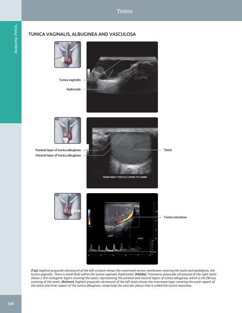
Testes Anatomy: Pelvis TUNICA VAGINALIS, ALBUGINEA AND VASCULOSA Tunica vaginalis Hydrocele Parietal layer of tunica albuginea Visceral layer of tunica albuginea Testis Tunica vasculosa (Top) Sagittal grayscale ultrasound of the left scrotum shows the outermost serous membrane covering the testis and epididymis, the tunica vaginalis. There is small fluid within the tunica vaginalis (hydrocele). (Middle) Transverse grayscale ultrasound of the right testis shows 2 thin echogenic layers covering the testis, representing the parietal and visceral layers of tunica albuginea, which is the fibrous covering of the testis. (Bottom) Sagittal grayscale ultrasound of the left testis shows the innermost layer covering the outer aspect of the testis and inner aspect of the tunica albuginea, comprising the vascular plexus that is called the tunica vasculosa. 138
- Page 108 and 109: Abdominal Lymph Nodes LYMPHANGIOGRA
- Page 110 and 111: Peritoneal Spaces and Structures PE
- Page 112 and 113: Peritoneal Spaces and Structures PE
- Page 114 and 115: Peritoneal Spaces and Structures Li
- Page 116 and 117: Peritoneal Spaces and Structures IN
- Page 118 and 119: Abdominal Wall ANTERIOR ABDOMINAL W
- Page 120 and 121: Abdominal Wall MUSCLES OF BACK IN S
- Page 122 and 123: Abdominal Wall Subcutaneous fat Rig
- Page 124 and 125: Abdominal Wall Right lobe of liver
- Page 126 and 127: Abdominal Wall Right rectus abdomin
- Page 128 and 129: Abdominal Wall Subcutaneous fat Rig
- Page 130 and 131: PART I SECTION 2 Pelvis Ureters and
- Page 132 and 133: Ureters and Bladder - Distended bla
- Page 134 and 135: Ureters and Bladder URINARY BLADDER
- Page 136 and 137: Ureters and Bladder CT UROGRAM CORR
- Page 138 and 139: Ureters and Bladder Liver URETER An
- Page 140 and 141: Ureters and Bladder WEIGERT-MEYER L
- Page 142 and 143: Prostate ○ Sac-like structures su
- Page 144 and 145: Prostate ZONAL ANATOMY OF THE PROST
- Page 146 and 147: Prostate SEMINAL VESICLES AND VAS D
- Page 148 and 149: Prostate PROSTATE ANATOMY Anatomy:
- Page 150 and 151: Testes ○ Internal oblique muscle
- Page 152 and 153: Testes EPIDIDYMIS AND SCROTAL WALL
- Page 154 and 155: Testes TESTIS, SAGITTAL VIEW Anatom
- Page 156 and 157: Testes Scrotal wall EPIDIDYMIS, HEA
- Page 160 and 161: Testes ARTERIAL AND VENOUS SUPPLY A
- Page 162 and 163: Uterus ARTERIES OF UTERUS AND ADJAC
- Page 164 and 165: Uterus NORMAL VARIATIONS, UTERINE P
- Page 166 and 167: Uterus UTERINE VARIATIONS WITH AGE
- Page 168 and 169: Uterus CYCLIC CHANGES OF ENDOMETRIU
- Page 170 and 171: Uterus FALLOPIAN TUBE Anatomy: Pelv
- Page 172 and 173: Cervix GRAPHICS OF CERVIX ANATOMY A
- Page 174 and 175: Cervix TRANSVAGINAL ULTRASOUND OF C
- Page 176 and 177: Cervix CHANGES OF CERVIX DURING PRE
- Page 178 and 179: Vagina GRAPHICS OF NORMAL VAGINAL A
- Page 180 and 181: Vagina Urinary bladder TRANSVERSE U
- Page 182 and 183: Vagina SPECTRAL WAVEFORM OF VAGINAL
- Page 184 and 185: Ovaries Mesosalpinx LIGAMENTOUS SUP
- Page 186 and 187: Ovaries Transvaginal transducer NOR
- Page 188 and 189: Ovaries Ovarian artery SPECTRAL WAV
- Page 190 and 191: Ovaries CYCLIC CHANGES OF OVARY Ana
- Page 192 and 193: Ovaries CYCLIC CHANGES OF INTRAOVAR
- Page 194 and 195: Liver Transplant Hepatic Artery Ste
- Page 196 and 197: Approach to Hepatic Sonography (Lef
- Page 198 and 199: Approach to Hepatic Sonography (Lef
- Page 200 and 201: Acute Hepatitis TERMINOLOGY Definit
- Page 202 and 203: Acute Hepatitis (Left) Transverse g
- Page 204 and 205: Hepatic Cirrhosis TERMINOLOGY Defin
- Page 206 and 207: Hepatic Cirrhosis (Left) Longitudin
Testes<br />
Anatomy: <strong>Pelvis</strong><br />
TUNICA VAGINALIS, ALBUGINEA AND VASCULOSA<br />
Tunica vaginalis<br />
Hydrocele<br />
Parietal layer of tunica albuginea<br />
Visceral layer of tunica albuginea<br />
Testis<br />
Tunica vasculosa<br />
(Top) Sagittal grayscale ultrasound of the left scrotum shows the outermost serous membrane covering the testis <strong>and</strong> epididymis, the<br />
tunica vaginalis. There is small fluid within the tunica vaginalis (hydrocele). (Middle) Transverse grayscale ultrasound of the right testis<br />
shows 2 thin echogenic layers covering the testis, representing the parietal <strong>and</strong> visceral layers of tunica albuginea, which is the fibrous<br />
covering of the testis. (Bottom) Sagittal grayscale ultrasound of the left testis shows the innermost layer covering the outer aspect of<br />
the testis <strong>and</strong> inner aspect of the tunica albuginea, comprising the vascular plexus that is called the tunica vasculosa.<br />
138


