Diagnostic Ultrasound - Abdomen and Pelvis
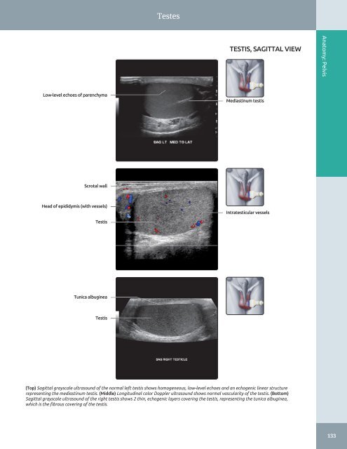
Testes TESTIS, SAGITTAL VIEW Anatomy: Pelvis Low-level echoes of parenchyma Mediastinum testis Scrotal wall Head of epididymis (with vessels) Intratesticular vessels Testis Tunica albuginea Testis (Top) Sagittal grayscale ultrasound of the normal left testis shows homogeneous, low-level echoes and an echogenic linear structure representing the mediastinum testis. (Middle) Longitudinal color Doppler ultrasound shows normal vascularity of the testis. (Bottom) Sagittal grayscale ultrasound of the right testis shows 2 thin, echogenic layers covering the testis, representing the tunica albuginea, which is the fibrous covering of the testis. 133
Testes Anatomy: Pelvis TESTIS, TRANSVERSE VIEW Tunica albuginea Rete testis Mediastinum testis Scrotal wall Epididymis, body Vessels in pampiniform plexus Right testis Transmediastinal artery Low resistance arterial flow (Top) Transverse grayscale ultrasound of the normal testis shows diffuse low-level internal echoes in the parenchyma, mediastinum testis, and the striated pattern of rete testis. The testis is covered with 2 thin, echogenic layers of tunica albuginea. (Middle) Transverse color Doppler ultrasound again shows normal paucity of testicular vascularity. Some vessels are also identified in the epididymis. More vessels can be identified along the mediastinum testis. (Bottom) Pulsed Doppler ultrasound of an intratesticular artery and the transmediastinal artery shows low resistance with low systolic and end-diastolic flow velocities. 134
- Page 104 and 105: Bowel Urinary bladder RECTOSIGMOID
- Page 106 and 107: Abdominal Lymph Nodes RETROPERITONE
- Page 108 and 109: Abdominal Lymph Nodes LYMPHANGIOGRA
- Page 110 and 111: Peritoneal Spaces and Structures PE
- Page 112 and 113: Peritoneal Spaces and Structures PE
- Page 114 and 115: Peritoneal Spaces and Structures Li
- Page 116 and 117: Peritoneal Spaces and Structures IN
- Page 118 and 119: Abdominal Wall ANTERIOR ABDOMINAL W
- Page 120 and 121: Abdominal Wall MUSCLES OF BACK IN S
- Page 122 and 123: Abdominal Wall Subcutaneous fat Rig
- Page 124 and 125: Abdominal Wall Right lobe of liver
- Page 126 and 127: Abdominal Wall Right rectus abdomin
- Page 128 and 129: Abdominal Wall Subcutaneous fat Rig
- Page 130 and 131: PART I SECTION 2 Pelvis Ureters and
- Page 132 and 133: Ureters and Bladder - Distended bla
- Page 134 and 135: Ureters and Bladder URINARY BLADDER
- Page 136 and 137: Ureters and Bladder CT UROGRAM CORR
- Page 138 and 139: Ureters and Bladder Liver URETER An
- Page 140 and 141: Ureters and Bladder WEIGERT-MEYER L
- Page 142 and 143: Prostate ○ Sac-like structures su
- Page 144 and 145: Prostate ZONAL ANATOMY OF THE PROST
- Page 146 and 147: Prostate SEMINAL VESICLES AND VAS D
- Page 148 and 149: Prostate PROSTATE ANATOMY Anatomy:
- Page 150 and 151: Testes ○ Internal oblique muscle
- Page 152 and 153: Testes EPIDIDYMIS AND SCROTAL WALL
- Page 156 and 157: Testes Scrotal wall EPIDIDYMIS, HEA
- Page 158 and 159: Testes TESTICULAR AND EPIDIDYMAL AP
- Page 160 and 161: Testes ARTERIAL AND VENOUS SUPPLY A
- Page 162 and 163: Uterus ARTERIES OF UTERUS AND ADJAC
- Page 164 and 165: Uterus NORMAL VARIATIONS, UTERINE P
- Page 166 and 167: Uterus UTERINE VARIATIONS WITH AGE
- Page 168 and 169: Uterus CYCLIC CHANGES OF ENDOMETRIU
- Page 170 and 171: Uterus FALLOPIAN TUBE Anatomy: Pelv
- Page 172 and 173: Cervix GRAPHICS OF CERVIX ANATOMY A
- Page 174 and 175: Cervix TRANSVAGINAL ULTRASOUND OF C
- Page 176 and 177: Cervix CHANGES OF CERVIX DURING PRE
- Page 178 and 179: Vagina GRAPHICS OF NORMAL VAGINAL A
- Page 180 and 181: Vagina Urinary bladder TRANSVERSE U
- Page 182 and 183: Vagina SPECTRAL WAVEFORM OF VAGINAL
- Page 184 and 185: Ovaries Mesosalpinx LIGAMENTOUS SUP
- Page 186 and 187: Ovaries Transvaginal transducer NOR
- Page 188 and 189: Ovaries Ovarian artery SPECTRAL WAV
- Page 190 and 191: Ovaries CYCLIC CHANGES OF OVARY Ana
- Page 192 and 193: Ovaries CYCLIC CHANGES OF INTRAOVAR
- Page 194 and 195: Liver Transplant Hepatic Artery Ste
- Page 196 and 197: Approach to Hepatic Sonography (Lef
- Page 198 and 199: Approach to Hepatic Sonography (Lef
- Page 200 and 201: Acute Hepatitis TERMINOLOGY Definit
- Page 202 and 203: Acute Hepatitis (Left) Transverse g
Testes<br />
TESTIS, SAGITTAL VIEW<br />
Anatomy: <strong>Pelvis</strong><br />
Low-level echoes of parenchyma<br />
Mediastinum testis<br />
Scrotal wall<br />
Head of epididymis (with vessels)<br />
Intratesticular vessels<br />
Testis<br />
Tunica albuginea<br />
Testis<br />
(Top) Sagittal grayscale ultrasound of the normal left testis shows homogeneous, low-level echoes <strong>and</strong> an echogenic linear structure<br />
representing the mediastinum testis. (Middle) Longitudinal color Doppler ultrasound shows normal vascularity of the testis. (Bottom)<br />
Sagittal grayscale ultrasound of the right testis shows 2 thin, echogenic layers covering the testis, representing the tunica albuginea,<br />
which is the fibrous covering of the testis.<br />
133


