Diagnostic Ultrasound - Abdomen and Pelvis
Abdominal Wall MUSCLES OF BACK IN SITU Anatomy: Abdomen Spinous process Spinalis thoracis muscle Longissimus thoracis muscle Iliocostalis muscle Serratus posterior inferior muscle Transversus abdominis (muscle and tendon) Internal oblique muscle External oblique muscle Iliac crest Graphic shows the paraspinal muscles and muscles of the back. The latissimus dorsi muscles are not included. The erector spinae have thick tendinous origins from the sacral and iliac crests and the lumbar and 11th to 12th thoracic spinous processes. Superiorly, the muscle becomes fleshy, and in the upper lumbar region subdivides to become the iliocostalis, longissimus, and spinalis muscles (from lateral to medial), tapering as they insert into the vertebrae and ribs. The erector muscles flank the spinous processes and span the length of the posterior thorax and abdomen. They are responsible for extension of the vertebral column. 99
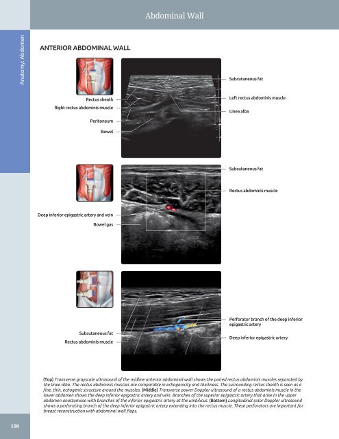
Abdominal Wall Anatomy: Abdomen ANTERIOR ABDOMINAL WALL Subcutaneous fat Rectus sheath Right rectus abdominis muscle Peritoneum Bowel Left rectus abdominis muscle Linea alba Subcutaneous fat Rectus abdominis muscle Deep inferior epigastric artery and vein Bowel gas Subcutaneous fat Rectus abdominis muscle Perforator branch of the deep inferior epigastric artery Deep inferior epigastric artery (Top) Transverse grayscale ultrasound of the midline anterior abdominal wall shows the paired rectus abdominis muscles separated by the linea alba. The rectus abdominis muscles are comparable in echogenicity and thickness. The surrounding rectus sheath is seen as a fine, thin, echogenic structure around the muscles. (Middle) Transverse power Doppler ultrasound of a rectus abdominis muscle in the lower abdomen shows the deep inferior epigastric artery and vein. Branches of the superior epigastric artery that arise in the upper abdomen anastomose with branches of the inferior epigastric artery at the umbilicus. (Bottom) Longitudinal color Doppler ultrasound shows a perforating branch of the deep inferior epigastric artery extending into the rectus muscle. These perforators are important for breast reconstruction with abdominal wall flaps. 100
- Page 70 and 71: Kidneys RENAL FASCIA AND PERIRENAL
- Page 72 and 73: Kidneys Right hemidiaphragm RIGHT K
- Page 74 and 75: Kidneys RIGHT KIDNEY, CT CORRELATIO
- Page 76 and 77: Kidneys Right erector spinae muscle
- Page 78 and 79: Kidneys RIGHT INTRARENAL ARTERY AND
- Page 80 and 81: Kidneys LEFT KIDNEY, CT CORRELATION
- Page 82 and 83: Kidneys LEFT KIDNEY, CT CORRELATION
- Page 84 and 85: Kidneys Subcutaneous fat Left latis
- Page 86 and 87: Kidneys LEFT MAIN RENAL ARTERY AND
- Page 88 and 89: Kidneys Right lobe of liver MULTIPL
- Page 90 and 91: Bowel - Forms an incomplete ring in
- Page 92 and 93: Bowel Falciform ligament STOMACH AN
- Page 94 and 95: Bowel SMALL INTESTINE Anatomy: Abdo
- Page 96 and 97: Bowel Abdominal wall STOMACH Anatom
- Page 98 and 99: Bowel Rectus muscle SMALL BOWEL Ana
- Page 100 and 101: Bowel Abdominal wall musculature Ce
- Page 102 and 103: Bowel LARGE BOWEL Abdominal wall mu
- Page 104 and 105: Bowel Urinary bladder RECTOSIGMOID
- Page 106 and 107: Abdominal Lymph Nodes RETROPERITONE
- Page 108 and 109: Abdominal Lymph Nodes LYMPHANGIOGRA
- Page 110 and 111: Peritoneal Spaces and Structures PE
- Page 112 and 113: Peritoneal Spaces and Structures PE
- Page 114 and 115: Peritoneal Spaces and Structures Li
- Page 116 and 117: Peritoneal Spaces and Structures IN
- Page 118 and 119: Abdominal Wall ANTERIOR ABDOMINAL W
- Page 122 and 123: Abdominal Wall Subcutaneous fat Rig
- Page 124 and 125: Abdominal Wall Right lobe of liver
- Page 126 and 127: Abdominal Wall Right rectus abdomin
- Page 128 and 129: Abdominal Wall Subcutaneous fat Rig
- Page 130 and 131: PART I SECTION 2 Pelvis Ureters and
- Page 132 and 133: Ureters and Bladder - Distended bla
- Page 134 and 135: Ureters and Bladder URINARY BLADDER
- Page 136 and 137: Ureters and Bladder CT UROGRAM CORR
- Page 138 and 139: Ureters and Bladder Liver URETER An
- Page 140 and 141: Ureters and Bladder WEIGERT-MEYER L
- Page 142 and 143: Prostate ○ Sac-like structures su
- Page 144 and 145: Prostate ZONAL ANATOMY OF THE PROST
- Page 146 and 147: Prostate SEMINAL VESICLES AND VAS D
- Page 148 and 149: Prostate PROSTATE ANATOMY Anatomy:
- Page 150 and 151: Testes ○ Internal oblique muscle
- Page 152 and 153: Testes EPIDIDYMIS AND SCROTAL WALL
- Page 154 and 155: Testes TESTIS, SAGITTAL VIEW Anatom
- Page 156 and 157: Testes Scrotal wall EPIDIDYMIS, HEA
- Page 158 and 159: Testes TESTICULAR AND EPIDIDYMAL AP
- Page 160 and 161: Testes ARTERIAL AND VENOUS SUPPLY A
- Page 162 and 163: Uterus ARTERIES OF UTERUS AND ADJAC
- Page 164 and 165: Uterus NORMAL VARIATIONS, UTERINE P
- Page 166 and 167: Uterus UTERINE VARIATIONS WITH AGE
- Page 168 and 169: Uterus CYCLIC CHANGES OF ENDOMETRIU
Abdominal Wall<br />
Anatomy: <strong>Abdomen</strong><br />
ANTERIOR ABDOMINAL WALL<br />
Subcutaneous fat<br />
Rectus sheath<br />
Right rectus abdominis muscle<br />
Peritoneum<br />
Bowel<br />
Left rectus abdominis muscle<br />
Linea alba<br />
Subcutaneous fat<br />
Rectus abdominis muscle<br />
Deep inferior epigastric artery <strong>and</strong> vein<br />
Bowel gas<br />
Subcutaneous fat<br />
Rectus abdominis muscle<br />
Perforator branch of the deep inferior<br />
epigastric artery<br />
Deep inferior epigastric artery<br />
(Top) Transverse grayscale ultrasound of the midline anterior abdominal wall shows the paired rectus abdominis muscles separated by<br />
the linea alba. The rectus abdominis muscles are comparable in echogenicity <strong>and</strong> thickness. The surrounding rectus sheath is seen as a<br />
fine, thin, echogenic structure around the muscles. (Middle) Transverse power Doppler ultrasound of a rectus abdominis muscle in the<br />
lower abdomen shows the deep inferior epigastric artery <strong>and</strong> vein. Branches of the superior epigastric artery that arise in the upper<br />
abdomen anastomose with branches of the inferior epigastric artery at the umbilicus. (Bottom) Longitudinal color Doppler ultrasound<br />
shows a perforating branch of the deep inferior epigastric artery extending into the rectus muscle. These perforators are important for<br />
breast reconstruction with abdominal wall flaps.<br />
100


