Diagnostic Ultrasound - Abdomen and Pelvis
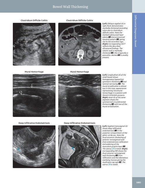
Bowel Wall Thickening Clostridium Difficile Colitis Clostridium Difficile Colitis (Left) Oblique sagittal US in right flank demonstrates markedly thickened ascending colon due to clostridium difficile colitis. Note the marked submucosal layer edema ſt and pronounced haustral pattern st, giving rise to the accordion sign. (Right) Corresponding CECT reflects the described ultrasound findings. The colonic wall is markedly thickened ſt; this is typically a pancolitis. Ascites is usually present. Differential Diagnoses: Bowel Mural Hemorrhage Mural Hemorrhage (Left) Longitudinal US of the small bowel shows homogeneous hypoechoic symmetric thickening ſt and luminal narrowing . The mural stratification is almost lost in this case, appearances representing intramural hemorrhage in a patient with Henoch-Schönlein purpura. (Right) Axial US of the same segment shows the symmetrical circumferential thickening ſt with loss of the mural stratification. Deep Infiltrative Endometriosis Deep Infiltrative Endometriosis (Left) Sagittal transvaginal US shows deep infiltrative endometriosis ſt in the posterior compartment of the pelvic cul-de-sac. Note the focal eccentric thickening of anterior rectosigmoid wall from endometriosis infiltration and widening of the muscularis propria layer . The uterus is noted. (Right) Corresponding MR shows the focal anterior rectosigmoid wall thickening ſt from infiltration and the edematous overlying mucosa giving the mushroom cap sign . The uterus is noted. 1005
This page intentionally left blank
- Page 976 and 977: Small Kidney Postobstructive Atroph
- Page 978 and 979: Hypoechoic Kidney • Multiple Myel
- Page 980 and 981: Hypoechoic Kidney Acute Renal Arter
- Page 982 and 983: Hyperechoic Kidney ○ Echogenic co
- Page 984 and 985: Hyperechoic Kidney Chronic Glomerul
- Page 986 and 987: Cystic Renal Mass ○ Associated wi
- Page 988 and 989: Cystic Renal Mass Multicystic Dyspl
- Page 990 and 991: Solid Renal Mass • Horseshoe Kidn
- Page 992 and 993: Solid Renal Mass Renal Lymphoma Ren
- Page 994 and 995: Renal Pseudotumor Column of Bertin
- Page 996 and 997: Dilated Renal Pelvis • Intrarenal
- Page 998 and 999: Dilated Renal Pelvis Pyonephrosis P
- Page 1000 and 1001: PART III SECTION 7 Abdominal Wall/P
- Page 1002 and 1003: Diffuse Peritoneal Fluid Hemoperito
- Page 1004 and 1005: Solid Peritoneal Mass - Higher dens
- Page 1006 and 1007: Solid Peritoneal Mass Mimics Benign
- Page 1008 and 1009: Cystic Peritoneal Mass ○ Women of
- Page 1010 and 1011: Cystic Peritoneal Mass Pseudomyxoma
- Page 1012 and 1013: PART III SECTION 8 Prostate Enlarge
- Page 1014 and 1015: Enlarged Prostate Benign Prostatic
- Page 1016 and 1017: Focal Lesion in Prostate ○ Variab
- Page 1018 and 1019: Focal Lesion in Prostate Müllerian
- Page 1020 and 1021: PART III SECTION 9 Bowel Bowel Wall
- Page 1022 and 1023: Bowel Wall Thickening - Distal ileu
- Page 1024 and 1025: Bowel Wall Thickening Crohn Disease
- Page 1028 and 1029: PART III SECTION 10 Scrotum 1008
- Page 1030 and 1031: Diffuse Testicular Enlargement Test
- Page 1032 and 1033: Decreased Testicular Size Testicula
- Page 1034 and 1035: Testicular Calcifications Sertoli C
- Page 1036 and 1037: Focal Testicular Mass - Most common
- Page 1038 and 1039: Focal Testicular Mass Testicular Ly
- Page 1040 and 1041: Focal Extratesticular Mass - 3-50 m
- Page 1042 and 1043: Focal Extratesticular Mass Inguinal
- Page 1044 and 1045: Focal Extratesticular Mass Liposarc
- Page 1046 and 1047: Extratesticular Cystic Mass Varicoc
- Page 1048 and 1049: PART III SECTION 11 Female Pelvis
- Page 1050 and 1051: Cystic Adnexal Mass □ Hemorrhagic
- Page 1052 and 1053: Cystic Adnexal Mass Dermoid (Mature
- Page 1054 and 1055: Solid Adnexal Mass - May masquerade
- Page 1056 and 1057: Solid Adnexal Mass Fibrothecoma Hem
- Page 1058 and 1059: Extraovarian Adnexal Mass Helpful C
- Page 1060 and 1061: Extraovarian Adnexal Mass Paraovari
- Page 1062 and 1063: Extraovarian Adnexal Mass Lymph Nod
- Page 1064 and 1065: Enlarged Ovary - Often bilateral (5
- Page 1066 and 1067: Enlarged Ovary Theca Lutein Cysts T
- Page 1068 and 1069: Enlarged Uterus Leiomyoma Adenomyos
- Page 1070 and 1071: Abnormal Endometrium ○ Multiple e
- Page 1072 and 1073: Abnormal Endometrium Pregnancy and
- Page 1074 and 1075: Abnormal Endometrium Tamoxifen-Indu
Bowel Wall Thickening<br />
Clostridium Difficile Colitis<br />
Clostridium Difficile Colitis<br />
(Left) Oblique sagittal US in<br />
right flank demonstrates<br />
markedly thickened ascending<br />
colon due to clostridium<br />
difficile colitis. Note the<br />
marked submucosal layer<br />
edema ſt <strong>and</strong> pronounced<br />
haustral pattern st, giving<br />
rise to the accordion sign.<br />
(Right) Corresponding CECT<br />
reflects the described<br />
ultrasound findings. The<br />
colonic wall is markedly<br />
thickened ſt; this is typically a<br />
pancolitis. Ascites is usually<br />
present.<br />
Differential Diagnoses: Bowel<br />
Mural Hemorrhage<br />
Mural Hemorrhage<br />
(Left) Longitudinal US of the<br />
small bowel shows<br />
homogeneous hypoechoic<br />
symmetric thickening ſt <strong>and</strong><br />
luminal narrowing . The<br />
mural stratification is almost<br />
lost in this case, appearances<br />
representing intramural<br />
hemorrhage in a patient with<br />
Henoch-Schönlein purpura.<br />
(Right) Axial US of the same<br />
segment shows the<br />
symmetrical circumferential<br />
thickening ſt with loss of the<br />
mural stratification.<br />
Deep Infiltrative Endometriosis<br />
Deep Infiltrative Endometriosis<br />
(Left) Sagittal transvaginal US<br />
shows deep infiltrative<br />
endometriosis ſt in the<br />
posterior compartment of the<br />
pelvic cul-de-sac. Note the<br />
focal eccentric thickening of<br />
anterior rectosigmoid wall<br />
from endometriosis infiltration<br />
<strong>and</strong> widening of the<br />
muscularis propria layer .<br />
The uterus is noted. (Right)<br />
Corresponding MR shows the<br />
focal anterior rectosigmoid<br />
wall thickening ſt from<br />
infiltration <strong>and</strong> the edematous<br />
overlying mucosa giving the<br />
mushroom cap sign . The<br />
uterus is noted.<br />
1005


