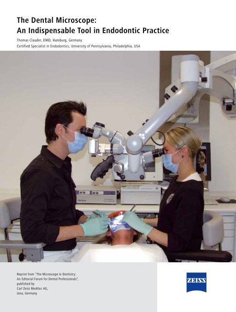
The Dental Microscope: An Indispensable Tool in Endodontic Practice
The Dental Microscope: An Indispensable Tool in Endodontic Practice
The Dental Microscope: An Indispensable Tool in Endodontic Practice

Create successful ePaper yourself
Turn your PDF publications into a flip-book with our unique Google optimized e-Paper software.
<strong>The</strong> <strong>Dental</strong> <strong>Microscope</strong>:<br />
<strong>An</strong> <strong>Indispensable</strong> <strong>Tool</strong> <strong>in</strong> <strong>Endodontic</strong> <strong>Practice</strong><br />
Thomas Clauder, DMD, Hamburg, Germany<br />
Certified Specialist <strong>in</strong> <strong>Endodontic</strong>s, University of Pennsylvania, Philadelphia, USA<br />
Repr<strong>in</strong>t from “<strong>The</strong> <strong>Microscope</strong> <strong>in</strong> Dentistry:<br />
<strong>An</strong> Editorial Forum for <strong>Dental</strong> Professionals“,<br />
published by<br />
Carl Zeiss Meditec AG,<br />
Jena, Germany
2<br />
<strong>The</strong> <strong>Dental</strong> <strong>Microscope</strong>: <strong>An</strong> <strong>Indispensable</strong> <strong>Tool</strong> <strong>in</strong> <strong>Endodontic</strong> <strong>Practice</strong><br />
Thomas Clauder, DMD<br />
<strong>The</strong> <strong>Microscope</strong> <strong>in</strong> Dentistry • <strong>Endodontic</strong>s<br />
High-quality endodontic therapy is the basis for long-term function and biologic success, ensur<strong>in</strong>g<br />
that patients rema<strong>in</strong> free of pa<strong>in</strong>. State-of-the-art equipment and thorough cl<strong>in</strong>ical know-how are<br />
vitally important to reach this goal. Today, the world‘s lead<strong>in</strong>g practic<strong>in</strong>g dentists and researchers<br />
are largely <strong>in</strong> agreement that <strong>in</strong> endodontics the dental microscope has pushed the limits of<br />
treatment potential a long way towards enhanc<strong>in</strong>g long-term patient outcomes.<br />
Nowadays, teeth that require endodontic<br />
therapy can provide a basis for many aesthetically<br />
demand<strong>in</strong>g prosthetic restorations.<br />
Rout<strong>in</strong>e endodontic practice, how-ever,<br />
confronts the practitioner with an <strong>in</strong>creas<strong>in</strong>g<br />
number of challenges (Fig. 1, 2).<br />
Fig. 1 and 2: X-ray images of S-shaped and<br />
crooked root canals<br />
For example, anatomical variations are not as<br />
rare or exotic as is frequently assumed. Walter<br />
Hess described the complex anatomy of root<br />
canals <strong>in</strong> great detail as early as 1917. 4 Subsequent<br />
anatomical studies have s<strong>in</strong>ce been<br />
published <strong>in</strong> various countries and a broad<br />
range of populations. Many of these important<br />
structures cannot be readily detected or treated<br />
with traditional endodontic treatment methods.<br />
Failures <strong>in</strong> non-surgical and surgical endodontic<br />
therapy were frequent, and they still are. This is<br />
reflected <strong>in</strong> daily dental practice and cross-sectional<br />
epidemiological studies. <strong>The</strong> discrepancy<br />
between possible successful prognosis and<br />
reality is quite substantial.<br />
<strong>The</strong> <strong>in</strong>troduction of the dental microscope and<br />
the associated ability to <strong>in</strong>spect the root canals<br />
– both orthograde and retrograde – have<br />
fundamentally changed our understand<strong>in</strong>g of<br />
dental morphology and its complexity. However,<br />
follow<strong>in</strong>g the first publications there<br />
was no widespread acceptance of microscopic<br />
techniques among dentists, until the beg<strong>in</strong>n<strong>in</strong>g<br />
of the 1990s. Well-known specialists<br />
such as Prof. Syngcuk Kim (University of<br />
Pennsylvania, Philadelphia, USA) and Dr. Gary<br />
Carr (San Diego, USA) facilitated the establish-<br />
ment and widespread use of microscopic<br />
techniques. Prof. Kim‘s motto “You can only<br />
treat what you can see!“ has made dentists all<br />
over the world enthusiastic about microscopic<br />
treatment. In 1998, the American <strong>Dental</strong><br />
Association <strong>in</strong>stitut-ed microscope proficiency<br />
as obligatory for all endodontic specialist<br />
programs <strong>in</strong> the USA.<br />
As the use of dental microscopes <strong>in</strong>creased<br />
worldwide, new <strong>in</strong>struments became established,<br />
the utilization of which greatly facilitates<br />
a considerable amount of work under the<br />
microscope. For a restorative dentist or endodontic<br />
specialist, the dental microscope offers<br />
a large number of benefits:<br />
1. Better visualization<br />
Due to the magnification, and clear coaxial<br />
illum<strong>in</strong>ation of the work<strong>in</strong>g field, it is possible<br />
to address unique or specialized treatment<br />
situations more efficiently and with greater<br />
precision.<br />
2. Improved treatment quality<br />
Microscopic techniques are superior to<br />
traditional treatment concepts, as has been<br />
proven by various studies. 1,7,8,10<br />
3. Ideal treatment ergonomics<br />
Appropriate work<strong>in</strong>g posture and ergonomics<br />
play a key role <strong>in</strong> ma<strong>in</strong>ta<strong>in</strong><strong>in</strong>g the dentist‘s<br />
own health and personal well-be<strong>in</strong>g. For some<br />
colleagues, this is the ma<strong>in</strong> criterion for daily<br />
use <strong>in</strong> their practice.<br />
4. “Fun factor“ <strong>in</strong> the practice<br />
Cl<strong>in</strong>icians that utilize a dental microscope will<br />
f<strong>in</strong>d they have more enjoyment dur<strong>in</strong>g procedures<br />
due to the ideal work<strong>in</strong>g con-ditions and<br />
the predictable treatment outcomes. <strong>The</strong>y will<br />
be more motivated as treatment is experienced<br />
more <strong>in</strong>tensely and visualization is improved<br />
consider-ably. Dentists, assisted by illum<strong>in</strong>ation,<br />
magnification, and special <strong>in</strong>struments,<br />
will also gradually experience a greater level<br />
of personal satisfaction. This is driven by<br />
their ability to recognize much greater detail,<br />
visualize many more root canals and anomalies,<br />
treat them successfully, and ultimately achieve<br />
more therapy successes, particularly those with<br />
spectacular results. <strong>The</strong> dentist can expla<strong>in</strong> this<br />
to the patient and, through enthusiasm and<br />
fasc<strong>in</strong>ation, enable him or her to participate <strong>in</strong><br />
this positive effect.<br />
In all areas, from exposure of the access<br />
cavity and preparation to three-dimen-sional<br />
obturation and postendodontic management,<br />
the microscope provides major advantages over<br />
work<strong>in</strong>g without appropriate magnification. As<br />
a result, the use of the microscope can be expressly<br />
recommended for the follow<strong>in</strong>g specific<br />
<strong>in</strong>dications and special aspects:<br />
1. Diagnosis<br />
Microfractures and longitud<strong>in</strong>al fractures are<br />
often overlooked cl<strong>in</strong>ically and represent a<br />
cause of pa<strong>in</strong> that is difficult to diagnose (Figs.<br />
3, 4). Visualization under the dental microscope<br />
is the basis for further treatment plann<strong>in</strong>g.<br />
2. Canals/canal systems<br />
that are difficult to localize. If the radiographic<br />
image is exam<strong>in</strong>ed more closely, there are often<br />
signs of unusual root and/or canal shapes<br />
like those caused by changes <strong>in</strong> the course of<br />
canal anatomy or root surface. Interruptions<br />
<strong>in</strong> the canal shape are almost always a certa<strong>in</strong><br />
<strong>in</strong>dication of canal system splitt<strong>in</strong>g. <strong>An</strong> offcenter<br />
exposure or three dimensional image<br />
can provide further valuable <strong>in</strong>formation. Threerooted<br />
premolars, for example, are encountered<br />
<strong>in</strong> 6% of all first maxillary premolars (Figs. 5,<br />
6). 2 However, anatomical variations also <strong>in</strong>clude<br />
other complex structures like C-shaped canals.<br />
In the case of second mandibular molars, they<br />
account for approximately 7.6%. However, <strong>in</strong><br />
Asian populations such as <strong>in</strong> Koreans they can<br />
reach up to 31.3% (Figs. 7, 8). 5,9,12 Treatment of<br />
this anatomical variation can be highly com
Fig. 3: Microfracture diagnosed dur<strong>in</strong>g<br />
orthograde root canal treatment<br />
Fig. 6: Buccal separation of a three-rooted<br />
premolar<br />
plex. Without a doubt, the second mesiobuccal<br />
canal <strong>in</strong> maxil-lary molars, which is often<br />
difficult tolocalize and prepare, is the reason<br />
whythe failure rate is highest <strong>in</strong> first maxillary<br />
molars (Fig. 9). Depend<strong>in</strong>g on the literature<br />
source, the frequency of the fourth canal is<br />
determ<strong>in</strong>ed to be between 52% and 95.2% <strong>in</strong><br />
vitro and between 16% and 78% <strong>in</strong> vivo. 3<br />
Virtually all studies po<strong>in</strong>t to dist<strong>in</strong>ct advantages<br />
<strong>in</strong> the localization of second mesiobuccal<br />
canals when us<strong>in</strong>g a dental microscope.<br />
3. Obliterations and calcifications<br />
<strong>The</strong>se signs occur to a greater or lesser extent<br />
<strong>in</strong> 50% of all teeth, impair<strong>in</strong>g <strong>in</strong>-strumentation<br />
considerably or essentially prevent<strong>in</strong>g treatment<br />
of the canal system (Fig. 10).<br />
4. Denticles<br />
This specific form of calcification is also encountered<br />
very frequently. Denticles<br />
Fig. 9: Localization of the second mesiobuccal<br />
canal (MB II) of an upper first molar<br />
Fig. 4: Microfracture diagnosed dur<strong>in</strong>g<br />
microsurgical endodontic treatment<br />
Fig. 7: Excavation of a C-shaped root canal<br />
can be found even on the molars of young<br />
patients <strong>in</strong> 19.7% of cases. 6 <strong>The</strong>y can block<br />
the canal entrance or even ob-struct further<br />
<strong>in</strong>strumentation (Fig. 11).<br />
5. Open apex<br />
Modern apexification therapies call for special<br />
treatment techniques and materials, the<br />
manipulation of which is facilitated significantly<br />
under a dental microscope (Figs. 12-14).<br />
6. Perforation repair and removal<br />
of fractured <strong>in</strong>struments. Treatment of these<br />
iatrogenic problems and treatment prognosis<br />
chiefly <strong>in</strong>volve visualization of the problem so<br />
the microscope certa<strong>in</strong>ly plays a major role <strong>in</strong><br />
this context. If, for example, the fragment can<br />
be removed without any major loss of tooth<br />
structure, the prog-nosis for preservation of the<br />
tooth is quite good (Figs. 15, 16).<br />
Fig. 10: Obliterated canal orifices impair <strong>in</strong>strumentation<br />
or even prevent root canal treatment<br />
<strong>The</strong> <strong>Microscope</strong> <strong>in</strong> Dentistry • <strong>Endodontic</strong>s<br />
Fig. 5: X-ray images of a three-rooted premolar<br />
(pre- and postoperative)<br />
Fig. 8: Obturation of a C-shaped root canal<br />
3<br />
7. Microsurgical apicoectomy<br />
Modern techniques <strong>in</strong>volve: microsurgical<br />
flap design and suture techniques, atraumatic<br />
procedures dur<strong>in</strong>g resection, management<br />
of the bone structures, m<strong>in</strong>imally <strong>in</strong>vasive<br />
retrograde cavity preparation and retrograde<br />
fill<strong>in</strong>g of the canal system and all its branches.<br />
Modern microsurgical concepts were published<br />
by Prof. Kim <strong>in</strong> the 1990s. <strong>The</strong>y provide not only<br />
an atraumatic procedure and fewer complications<br />
for patients, but also a much better<br />
prognosis than traditional procedures (Fig. 17).<br />
While conventional apicoectomies can expect<br />
prognoses with a success rate of around 60%,<br />
the prognosis for a microsurgical procedure is<br />
significantly better.<br />
A very conv<strong>in</strong>c<strong>in</strong>g study concern<strong>in</strong>g the benefits<br />
of microsurgical procedures was reported by<br />
Rub<strong>in</strong>ste<strong>in</strong> and Kim <strong>in</strong> 1999. 7,8 While the shortterm<br />
<strong>in</strong>vestigation confirmed heal<strong>in</strong>g <strong>in</strong> 96.8%<br />
Fig. 11: Denticles may block the canal<br />
entrance
Fig. 12: X-ray images of the treatment<br />
of an open apex<br />
Fig. 15: Visualization of a fractured <strong>in</strong>strument<br />
is essential for retrieval<br />
of cases 7 , the follow-up after 5-7 years also atta<strong>in</strong>s<br />
an amaz<strong>in</strong>gly good heal<strong>in</strong>g success rate of<br />
91.5%. 8 This is well beyond the success rates of<br />
conventional apicoectomy procedures. <strong>An</strong>other<br />
study po<strong>in</strong>ts to an even greater discrepancy of<br />
44.2% for the traditional method and 91.1%<br />
for microsurgical techniques. 11<br />
<strong>The</strong> dental microscope not only offers many<br />
useful applications <strong>in</strong> the treatment of root<br />
canals and throughout the entire field of dentistry,<br />
but also improves the overall treatment<br />
quality. It encourages dentists to review and<br />
perfect their own treatment concepts result<strong>in</strong>g<br />
<strong>in</strong> a positive impact on the entire practice<br />
structure.<br />
Image courtesy: Dr. Thomas Clauder,<br />
Hamburg, Germany<br />
Manufacturer:<br />
Carl Zeiss Meditec AG<br />
Goeschwitzer Strasse 51–52<br />
07745 Jena<br />
Germany<br />
www.meditec.zeiss.com/dentistry<br />
www.meditec.zeiss.com/contacts<br />
Fig. 13: Localization of the root canal end<br />
Fig. 16: Cleaned out perforation site and visible<br />
bone <strong>in</strong> the furcation<br />
References:<br />
1. Baldassari-Cruz L. A., Lilly J. P., Rivera E. M. (1998):<br />
Effectiveness of mesiol<strong>in</strong>gual canal location with and<br />
without the use of the microscope. J Endod 1998; 24:<br />
287 (abstract OR 63)<br />
2. Carns E. J., Skidmore A. E. (1973): Configurations<br />
and deviations of root canals of maxillary first<br />
premolars. Oral Surg Oral Med Oral Pathol 1973;<br />
36: 880-886<br />
3. Görduysus M. Ö., Görduysus M., Friedman S. (2001):<br />
Operat<strong>in</strong>g microscope improves negotiation of second<br />
mesiobuccal canals <strong>in</strong> maxillary molars. J Endod 2001;<br />
27: 683-686<br />
4. Hess W. (1917): Zur <strong>An</strong>atomie der Wurzelkanäle<br />
des menschlichen Gebisses mit Berücksichtigung der<br />
fe<strong>in</strong>eren Verzweigungen am Foramen apicale. Schweiz<br />
Vierteljahrsschr Zahnheilk 1917; 27: 1-34<br />
5. Jafarzadeh H., Wu Y.-N. (2007): <strong>The</strong> C-shaped Root<br />
Canal Configuration: A Review. J Endod 2007; 33(5):<br />
517-523<br />
6. Ranjitkar S., Taylor J. A., Townsend G. C. (2002): A<br />
radiographic assessment of the prevalence of pulp<br />
Fig. 14: Creation of a barrier across the open<br />
apex, before obturation<br />
Fig. 17: Microsurgical retropreparation and<br />
retrofill with m<strong>in</strong>eral trioxide aggregate (MTA)<br />
stones <strong>in</strong> Australians. Aust Dent J 47, 36-40<br />
7. Rub<strong>in</strong>ste<strong>in</strong> R. A., Kim S. (1999): Short-term observation<br />
of the results of endodontic surgery with the use of<br />
a surgical operat<strong>in</strong>g microscope and Super-EBA as rootend<br />
fill<strong>in</strong>g material. J Endod 1999; 25: 43-48<br />
8. Rub<strong>in</strong>ste<strong>in</strong> R. A., Kim S. (2002): Long-term followup<br />
of cases considered healed one year after apical<br />
microsurgery. J Endod 2002; 28: 378-383<br />
9. Seo M. S., Park D. S. (2004): C-shaped root canals<br />
of mandibular second molars <strong>in</strong> a Korean population:<br />
cl<strong>in</strong>ical observation and <strong>in</strong> vitro analysis. Int Endod J<br />
2004; 37: 139–144<br />
10. Stropko J. J. (1999): Canal morphology of maxillary<br />
molars: cl<strong>in</strong>ical observations of canal configurations.<br />
J Endod 1999; 25: 446-450<br />
11. Tsesis I., Rosen E., Schwartz-Arad D., Fuss Z. (2006):<br />
Retrospective Evaluation of Surgical <strong>Endodontic</strong><br />
Treatment: Traditional versus Modern Technique.<br />
J Endod 2006; 32(5): 412-416<br />
12. We<strong>in</strong>e F. S. (1998): <strong>The</strong> C-shaped mandibular<br />
second molar: Incidence and other considerations.<br />
J Endod 1998; 24: 372-375<br />
Dr. Thomas Clauder is a graduate of the University of Hamburg, Germany. He has been work<strong>in</strong>g <strong>in</strong> a<br />
jo<strong>in</strong>t dental office <strong>in</strong> Hamburg s<strong>in</strong>ce 1997 and has used a dental microscope s<strong>in</strong>ce that time. After<br />
complet<strong>in</strong>g the International Program and obta<strong>in</strong><strong>in</strong>g his certification as a specialist from the University<br />
of Pennsylvania, USA, Department of <strong>Endodontic</strong>s, he devoted his attention solely to endodontics<br />
and endodontic microsurgery. Dr. Clauder is a Certified Member of the European Society of Endodontology<br />
(ESE) and member of the American Association of Endodontists (AAE). He is also a charter<br />
member and former Vice President of the German <strong>Endodontic</strong>s Association (DGEndo).<br />
Dr. Clauder can be reached at Thomas.Clauder@praxis-clauder.de | www.praxis-clauder.de