Askin's Tumor with Massive Hemoptysis - Tanaffos
Askin's Tumor with Massive Hemoptysis - Tanaffos
Askin's Tumor with Massive Hemoptysis - Tanaffos

Create successful ePaper yourself
Turn your PDF publications into a flip-book with our unique Google optimized e-Paper software.
Case Report<br />
�2012 NRITLD, National Research Institute of Tuberculosis and Lung Disease, Iran<br />
ISSN: 1735-0344 <strong>Tanaffos</strong> 2012; 11 (1): 63-66<br />
Askin’s <strong>Tumor</strong> <strong>with</strong> <strong>Massive</strong> <strong>Hemoptysis</strong><br />
Mohammad Reza Lashkarizadeh 1 , Mitra<br />
Samareh Fekri 2 , Zahra Farahmandinia 3<br />
1 Department of Surgery, 2 Department of Internal<br />
Medicine, 3 Department of Pediatrics, Afzalipour<br />
Hospital, Kerman, Iran.<br />
Received: 28 July 2011<br />
Accepted: 29 November 2011<br />
Correspondence to: Lashkarizadeh MR<br />
Address: Afzalipour Hospital, Imam Khomeini<br />
Highway, Kerman 16169391, Iran<br />
Email address: lashkarizadeh@kmu.ac.ir<br />
INTRODUCTION<br />
Primitive neuroectodermal tumors of the<br />
thoracopulmonary region are rare tumors found in<br />
children and young adults; they are referred to as “Askin’s<br />
tumors” (1). Here we report a case of an Askin’s tumor<br />
<strong>with</strong> massive hemoptysis. We focus on the clinical features,<br />
imaging, and histopathological aspects of this tumor.<br />
CASE SUMMARIES<br />
A 15-year-old female presented <strong>with</strong> hemoptysis of 5days<br />
duration to Afzalipour Hospital (Kerman, Iran). She<br />
had three episodes of hemoptysis while hospitalized, <strong>with</strong><br />
the final episode being massive. She had history of fever<br />
and malaise for one month before hospital admission. Her<br />
vital signs on admission were: heart rate 88 beats per<br />
minute, blood pressure 120/80 mmHg, and body<br />
temperature 37.5°C. On physical examination, crackles<br />
were heard in the right lung.<br />
TANAFFOS<br />
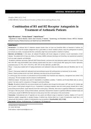
Askin’s tumor is a rare neoplasm of the chest wall <strong>with</strong> a dismal prognosis and<br />
is usually observed in young subjects. We describe a 15-year-old female <strong>with</strong><br />
massive hemoptysis who had an extensive thoracopulmonary tumor on chest<br />
CT. She underwent bronchoscopy which showed the location of the tumor in<br />
the bronchus intermedius. The biopsy obtained from the tumor enabled a<br />
diagnosis of Askin’s tumor to be made. After induction of chemotherapy,<br />
hemoptysis stopped and her constitutional symptoms improved. We focus on<br />
the clinical features, imaging, and histopathological characteristics of Askin’s<br />
tumor.<br />
Key words: Askin’s tumor, Chemotherapy, Ewing’s sarcoma,<br />
<strong>Hemoptysis</strong><br />
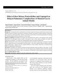
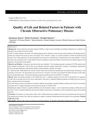
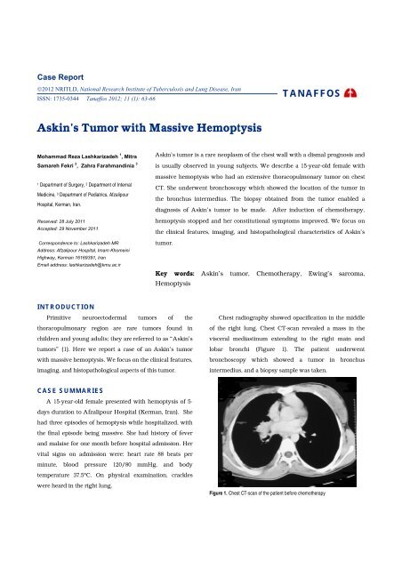
Chest radiography showed opacification in the middle<br />
of the right lung. Chest CT-scan revealed a mass in the<br />
visceral mediastinum extending to the right main and<br />
lobar bronchi (Figure 1). The patient underwent<br />
bronchoscopy which showed a tumor in bronchus<br />
intermedius, and a biopsy sample was taken.<br />
Figure 1. Chest CT-scan of the patient before chemotherapy
64 Askin’s <strong>Tumor</strong> <strong>with</strong> <strong>Massive</strong> <strong>Hemoptysis</strong><br />
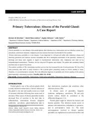
Biopsy samples were evaluated using histological and<br />
immunological assays. Histologically, a section from the<br />
bronchial mucosa showed a neoplastic growth comprising<br />
a nest of small-to-medium-sized cells <strong>with</strong> hyperchromatic<br />
nuclei, scant cytoplasm, and foci of necrosis (Figure 2).<br />
Figure 2. Hematoxylin and eosin staining (×400) of the lung biopsy specimen<br />
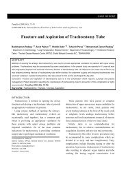
Immunohistochemistry revealed that groups of cells<br />
were negative for CD99 and CD45. <strong>Tumor</strong> cells were<br />
focally positive for creatine kinase (CK) and strongly<br />
positive for neuron-specific enolase (NSE) (Figure 3).<br />
Figure 3. Immunohistochemical staining (neuron-specific enolase) of the lung<br />
biopsy specimen<br />
Based on histopathological and immunohistochemical<br />
findings, a diagnosis of Askin’s tumor was made. The<br />
tumor could not be resected due to extensive involvement<br />
<strong>Tanaffos</strong> 2012; 11(1): 63-66<br />
of lung tissue and the mediastinum. The patient was<br />
offered chemotherapy <strong>with</strong> alternating drug regimens<br />
(vincristine, doxorubicin, and cyclophosphamide; and<br />
isophosphamide, etoposide) for 17 cycles. She has received<br />
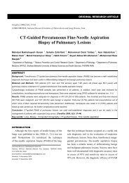
eight cycles of chemotherapy so far. After induction of<br />
chemotherapy, hemoptysis stopped and her constitutional<br />
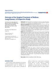
symptoms improved. Chest CT at six-month follow-up<br />
showed a significant improvement (Figure 4).<br />
Figure 4. Chest CT of the patient after chemotherapy<br />
DISCUSSION<br />
Ewing's sarcoma (ES) was initially believed to be of<br />
perivascular endothelial origin. The Ewing's sarcoma<br />
family of tumors (EFT) includes ES of bone (ESB),<br />
extraosseous ES (EES), peripheral primitive<br />
neuroectodermal tumor of bone (pPNET), and malignant<br />
small-cell tumor of thoracopulmonary region (Askin’s<br />
tumor). All of these tumors are now known to be<br />
neoplasms of neuroectodermal origin (2).<br />
Askin’s tumor is a rare neoplasm of the chest wall. It<br />
has a dismal prognosis and is usually observed in young<br />
subjects (3,4).<br />
The aggressive nature of Askin’s tumor results in its<br />
short clinical presentation. The diagnosis of Askin’s tumor<br />
is primarily by histopathologic examination. Imaging has<br />
only a complimentary role (5).<br />
PNET of the chest wall should be considered in a child<br />
<strong>with</strong> a chest wall mass. CT is valuable for evaluating tumor<br />
extension at diagnosis, the effects of chemotherapy, and
assessing tumor recurrence after surgery. However, CT can<br />
overestimate infiltration into the pleura, lung or<br />
diaphragm, and it would be better evaluated by<br />
ultrasonography. MRI is superior to CT for evaluation of<br />
tumor extension, and may be considered complementary<br />
to CT, particularly for very large tumors of the chest wall<br />
(6).<br />
Kabiri and colleagues emphasized on the difficult<br />
histological diagnosis, and demonstrated the importance of<br />
complete removal of the tumor for survival (7).<br />
Takanami and colleagues reported a case of a 16-yearold<br />
male who underwent surgery for excision of Askin’s<br />
tumor. He subsequently underwent six excisions of local<br />
Askin’s tumors due to recurrence, <strong>with</strong> postoperative<br />
chemotherapy and radiotherapy for a 7-year period (8).<br />
The established treatment of this tumor is neo-adjuvant<br />
chemotherapy followed by surgical excision of the tumor<br />
and post operative chemotherapy <strong>with</strong> or <strong>with</strong>out<br />
radiotherapy (9,10). The neo-adjuvant chemotherapy<br />
results in better regional management of the tumor, less<br />
extensive surgery and can treat the distant metastasis. The<br />
studies on Ewing’s sarcoma patients demonstrated that<br />
deferred surgical excision of tumor subsequent to<br />
chemotherapy leads to a more negative margin as<br />
compared to cases who underwent surgery alone (9).<br />
Chemotherapy in the past consisted of a combination of<br />
Vincristine, Actinomycin-D, and Cyclophosphamide.<br />
Currently, Doxorubicin is added to the regimen in the<br />
majority of protocols. Additional drugs administered in<br />
most patients are Ifosfamide and Etoposide (11). Operation<br />
<strong>with</strong> wide margins is ideal, but occasionally is feasible in<br />
patients <strong>with</strong> chest wall tumors. Patients <strong>with</strong> positive<br />
margin of tumor after surgery need post operative<br />
radiation. Due to the delayed complications of<br />
radiotherapy such as pulmonary impairment and<br />
amplified cardiac toxicity <strong>with</strong> the prescription of<br />
anthracyclines, adequate treatment should be planned<br />
ahead to avoid such complications (10, 12).<br />
The age of presented case was older than most patients<br />
<strong>with</strong> Askin’s tumor.<br />
<strong>Tanaffos</strong> 2012; 11(1): 63-66<br />
Lashkarizadeh MR, et al. 65<br />
Older age (>14 years) has constantly been correlated<br />
<strong>with</strong> a poorer survival rate from EWS (13). The reason for<br />
this is unclear, as recent surveys showed no difference in<br />
metastasis at diagnosis, or histological response to neoadjuvant<br />
therapy. Patients treated at referral centers<br />
showed to have better outcomes. Superior survival rate<br />
among children compared to adults may be related to<br />
higher familiarity of pediatric oncologists <strong>with</strong> this disease<br />
and its available treatment options (14).<br />
In our literature review, most articles about Askin’s<br />
tumor were case reports. These cases mainly presented as a<br />
mass in the chest wall <strong>with</strong> or <strong>with</strong>out pulmonary<br />
involvement, and the diagnosis was made by biopsy from<br />
the mass. In our case, we could diagnose Askin’s tumor by<br />
a minor procedure. The patient had advanced disease and<br />
the tumor could not be resected. She; therefore, underwent<br />
chemotherapy and a significant response was observed.<br />
In conclusion, if Askin’s tumor manifests <strong>with</strong> massive<br />
hemoptysis, the diagnosis can be reached <strong>with</strong><br />
bronchoscopy. If the tumor is extensive, chemotherapy can<br />
be used to stop hemoptysis.<br />
REFERENCES<br />
1. Takanami I, Imamura T. The treatment of Askin tumor:<br />
results of two cases. J Thorac Cardiovasc Surg 2002; 123 (2):<br />
391- 2.<br />
2. Carvajal R, Meyers P. Ewing's sarcoma and primitive<br />
neuroectodermal family of tumors. Hematol Oncol Clin<br />
North Am 2005; 19 (3): 501- 25, vi-vii.<br />
3. Katsenos S, Nikopoloulou M, Kokkonouzis I, Archondakis S.<br />
<strong>Askin's</strong> tumor: a rare chest wall neoplasm. Case report and<br />
short review. Thorac Cardiovasc Surg 2008; 56 (5): 308- 10.<br />
4. Ayadi H, Ayoub AK. Askin tumor: two cases. Rev Pneumol<br />
Clin 2002; 58 (6 Pt 1): 347- 50.<br />
5. Aggarwal M, Lakhhar B, Aggarwal BK, Anugu R. Askin<br />
tumor: a malignant small cell tumor. Indian J Pediatr 2000; 67<br />
(11): 853- 5.<br />
6. Sallustio G, Pirronti T, Lasorella A, Natale L, Bray A, Marano<br />
P. Diagnostic imaging of primitive neuroectodermal tumour<br />
of the chest wall (Askin tumour). Pediatr Radiol 1998; 28 (9):<br />
697- 702.
66 Askin’s <strong>Tumor</strong> <strong>with</strong> <strong>Massive</strong> <strong>Hemoptysis</strong><br />
7. Kabiri H, el Fakir Y, Mahassini N, Benamor J, Alaziz S,<br />
Elmaslout A, et al. Malignant small-cell thoracic pulmonary<br />
tumor (Askin tumor). Rev Pneumol Clin 1999; 55 (1): 21- 5.<br />
8. Takanami I, Imamura T, Naruke M, Kodaira S. Long-term<br />
survival after repeated resections of Askin tumor recurrences.<br />
Eur J Cardiothorac Surg 1998; 13 (3): 313- 5.<br />
9. Christiansen S, Semik M, Dockhorn-Dworniczak B, Rötker J,<br />
Thomas M, Schmidt C, et al. Diagnosis, treatment and<br />
outcome of patients <strong>with</strong> Askin-tumors. Thorac Cardiovasc<br />
Surg 2000; 48 (5): 311- 5.<br />
10. Parikh M, Samujh R, Kanojia RP, Mishra AK, Sodhi KS, Bal<br />
A. Peripheral primitive neuroectodermal tumor of the chest<br />
wall in childhood: clinico-pathological significance,<br />
management and literature review. Chang Gung Med J 2011;<br />
34 (2): 213- 7.<br />
11. van den Berg H, van Rijn RR, Merks JH. Management of<br />
tumors of the chest wall in childhood: a review. J Pediatr<br />
Hematol Oncol 2008; 30 (3): 214- 21.<br />
<strong>Tanaffos</strong> 2012; 11(1): 63-66<br />
12. Shamberger RC, Laquaglia MP, Krailo MD, Miser JS,<br />
Pritchard DJ, Gebhardt MC, et al. Ewing sarcoma of the rib:<br />
results of an intergroup study <strong>with</strong> analysis of outcome by<br />
timing of resection. J Thorac Cardiovasc Surg 2000; 119 (6):<br />
1154- 61.<br />
13. Cotterill SJ, Ahrens S, Paulussen M, Jürgens HF, Voûte PA,<br />
Gadner H, et al. Prognostic factors in Ewing's tumor of bone:<br />
analysis of 975 patients from the European Intergroup<br />
Cooperative Ewing's Sarcoma Study Group. J Clin Oncol<br />
2000; 18 (17): 3108- 14.<br />
14. Subbiah V, Anderson P, Lazar AJ, Burdett E, Raymond K,<br />
Ludwig JA. Ewing's sarcoma: standard and experimental<br />
treatment options. Curr Treat Options Oncol 2009; 10 (1-2):<br />
126- 40.