A study of histopathological effects and functional tests in liver and kidney of laboratory male mice treated with ammonium chloride
Abstract The present study was aimed to investigate the effects of ammonium chloride on mice. The animals were divided into three groups: groups 2 and 3were treated orally with ammonium chloride at doses of 2 and 4 mg/body weight, respectively for a period of three weeks, while group 1 served as a control group. The tissue sections which were made from mice organs (liver and kidney) which treated with 4mg/ body weight of ammonium chloride proved that this dose has toxic effect only on the liver and kidneys. In the liver, the histopathological effects are represented by hypertrophy and irregular shape of nucleus, degeneration of cytoplasm, congestion of sinusoid, bleeding and infiltration of inflammatory cells. In kidneys, the effects focus on renal tubules only which are represented by the degenerative changes, necrosis and infiltration of inflammatory cells. This study was also carried out to investigate the influence of ammonium chloride on levels of urea, creatinine, total protein, lipid (triglyceride and cholesterol), and liver marker enzymes such as AST, ALT and ALP. Oral administration of ammonium chloride (4mg/body weight) caused a significant increase in the levels of AST and a significant decrease in the level of ALP and total protein in mice. Treated mice with ammonium chloride at a dose of 2 and 4mg/ body weight for 21 days showed a significant decrease in levels of creatinine, triglyceride and cholesterol, while ALT and urea had no affect at two doses of ammonium chloride. In conclusion, ammonium chloride causes direct hepatotoxicity and nephrotoxicity.
Abstract
The present study was aimed to investigate the effects of ammonium chloride on mice. The animals were divided into three groups: groups 2 and 3were treated orally with ammonium chloride at doses of 2 and 4 mg/body weight, respectively for a period of three weeks, while group 1 served as a control group. The tissue sections which were made from mice organs (liver and kidney) which treated with 4mg/ body weight of ammonium chloride proved that this dose has toxic effect only on the liver and kidneys. In the liver, the histopathological effects are represented by hypertrophy and irregular shape of nucleus, degeneration of cytoplasm, congestion of sinusoid, bleeding and infiltration of inflammatory cells. In kidneys, the effects focus on renal tubules only which
are represented by the degenerative changes, necrosis and infiltration of inflammatory cells. This study was also carried out to investigate the influence of ammonium chloride on levels of urea, creatinine, total protein, lipid (triglyceride and cholesterol), and liver marker enzymes such as AST, ALT and ALP. Oral administration of ammonium chloride (4mg/body weight) caused a significant increase in the levels of AST and a significant decrease in the level of ALP and total protein in mice. Treated mice with ammonium chloride at a dose of 2 and
4mg/ body weight for 21 days showed a significant decrease in levels of creatinine, triglyceride and cholesterol, while ALT and urea had no affect at two doses of ammonium chloride. In conclusion, ammonium chloride causes direct hepatotoxicity and nephrotoxicity.

You also want an ePaper? Increase the reach of your titles
YUMPU automatically turns print PDFs into web optimized ePapers that Google loves.
Int. J. Biosci. 2016<br />
Oxidative stress <strong>in</strong>jury is actively <strong>in</strong>volved <strong>in</strong> the<br />
pathogenesis <strong>of</strong> <strong>ammonium</strong> <strong>chloride</strong> <strong>and</strong> <strong>in</strong>duced<br />
acute <strong>kidney</strong> <strong>in</strong>jury. Reactive oxygen species (ROS)<br />
directly act on cell components, <strong>in</strong>clud<strong>in</strong>g lipids,<br />
prote<strong>in</strong>s, <strong>and</strong> DNA, <strong>and</strong> destroy their structure.<br />
Increased levels <strong>of</strong> circulatory ammonia <strong>in</strong> <strong>mice</strong><br />
<strong>treated</strong> <strong>with</strong> <strong>ammonium</strong> <strong>chloride</strong> may be due to the<br />
<strong>liver</strong> damage caused by ammonia-<strong>in</strong>duced free radical<br />
generation. Reports have shown that excess ammonia<br />
<strong>in</strong>duces nitric oxide synthase, which lead to the<br />
enhanced production <strong>of</strong> the nitric oxide, lead<strong>in</strong>g <strong>in</strong><br />
turn to oxidative stress <strong>and</strong> <strong>liver</strong> damage (Kosenko et<br />
al., 2000; Schliess et al., 2002).<br />
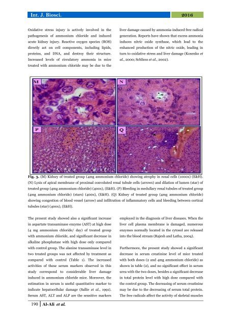
Fig. 3. (M) Kidney <strong>of</strong> <strong>treated</strong> group (4mg <strong>ammonium</strong> <strong>chloride</strong>) show<strong>in</strong>g atrophy <strong>in</strong> renal cells (1000x) (E&H).<br />
(N) Lysis <strong>of</strong> apical membrane <strong>of</strong> proximal convoluted renal tubule cells (arrows) <strong>and</strong> dilation <strong>of</strong> lumen (star) <strong>of</strong><br />
<strong>treated</strong> group (4mg <strong>ammonium</strong> <strong>chloride</strong>) (400x), (E&H). (P) Bleed<strong>in</strong>g <strong>in</strong> medullary renal tubules <strong>of</strong> <strong>treated</strong> group<br />
(4mg <strong>ammonium</strong> <strong>chloride</strong>) (stars) (400x), (E&H). (Q) Kidney <strong>of</strong> <strong>treated</strong> group (4mg <strong>ammonium</strong> <strong>chloride</strong>)<br />
show<strong>in</strong>g congestion <strong>of</strong> blood vessel (arrow) <strong>and</strong> <strong>in</strong>filtration <strong>of</strong> <strong>in</strong>flammatory cells <strong>and</strong> bleed<strong>in</strong>g between cortical<br />
tubules (star) (400x), (E&H).<br />
The present <strong>study</strong> showed also a significant <strong>in</strong>crease<br />
<strong>in</strong> aspartate transam<strong>in</strong>ase enzyme (AST) at high dose<br />
(4 mg <strong>ammonium</strong> <strong>chloride</strong>/ day) <strong>of</strong> <strong>treated</strong> group<br />
<strong>with</strong> <strong>ammonium</strong> <strong>chloride</strong>, <strong>and</strong> significant decrease <strong>in</strong><br />
alkal<strong>in</strong>e phosphatase <strong>with</strong> high dose only compared<br />
<strong>with</strong> control group. The alan<strong>in</strong>e transam<strong>in</strong>ase level <strong>in</strong><br />
two <strong>treated</strong> groups was not affected by treatment as<br />
compared <strong>with</strong> control (Table 1). The <strong>in</strong>creased<br />
activities <strong>of</strong> these serum markers observed <strong>in</strong> this<br />
<strong>study</strong> correspond to considerable <strong>liver</strong> damage<br />
<strong>in</strong>duced <strong>in</strong> <strong>ammonium</strong> <strong>chloride</strong> <strong>mice</strong>. Moreover, the<br />
estimation <strong>in</strong> serum is useful quantitative marker to<br />
<strong>in</strong>dicate hepatocellular damage (Sallie et al., 1991).<br />
Serum AST, ALT <strong>and</strong> ALP are the sensitive markers<br />
employed <strong>in</strong> the diagnosis <strong>of</strong> <strong>liver</strong> diseases. When the<br />
<strong>liver</strong> cell plasma membrane is damaged, numerous<br />
enzymes normally located <strong>in</strong> the cytosol are released<br />
<strong>in</strong>to the blood stream (Rajesh <strong>and</strong> Latha, 2004).<br />
Furthermore, the present <strong>study</strong> showed a significant<br />
decrease <strong>in</strong> serum creat<strong>in</strong><strong>in</strong>e level <strong>of</strong> <strong>mice</strong> <strong>treated</strong><br />
<strong>with</strong> both doses (2 <strong>and</strong> 4mg <strong>ammonium</strong> <strong>chloride</strong>) as<br />
shown <strong>in</strong> table (2), <strong>and</strong> no significant effect <strong>in</strong> serum<br />
urea <strong>with</strong> the two doses, besides a significant decrease<br />
<strong>in</strong> total prote<strong>in</strong> level <strong>with</strong> high dose compared <strong>with</strong><br />
the control group. The decreas<strong>in</strong>g <strong>of</strong> serum creat<strong>in</strong><strong>in</strong>e<br />
may be due to the decreas<strong>in</strong>g <strong>of</strong> serum total prote<strong>in</strong>.<br />
The free radicals affect the activity <strong>of</strong> skeletal muscles<br />
190 Al-Ali et al.

![Review on: impact of seed rates and method of sowing on yield and yield related traits of Teff [Eragrostis teff (Zucc.) Trotter] | IJAAR @yumpu](https://documents.yumpu.com/000/066/025/853/c0a2f1eefa2ed71422e741fbc2b37a5fd6200cb1/6b7767675149533469736965546e4c6a4e57325054773d3d/4f6e6531383245617a537a49397878747846574858513d3d.jpg?AWSAccessKeyId=AKIAICNEWSPSEKTJ5M3Q&Expires=1723384800&Signature=%2BHiew7TFNV8H1Mw2Ftca5xvIsVY%3D)