FISH Protocols - Silva
FISH Protocols - Silva
FISH Protocols - Silva

Create successful ePaper yourself
Turn your PDF publications into a flip-book with our unique Google optimized e-Paper software.
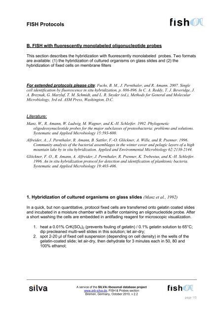
<strong>FISH</strong> <strong>Protocols</strong><br />
B. <strong>FISH</strong> with fluorescently monolabeled oligonucleotide probes<br />
This section describes the hybridization with fluorescently monolabeled probes. Two formats<br />
are available: (1) the hybridization of cultured organisms on glass slides and (2) the<br />
hybridization of fixed cells on membrane filters<br />
For extended protocols please cite: Fuchs, B. M., J. Pernthaler, and R. Amann. 2007. Single<br />
cell identification by fluorescence in situ hybridization, p. 886-896. In C. A. Reddy, T. J. Beveridge, J.<br />
A. Breznak, G. Marzluf, T. M. Schmidt, and L. R. Snyder (ed.), Methods for General and Molecular<br />
Microbiology, 3rd ed. ASM Press, Washington, D.C.<br />
Literature:<br />
Manz, W., R. Amann, W. Ludwig, M. Wagner, and K.-H. Schleifer. 1992. Phylogenetic<br />
oligodeoxynucleotide probes for the major subclasses of proteobacteria: problems and solutions.<br />
Systematic and Applied Microbiology 15:593-600.<br />
Alfreider, A., J. Pernthaler, R. Amann, B. Sattler, F.-O. Glöckner, A. Wille, and R. Psenner. 1996.<br />
Community analysis of the bacterial assemblages in the winter cover and pelagic layers of a high<br />
mountain lake by in situ hybridization. Applied and Environmental Microbiology 62:2138-2144.<br />
Glöckner, F. O., R. Amann, A. Alfreider, J. Pernthaler, R. Psenner, K. Trebesius, and K.-H. Schleifer.<br />
1996. An in situ hybridization protocol for detection and identification of planktonic bacteria.<br />
Systematic and Applied Microbiology 19:403-406.<br />
1. Hybridization of cultured organisms on glass slides (Manz et al., 1992)<br />
In a quick, but non quantitative, protocol fixed cells are transferred onto gelatin coated slides<br />
and incubated in a moisture chamber with a buffer containing an oligonucleotide probe. After<br />
a short washing the cells are embedded in antifading reagent for microscopic visualization.<br />
1. heat a 0.01% CrK(SO4)2 (prevents fouling of gelatin) / 0.1% gelatin solution to 65°C;<br />
dip precleaned multi-well slides in this solution; let air-dry;<br />
2. spot 2-20 µl of fixed cell suspension (depending on cell density) in the wells of the<br />
gelatin-coated slide; let air-dry, then dehydrate for 3 minutes each in 50, 80 and<br />
100% ethanol;<br />
A service of the SILVA ribosomal database project<br />
www.arb-silva.de, <strong>FISH</strong> & Probes section<br />
Bremen, Germany, October 2010, v 2.2<br />
page 1/5
3. prepare hybridization buffer: see table 1;<br />
Stock reagent Volume<br />
5 M NaCl 360 µl 900 mM<br />
1 M Tris / HCl 40 µl 20 mM<br />
Formamide % depending on probe<br />
distilled H2O add to 2 ml<br />
10% SDS 2 µl 0.01%<br />
(add SDS last to avoid precipitation)<br />
Table 1: Standard hybridization buffer<br />
A service of the SILVA ribosomal database project<br />
www.arb-silva.de, <strong>FISH</strong> & Probes section<br />
Bremen, Germany, October 2010, v 2.2<br />
final concentration in<br />
hybridization buffer<br />
Note: The formamide concentration is dependent on the probe used and determines the<br />
stringency of the hybridization. Hybridization stringency may also be adjusted by temperature<br />
rather than by the chemical composition of buffers. We find that it is more convenient to keep<br />
incubator and water bath at one set temperature and to modulate the stringency by adding<br />
formamide.<br />
4. for the hybridization mixtures add 1 volume of probe working solution (50 ng DNA µl -1 )<br />
to 9 volume of hybridization buffer in a 0.5-ml microfuge tube; keep probe solutions<br />
dark and on ice;<br />
Note: The probe is fluorescently labeled and can be e.g. purchased from biomers.net (Ulm,<br />
Germany). The probe stock usually comes dried in a tube and needs to be diluted with sterile<br />
water according to the manufacturer’s instructions. Alternatively, measure a small aliquot in<br />
the photometer and use the formula: 1 OD ~ 20 ng DNA µl -1 (see webpage).<br />
5. prepare hybridization vessels from 50 ml polyethylene tubes: insert a piece of blotting<br />
paper into a polyethylene tube and soak it with the remaining hybridization buffer; use<br />
separate tubes for each concentration of formamide;<br />
6. add 10 µl of hybridization mix to the samples in each well and place the slide into the<br />
polyethylene tube (in a horizontal position);<br />
7. incubate at 46°C for at least 90 min (maximum: 3 hours);<br />
8. prepare 50 ml of washing buffer (see table 2) in a polyethylene tube and preheat in a<br />
48°C water bath<br />
page 2/5
Stock reagent Volume final concentration in<br />
hybridization buffer<br />
5 M NaCl concentration depending on % formamide in hybridization<br />
buffer (see table A in appendix)<br />
1 M Tris / HCl 1 ml 20 mM<br />
0.5 M EDTA (only if ≥ 20%<br />
formamide in hybridization!)<br />
distilled H2O add to 50 ml<br />
(500 µl) 5 mM<br />
10% SDS 50 µl 0.01%<br />
(add SDS last to avoid precipitation)<br />
Table 2: Standard washing buffer<br />
Note: The stringency in the washing buffer is achieved by adjusting the NaCl concentration.<br />
This avoids the use of excess amounts of formamide.<br />
9. quickly rinse the slide carefully with a bit of washing buffer, transfer slide into<br />
preheated washing buffer and incubate for 25 min at 48°C (water bath);<br />
10. rinse slide with distilled H2O, let air-dry;<br />
11. for counterstaining cover each well with 10 µl of a 1 µg ml-1 DAPI solution, and<br />
incubate for 3 minutes; rinse slide with distilled H2O let air-dry;<br />
12. samples are mounted in a 4:1 mix of Citifluor (Citifluor Ltd, London, U.K) and Vecta<br />
Shield (Vector Laboratories, Inc., Burlingame, CA); Vecta Shield contains a superior<br />
antibleaching reagent, but quenches DAPI fluorescence; the wells have to be<br />
completely dry before embedding, otherwise a fraction of cells will detach during<br />
inspection;<br />
13. double stained and air dried preparations as well as mounted slides can be stored in<br />
the dark at -20°C for several days without substantial loss of probe fluorescence;<br />
14. probe-conferred fluorescence fades much more rapidly than DAPI fluorescence in the<br />
microscopic image, and UV excitation will also bleach the probe signal; for counting, it<br />
is, therefore, safer to first quantify probe stained cells and subsequently all cells from<br />
the same field of vision in UV excitation;<br />
A service of the SILVA ribosomal database project<br />
www.arb-silva.de, <strong>FISH</strong> & Probes section<br />
Bremen, Germany, October 2010, v 2.2<br />
page 3/5
2. Hybridization of fixed cells on membrane filters (Glöckner et al., 1996)<br />
Notes:<br />
1. Cut sections from membrane filters with a razor blade and label filter sections with a<br />
pencil, e.g., by numbering them.<br />
2. Put filter sections on glass slides (cells facing up!), several filter sections can be<br />
placed on one slide and for simultaneous hybridization with the same probe.<br />
3. Prepare 2 ml of hybridization buffer in a microfuge tube (360 µl 5 M NaCl, 40 µl 1M<br />
Tris/HCl, formamide % depending on probe, add water to 2 ml, add 2 µl SDS (10%)).<br />
4. Remove an aliquot of 20 µl per filter piece into a separate cap and add 2 µl probe<br />
working solution (concentration: 50 ng DNA µl -1 ) per filter piece.<br />
5. Prepare moisture chamber by putting a piece of blotting paper into a 50 ml<br />
polyethylene tube and soaking it with the remaining hybridization buffer without probe<br />
(see above).<br />
6. Carefully cover the filter section with the hybridization mix and place the slide with<br />
filter sections into the polyethylene tube (in a horizontal position).<br />
7. Incubate at 46°C for at least 90 min (maximum: 3 hours).<br />
8. Meanwhile prepare 50 ml of washing buffer in a polyethylene tube (X ml 5M NaCl,<br />
depending on formamide concentration in the hybridization buffer (see table A in<br />
Appendix), 1 ml 1 M Tris/ HCl, 500µl 0.5M EDTA (if formamide concentration of the<br />
hybridization buffer was ≥20%), add to 50 ml with water, 50 µl 10% SDS).<br />
9. Quickly transfer filter sections into preheated washing buffer and incubate for 15 min<br />
at 48°C (water bath).<br />
10. Pour washing buffer with filter sections into a petri dish. Pick filter sections and rinse<br />
them by placing them into a petri dish with distilled H2O for several seconds, then let<br />
them air-dry on blotting paper.<br />
11. For counterstaining put filter sections on a glass plate, cover with app. 50 µl of DAPI<br />
solution (1 µg ml -1 ), and incubate for 3 min. Afterwards wash filter sections<br />
subsequently for 1 min. in distilled H2O and for 1 min. in 80% ethanol to remove<br />
unspecific staining. Let air-dry.<br />
12. Samples are mounted in a 4:1 mix of Citifluor and Vecta Shield. The filter sections<br />
have to be completely dry before embedding, otherwise part of the cells might detach<br />
during inspection.<br />
13. Double stained and air dried preparations as well as filters mounted on slides can be<br />
stored in the dark at -20°C for several days without substantial loss of probe<br />
fluorescence.<br />
• We find that, following our procedure, 80-90% of the initial bacterial cell numbers are<br />
recovered after hybridizations of bacterioplankton on membrane filters. This fraction<br />
may, however, depend on the type of sample and should be verified experimentally.<br />
• Therefore, do not attempt to determine absolute cell counts from filters after<br />
hybridization, but only the percentage of hybridized cells. Additionally, the distribution<br />
of cells on sections of a 47 mm diameter membrane filter is never as even as on a<br />
small filter, resulting in a higher error of the total DAPI counts.<br />
• At least 500 DAPI-stained cells should be counted per hybridized filter piece to<br />
reduce the counting error
Appendix<br />
Table A: NaCl concentration in the washing buffer according to % formamide of the<br />
hybridization buffer (depending on probe).<br />
% formamide in<br />
hybridisation<br />
buffer<br />
[NaCl] in M<br />
endconcentration<br />
Washing at 48°C<br />
µl 5 M NaCl in<br />
50 ml<br />
0 0.900 9000<br />
5 0.636 6300<br />
10 0.450 4500<br />
15 0.318 3180<br />
20 0.225 2150<br />
25 0.159 1490<br />
30 0.112 1020<br />
35 0.080 700<br />
40 0.056 460<br />
45 0.040 300<br />
50 0.028 180<br />
55 0.020 100<br />
60 0.014 40<br />
65 - -<br />
70 - -<br />
A service of the SILVA ribosomal database project<br />
www.arb-silva.de, <strong>FISH</strong> & Probes section<br />
Bremen, Germany, October 2010, v 2.2<br />
page 5/5