Hafnia alvei - Clinical Infectious Diseases
Hafnia alvei - Clinical Infectious Diseases
Hafnia alvei - Clinical Infectious Diseases

You also want an ePaper? Increase the reach of your titles
YUMPU automatically turns print PDFs into web optimized ePapers that Google loves.
1040<br />
<strong>Clinical</strong> Significance of Extraintestinal <strong>Hafnia</strong> <strong>alvei</strong> Isolates from 61 Patients and<br />
Review of the Literature<br />
Huldrych Giinthard and Andreas Pennekamp From the Department ofInternal Medicine, Stadtspital Triemli, and the<br />
Department ofMedical Microbiology ofthe University ofZurich,<br />
Zurich, Switzerland<br />
<strong>Hafnia</strong> <strong>alvei</strong> is a gram-negative bacterium that is rarely isolated from human specimens and is<br />
rarely considered to be pathogenic. It has been associated with gastroenteritis, meningitis, bacteremia,<br />
pneumonia, nosocomial wound infections, endophthalmitis, and a buttock abscess. We studied<br />
80 H. <strong>alvei</strong> isolates recovered from 61 patients within a period of 30 months. H. <strong>alvei</strong> was cultured<br />
from sites that included the respiratory tract (n = 38), the gastrointestinal tract (n = 16), and the<br />
urogenital tract (n = 12); the organism was found in blood cultures (n = 8), on central venous<br />
catheters (n = 3), and on the skin (n = 3). Only 25% of H. <strong>alvei</strong> isolates were recovered in pure<br />
cultures. Fifty-seven (93.4%) of the patients had an underlying illness. H. <strong>alvei</strong> proved to be the<br />
etiologic agent in two episodes of septicemia and in one episode of peritonitis and was probably<br />
responsible for septicemia in two other patients and pneumonia in one. All six of these patients<br />
recovered after receiving antibiotic treatment and/or standard surgical treatment, when needed.<br />
Three of these infections were nosocomial, and three were community acquired. Of the strains of<br />
H. <strong>alvei</strong> tested in our study, 100% were susceptible to netilmicin, ciprofloxacin, and imipenem; 92%<br />
were susceptible to piperacillin; 90% were susceptible to co-trimoxazole; and 88% were susceptible<br />
to ceftriaxone and ceftazidime. In this study, we found H. <strong>alvei</strong> to be a rare but significant etiologic<br />
agent of nosocomial and community-acquired infections.<br />
<strong>Hafnia</strong> <strong>alvei</strong> is a gram-negative facultative rod-shaped anaerobe<br />
that belongs to the Enterobacteriaceae; the organism was<br />
formerly named Enterobacter hafniae. H. <strong>alvei</strong> is rarely considered<br />
to be pathogenic; rather, H <strong>alvei</strong> has been found to be<br />
related to the enteropathogenic Escherichia coli on the basis<br />
of a single virulence factor. Electron microscopy has demonstrated<br />
inflammation and mucosal invasion by H <strong>alvei</strong> in rabbit<br />
bowels, and an attachment-effacement gene like that detected<br />
in enteropathogenic E. coli was found in H. <strong>alvei</strong> by hybridization<br />
[1, 2]. No other distinct virulence factors have been noted<br />
in this organism so far [3]. Stool specimens are generally not<br />
examined for the presence of H <strong>alvei</strong>.<br />
Cases of diarrhea due to H <strong>alvei</strong> have occurred mainly in<br />
children. H. <strong>alvei</strong> was reported to have caused acute gastroenteritis<br />
in children from Bangladesh and Spain [1-2]. In one<br />
study, 16% of Finnish tourists with acute gastroenteritis and<br />
diarrhea who were returning from Morocco excreted H <strong>alvei</strong><br />
in their stools [4]. Seven strains isolated from children with<br />
diarrhea expressed the attachment-effacement gene [2]. In two<br />
case reports, H <strong>alvei</strong> was mentioned as a cause of nonbloody<br />
diarrhea. One of these cases was presumably associated with<br />
reactive arthritis [5, 6]; however, cultures of joint aspirates<br />
Received 3 October 1995; revised 16 January 1996.<br />
Reprints or correspondence: Dr. Andreas Pennekamp, Department ofMedical<br />
Microbiology of the University of Zurich, Gloriastrasse 32, CH-S028 Zurich,<br />
Switzerland.<br />
<strong>Clinical</strong> <strong>Infectious</strong> <strong>Diseases</strong> 1996;22:1040-5<br />
© 1996 by The University of Chicago. All rights reserved.<br />
1058-4838/96/2206-0021$02.00<br />
were negative. A single case of H <strong>alvei</strong> meningitis in a 1year-old<br />
girl [7] and a case ofnecrotizing H <strong>alvei</strong> enterocolitis<br />
with septicemia in a 20-day-old boy [8] have also been described.<br />
In adults, H <strong>alvei</strong> has caused bacteremia [9, 10], pneumonia,<br />
and nosocomial wound infections [11-13]. H. <strong>alvei</strong><br />
was recovered with Salmonella arizonae from a patient with<br />
endogenous endophthalmitis who was receiving steroids [14].<br />
Recently, H <strong>alvei</strong> was isolated from a buttock abscess that<br />
resulted from skin puncture with carpet nails in an otherwise<br />
healthy middle-aged man [15].<br />
Until now, there have been no data published about the true<br />
rate of isolation of H. <strong>alvei</strong> from clinical specimens, and the<br />
clinical significance of this bacterium remains to be defined.<br />
Therefore, we performed a study on the frequency of isolation<br />
of H <strong>alvei</strong> from clinical specimens (except stools) and correlated<br />
the microbiological findings with the clinical data.<br />
Materials and Methods<br />
Patient evaluation. Patients from whom isolates ofH. <strong>alvei</strong><br />
were recovered during routine diagnostic testing at the Department<br />
of Medical Microbiology, University of Zurich (Zurich)<br />
and at the Microbiology Laboratory of the Stadtspital Triemli<br />
(Zurich) between January 1992 and May 1995 were identified<br />
by reviewing the laboratories' records. The patients' charts<br />
were reviewed for data on isolation sites, underlying illnesses,<br />
cocultivation of other bacteria, and discrimination between<br />
nosocomial infections or community-acquired infections.<br />
Isolation and identification. H. <strong>alvei</strong> was isolated from normally<br />
sterile body fluids such as blood, ascitic fluid, and ab-<br />
Downloaded from<br />
http://cid.oxfordjournals.org/<br />
by guest on December 14, 2012
em 1996;22 (June) Extraintestinal H <strong>alvei</strong> Infection 1041<br />
dominal aspirates as well as from postoperative indwelling<br />
catheters, central venous lines, and urine collected from catheters.<br />
Respiratory tract isolates were cultured from sputum and<br />
tracheal and bronchial aspirates. Samples other than blood were<br />
plated on selective and differential media, and colonies of Enterobacteriaceae<br />
were detected on MacConkey agar (Becton<br />
Dickinson, Basel, Switzerland); blood was cultured with use<br />
of the BacT/Alert blood culture system (Organon Teknika,<br />
Basel).<br />
The identification of Enterobacteriaceae was based on biochemical<br />
data obtained by means of commercial identification<br />
systems (API20E and RapID 32E; bioMerieux, Geneva, Switzerland)<br />
and on lack of lactose fermentation on MacConkey<br />
agar. Biochemical reactions for which H. <strong>alvei</strong> was positive<br />
included lysine decarboxylase, ornithine decarboxylase, and<br />
mannitol fermentation, whereas it was negative for fermentation<br />
ofsorbitol, inositol, sucrose, and melibiose; these negative<br />
reactions differentiated H. <strong>alvei</strong> from Enterobacter aerogenes.<br />
Strains with T values of
1042 Glinthard and Pennekamp<br />
Table 1. Isolation of <strong>Hafnia</strong> <strong>alvei</strong> in pure or mixed cultures of specimens from different body sites<br />
of 61 patients.<br />
Site involved<br />
Respiratory tract*<br />
Gastrointestinal tract t<br />
Blood<br />
Central venous catheter<br />
Urogenital tract t<br />
Skin<br />
No. of isolates recovered<br />
in pure cultures<br />
8<br />
5<br />
3<br />
1<br />
3<br />
o<br />
No. of isolates recovered<br />
in mixed cultures<br />
30<br />
11<br />
5<br />
2<br />
9<br />
3<br />
Total no. (%) from indicated<br />
site (n = 80)<br />
38 (47.5)<br />
16 (20)<br />
8 (10)<br />
3 (3.75)<br />
12 (15)<br />
3 (3.75)<br />
NOTE. Twenty (25%) ofthe isolates were recovered in pure cultures, and 60 (75%) ofthe isolates were recovered<br />
in mixed cultures.<br />
* The following specimens were obtained: tracheal aspirates (16), pharyngeal smears (4), bronchoalveolar lavage<br />
fluid (2), nasal smears (1), bronchial aspirates (7), and sputum (8).<br />
t The following specimens were obtained: ascitic fluid aspirates (2), gallbladder (1), abscesses and deep abdominal<br />
wounds (10), and pancreatic pseudocysts (3); no stool samples were included.<br />
t The following specimens were obtained: urine collected from indwelling catheters (6), clean-catch urine (3),<br />
scrotal smears (2), and episiotomy wound (1).<br />
underlying illnesses [11-12], except when it is an enteric<br />
pathogen [1, 4-6, 8, 17].<br />
Thirty-eight H <strong>alvei</strong> isolates were recovered from respiratory<br />
specimens, 16 were recovered from the gastrointestinal<br />
tract, 12 were recovered from the urogenital tract, eight were<br />
recovered from blood, three were recovered from intravenous<br />
catheters, and three were recovered from the skin. Only 25%<br />
of all H. <strong>alvei</strong> isolates grew in pure culture. H. <strong>alvei</strong> was<br />
CID 1996;22 (June)<br />
cultured with a variety ofother bacteria and fungi in the respiratory<br />
and gastrointestinal specimens.<br />
H. <strong>alvei</strong> was the only identified pathogen causing two cases<br />
of septicemia and one case of acute peritonitis. The peritonitis<br />
occurred after a patient with stenosis of the choledochus duct<br />
underwent endoscopic retrograde cholangiopancreatography,<br />
and H. <strong>alvei</strong> was isolated from his blood and peritoneal fluid.<br />
This circumstance indicates that the bacterium's portal ofentry<br />
Table 2. Isolation of <strong>Hafnia</strong> <strong>alvei</strong> from different body sites of 61 patients with and without underlying conditions.<br />
Underlying conditions<br />
Malignancies<br />
Leukemia or lymphoma<br />
Abdominal carcinoma<br />
Throat or bronchial carcinoma<br />
Others<br />
Total t<br />
Abdominal surgeryt<br />
Trauma§<br />
Heart surgery II<br />
Pancreatitis<br />
Chronic obstructive pulmonary disease<br />
Lung transplantation<br />
None<br />
Respiratory<br />
tract*<br />
4/1<br />
3/1<br />
1/0<br />
5/4<br />
8/7<br />
2/2<br />
2/0<br />
1/1<br />
4/0<br />
Gastrointestinal<br />
tract<br />
2<br />
4<br />
6<br />
Site involved<br />
Blood and central<br />
venous catheter<br />
2<br />
2<br />
Urogenital<br />
tract<br />
2<br />
1<br />
1<br />
2<br />
1<br />
1<br />
Skin Total (%)<br />
6<br />
7<br />
5<br />
2<br />
20 (33)<br />
11 (18)<br />
9 (14.8)<br />
9 (14.8)<br />
4 (6.6)<br />
3 (4.9)<br />
1 (1.6)<br />
4 (6.6)<br />
NOTE. Only one isolate per patient was taken into account. Of the 61 patients, 35 (18 ofwhom were intubated) had isolates recovered from the respiratory<br />
tract; nine, from the gastrointestinal tract; seven, from blood and central venous catheters; seven, from the urogenital tract; and three, from the skin.<br />
* Total no. of isolates/no. of isolates from intubated patients.<br />
t Includes carcinoma (13 patients), acute myelocytic leukemia (3), plasmocytoma (1), non-Hodgkin's lymphoma (1), sarcoma (1), and aplastic anemia of<br />
unknown etiology (1).<br />
t Includes incarcerated hernia (3 patients), perforation of the sigmoid colon (2), small bowel ileus (2), infrarenal aortic aneurysm (2), intra-abdominal abscess<br />
(2), delivery of infant by vacuum extraction (1), and endoscopic retrograde cholangiopancreatography (1).<br />
§ Includes subarachnoidal bleeding (3 patients), polytrauma (2), knife injuries (2), bums (1), femur fracture (1).<br />
II Includes aortocoronary bypass surgery (8 patients) and aortic valve replacement (1).<br />
Downloaded from<br />
http://cid.oxfordjournals.org/ by guest on December 14, 2012
cm 1996; 22 (June) Extraintestinal H <strong>alvei</strong> Infection 1043<br />
Table 3. <strong>Clinical</strong> characteristics of patients with <strong>Hafnia</strong> <strong>alvei</strong> infection.<br />
First day<br />
Type of organism isolated Antibiotic treatment,<br />
Age (y)/sex infection Site(s) involved after admission Underlying disease <strong>Clinical</strong> presentation outcome*<br />
371M Septicemia Blood (X2),t pancreatic 38 Acute pancreatitis Pancreatic pseudocysts, Imipenemlcilastatin,<br />
pseudocyst fluid perforation of transverse recovered<br />
colon, sepsis syndrome<br />
49/F Septicemia Blood (X2),t central 28 Sigmoid diverticulitis Perforation of the sigmoid colon Imipenemlcilastatin,<br />
venous catheter with sepsis syndrome recovered<br />
501M Peritonitis Peritoneal fluid Chronic pancreatitis Stenosis of the choledochus duct, Imipenemlcilastatin,<br />
acute abdomen recovered<br />
* The surgical treatment in these cases is not included because standard procedures were performed; no surgical interventions were primarily done for infection with<br />
H. <strong>alvei</strong>.<br />
t Organism isolated in two separate blood cultures.<br />
might have been the gastrointestinal tract, where it is considered<br />
to be a commensal [16]. Earlier reports have also mentioned<br />
a correlation between abdominal wounds or abdominal<br />
surgery and the isolation of H <strong>alvei</strong> in blood [10]. H <strong>alvei</strong><br />
was isolated along with other pathogens from three other patients<br />
who had septicemia, pneumonia, and cholangitis.<br />
Overall, 11.5% of the patients in our study population had<br />
proven (or at least probable) infections caused by H <strong>alvei</strong>, and<br />
57 ofthem presented with an underlying illness. Ofsix patients<br />
with H. <strong>alvei</strong> infections, three had nosocomial infections [19],<br />
and three had community-acquired infections. The number of<br />
Table 4. Antimicrobial susceptibility test results for <strong>Hafnia</strong> <strong>alvei</strong> isolates.<br />
infections might even have been higher, but the retrospective<br />
nature of our study did not allow correlation of the other<br />
H <strong>alvei</strong> isolates with clinically significant infections. However,<br />
10 additional patients from whom H. <strong>alvei</strong> was isolated were<br />
treated with broad-spectrum antibiotics in the absence of a<br />
clear diagnosis of infection. This circumstance supports the<br />
hypothesis that there was a higher rate of significant infections<br />
in our study population. In all six of our patients with proven<br />
or probable H. <strong>alvei</strong> infections, the infections were successfully<br />
treated with antibiotics chosen according to the results of susceptibility<br />
testing.<br />
Present report Literature review t<br />
No. of strains Percent No. of strains Percent<br />
Antibiotic tested susceptible* tested susceptible<br />
Amoxicillinlclavulanic acid 77 1 NA NA<br />
Ampicillin 76 9 23 9<br />
Cephalothin 77 a 23 17<br />
Cefamandole 71 79 NA NA<br />
Ceftriaxone 73 88 NA NA<br />
Ceftazidime 73 88 12 42<br />
Cefuroxime 69 77 NA NA<br />
Gentamicin NA NA 23 100<br />
Amikacin NA NA 13 100<br />
Netilmicin 75 100 NA NA<br />
Tobramycin 73 99 NA NA<br />
Ciprofloxacin 75 100 7 100<br />
Piperacillin 64 92 13 85<br />
Tetracycline NA NA 5 84<br />
Piperacillinltazobactam 19 89 NA NA<br />
Trimethoprim-sulfamethoxazole 76 90 13 92<br />
Imipenemlcilastatin 73 99 8 100<br />
Colistin 72 75 NA NA<br />
NOTE. NA = not available.<br />
* Susceptibility testing was performed according to the National Committee for <strong>Clinical</strong> Laboratory Standards<br />
(NCCLS) for the disk diffusion method and interpreted according to NCCLS guidelines.<br />
tData are from [6,16-19].<br />
Downloaded from<br />
http://cid.oxfordjournals.org/<br />
by guest on December 14, 2012
1044 Giinthard and Pennekamp cm 1996;22 (June)<br />
As shown in table 1, 38 (47.5%) of all isolates recovered<br />
from 35 patients were of respiratory origin. Eighteen of the<br />
patients were intubated at the time that samples were obtained,<br />
and 17 were not intubated during their hospital courses. Intubated<br />
patients who are immunosuppressed as a result ofcardiac<br />
surgery or trauma seem to harbor H. <strong>alvei</strong> in their respiratory<br />
tracts more often than do patients who have previously undergone<br />
abdominal surgery. Whether the oropharyngeal-tracheal<br />
regions of these patients had been colonized previously or<br />
whether they became colonized as a result of hospital procedures<br />
(e.g., intubation) cannot be answered sufficiently. We<br />
do not know the prevalence of H. <strong>alvei</strong> colonization of the<br />
oropharyngeal-tracheal regions and other bodily sites in healthy<br />
persons or patients with underlying diseases. In large studies<br />
[20-21], this bacterium was rarely recovered from any site,<br />
making nosocomial infection improbable and emphasizing the<br />
likelihood of endogenous colonization in most patients before<br />
admission to the hospital.<br />
On the other hand, only eight of 30 respiratory tract isolates<br />
were recovered from pure cultures, compared with 22 isolates<br />
that were recovered from mixed cultures; these mixed cultures<br />
mainly consisted of other gram-negative bacteria that are<br />
known to colonize the oropharyngeal-tracheal tract in severely<br />
ill patients [22]. The presence of H. <strong>alvei</strong> in feces reflects the<br />
fact that the organism is part of the normal bowel flora. Case<br />
reports of H. <strong>alvei</strong> as the etiologic agent of diarrhea must be<br />
evaluated critically because no toxin production or toxic mucosal<br />
changes in humans have been detected so far. However,<br />
it is worth mentioning that H. <strong>alvei</strong> might cause diarrhea in<br />
immunocompromised patients, as case reports ofneonates and<br />
malnourished children with diarrhea have shown [1, 7, 17].<br />
In comparing the susceptibilities of H. <strong>alvei</strong> isolates, we<br />
observed a higher rate ofsusceptibility to ampicillinJamoxicillin<br />
and cephalothin among the strains described in the literature.<br />
In conducting this study we used susceptibility to amoxicillin<br />
and first-generation cephalosporins as selection criteria; therefore,<br />
these data are not comparable to those from previous<br />
reports.<br />
In our study the most active antimicrobials were netilmicin<br />
(100% of isolates were susceptible), ciprofloxacin (100% susceptible),<br />
and imipenem (99% susceptible). These results are<br />
similar to the scant results reported in the literature (only a<br />
few strains have been tested), where 100% of the H. <strong>alvei</strong><br />
strains were found to be susceptible to amikacin, gentamicin,<br />
ciprofloxacin, and imipenem [23 - 26].<br />
The susceptibility to ceftazidime observed in our study was<br />
significantly different from that reported in the literature (P <<br />
.001). This difference cannot be explained but is probably not<br />
caused by the larger number of strains tested in our study<br />
because the results were otherwise almost congruent. Another<br />
slight difference was observed when susceptibilities to piperacillin<br />
were compared: 92% of our strains (64 tested) were<br />
susceptible, while 85% of strains in other reports (13 tested)<br />
were susceptible. However, the fact that susceptibility results<br />
from other reports as well as from our study were comparable<br />
for most antimicrobials and that the biochemical features were<br />
similar indicates that H. <strong>alvei</strong> was correctly identified as the<br />
pathogen.<br />
In summary, this study shows that H. <strong>alvei</strong> is a rare human<br />
pathogen; however, the organism may be responsible for serious<br />
nosocomial and community-acquired infections. Infections<br />
mainly occur in patients with underlying illnesses, and H. <strong>alvei</strong><br />
is often isolated in coculture with different gram-negative rods,<br />
which may be due to endogenous colonization of the bowel.<br />
Treatment of H. <strong>alvei</strong> infection on the basis of antimicrobial<br />
susceptibility testing results is effective. In severe cases, treatment<br />
with imipenem or a third-generation cephalosporin in<br />
combination with an aminoglycoside is recommended. More<br />
data on the role of H. <strong>alvei</strong> and its pathogenicity are needed.<br />
In addition, the colonizing rate for this organism in different<br />
sites in healthy adults must be determined.<br />
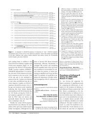
Acknowledgments<br />
The authors are indebted to Professor A. von Graevenitz and<br />
Dr. J. Gubler for their helpful comments and careful review ofthe<br />
manuscript.<br />
References<br />
1. Albert MJ, Alam K, Islam M, et al. <strong>Hafnia</strong> <strong>alvei</strong>, a probable cause of<br />
diarrhea in humans. Infect Immun 1991;59:1507-13.<br />
2. Albert MJ, Faruque SM, Ansaruzzaman M, et al. Sharing of virulence<br />
associated properties at the phenotypic and genetic levels between enteropathogenic<br />
Escherichia coli and <strong>Hafnia</strong> <strong>alvei</strong>. J Med Microbiol<br />
1992;37:310-4.<br />
3. Ridell J, Siitonen A, Paulin L, Lindroos 0, Korkeala H, Albert 1. Characterization<br />
of<strong>Hafnia</strong> <strong>alvei</strong> by biochemical tests, random amplified polymorphic<br />
DNA PCR, and partial sequencing of 16S rRNA gene. J Clin<br />
Microbiol 1995;33:2372-6.<br />
4. Ridell J, Siitonen A, Paulin L, Mattila L, Korkeala H, Albert MJ. <strong>Hafnia</strong><br />
<strong>alvei</strong> in stool specimens from patients with diarrhea and healthy controls.<br />
J Clin Microbiol 1994; 32:2335-7.<br />
5. Westblom TU, Milligan TW. Acute bacterial gastroenteritis caused by<br />
<strong>Hafnia</strong> <strong>alvei</strong> [letter]. Clin Infect Dis 1992; 14:1271-2.<br />
6. Newmark 11, Hobbs WN, Wilson BE. Reactive arthritis associated with<br />
<strong>Hafnia</strong> <strong>alvei</strong> enteritis. Arthritis Rheum 1994;37:960.<br />
7. Mojtabaee A, Siadati A. Enterobacter hafnia meningitis. J Pediatr 1978;<br />
93:1062-3.<br />
8. Ginsberg HG, Goldsmith JP. <strong>Hafnia</strong> <strong>alvei</strong> septicemia in an infant with<br />
necrotizing enterocolitis. J Perinatol 1988; 8: 122-3.<br />
9. Englund GW. Persistent septicemia due to <strong>Hafnia</strong> <strong>alvei</strong>. Report of a case.<br />
Am J Clin PathoI1969;51:717-9.<br />
10. Jennis F, Mccarthy SW. <strong>Hafnia</strong>: an unusual cause of postoperative gramnegative<br />
bacteriae. Med J Aust 1967; 1:286-7.<br />
11. Klapholz A, Lessnau KD, Huang B, Talavera W, Boyle JF. <strong>Hafnia</strong> <strong>alvei</strong>.<br />
Respiratory tract isolates in a community hospital over a three-year<br />
period and a literature review. Chest 1994; 105:1098-1100:<br />
12. Frick T, KUDZ M, Vogt M, Turina M. Typical nosocomial infection with<br />
an unusual cause: <strong>Hafnia</strong> <strong>alvei</strong>. Report of 2 cases and literature review.<br />
Schweiz Rundsch Med Prax 1990;79:1092-4.<br />
13. Berger SA, Edberg SC, Klein RS. Enterobacter hafnaie infection: report<br />
of two cases and review of the literature. Am J Med Sci 1977;273:<br />
101-4.<br />
Downloaded from<br />
http://cid.oxfordjournals.org/<br />
by guest on December 14, 2012