MGIT TM Procedure Manual - Foundation for Innovative New ...
MGIT TM Procedure Manual - Foundation for Innovative New ...
MGIT TM Procedure Manual - Foundation for Innovative New ...

Create successful ePaper yourself
Turn your PDF publications into a flip-book with our unique Google optimized e-Paper software.
Prepared <strong>for</strong> the <strong>Foundation</strong> <strong>for</strong> <strong>Innovative</strong> <strong>New</strong> Diagnostics<br />
<strong>MGIT</strong> <strong>TM</strong><br />
<strong>Procedure</strong> <strong>Manual</strong><br />
For BACTEC <strong>MGIT</strong> 960 TB System<br />
(Also applicable <strong>for</strong> <strong>Manual</strong> <strong>MGIT</strong>)<br />
Mycobacteria Growth Indicator Tube (<strong>MGIT</strong>)<br />
Culture and Drug Susceptibility Demonstration Projects<br />
by<br />
Salman H. Siddiqi, Ph.D.<br />
(BD Fellow, Sparks, Maryland, USA )<br />
and<br />
Sabine Rüsch-Gerdes, Ph.D.<br />
(Director, National Reference Center <strong>for</strong><br />
Mycobacteria, Borstel, Germany)<br />
July 2006
Section I: Principle of <strong>Procedure</strong>s<br />
TABLE OF CONTENTS<br />
A. Introduction<br />
B. Principle of the BACTEC <strong>MGIT</strong> 960 TB System<br />
1. <strong>MGIT</strong> medium<br />
2. Principle of detection and susceptibility testing<br />
Section II: <strong>Procedure</strong> <strong>for</strong> Primary Isolation<br />
A. Introduction<br />
B. Important Safety Precautions<br />
C. Specimen Handling<br />
1. Collection<br />
2. Transportation<br />
3. Storage<br />
D. Digestion, Decontamination and Concentration<br />
1. Sputum<br />
a. NaOH-NALC procedure<br />
b. Other procedures<br />
c. Important points<br />
2. Specimens other than sputum (extra-pulmonary)<br />
a. Pus and other mucopurulent specimens<br />
b. Gastric aspirates<br />
c. Bronchial washings<br />
d. Laryngeal swabs<br />
e. Tissue<br />
f. Urine<br />
g. Other body fluids<br />
E. Smears <strong>for</strong> Acid-Fast Bacteria (AFB)<br />
1. Smear preparation<br />
2. Staining methods<br />
a. Ziehl-Neelsen staining<br />
b. Kinyoun’s staining<br />
c. Two-step staining<br />
d. Fluorochrome acid-fast staining<br />
3. Microscopy<br />
4. Reporting<br />
<strong>MGIT</strong> <strong>TM</strong> <strong>Procedure</strong> <strong>Manual</strong> 3
TABLE OF CONTENTS<br />
F. Preparation and Inoculation <strong>for</strong> Culture<br />
1. Reagents<br />
a. <strong>MGIT</strong> medium<br />
b. <strong>MGIT</strong> growth supplement (enrichment)<br />
c. <strong>MGIT</strong> PANTA<br />
2. <strong>Procedure</strong>s<br />
a. Reconstituting PANTA<br />
b. Inoculation of <strong>MGIT</strong> medium<br />
c. Inoculation of additional media<br />
d. Precautions<br />
e. Incubation<br />
f. Detection of positive growth<br />
G. Work-up of Positive Cultures<br />
1. AFB smear from a positive <strong>MGIT</strong> tube<br />
2. Dealing with contamination<br />
a. Bacterial contamination<br />
b. Isolation of mycobacteria from contaminated or mixed cultures<br />
c. Isolation of mixed mycobacterial culture on Middlebrook Agar Plate<br />
d. Cross-contamination<br />
3. Sub-culturing a positive <strong>MGIT</strong> tube<br />
4. Identification of isolated mycobacteria<br />
H. Results / Reporting<br />
I. Per<strong>for</strong>mance Characteristics<br />
J. Limitations of the <strong>Procedure</strong><br />
K. Quality Control<br />
1. Quality control of AFB smear staining<br />
2. Quality control (QC) testing of <strong>MGIT</strong> medium<br />
a. QC strains<br />
b. Preparation of culture suspension<br />
c. Preparation of dilutions<br />
d. Inoculation/incubation<br />
e. Expected results<br />
f. Precautions<br />
3. Quality control of laboratory procedures<br />
a. Positive and negative controls<br />
b. Quality control with laboratory data<br />
4. Record keeping<br />
<strong>MGIT</strong> <strong>TM</strong> <strong>Procedure</strong> <strong>Manual</strong> 4
TABLE OF CONTENTS<br />
Section III: Drug Susceptibility Testing<br />
A. Primary Drug Susceptibility Testing (SIRE)<br />
1. Introduction<br />
2. Principles of the test<br />
3. Reagents<br />
a. Drugs<br />
b. SIRE supplement<br />
c. Storage<br />
4. <strong>Procedure</strong>s<br />
a. Reconstitution of lyophilized drugs<br />
b. Addition of a drug to the medium<br />
c. Preparation of the inoculum<br />
d. Inoculation and incubation<br />
5. Testing at higher drug concentrations<br />
6. Results<br />
7. Reporting<br />
8. Quality control (QC)<br />
B. Pyrazinamide (PZA) Susceptibility Testing<br />
1. Introduction<br />
2. Principles of the test<br />
3. Reagents<br />
4. <strong>Procedure</strong>s<br />
a. Reconstitution of lyophilized PZA drug<br />
b. Preparation of the inoculum<br />
c. Inoculation and incubation<br />
5. Results<br />
6. Reporting<br />
7. Quality control<br />
C. Secondline Drug Susceptibility Testing<br />
Section IV: References<br />
<strong>MGIT</strong> <strong>TM</strong> <strong>Procedure</strong> <strong>Manual</strong> 5
Section V: Appendices<br />
TABLE OF CONTENTS<br />
Appendix A: Supplies from Becton, Dickinson & Company (BD)<br />
1. Supplies <strong>for</strong> culture work<br />
2. Staining kits<br />
3. Supplies <strong>for</strong> drug susceptibility testing<br />
Appendix B: Miscellaneous <strong>Procedure</strong>s and In<strong>for</strong>mation<br />
1. Stains of mycobacteria commonly used<br />
2. McFarland turbidity standard<br />
3. AFB stains<br />
a. Ziehl-Neelsen stain<br />
b. Kinyoun’s stain<br />
c. Auramine O fluorescent (fluorochrome) stain<br />
4. Reagents <strong>for</strong> digestion decontamination<br />
a. NaOH – NALC reagents<br />
b. Sodium hydroxide solution<br />
c. Phosphate buffer (pH 6.8, 0.067 M)<br />
Appendix C: <strong>Procedure</strong>s <strong>for</strong> Troubleshooting<br />
1. Decrease or no recovery of mycobacteria<br />
2. Delay in the detection time<br />
3. Guidelines to control high contamination<br />
4. Cross-contamination<br />
Appendix D: Guidelines <strong>for</strong> Susceptibility Testing<br />
1. Initial start and evaluation of BACTEC <strong>MGIT</strong> 960 susceptibility testing<br />
a. Introduction<br />
b. Planning the evaluation<br />
c. Preparation of inoculum<br />
d. Addition of supplement into <strong>MGIT</strong> tubes<br />
e. Addition of drugs to the medium<br />
f. Addition of inoculum<br />
g. Loading <strong>MGIT</strong> tubes in the instrument<br />
h. Interpretation of results<br />
i. How to evaluate BACTEC <strong>MGIT</strong> 960 susceptibility results<br />
2. Susceptibility testing and second line drugs<br />
<strong>MGIT</strong> <strong>TM</strong> <strong>Procedure</strong> <strong>Manual</strong> 6
PREFACE<br />
The purpose of this procedure manual is to provide additional procedures and comprehensive<br />
instructions which may not be included in the package inserts of the BACTEC <strong>MGIT</strong><br />
960 System. These measures and instructions should help in starting a new liquid culture<br />
system in a laboratory, especially in developing countries. This procedure manual provides<br />
guidelines <strong>for</strong> the BBL® <strong>MGIT</strong> System, which is a manual system, and <strong>for</strong> BACTEC<br />
<strong>MGIT</strong> 960 TB System, which is an automatic instrument system. Because contamination of<br />
liquid media is a concern <strong>for</strong> new users, special emphasis is given and guidelines <strong>for</strong><br />
contamination control have been provided in different sections of the manual.<br />
Every laboratory using the Mycobacteria Growth Indicator Tube (<strong>MGIT</strong>) System should<br />
keep this manual readily available and should use this document as a reference <strong>for</strong><br />
mycobacteriology procedures, particularly <strong>for</strong> the <strong>MGIT</strong> System. Optimal per<strong>for</strong>mance is<br />
achievable only if these procedures are strictly followed.<br />
Further changes in the procedure or products, if needed, will be communicated with<br />
replacement pages which should be inserted into your copy of this manual. For further<br />
procedural details used conventionally in mycobacteriology laboratories, please refer to<br />
Clinical Microbiology <strong>Procedure</strong> Handbook (Section 7) and Public Health Mycobacteriology<br />
1, 2<br />
Level III Guide, CDC Handbook.<br />
<strong>MGIT</strong> <strong>TM</strong> <strong>Procedure</strong> <strong>Manual</strong> 7
<strong>MGIT</strong> <strong>TM</strong> <strong>Procedure</strong> <strong>Manual</strong> 8
Section I: Principle of <strong>Procedure</strong><br />
A. Introduction<br />
Demonstration of acid-fast bacilli (AFB) in a smear made from a clinical specimen provides<br />
a preliminary diagnosis of mycobacterial disease, while the isolation of mycobacteria on<br />
culture provides a definite diagnosis of tuberculosis or disease due to mycobacteria other<br />
than M. tuberculosis (MOTT bacilli) or non-tuberculous mycobacteria (N<strong>TM</strong>). As much as<br />
50-60% of AFB culture-positive clinical specimens may fail to reveal AFB on smear made<br />
from the specimen. As a consequence, culture techniques play a key role in the diagnosis of<br />
mycobacterial disease.<br />
Egg-based media, such as Lowenstein-Jensen (LJ) or Ogawa have been used <strong>for</strong> cultivation<br />
of mycobacteria <strong>for</strong> several decades. In 1958, Middlebrook and Cohn described an agarbased<br />
medium to permit more rapid detection of mycobacterial growth. 3 However, it still<br />
required an average of 3-4 weeks to recover mycobacteria from clinical specimens.<br />
In 1969, Deland and Wagner developed a technique <strong>for</strong> semi-automated detection of the<br />
metabolism of bacteria by measuring the 14 CO2 liberated during the growth and<br />
decarboxylation of 14 C-labeled substrate incorporated in the growth medium. 4 This<br />
radiometric technique was widely used <strong>for</strong> blood culture using the BACTEC 460 instrument.<br />
In 1980, this technique was introduced commercially <strong>for</strong> mycobacterial recovery from<br />
clinical specimens and drug susceptibility testing. A large number of clinical trials were<br />
carried out to compare the radiometric BACTEC 460 TB System with solid media <strong>for</strong><br />
primary isolation and drug susceptibility testing. Several evaluations of the BACTEC 460 TB<br />
System published between 1980 and 1985 demonstrated excellent results with significant<br />
time savings, 5,6,7,8.9,10 especially from smear negative specimens. 6<br />
The BACTEC 460 TB System has been reported to yield 15-20% increased culture positivity<br />
of clinical specimens as compared to conventional solid media such as LJ medium, with an<br />
average time-to-detection of positive growth from 8 to 14 days as compared to 3 to 5 weeks<br />
on solid media. The introduction of the BACTEC 460 TB System revolutionized laboratory<br />
testing <strong>for</strong> mycobacteria and has established itself as the gold standard <strong>for</strong> culture and<br />
susceptibility testing.<br />
The high efficiency of the BACTEC TB System is due to the use of liquid medium.<br />
Moreover, a growth enhancing substance is added to the medium to further reduce the<br />
detection time. 11 Since the introduction of the BACTEC 460 TB System, it has been<br />
established that liquid medium is far superior to solid media <strong>for</strong> recovery, time-to-detection<br />
and drug susceptibility testing. Certain species of mycobacteria are reported to grow in<br />
liquid medium only, thus failing to be detected on solid media. 12 In 1993, the Centers <strong>for</strong><br />
Disease Control and Prevention (CDC) recommended that every clinical laboratory must use<br />
a liquid medium to isolate mycobacteria in conjunction with solid media. 13 A follow-up<br />
survey indicated an increasing trend of using liquid medium <strong>for</strong> achieving rapid and<br />
maximum recovery of mycobacteria from clinical specimens. 14<br />
<strong>MGIT</strong> <strong>TM</strong> <strong>Procedure</strong> <strong>Manual</strong> 9
Section I: Principle of <strong>Procedure</strong>s<br />
Liquid media is more prone to contamination with bacteria that are commonly present as<br />
normal flora in certain types of clinical specimens and sometimes survive the<br />
decontamination process. Thus, addition of antimicrobials is needed to suppress<br />
contamination in liquid media. With the BACTEC 460 TB System, an antimicrobial mixture<br />
called PANTA (Polymyxin B, Amphotericin B, Nalidixic Acid, Trimethoprim, Azlocillin)<br />
is used <strong>for</strong> this purpose and reduces the contamination rate close to that generally<br />
experienced with solid media. 15 Some PANTA <strong>for</strong>mulation is also used in newer liquid<br />
media that have been developed in recent years.<br />
One of the disadvantages of the BACTEC 460 TB System is the use of 14 C-Labeled<br />
radioactive substrate. Because of the strict regulations of handling and waste disposal of<br />
radioactive material, it became necessary to develop a non-radiometric technique <strong>for</strong><br />
mycobacterial culture and susceptibility testing. Becton, Dickinson and Company (BD)<br />
developed a new system called Mycobacteria Growth Indicator Tube (<strong>MGIT</strong>), which is<br />
non-radiometric and offers the same rapid, sensitive and reliable methods of testing as the<br />
BACTEC 460 TB System. BBL <strong>MGIT</strong> System is the manual system while BACTEC<br />
<strong>MGIT</strong> 960 (<strong>MGIT</strong> 960) is the fully automatic system <strong>for</strong> detection of mycobacterial growth<br />
and drug susceptibility testing of M. tuberculosis. Numerous studies have been carried out<br />
using <strong>MGIT</strong> System <strong>for</strong> primary isolation of mycobacteria as compared with LJ and Ogawa<br />
media 16, 17, 18, 19, 20, 21, 22, 23, 24, 25, 26 27, 28, 29, 30, 31, 32, 33, 34, 35, 24, 36, 25, 37,<br />
and with BACTEC 460 TB.<br />
38, 39, 40 41,<br />
Similarly, testing <strong>for</strong> drug susceptibility by <strong>MGIT</strong> has been thoroughly evaluated.<br />
42, 43, 44, 45, 46, 4748, 49, 33, 50, 51, 52<br />
<strong>MGIT</strong> susceptibility testing <strong>for</strong> PZA produces results similar to<br />
the BACTEC 460 TB system. 53, 54 Some investigators have also evaluated <strong>MGIT</strong> 960 <strong>for</strong><br />
second-line and M. avium complex drug susceptibility testing. 55, 56 Better per<strong>for</strong>mance of<br />
<strong>MGIT</strong>, as compared with other commercially available TB liquid culture and molecular<br />
57, 58, 59, 60<br />
amplification systems, has also been reported.<br />
B. Principle of the BACTEC <strong>MGIT</strong> 960 System<br />
1. <strong>MGIT</strong> medium<br />
The <strong>MGIT</strong> (Mycobacteria Growth Indicator Tube) consists of liquid broth medium that is<br />
known to yield better recovery and faster growth of mycobacteria. The <strong>MGIT</strong> contains 7.0<br />
ml of modified Middlebrook 7H9 broth base. This medium is terminally sterilized by<br />
autoclaving. An enrichment, <strong>MGIT</strong> OADC (Oleic acid, Albumin, Dextrose and Catalase) or<br />
<strong>MGIT</strong> 960 Growth Supplement, is added to make the medium complete. This Growth<br />
Supplement is essential <strong>for</strong> growth of many mycobacteria, especially those belonging to M.<br />
tuberculosis complex. Addition of the <strong>MGIT</strong> PANTA is necessary to suppress<br />
contamination.<br />
<strong>MGIT</strong> <strong>TM</strong> <strong>Procedure</strong> <strong>Manual</strong> 10
2. Principle of detection and drug susceptibility testing<br />
Section I: Principle of <strong>Procedure</strong>s<br />
In addition to Middlebrook 7H9 liquid media, the <strong>MGIT</strong> tube contains an oxygen-quenched<br />
fluorochrome, tris 4, 7-diphenyl-1, 10-phenonthroline ruthenium chloride pentahydrate,<br />
embedded in silicone at the bottom of the tube. During bacterial growth within the tube, the<br />
free oxygen is utilized and is replaced with carbon dioxide. With depletion of free oxygen,<br />
the fluorochrome is no longer inhibited, resulting in fluorescence within the <strong>MGIT</strong> tube when<br />
visualized under UV light. The intensity of fluorescence is directly proportional to the<br />
extent of oxygen depletion.<br />
<strong>MGIT</strong> tubes may be incubated at 37ºC and read manually under a UV light or entered into a<br />
<strong>MGIT</strong> 960 instrument where they are incubated and monitored <strong>for</strong> increasing fluorescence<br />
every 60 minutes. Growth of bacteria as well as mycobacteria increases the fluorescence. In<br />
case of M. tuberculosis, at the time of positivity, there are approximately 10 5 – 10 6 colony<strong>for</strong>ming<br />
units (CFU) per ml of medium. The instrument declares a tube negative if it remains<br />
negative <strong>for</strong> six weeks (42 days). The detection of growth can also be visually observed by<br />
the presence of a non-homogeneous light turbidity or small granular/flaky appearance in the<br />
medium. Growth of some N<strong>TM</strong> (most commonly rapid growers) results in light turbidity,<br />
while contaminating bacteria generally produce heavy turbidity.<br />
Drug susceptibility testing can be per<strong>for</strong>med based on the same principle. Two <strong>MGIT</strong> tubes<br />
are inoculated with the test culture. A known concentration of a test drug is added to one of<br />
the <strong>MGIT</strong> tubes, and growth is compared with the <strong>MGIT</strong> tube without the drug (growth<br />
control). If the test drug is active against the isolated mycobacteria, it will inhibit the growth<br />
and thus there will be suppression of fluorescence, while the growth control will grow<br />
uninhibited and will have increasing fluorescence. Growth is monitored by the BACTEC<br />
960 instrument which automatically interprets results as susceptible or resistant.<br />
<strong>MGIT</strong> <strong>TM</strong> <strong>Procedure</strong> <strong>Manual</strong> 11
<strong>MGIT</strong> <strong>TM</strong> <strong>Procedure</strong> <strong>Manual</strong> 12
Section II: <strong>Procedure</strong> <strong>for</strong> primary isolation<br />
A. Introduction<br />
Mycobacteria Growth Indicator Tube (BBL <strong>MGIT</strong>) contains modified Middlebrook 7H9<br />
broth base. When supplemented with <strong>MGIT</strong> Growth Supplement and PANTA, it provides an<br />
optimum medium <strong>for</strong> growth of a majority of mycobacterial species. All types of specimens,<br />
pulmonary as well as extra-pulmonary (except blood), can be inoculated into <strong>MGIT</strong> <strong>for</strong><br />
primary isolation of mycobacteria. Urine specimens have not been evaluated by BD but<br />
other investigators have reported successful isolation of mycobacteria from urine specimens.<br />
Mucoid specimens are expected to contain contaminating bacteria as normal flora and must<br />
be digested (liquefaction) and decontaminated be<strong>for</strong>e inoculation. On the other hand,<br />
aseptically collected body fluids or tissue biopsies do not need to be decontaminated.<br />
However, since it is difficult to maintain sterile conditions throughout the collection of<br />
specimens, it is recommended that all specimens be decontaminated. Aseptically collected<br />
specimens need only light decontamination. Clinical specimens collected in large volumes<br />
(of more than 10 ml) require centrifugation be<strong>for</strong>e decontamination to reduce the overall<br />
volume and to concentrate mycobacteria present in the specimens into a smaller volume.<br />
After decontamination, the specimen should be centrifuged again and the sediment used <strong>for</strong><br />
preparation of smear and inoculation <strong>for</strong> culture.<br />
B. Important Safety Precautions<br />
Per<strong>for</strong>m all procedures, such as processing of specimens, smear preparation, inoculum<br />
preparation, making dilutions, inoculation of media, and subculturing in a suitable biological<br />
safety cabinet in a room dedicated <strong>for</strong> mycobacterial work. The CDC has recommended a<br />
Biosafety Level (BSL) 2 laboratory with negative air pressure and with an appropriate<br />
ventilation system <strong>for</strong> mycobacterial work . 2 More recently, the CDC has recommended that<br />
work involving manipulation of TB cultures, such as DST, be done in a BSL 3 laboratory. 61<br />
However, this work may be done in a BSL 2 laboratory providing the exhaust air from the<br />
laboratory is discharged to the outdoors, the ventilation is balanced to provide directional<br />
airflow into the room, access to the room is restricted when work is in progress, and the<br />
practices and equipment recommended <strong>for</strong> BSL-3 are followed. This includes use of proper<br />
protective gowns, gloves and respirator masks (approved by OSHA) while handling<br />
specimens and mycobacterial cultures. International Safety Standards, along with the local<br />
specifications, may also be followed.<br />
Use an appropriate mycobacterial disinfectant such as Amphyl® <strong>for</strong> cleaning the work area.<br />
The CDC states: “With so many disinfectants available, it is important to consult the product<br />
brochures to make certain the disinfectant is bactericidal <strong>for</strong> mycobacteria.” 2<br />
<strong>MGIT</strong> <strong>TM</strong> <strong>Procedure</strong> <strong>Manual</strong> 13
Section II: <strong>Procedure</strong> <strong>for</strong> Primary Isolation<br />
Prior to use, examine all <strong>MGIT</strong> tubes <strong>for</strong> evidence of damage. Do not use any tube that is<br />
cracked or has other defects. Do not use a tube if the medium is discolored, cloudy or<br />
appears to be contaminated. Comprehensive reviews of laboratory safety procedures may be<br />
found in recognized publications of the Centers <strong>for</strong> Disease Control, 2, 61 the American<br />
Society <strong>for</strong> Microbiology 1 or other International and National guidelines.<br />
C. Specimen Handling<br />
1. Collection<br />
Specimens should be collected in clean, preferably sterile containers with a tight-fitted lid or<br />
cap. At least two morning specimens collected on separate days should be processed <strong>for</strong><br />
each new case. For patients with respiratory symptoms, the specimens should be<br />
expectorated sputum and not saliva, with a volume of about 2-10 ml each.<br />
2. Transportation<br />
Specimens should be transported to the laboratory as quickly as possible. Delays in<br />
transportation, especially in hot weather, result in an increase in contaminating bacteria that<br />
result in higher contamination rate of the medium. Specimens should be transported in a<br />
container, such as an ice box, in which temperature is maintained as low as possible. This is<br />
especially important in countries with high ambient temperatures.<br />
3. Storage<br />
Upon receipt, the specimens should be refrigerated and processed as soon as possible.<br />
D. Digestion, Decontamination and Concentration<br />
Numerous procedures <strong>for</strong> digestion and decontamination have been in use throughout the<br />
world. Some procedures are known to be compatible with egg-based media only and may<br />
not be used with any other medium not containing egg yolk. These procedures include<br />
Zephiran-Trisodium Phosphate (Z-TSP), Sodium Lauryl Sulphate, Cetylpyridinium chloride<br />
(CPC) or other quaternary ammonium compounds.<br />
It is extremely important to follow the standard procedure <strong>for</strong> decontamination recommended<br />
<strong>for</strong> <strong>MGIT</strong> in order to obtain optimal results. Detection of growth in <strong>MGIT</strong> is based on an<br />
oxygen sensor system, and high concentration of N-Acetyl L-Cysteine (NALC) or sodium<br />
hydroxide (NaOH) may result in false fluorescence. Processing of specimens may vary<br />
according to their type. The following is a general outline of procedures <strong>for</strong> different types of<br />
1, 2<br />
clinical specimens.<br />
<strong>MGIT</strong> <strong>TM</strong> <strong>Procedure</strong> <strong>Manual</strong> 14
1. Sputum<br />
Section II: <strong>Procedure</strong> <strong>for</strong> Primary Isolation<br />
Proper sputum collection is extremely critical <strong>for</strong> best results, and early morning specimens<br />
are preferred. The specimen should be expectorated sputum and not saliva that often would<br />
not yield correct results. A specimen should be between 2-10 ml in volume. Ideally, from a<br />
new patient, three specimens should be collected on consecutive days and should be<br />
processed separately. WHO recommends two morning specimens and a third spot specimen<br />
when a patient visits the clinic. Pooled specimens are not recommended.<br />
a. NaOH-NALC procedure<br />
This is the standard recommended procedure to be used with <strong>MGIT</strong>, which is also<br />
recommended by CDC. 2 In this procedure, the initial concentration of NaOH is 4%. This<br />
4% NaOH solution is mixed with an equal quantity of sodium citrate solution (2.9%) to make<br />
a working solution (NaOH concentration in this solution is 2%). When an equal quantity of<br />
NaOH-NALC-citrate and sputum are mixed, the final concentration of NaOH in the<br />
specimen is 1%. (For the procedure <strong>for</strong> the preparation of NaOH-NALC, see Appendix B).<br />
Commercially prepared NaOH-NALC-sodium citrate solution and phosphate buffer are also<br />
available (BD MycoPrep). Use of MycoPrep would minimize the use of non-standard,<br />
digestion solutions (Cat. No. 240862, 240863).<br />
Materials and Methods<br />
Materials Required:<br />
• Disposable 50 ml plastic tubes (Falcon tubes)<br />
• Sterile NaOH-NALC-sodium citrate solution, preferably, MycoPrep<br />
• Phosphate buffer pH 6.8 (0.067M). Commercially prepared (MycoPrep) or lab<br />
prepared and sterilized<br />
• Centrifuge with a minimum 3000-3500x g <strong>for</strong>ce and safety shield (refrigerated<br />
centrifuge is preferred)<br />
• Vortex mixer, shaker<br />
• Timer<br />
• Pipettes/transfer pipettes or a pipettor with cotton plugged pipette tips<br />
<strong>MGIT</strong> <strong>TM</strong> <strong>Procedure</strong> <strong>Manual</strong> 15
Section II: <strong>Procedure</strong> <strong>for</strong> Primary Isolation<br />
• If specimen is not collected in a 50 ml centrifuge tube, transfer it to a 50 ml centrifuge<br />
tube with a screw cap.<br />
• Add NaOH-NALC-sodium citrate solution in a volume equal to the quantity of<br />
specimen. Tighten the cap.<br />
• Vortex lightly or hand mix <strong>for</strong> about 15-30 seconds. Invert the tube so the whole<br />
tube is exposed to the NaOH-NALC solution.<br />
• Wait 15-20 minutes (up to 25 minutes maximum) after adding the NaOH-NALC<br />
solution. Vortex lightly or hand mix/invert every 5-10 minutes or put the tubes on a<br />
shaker and shake lightly during the whole time.<br />
• Make sure the specimen is completely liquefied. If still mucoid, add a small<br />
quantity of NALC powder (30-35 grams) directly to the specimen tube. Mix well.<br />
• At the end of 15-20 minutes, add phosphate buffer (pH 6.8) up to the top ring on the<br />
centrifuge tube (plastic tube has a ring <strong>for</strong> 50 ml mark). Mix well (lightly vortex or<br />
invert several times). Addition of sterile water is not a suitable alternative <strong>for</strong> the<br />
phosphate buffer.<br />
• Centrifuge the specimen at a speed of 3000 g or more <strong>for</strong> 15-20 minutes. Use of<br />
refrigerated centrifugation at a higher speed is known to increase recovery of<br />
2, 62<br />
mycobacteria.<br />
• After centrifugation, allow tubes to sit <strong>for</strong> 5 minutes to allow aerosols to settle.<br />
Then carefully decant the supernatant into a suitable container containing a<br />
mycobactericidal disinfectant. Make sure the sediment is not lost during decanting of<br />
the supernatant fluid. Add a small quantity (1-2 ml) phosphate buffer (pH 6.8) and<br />
resuspend the sediment with the help of a pipette or vortex mixer.<br />
• Use the resuspended pellet <strong>for</strong> making smears and <strong>for</strong> inoculation of <strong>MGIT</strong> tubes and<br />
other media.<br />
b. Other procedures<br />
These methods are routinely used in laboratories <strong>for</strong> other culture systems.<br />
• NaOH Method: Sodium hydroxide (NaOH) alone (Petroff’s Method) is used<br />
with the starting concentration of 3-4% NaOH. A higher concentration of NaOH could<br />
be toxic to mycobacteria and could affect the oxygen sensor adversely. The procedure<br />
is the same as the one <strong>for</strong> NaOH-NALC. Add buffer after 20 minutes of<br />
decontamination and after centrifugation to reduce the pH.<br />
<strong>MGIT</strong> <strong>TM</strong> <strong>Procedure</strong> <strong>Manual</strong> 16
Section II: <strong>Procedure</strong> <strong>for</strong> Primary Isolation<br />
• Oxalic Acid Method: Conventionally, oxalic acid (5% aqueous solution) is used<br />
only <strong>for</strong> those specimens which have a persistent Pseudomonas contamination<br />
problem. Neutralization of the specimen with an alkali after digestion and<br />
decontamination is preferred. 2 This method has not been validated <strong>for</strong> <strong>MGIT</strong>.<br />
• Sulfuric Acid Method: This method is not commonly used and has not been<br />
validated <strong>for</strong> <strong>MGIT</strong>. It is recommended only <strong>for</strong> urine specimens or those specimens<br />
which cannot be processed by NaOH or NaOH-NALC method due to persistent<br />
problems with bacteria contamination.<br />
• Zephirain-Trisodium Phosphate Method (Z-TSP): This method is compatible<br />
with egg-based media only and does not work with any other solid or liquid media.<br />
DO NOT USE THIS METHOD FOR <strong>MGIT</strong>!<br />
• Cetylpyridinium Chloride (CPC) Method: A mixture of 1.0% CPC and 2.0%<br />
NaCl is used as a transport medium as well as decontamination reagent. This is a<br />
slow acting decontamination reagent. This method is not compatible with a non<br />
egg-based medium.<br />
DO NOT USE THIS METHOD FOR <strong>MGIT</strong>!<br />
• Benzalkonium Chloride and Lauryl Sulfate Methods: These methods are not<br />
compatible with non-egg-based media and should not be used with the <strong>MGIT</strong> system.<br />
c. Important points<br />
• NaOH is bactericidal <strong>for</strong> contaminating bacteria. It is also harmful <strong>for</strong> mycobacteria<br />
but to a much lesser extent. NaOH also helps in liquefying the specimen.<br />
• NALC only liquefies the specimen and has no decontamination properties.<br />
• The final pH of the specimen concentrate greatly affects the recovery and time-todetection<br />
of mycobacteria.<br />
• High pH will lower the positivity rate and increase the time-to-detection of positive<br />
culture.<br />
• High pH may also cause transient false fluorescence.<br />
• Keep the pH as close to neutral as possible. It is not necessary to neutralize the<br />
processed specimen, especially with the NaOH-NALC method. Some laboratories<br />
routinely neutralize the processed specimen. The neutralization step needs to be<br />
controlled very carefully.<br />
<strong>MGIT</strong> <strong>TM</strong> <strong>Procedure</strong> <strong>Manual</strong> 17
Section II: <strong>Procedure</strong> <strong>for</strong> Primary Isolation<br />
• With NaOH-NALC digestion, do not agitate the tube vigorously. Extensive aeration<br />
causes oxidation of NALC and makes it ineffective.<br />
• If the specimen has some blood mixed with it, do not use NaOH-NALC method<br />
because NALC does not work in the presence of blood. Use the NaOH method 2<br />
instead.<br />
• Mycobacteria, being hydrophobic, are hard to centrifuge down. Lower centrifugation<br />
speed (g-<strong>for</strong>ce) would not sediment mycobacteria very well and some bacteria would<br />
be lost during decanting the supernatant, which will affect the positivity rate. Higher<br />
centrifugation speeds and longer time (maximum 25 minutes) result in a better<br />
concentration of mycobacteria, which positively affects smear and culture positivity. 62<br />
• Temperature increase during centrifugation increases the killing effect on mycobacteria<br />
which will decrease the positivity rate and increase time-to-detection. 2<br />
• A refrigerated centrifuge with at least 3000x g <strong>for</strong>ce is ideal. If a refrigerated<br />
centrifuge is not available, avoid temperature build-up, especially if the room<br />
temperature is high. Add refrigerated (chilled) phosphate buffer be<strong>for</strong>e<br />
centrifugation which should help in keeping the temperature low.<br />
• Other reagents during the digestion/decontamination step should not be refrigerated but<br />
kept at room temperature. Lower temperatures reduce the digestion decontamination<br />
process of NaOH-NALC.<br />
2. Specimens other than sputum (extra-pulmonary)<br />
a. Pus and other mucopurulent specimens<br />
If the specimen is thick or mucoid and less than 10 ml in volume, digest and decontaminate<br />
with NaOH-NALC method similar to the procedure used <strong>for</strong> sputum specimens. If the<br />
specimen is not thick, it may be treated with 2-4% NaOH. The concentration of NaOH<br />
depends upon the contaminating bacteria expected to be present in the specimen. If the<br />
volume is over 10-12 ml, process only 10 ml or first concentrate by centrifugation at 3000x g<br />
<strong>for</strong> 15-20 minutes. In such a situation, if the specimen is thick, liquefy the specimen by<br />
adding a small quantity of NALC only (50-100 mg powder) and mix well. After the<br />
concentration step, resuspend the sediment in 5 ml sterile water, decontaminate with NaOH<br />
and concentrate again by configuration. Always resuspend the sediment (pellet) in buffer to<br />
reduce the pH.<br />
<strong>MGIT</strong> <strong>TM</strong> <strong>Procedure</strong> <strong>Manual</strong> 18
. Gastric aspirates<br />
Section II: <strong>Procedure</strong> <strong>for</strong> Primary Isolation<br />
Concentrate by centrifugation be<strong>for</strong>e decontaminating. Resuspend the sediment in about 5<br />
ml of sterile water and decontaminate with NaOH-NALC or 2-4% NaOH as recommended<br />
<strong>for</strong> sputum. After decontamination, concentrate again prior to inoculation of the sediment<br />
into culture media. Due to the low pH, gastric aspirates should be processed as soon as<br />
possible (within 4 hours of collection). If the specimen cannot be processed quickly, it<br />
should be neutralized with NaOH be<strong>for</strong>e transportation or storage.<br />
c. Bronchial washings<br />
All other pulmonary specimens, such as bronchial washings (BAL) may be treated as<br />
sputum. If the specimen is up to 10 ml in volume, process the whole specimen. For larger<br />
volumes, concentrate the specimen by centrifugation (3000x g, 15-20 minutes). If the<br />
specimen is thick or mucoid, liquefy by adding a small quantity of NALC powder (50-100<br />
mg). After centrifugation, resuspend the sediment in 5 ml sterile water and decontaminate<br />
like sputum.<br />
d. Laryngeal swabs<br />
Transfer the swab into a sterile centrifuge tube and add 2 ml sterile water. If necessary,<br />
break off the swab stick so the cap of the centrifuge tube can be placed on it and tightened.<br />
Add 2 ml of NaOH-NALC solution replace the cap and mix well in a vortex mixer. Let<br />
stand <strong>for</strong> 15 minutes. Remove the swab by with <strong>for</strong>ceps, squeezing the liquid out of the swab<br />
and discarding it. Fill the tube with phosphate buffer. Mix and centrifuge at about 3000x to<br />
3500 g <strong>for</strong> 15-20 minutes. Discard the supernatant fluid and resuspend the sediment in 1-2<br />
ml sterile buffer. Use this suspension <strong>for</strong> smear and culture.<br />
e. Tissue<br />
Tissue biopsies are generally collected aseptically and there<strong>for</strong>e decontamination procedures<br />
are not required. Homogenize the tissue in a tissue grinder with a small quantity of sterile<br />
saline or water (2-4 ml). All steps must be done in a biological safety cabinet (BSC) and all<br />
equipment must be sterile. Decontaminate the homogenized specimen following the same<br />
NaOH-NALC procedure as in sputum. After resuspension of the sediment with phosphate<br />
buffer, inoculate 0.5 ml <strong>MGIT</strong> tube. If the tissue grinder is not available, use a mortar and<br />
pestle. Tissue may also be placed in a Petri dish with sterile water (2-4 ml) and be torn apart<br />
with the help of two sterile needles. Work under the hood and use sterilized materials.<br />
<strong>MGIT</strong> <strong>TM</strong> <strong>Procedure</strong> <strong>Manual</strong> 19
f. Urine<br />
Section II: <strong>Procedure</strong> <strong>for</strong> Primary Isolation<br />
Isolation of mycobacteria from urine specimens has not been validated due to a very small<br />
number of urine specimens in BD clinical trials. Some investigators have successfully used<br />
BACTEC 460 TB and <strong>MGIT</strong> medium <strong>for</strong> isolation of mycobacteria from urine. 63, 64 As a<br />
routine isolation method, a totally voided, early morning urine specimen is used <strong>for</strong><br />
mycobacterial culture. Pooled or mid-stream urine specimens are not recommended. The<br />
specimen is concentrated by centrifugation using several 50 ml centrifuge tubes (with screw<br />
caps) <strong>for</strong> at least 20-25 minutes. Resuspend the sediment in each tube with 1-2 ml sterile<br />
water and then pool together (total volume 5-10 ml). Decontaminate the concentrated<br />
specimens with 4% NaOH <strong>for</strong> 15-20 minutes. After decontamination, proceed in a manner<br />
similar to sputum.<br />
g. Other body fluids<br />
Body fluids, such as CSF, synovial fluid and pleural fluid are collected aseptically and thus<br />
can be inoculated into <strong>MGIT</strong> medium without decontamination (with the addition of<br />
PANTA). However, since sterility is not guaranteed, it is recommended these specimens<br />
should be lightly decontaminated. If the specimen volume is more than 10 ml, concentrate<br />
by centrifugation at about 3000-3500x g <strong>for</strong> 15-20 minutes. Liquefy thick or mucoid<br />
specimens prior to centrifugation by adding NALC powder (50-100 mg). After<br />
centrifugation, resuspend the sediment in about 5 ml of saline and then decontaminate<br />
following the procedure similar to that <strong>for</strong> sputum. Isolation of mycobacteria from blood<br />
specimens has not been evaluated thoroughly. A few studies have been published or<br />
presented where blood was used with <strong>MGIT</strong> System after lysis centrifugation. 65 BACTEC<br />
Myco/F Lytic medium is recommended <strong>for</strong> isolation of mycobacteria and fungi from blood<br />
samples.<br />
E. Smears <strong>for</strong> Acid-Fast Bacteria (AFB)<br />
1. Smear preparation<br />
Prepare smears from all processed specimens be<strong>for</strong>e inoculation into medium. Details of<br />
the procedure are given in the CDC <strong>Procedure</strong> Handbook 2 or any other reference<br />
mycobacteriology book. 1 The procedure is outlined as follows:<br />
a. After digestion/decontamination, concentration and resuspension of the pellet mix<br />
the specimen well with a pipette and place about one drop or 2-3 loopfulls on a clean<br />
microscope slide.<br />
b. Spread the smear about 1½ cm x 1 cm.<br />
c. Allow the smear to air dry completely.<br />
<strong>MGIT</strong> <strong>TM</strong> <strong>Procedure</strong> <strong>Manual</strong> 20
Section II: <strong>Procedure</strong> <strong>for</strong> Primary Isolation<br />
d. Heat-fix the smear either by passing over the flame three to four times or by heating<br />
on a slide warmer at 65-75ºC <strong>for</strong> 2-3 hours or overnight. Do not overheat or expose<br />
smear to UV light.<br />
e. Per<strong>for</strong>m all the above procedures in a biological safety cabinet. Handle the smear<br />
carefully since mycobacteria may still be viable.<br />
2. Staining methods<br />
Commercially prepared staining kits are available; <strong>for</strong> example, BD, Ziehl-Neelsen Staining<br />
Kit, BD Kinyoun’s Staining Kit, BBL Two-Step Quick Stain, BBL Fluorescent Staining Kit<br />
(see Appendix A <strong>for</strong> details). These stains would give optimal results because they have<br />
been quality controlled. However, if the stains are to be made in the laboratory, follow<br />
procedures given in Appendix B. If commercially prepared stains are used, follow<br />
manufacturer’s recommendations. In<strong>for</strong>mation about different stains is given in Appendix B.<br />
a. Ziehl-Neelsen staining<br />
This method is used <strong>for</strong> staining smears made from specimen if fluorochrome staining is not<br />
available. It is also used to stain fluorochrome positive smears <strong>for</strong> confirmation, and <strong>for</strong><br />
staining smears made from positive cultures.<br />
• Flood the slide with carbol fuchsin stain. You may stain by covering the smear<br />
with filter paper and flooding the smear with the stain.<br />
• Heat gently until steam rises (electric stainer may be used <strong>for</strong> heating.) Do not<br />
allow the stain to boil or dry. Keep adding stain as it dries during heating.<br />
• Stain <strong>for</strong> 5-10 minutes; allow cooling.<br />
• Wash gently with water.<br />
• Decolorize with acid alcohol <strong>for</strong> 2 minutes or until no more color appears with<br />
acid alcohol.<br />
• Wash gently with water. Drain excess water.<br />
• Pour Methylene Blue (counter stain) on the smear and leave <strong>for</strong> 2 minutes.<br />
• Wash gently with water. Drain excess water.<br />
• Air-dry and observe under microscope. Do not blot dry as it may remove smear<br />
accidentally.<br />
<strong>MGIT</strong> <strong>TM</strong> <strong>Procedure</strong> <strong>Manual</strong> 21
. Kinyoun’s staining<br />
Section II: <strong>Procedure</strong> <strong>for</strong> Primary Isolation<br />
The procedure <strong>for</strong> staining is the same as with the Ziehl-Neelsen method, except heating<br />
carbol fuchsin is not necessary since it is a cold staining method (see Appendix B).<br />
Kinyoun’s stain is used in place of the Ziehl-Neelsen (ZN) method.<br />
c. Two-step staining<br />
The BD Quick Staining Kit is available and does not require heating or decolorizing (see<br />
Appendix A). This is a superior method because the decolorizing step has been eliminated<br />
by adding the decolorizing agent into the counter stain. This is a replacement <strong>for</strong> Ziehl-<br />
Neelsen or Kinyoun’s staining method. The procedure <strong>for</strong> staining is the same as the<br />
Kinyoun’s method except there is no decolonization step. After washing the carbol fuchsin<br />
stain, apply the counter stain. Follow the procedure recommended by the manufacturer.<br />
d. Fluorochrome acid-fast staining<br />
This method is recommended <strong>for</strong> quick screening of large numbers of specimens <strong>for</strong> the<br />
presence of acid-fast bacteria (AFB). Commercially prepared stains are available <strong>for</strong><br />
fluorescent staining (BD Brand Fluorescent Stain – see Appendix A). Among their<br />
advantages, fluorochrome stained smears are much quicker to screen, which offers time and<br />
labor savings over Ziehl-Neelsen or other carbol fuchsin methods. Screening can be done<br />
faster using lower magnification (250x to 450x magnification) compared to the ZN staining<br />
(800x-1000x magnification) and thus, a larger area can be covered within the same<br />
timeframe. Fluorochrome staining is considered more sensitive than the ZN staining method<br />
<strong>for</strong> detecting AFB on a smear. However, it requires an expensive UV microscope, and since<br />
the stained smears are not stable they should be read preferably the same day (overnight<br />
refrigerated storage is acceptable). It is recommended that fluorochrome smears that are<br />
positive <strong>for</strong> AFB should be re-stained with any of the carbol fuchsin staining methods to<br />
confirm the results, looking <strong>for</strong> AFB morphology which cannot be easily detected in<br />
fluorochrome stains. 2 This practice should be followed at least during the initial phase of<br />
starting fluorochrome staining and also <strong>for</strong> periodic quality control checking of this method.<br />
Fluorochrome staining is not recommended <strong>for</strong> smears made from positive cultures.<br />
Two fluorescent stains commonly used are Auramine O and Auramine-Rhodamine. With<br />
Auramine O staining, mycobacteria appear bright yellow fluorescent color while with<br />
Auramine-Rhodamine staining mycobacteria develop yellow-orange fluorescent color. The<br />
staining procedure is the same <strong>for</strong> both stains.<br />
<strong>MGIT</strong> <strong>TM</strong> <strong>Procedure</strong> <strong>Manual</strong> 22
<strong>Procedure</strong>s<br />
Section II: <strong>Procedure</strong> <strong>for</strong> Primary Isolation<br />
• Shake the bottle of stain and flood the slide with the stain covering the whole smear all<br />
the time (no filter paper, no heating).<br />
• After 15 minutes, rinse the slide gently with water. Drain excess water.<br />
• Flood the slide with 0.5% acid alcohol (decolorizer) and leave <strong>for</strong> 2 minutes.<br />
• Gently rinse the slide with water again. Drain.<br />
• Cover the smear with potassium permanganate solution (counter stain) and leave <strong>for</strong> 2<br />
minutes. Do not over expose the smear to counter stain.<br />
• Rinse with water again.<br />
• Air-dry and examine the smear under 10x, then 40x objective using a UV light<br />
microscope <strong>for</strong> acid-fast bacilli.<br />
Caution: Use chlorine-free water as chlorine may interfere in the fluorescence. Preferably,<br />
use distilled or deionized water. The time <strong>for</strong> counter stain is critical. Do not exceed 2<br />
minutes with potassium permanganate. Do not blot dry.<br />
3. Microscopy<br />
It is important to examine smears very carefully. Use a good binocular light microscope with<br />
oil immersion (100x) objective and a 10x eye piece (total magnification 1000x <strong>for</strong> ZN<br />
smears). Examine a minimum of 100 fields <strong>for</strong> each smear be<strong>for</strong>e reporting as negative. For<br />
smears found 3+ to 4+, only a few fields may need to be examined. Report results as soon as<br />
possible.<br />
Below is the recommended procedure of smear examination. Lines with arrows indicate<br />
movement of the field observation by moving the slide under the lens of the microscope.<br />
<strong>MGIT</strong> <strong>TM</strong> <strong>Procedure</strong> <strong>Manual</strong> 23
4. Reporting<br />
Section II: <strong>Procedure</strong> <strong>for</strong> Primary Isolation<br />
Report smear results as soon as they are available. Negative as well as positive results<br />
should be reported. The CDC recommends the smear result should be reported within 24<br />
hours of receiving the specimen. 13<br />
There are different criteria <strong>for</strong> degree of positivity which may be followed to quantitatively<br />
report the number of AFB seen on a smear. One of the quantitative reporting procedures<br />
recommended by the CDC is as follows. 2<br />
Number of AFB Seen Report<br />
0 Negative<br />
1-2 AFB / Whole Smear Doubtful positive. Confirm by observing<br />
another<br />
smear from the same specimen or from another<br />
specimen from the same patient.<br />
1-9 AFB/100 Field 1+<br />
1-9 AFB/10 Field 2+<br />
1-9 AFB/Field 3+<br />
>9/Field 4+<br />
If smear positivity is doubtful with only 1-2 AFB seen on the whole smear, stain and<br />
examine another smear made from the same patient. Doubtful fluorochrome stained positive<br />
smears should be confirmed by Ziehl-Neelsen or any other carbol fuschin method.<br />
It is important to run a positive and a negative quality control slide with each batch of stains.<br />
(See Section II-K <strong>for</strong> Quality Control.)<br />
F. Preparation and Inoculation <strong>for</strong> Culture<br />
1. Reagents<br />
a. <strong>MGIT</strong> medium<br />
The <strong>MGIT</strong> 960 tube contains 7.0 ml of modified 7H9 broth base. (Note: The manual <strong>MGIT</strong><br />
tube is different in that it contains 4.0 ml of the medium.) The approximate <strong>for</strong>mula, per<br />
1000 ml of purified water, contains:<br />
� Modified Middlebrook 7H9 broth base 5.9 gm<br />
� Casein peptone 1.25g<br />
<strong>MGIT</strong> <strong>TM</strong> <strong>Procedure</strong> <strong>Manual</strong> 24
Section II: <strong>Procedure</strong> <strong>for</strong> Primary Isolation<br />
Adjusted and/or supplemented as required to meet the per<strong>for</strong>mance criteria. There is a<br />
fluorescent sensor embedded in silicone on the bottom of the tube. The tube is flushed with<br />
10% CO2 at the time of filling and then capped with polypropylene screw caps. Keep the<br />
caps closed until you are ready to make any addition to the medium. Open the cap <strong>for</strong> as<br />
little time as possible.<br />
b. <strong>MGIT</strong> growth supplement (enrichment)<br />
<strong>MGIT</strong> growth supplement is provided <strong>for</strong> the BACTEC <strong>MGIT</strong> 960, 7 ml tube. For manual<br />
<strong>MGIT</strong>, a different enrichment (BBL <strong>MGIT</strong> OADC, 15 ml) is used. The enrichment must be<br />
added to the <strong>MGIT</strong> medium prior to inoculation of a specimen. <strong>MGIT</strong> growth supplement<br />
contains 15 ml of the following approximate <strong>for</strong>mula:<br />
� Bovine Albumin -------------------50.0 gm<br />
� Dextrose-----------------------------20.0 gm<br />
� Catalase-------------------------------0.03 gm<br />
� Oleic Acid--------------------------- 0.1 gm<br />
� Polyoxyethyline state (POES) ---- 1.1 gm<br />
<strong>MGIT</strong> growth supplement, or OADC enrichment, is a sterile product. Handle aseptically.<br />
Do not use if turbid or if it appears to be contaminated.<br />
Do not leave <strong>MGIT</strong> tube caps open after adding OADC. It is important to add the growth<br />
supplement in the biological safety cabinet to avoid contaminating the medium.<br />
c. <strong>MGIT</strong> PANTA<br />
Contamination can be reduced by supplementing the medium with a mixture of antimicrobial<br />
PANTA prior to the inoculation of specimen.<br />
Each vial of <strong>MGIT</strong> PANTA (<strong>for</strong> <strong>MGIT</strong> 960) contains a lyophilized mixture of the<br />
antimicrobials with the concentrations, at the time of production, as follows:<br />
� Polymyxin B-----------------------6,000 units<br />
� Amphotericin B-------------------- 600 µg<br />
� Nalidixic Acid----------------------2,400 µg<br />
� Trimethoprim----------------------- 600 µg<br />
� Azlocillin---------------------------- 600 µg<br />
For manual <strong>MGIT</strong>, the procedure <strong>for</strong> adding PANTA differs but the final concentrations of<br />
PANTA antimicrobials in the medium are the same in both the systems.<br />
<strong>MGIT</strong> <strong>TM</strong> <strong>Procedure</strong> <strong>Manual</strong> 25
2. <strong>Procedure</strong>s<br />
a. Reconstituting PANTA<br />
Section II: <strong>Procedure</strong> <strong>for</strong> Primary Isolation<br />
Reconstitute <strong>MGIT</strong> PANTA with 15.0 ml <strong>MGIT</strong> growth supplement. Mix until completely<br />
dissolved. Add 0.8 ml of this enrichment to each <strong>MGIT</strong> tube. The enrichment with<br />
reconstituted PANTA should be added to the <strong>MGIT</strong> medium prior to inoculation of specimen<br />
in <strong>MGIT</strong> tube. Do not add PANTA/enrichment after the inoculation of specimen. Do not<br />
store <strong>MGIT</strong> tube after the addition of enrichment/ PANTA.<br />
b. Inoculation of <strong>MGIT</strong> medium<br />
• Label <strong>MGIT</strong> tubes with specimen number.<br />
• Unscrew the cap and aseptically add 0.8 ml of <strong>MGIT</strong> growth supplement/PANTA<br />
to each <strong>MGIT</strong> tube. Use of an adjustable pipettor is recommended.<br />
• Using a sterile pipette or a transfer pipette, add up to 0.5 ml of a well mixed<br />
processed/concentrated specimen to the appropriately labeled <strong>MGIT</strong> tube. Use<br />
separate pipette or pipette tip <strong>for</strong> each specimen.<br />
• Immediately recap the tube tightly and mix by inverting the tube several times.<br />
• Wipe tubes and caps with a mycobactericidal disinfectant and leave inoculated<br />
tubes at room temperature <strong>for</strong> 30 minutes.<br />
• Work under the biologic safety cabinet <strong>for</strong> the specimen inoculation.<br />
c. Inoculation of additional media<br />
It is customary to use two different types of media <strong>for</strong> maximum recovery of mycobacteria.<br />
With the <strong>MGIT</strong> system, maximum recovery of mycobacteria may be achieved by using an<br />
additional solid medium, most commonly an egg-based medium such as LJ is used. 13 The<br />
decision to use <strong>MGIT</strong> medium alone or in combination with an additional conventional solid<br />
medium should be made after reviewing each institution’s own experience and requirements.<br />
Usually 0.1 to 0.25 ml of processed/concentrated specimen is inoculated onto solid medium.<br />
d. Precautions<br />
• One of the major sources of contamination in <strong>MGIT</strong> medium is environmental<br />
contaminants introduced during addition of growth supplement.<br />
<strong>MGIT</strong> <strong>TM</strong> <strong>Procedure</strong> <strong>Manual</strong> 26
• Make all additions inside a biosafety hood.<br />
• Do not open several tubes at a time.<br />
• Open <strong>MGIT</strong> tube <strong>for</strong> as short a period of time as possible.<br />
Section II: <strong>Procedure</strong> <strong>for</strong> Primary Isolation<br />
• A repeat pipettor is very helpful when adding the growth supplement.<br />
• Always recap the tube tightly. If the cap is left loose, it may affect the detection of<br />
fluorescence.<br />
• Volumes greater than 0.5 ml of decontaminated specimen may disturb the pH of the<br />
medium and may cause false fluorescence. This may also increase contamination<br />
or otherwise adversely affect the per<strong>for</strong>mance of the <strong>MGIT</strong> medium.<br />
e. Incubation<br />
Incubation Temperature: All inoculated <strong>MGIT</strong> (7mL) tubes should be entered in the<br />
BACTEC <strong>MGIT</strong> 960 instrument after scanning each tube (please refer to the BACTEC<br />
<strong>MGIT</strong> 960 Instrument <strong>Manual</strong> <strong>for</strong> details). It is important to keep the cap tightly closed and<br />
not to shake the tube during the incubation. This helps in maintaining the oxygen gradient in<br />
the medium. The instrument maintains 37ºC + 1ºC temperature. Since the optimum<br />
temperature <strong>for</strong> growth of M. tuberculosis is 37ºC, make sure the temperature is close to<br />
37ºC.<br />
Note: If a specimen is suspected of containing mycobacteria which require an optimum<br />
temperature other than 37°C (<strong>for</strong> example, M. haemophilum, M. marinum, M. chelonae and<br />
M. ulcerans require 30°C), then two sets of media should be inoculated, one in the<br />
instrument at 37ºC and the other in an outside incubator at 30ºC. These tubes can be<br />
monitored by using a UV light source (Wood’s lamp) and can also be checked visually (refer<br />
to the BACTEC <strong>MGIT</strong> 960 <strong>Manual</strong>). Specimens from skin and open wounds should always<br />
be inoculated into duplicate <strong>MGIT</strong> tubes, one <strong>for</strong> 37ºC and the other <strong>for</strong> 30ºC.<br />
Length of incubation: <strong>MGIT</strong> tubes should be incubated until the instrument flags them<br />
positive. After a maximum of six (6) weeks, the instrument flags the tubes negative if there<br />
is no growth. Some species such as M. ulcerans and M. genavense may require extended<br />
incubation time. If such species are expected to be present, incubate further <strong>for</strong> 2-3 weeks.<br />
<strong>MGIT</strong> <strong>TM</strong> <strong>Procedure</strong> <strong>Manual</strong> 27
f. Detection of positive growth<br />
Section II: <strong>Procedure</strong> <strong>for</strong> Primary Isolation<br />
The instrument signals a tube positive <strong>for</strong> growth, and an indicator green light shows the<br />
exact location of the positive tube in the drawer of the instrument. At this point, the tube<br />
should be removed and scanned outside the instrument. The tube should be observed<br />
visually. Mycobacterial growth appears granular and not very turbid while contaminating<br />
bacterial growth appears very turbid. Growth, especially of the M. tuberculosis complex,<br />
settles at the bottom of the tube.<br />
In<strong>for</strong>mation about the time-to-detection of positive growth can be retrieved from the<br />
unloaded positives report. Time-to-detection of positive growth depends on several factors,<br />
such as:<br />
• the number of viable AFB inoculated into <strong>MGIT</strong> tube;<br />
• the type of species of mycobacteria, such as M. tuberculosis and M. bovis that<br />
grow more slowly than N<strong>TM</strong>, such as M. avium complex;<br />
• certain types of specimens, such as specimens from extra-pulmonary sites, usually<br />
take a longer time to turn positive due to lower numbers of AFB present in the<br />
specimens;<br />
• the treatment status of patients also plays an important role. Specimens from<br />
chronically treated patients with drug–resistant TB take a longer time to grow;<br />
• processing of specimen influences the positivity as well. A high pH or very low pH<br />
may cause injury or death to mycobacteria during processing of the specimen. Thus, it<br />
takes longer <strong>for</strong> revival and growth of viable mycobacteria. In some instances, as many<br />
as 60-70% of the mycobacteria are killed during processing. 2<br />
• loss of mycobacteria during centrifugation is also significant. 62 If the centrifuge<br />
generates heat it will accelerate killing of mycobacteria during centrifugation.<br />
Insufficient centrifugation speed may not bring down all mycobacteria into the<br />
sediment since mycobacteria, being hydrophobic, are difficult to concentrate.<br />
It has been reported that in a broth system, most of the mycobacterial species grow better and<br />
faster compared to the solid media. 25, 58, 66 This is especially true <strong>for</strong> slow growing<br />
mycobacteria such as the M. tuberculosis complex, particularly from treated patients. M.<br />
avium, which is a slow growing N<strong>TM</strong> on solid medium, grows much faster in liquid medium.<br />
<strong>MGIT</strong> <strong>TM</strong> <strong>Procedure</strong> <strong>Manual</strong> 28
Section II: <strong>Procedure</strong> <strong>for</strong> Primary Isolation<br />
Note: In some instances, especially if mycobacterial growth is extremely slow or there is<br />
less oxygen consumption during mycobacterial growth, there may be growth in the <strong>MGIT</strong><br />
broth without the presence of fluorescence. It is recommended that at the termination of<br />
incubation protocol, all negative tubes should be observed visually <strong>for</strong> turbidity and growth<br />
be<strong>for</strong>e discarding. If there is any suspicion of growth, an AFB smear and subculture should<br />
be done. This eliminates chances of reporting false negatives. If <strong>MGIT</strong> results are not<br />
satisfactory due to poor recovery, delay in detection or high contamination rate, follow the<br />
instructions <strong>for</strong> troubleshooting (Appendix C). Quality Control <strong>for</strong> the reagents and products<br />
used in the isolation, as well as <strong>for</strong> the test procedure, is critically important <strong>for</strong><br />
mycobacteriology laboratories (see Section II-K, Quality Control).<br />
G. Work-up of Positive Cultures<br />
1. AFB smear from a positive <strong>MGIT</strong> tube<br />
Once a <strong>MGIT</strong> tube is positive by fluorescence or by visual observation, prepare a smear and<br />
stain with carbol fuchsin stain.<br />
<strong>Procedure</strong><br />
• Use a clean slide.<br />
• Mix the broth by vortexing and then by using a sterile pipette, remove and aliquot.<br />
Place 1-2 drops on the slide and spread over a small area (approx. 1½ x 1 cm).<br />
• Let the smear air dry.<br />
• Heat-fix the smear by passing it over a flame a few times or by using an electric<br />
warmer at 65ºC -70ºC <strong>for</strong> 2 hours to overnight. Do not leave the smear openly<br />
exposed to the UV light of the safety cabinet.<br />
• Stain the smear with Ziehl-Neelsen, Kinyoun’s or BBL® Quick Stain. Fluorochrome<br />
stain is NOT recommended. Air dry but do not blot dry.<br />
• Place a drop of oil on the stained and completely dried smear and screen under a low<br />
power objective to locate stained bacteria. Switch to an oil immersion objective lens<br />
<strong>for</strong> detailed observation.<br />
• If the broth appears turbid or contaminated, irrespective of AFB smear results,<br />
subculture on a blood or chocolate agar, or TSI, to rule out the presence of<br />
contaminating bacteria.<br />
• If the smear is negative <strong>for</strong> AFB and the tube does not appear to be contaminated, i.e.<br />
broth is clear, re-enter the tube into the instrument <strong>for</strong> further monitoring. Repeat<br />
AFB smears after 1-3 days.<br />
<strong>MGIT</strong> <strong>TM</strong> <strong>Procedure</strong> <strong>Manual</strong> 29
2. Dealing with contamination<br />
Section II: <strong>Procedure</strong> <strong>for</strong> Primary Isolation<br />
Liquid media are more prone to contamination than solid media. It is extremely important to<br />
process specimens with extreme care, adhering very closely to procedures and<br />
recommendations. Following are guidelines <strong>for</strong> controlling excessive media contamination<br />
(<strong>for</strong> further details, please refer to Appendix C-3 Troubleshooting).<br />
a. Bacterial contamination<br />
The incidence of contamination with bacteria (other than mycobacteria) varies from<br />
laboratory to laboratory depending upon several factors. According to the CDC guidelines,<br />
up to 5% contamination rate is acceptable in cultures of clinical specimens on solid media. 2<br />
A general recommendation is that 5% + 2% is acceptable <strong>for</strong> all media types. However, <strong>for</strong><br />
liquid media, slightly higher contamination may be accepted (up to 7-8%). Very low<br />
contamination rate (less than 3%) may indicate too harsh a decontamination process, which<br />
would also affect growth of mycobacteria and may reduce the positivity rate and increase<br />
time-to-detection of positive mycobacterial culture. On the other hand, a higher<br />
contamination rate (above 8%) may be due to the following reasons:<br />
• Improper or under decontamination of specimen.<br />
• Very mucoid specimens that are hard to liquefy may result in high contamination.<br />
• Long storage and transportation time of the specimen after collection. In such<br />
situations, especially in hot weather, bacteria tend to overgrow and then are hard to kill<br />
by routine decontamination procedure.<br />
• Use of non-sterile materials such as pipettes, tubes, etc.<br />
Sometimes if reagents are prepared, stored in bulk and used <strong>for</strong> long periods of time, they<br />
may become contaminated. (For details of troubleshooting, refer to Appendix C-3).<br />
Detection of contamination: Growth of contaminated bacteria will result in positive<br />
fluorescence. It is important to observe all fluorescent positive <strong>MGIT</strong> tubes visually <strong>for</strong><br />
turbidity and to make an AFB smear.<br />
If a <strong>MGIT</strong> tube broth is heavily turbid, contamination is suspected even if the AFB smear is<br />
positive. Usually contaminating bacteria cause heavy turbidity, although M. tuberculosis<br />
growth appears as particles without any significant turbidity, while some of the N<strong>TM</strong> may<br />
produce light turbidity. Contamination may be confirmed by the following method:<br />
• Make a smear and stain with Ziehl-Neelsen stain. Presence of non-acid-fast<br />
contaminated bacteria on smear confirms contamination.<br />
<strong>MGIT</strong> <strong>TM</strong> <strong>Procedure</strong> <strong>Manual</strong> 30
Section II: <strong>Procedure</strong> <strong>for</strong> Primary Isolation<br />
• Sub-culture a loopful of blood agar. If blood agar is not available use chocolate<br />
agar or brain heart infusion (BHI) agar plate. Several specimens (4) may be carefully<br />
inoculated on a plate (small streak <strong>for</strong> each specimen, properly labeled). Divide the<br />
plate and identify specimen number by a marker. Incubate these subcultures at<br />
35ºC+1ºC and observe after 24-48 hours. If contaminating growth appears, confirm<br />
again by gram and ZN stain.<br />
• If contamination is confirmed with negative AFB smear from the broth, discard the<br />
specimen and report as contaminated. If contamination is confirmed with positive AFB<br />
smear from the broth, repeat the isolation procedure (see below).<br />
How to control high contamination rate: The following are steps to help reduce a high<br />
contamination rate during isolation of mycobacteria from clinical specimens.<br />
Review all the procedures and make sure all recommended steps are followed closely. If<br />
high contamination persists, take the following measures:<br />
• Increase the NaOH concentration (not more than 1.5% final concentration in the<br />
specimen). The increase in NaOH is known to decrease contamination rate. Do not<br />
increase the exposure time more than 25 minutes to NaOH-NALC solution.<br />
• Increase the concentration of PANTA.<br />
• PANTA concentration may be increased by reconstituting with a smaller volume of<br />
Growth Supplement. However, the increase of PANTA concentration should be<br />
carefully evaluated since higher concentration of some antimicrobials present in<br />
PANTA may adversely affect growth of some species of mycobacteria other than M.<br />
tuberculosis. Instead of 15.0 ml use 10.0 ml to reconstitute PANTA. Add the regular<br />
0.8 ml volume in the <strong>MGIT</strong> tube.<br />
• Do not change the NaOH concentration and PANTA at the same time. Try one<br />
procedure at a time and document improvement of results.<br />
• If there seems to be a common source of contaminating bacteria (same kind of bacteria<br />
contaminating repeatedly), check sterility of all reagents. It is a good practice to<br />
dispense small quantities of reagents and use only one at a time. Leftovers should be<br />
discarded or re-sterilized.<br />
• Try to reduce time between collection of specimens and processing. If a specimen<br />
needs to be stored, use refrigeration.<br />
• Transport specimen with ice and in an insulated chest, especially in hot weather.<br />
• Inverting the tube during the decontamination process helps decontaminate the<br />
inside surface of the top of the tube.<br />
<strong>MGIT</strong> <strong>TM</strong> <strong>Procedure</strong> <strong>Manual</strong> 31
Section II: <strong>Procedure</strong> <strong>for</strong> Primary Isolation<br />
• If there is a persistent Pseudomonas contamination problem, oxalic acid procedure is<br />
known to be more efficient <strong>for</strong> killing these bacteria. However, it has not been<br />
validated <strong>for</strong> <strong>MGIT</strong>. Azlocillin in PANTA is very effective in the inhibition of<br />
pseudomonas growth; increasing the PANTA concentration may help.<br />
b. Isolation of mycobacteria from contaminated or mixed cultures<br />
Decontamination of contaminated culture: Usually more than one specimen is collected<br />
from a patient and it is not necessary to salvage a contaminated specimen if other specimens<br />
from the same patient are positive and not contaminated. However, if it is critical to have<br />
results on a particular specimen that was contaminated, the contaminated broth may be<br />
reprocessed to recover mycobacteria.<br />
• Transfer the entire <strong>MGIT</strong> broth into a 50 ml centrifuge tube.<br />
• Add an equal quantity of 4% sterile NaOH solution.<br />
• Mix well and let stand <strong>for</strong> 15-20 minutes, mixing and inverting the tube<br />
periodically.<br />
• Add phosphate buffer pH 6.8 after 15-20 minutes up to 40 ml mark. Mix well.<br />
• Centrifuge at least at 3000x g <strong>for</strong> 15-20 minutes.<br />
• Pour off the supernatant fluid.<br />
• Re-suspend the sediment in 0.5 ml of buffer and mix well.<br />
• Inoculate 0.5 ml into a fresh <strong>MGIT</strong> tube supplemented with <strong>MGIT</strong> growth<br />
supplement/PANTA.<br />
• Leave inoculated tubes at room temperature <strong>for</strong> 30 minutes, and the place in the<br />
instrument and follow <strong>for</strong> observation of growth.<br />
c. Isolation of mixed mycobacterial culture on Middlebrook Agar Plate<br />
AFB-positive cultures with more than one mycobacterial species may be separated by<br />
streaking a small loopful of positive broth on a Middlebrook 7H10 or 7H11 agar plate.<br />
Spread the loopful on the full surface of the plate to achieve isolated colonies. Incubate the<br />
inoculated plate at 37ºC + 1C in a plastic bag. Periodically observe <strong>for</strong> growth (up to 6<br />
weeks).<br />
<strong>MGIT</strong> <strong>TM</strong> <strong>Procedure</strong> <strong>Manual</strong> 32
d. Cross-contamination<br />
Section II: <strong>Procedure</strong> <strong>for</strong> Primary Isolation<br />
Cross-contamination of mycobacteria from specimen to specimen is also known in<br />
mycobacteriology laboratories. Usually it happens during the processing of specimens,<br />
especially at the time when a NaOH-NALC solution is added to the specimen or when a<br />
buffer is added to the tubes. Aerosol generation or splashing during the addition causes<br />
cross-contamination by contaminating the next tube or by contaminating the reagent stock<br />
solution. Touching the lip of the specimen tube with the reagent container during pouring or<br />
adding of the reagent may also lead to high contamination. Sometimes stock solution of a<br />
reagent gets contaminated with mycobacteria commonly found in water (M. gordonae, M.<br />
xenopi). Aliquoting small quantities reduce the chances of cross-contamination. In the event<br />
of a cross-contamination episode, all reagents, equipment and biosafety cabinets must be<br />
thoroughly checked.<br />
3. Sub-culturing a positive <strong>MGIT</strong> tube<br />
It is always helpful to subculture a <strong>MGIT</strong> tube positive <strong>for</strong> mycobacteria on an LJ slant. At<br />
the time positive mycobacterial growth is detected, there is sufficient biomass to use this<br />
growth <strong>for</strong> making a smear, per<strong>for</strong>ming drug susceptibility testing, or per<strong>for</strong>ming other tests<br />
such as species identification. However, growth on solid medium is important <strong>for</strong><br />
observation of colony morphology and chromogenicity, <strong>for</strong> biochemical testing, and<br />
speciation, or <strong>for</strong> a future reference. For subculture, inoculate approximately 0.1-0.2 ml of a<br />
well mixed positive <strong>MGIT</strong> broth on an LJ medium. This is especially important if <strong>MGIT</strong> is<br />
used as a stand-alone system. All subcultures should be incubated at 37ºC + 1ºC and be<br />
examined periodically until good growth is detected.<br />
4. Identification of isolated mycobacteria<br />
Tentative differentiation may be made by the following observations:<br />
a. Rate of growth. Generally, M. tuberculosis, M. bovis and, to some extent, M. kansasii<br />
are slow growers and take a longer time to turn positive in a <strong>MGIT</strong> tube as compared to<br />
other mycobacteria (N<strong>TM</strong>).<br />
b. In liquid medium growth, M. tuberculosis appears granular, while growth of most N<strong>TM</strong><br />
does not show flakes or granules; rather it <strong>for</strong>ms uni<strong>for</strong>m slight turbidity (except M.<br />
kansasii).<br />
c. Smear made from a positive <strong>MGIT</strong> broth also helps in tentative differentiation of M.<br />
tuberculosis complex from other mycobacteria. Cultures belonging to the TB complex<br />
<strong>for</strong>m typical clumps and serpentine cords while other mycobacteria appear as loose,<br />
smaller clumps and cording or single cells. M. kansasii may be difficult to differentiate<br />
because its morphology is closer to M. tuberculosis. 67 The identification of isolates<br />
grown in <strong>MGIT</strong> tube by molecular methods such as AccuProbe® (GenProbe Corp.) has<br />
been reported with good results. 57, 68, 69, 70 According to published reports, AccuProbe<br />
testing of positive specimens can be per<strong>for</strong>med within 24 hours of confirmation of<br />
<strong>MGIT</strong> <strong>TM</strong> <strong>Procedure</strong> <strong>Manual</strong> 33
Section II: <strong>Procedure</strong> <strong>for</strong> Primary Isolation<br />
positivity. The lateral flow immunochromatography test (CapiliaTB test) has also<br />
been reported <strong>for</strong> differentiation of the M. tuberculosis complex from N<strong>TM</strong>. 71 For<br />
complete speciation, other methods such as biochemical testing can be used. 2 Although<br />
HPLC analysis has been successfully carried out on positive 12B vials, HPLC analysis<br />
of growth in the <strong>MGIT</strong> tube has not been published yet.<br />
H. Results / Reporting<br />
If viable mycobacteria are present in an inoculated specimen, they will grow in the <strong>MGIT</strong><br />
medium and will be detected visually as well as by fluorescence. Report results only when a<br />
<strong>MGIT</strong> tube is positive by the instrument and smear made from the positive broth is also<br />
positive <strong>for</strong> AFB. Do not report a positive culture unless smear made from the positive tube<br />
is definitely positive <strong>for</strong> AFB. In rare cases, a <strong>MGIT</strong> tube may be negative in the instrument<br />
but will be positive by AFB smear and/or subculture. In such a case, report positive results.<br />
Reports should be sent as soon as results are ready. In case the identification requires<br />
additional time, results may be reported as culture positive <strong>for</strong> AFB, identification pending.<br />
If possible, it is better to identify the M. tuberculosis complex by molecular probe or other<br />
rapid tests and report results once identification is known.<br />
Negative cultures should be reported after completing the incubation protocol of the<br />
instrument and visual observation of the negative tubes. Contaminated cultures must be<br />
reported as contaminated after confirmation by smear/or subculture on bacterial medium.<br />
Conventionally, reports are sent at the following points 2 :<br />
a. Smear from specimen (fluorochrome or ZN) – Report positive or negative and the<br />
staining method used. (CDC recommendation within 24 hours of receipt of specimen.)<br />
b. Culture – Positive (with confirmation by AFB smear). Preferably after completion of<br />
identification – M. tuberculosis complex or MOTT bacilli (CDC recommendation<br />
within an average of 14 days.) Speciate mycobacteria later and report.<br />
c. Upon completion of the drug susceptibility test – susceptible or resistant to each test<br />
drug. (CDC recommendation within an average of 28 days.)<br />
d. Culture – Negative upon completion of the incubation protocol (42 days).<br />
I. Per<strong>for</strong>mance Characteristics<br />
Generally, liquid media are reported to yield more positive cultures than the solid media with<br />
significant savings in time-to-detection of positive growth. Numerous studies have been<br />
presented in scientific meetings or have been published in journals comparing per<strong>for</strong>mance<br />
of BACTEC <strong>MGIT</strong> 960 with BACTEC 460TB System and with conventional solid media. 16,<br />
17, 18, 19, 20, 21, 22, 24, 25, 26, 32, 66, 72<br />
<strong>MGIT</strong> <strong>TM</strong> <strong>Procedure</strong> <strong>Manual</strong> 34
J. Limitations of the <strong>Procedure</strong><br />
Section II: <strong>Procedure</strong> <strong>for</strong> Primary Isolation<br />
a. Colony morphology and pigmentation cannot be observed in a liquid medium.<br />
b. Even if a single viable contaminating bacterium survives the decontamination and<br />
PANTA inhibition, it may contaminate the entire medium. Contamination may<br />
mask mycobacterial growth.<br />
c. A positive culture from a clinical specimen cannot be correlated with colony<br />
<strong>for</strong>ming units (CFU) present in the specimen which sometimes is used to establish<br />
important N<strong>TM</strong> infection.<br />
d. <strong>MGIT</strong> tube that appears positive may contain a mixed growth of more than one<br />
type of mycobacteria. Faster growing mycobacteria may develop positive<br />
fluorescence prior to slower growing mycobacteria. There<strong>for</strong>e, it is important to<br />
subculture positive <strong>MGIT</strong> tube on a Middlebrook agar plate if there is any<br />
indication of the presence of more than one species of mycobacteria on the AFB<br />
smear made from the culture.<br />
e. Sometimes, excessive carryover of reducing agent or alkali may cause false<br />
fluorescence of the sensor <strong>for</strong> a short time.<br />
f. The use of PANTA antibiotic mixture, although necessary <strong>for</strong> suppression of<br />
contaminating bacteria, may have some inhibitory effect on some mycobacteria<br />
other than M. tuberculosis complex. This inhibition varies from species to species<br />
and isolates within a species. However, overall isolation of N<strong>TM</strong> is higher in<br />
liquid as compared to solid media.<br />
K. Quality Control<br />
1. Quality control of AFB smear staining<br />
Some laboratories only per<strong>for</strong>m quality control with a fresh batch of stain. However, it is<br />
recommended to include a positive control and a negative control with each batch of slides<br />
<strong>for</strong> staining.<br />
Prepare smears from positive cultures of M. tuberculosis (H37Rv, ATCC #27294 or H37Ra<br />
ATCC 21577); mycobacteria other than M. tuberculosis complex (N<strong>TM</strong>) may also be used<br />
<strong>for</strong> positive control. A suspension equivalent to McFarland number 0.5-1.0 turbidity<br />
standards must be used <strong>for</strong> making a positive smear. Smears can also be made from growth<br />
on solid medium. Bacterial suspension such as E. coli can be used <strong>for</strong> the negative control.<br />
All the controls should be examined be<strong>for</strong>e the slides from clinical specimens. The positive<br />
control should show good staining of AFB while the negative control should not show any<br />
AFB. Certain mycobacteria other than M. tuberculosis, especially the rapid growers, are<br />
easily decolorized and may give faint staining reactions. To monitor the quality of<br />
<strong>MGIT</strong> <strong>TM</strong> <strong>Procedure</strong> <strong>Manual</strong> 35
Section II: <strong>Procedure</strong> <strong>for</strong> Primary Isolation<br />
microscopy techniques, have a second person look at a selected number of positive and<br />
negative smears, as well as those that have very few AFB on the smear. Compare results of<br />
the two technicians.<br />
For external quality control (EQC) follow the standard procedures established in the<br />
laboratory.<br />
2. Quality control (QC) testing of <strong>MGIT</strong> medium<br />
Every new lot of <strong>MGIT</strong> medium and every new lot of the enrichment should be quality<br />
control tested by the user upon receipt and be<strong>for</strong>e it is used routinely.<br />
a. QC strains<br />
Cultures: The following three mycobacterial cultures are recommended <strong>for</strong> quality control<br />
testing.<br />
� M. tuberculosis, H37Rv ATCC 27294<br />
� M. kansasii ATCC 12478<br />
� M. <strong>for</strong>tuitum ATCC 6841<br />
If the ATCC reference strains of M. kansasii or M. <strong>for</strong>tuitum cannot be obtained, then a<br />
laboratory isolate which is well-characterized may be used as a quality control strain. Wellcharacterized<br />
strains will be available from the mycobacterial strain bank of TDR/WHO in<br />
late 2006. 73<br />
b. Preparation of culture suspension<br />
• Subculture the above mycobacteria on several LJ slants.<br />
• Incubate at 37ºC + 1ºC.<br />
• Observe growth visually.<br />
• As soon as there is good, confluent and pure growth, use this growth <strong>for</strong> making<br />
suspension.<br />
• Growth should appear within 10-15 days of subculturing and should be used<br />
within this period. Aged cultures would not give reliable results.<br />
• Remove growth from the slant by carefully scraping the colonies off the slant with a<br />
sterile loop or sterile spatula made from wooden applicator sticks. Take extreme<br />
precaution not to scrape off any culture medium (which gives false turbidity<br />
measurement).<br />
<strong>MGIT</strong> <strong>TM</strong> <strong>Procedure</strong> <strong>Manual</strong> 36
Section II: <strong>Procedure</strong> <strong>for</strong> Primary Isolation<br />
• Transfer growth into a screw cap tube containing 4 ml of sterile 7H9 broth and<br />
glass beads (6-10 beads, 1-2 mm diameter), which helps to break up clumps<br />
(Tube A).<br />
• Vortex the tube <strong>for</strong> at least 1-2 minutes. Make sure the suspension is well dispensed<br />
and very turbid (greater than McFarland #1 turbidity).<br />
• Let the suspension stand undisturbed <strong>for</strong> 20 minutes.<br />
• Using a transfer pipette, carefully transfer the supernatant to another sterile screw<br />
cap glass tube (Tube B). Avoid picking up any sediment.<br />
• Let this stand undisturbed <strong>for</strong> 15 minutes.<br />
• Carefully transfer the supernatant into another screw cap glass tube (Tube C)<br />
without taking any sediment.<br />
• Adjust the turbidity of suspension in Tube C to McFarland #0.5 turbidity standard<br />
by adding more 7H9 broth or sterile saline/deionized water and mix well. If the<br />
suspension is too turbid, transfer some of the suspension to another sterile tube and<br />
adjust the turbidity to McFarland #0.5 standard. This is the working suspension <strong>for</strong><br />
QC testing. This suspension may be frozen in small aliquots (1-2 ml) in appropriate<br />
tubes/vials at -70ºC + 10ºC. The frozen suspension may be used up to 6 months.<br />
Once thawed, do not refreeze.<br />
c. Preparation of dilutions<br />
• Dilute the working suspension McFarland #0.5, freshly prepared or frozen) 1:5 by<br />
taking 1.0 ml of suspension and adding into 4.0 ml of sterile water or saline. Mix<br />
well (Tube 1).<br />
• Dilute two more times 1:10 by adding 0.5 ml of suspension Tube 1 into 4.5 ml of<br />
sterile water or saline (Tube 2). Mix well and then again add 0.5 ml from Tube 2 to<br />
4.5 ml of sterile saline or distilled/deionized water (Tube 3). Mix well. Final<br />
dilution 1:500 (Tube 3). Stop here <strong>for</strong> M. tuberculosis and use Tube 3 <strong>for</strong> QC<br />
testing.<br />
• For M. <strong>for</strong>tuitum, further dilute Tube 3 1:10. Take 0.5 ml of suspension from Tube<br />
3 and add to 4.5 ml of sterile water or saline and mix well. Final dilution 1:5000<br />
(Tube 4). Use Tube 4 <strong>for</strong> QC testing.<br />
• For M. kansasii, dilute Tube 4 once again 1:10 by adding 0.5 ml from Tube 4 to 4.5<br />
ml of sterile saline/water, mix well. Final dilution 1:50,000 (Tube 5). Use Tube 5<br />
<strong>for</strong> QC testing.<br />
<strong>MGIT</strong> <strong>TM</strong> <strong>Procedure</strong> <strong>Manual</strong> 37
d. Inoculation/incubation<br />
Section II: <strong>Procedure</strong> <strong>for</strong> Primary Isolation<br />
• Supplement <strong>MGIT</strong> medium with Growth Supplement and PANTA as<br />
recommended.<br />
• Inoculate 0.5 ml from Tube 3 to each of two <strong>MGIT</strong> tubes <strong>for</strong> M. tuberculosis.<br />
Similarly, inoculate 0.5 ml from Tube 4 <strong>for</strong> M. <strong>for</strong>tuitum and Tube 5 <strong>for</strong> M.<br />
kansasii into each of the two labeled <strong>MGIT</strong> tubes. Mix.<br />
• Enter the inoculated tubes in the <strong>MGIT</strong> 960 instrument. Take the tubes out when<br />
indicated positive by the instrument. Retrieve data <strong>for</strong> time to positive.<br />
e. Expected results<br />
� M. tuberculosis Tube fluorescence positive in 6 to 10 days<br />
� M. kansasii Tube fluorescence positive in 7 to 11 days<br />
� M. <strong>for</strong>tuitum Tube fluorescence positive in 1 to 3 days<br />
If the above criteria are not met, repeat the test. If QC test still does not give satisfactory<br />
results, check the viability of the inoculum, age of the culture if stored frozen and other<br />
procedures. If everything meets the established specifications, contact Technical Services at<br />
BD Diagnostic Systems.<br />
f. Precautions<br />
• Use freshly prepared suspension adjusted to McFarland #0.5 standard. If frozen<br />
(-70ºC + 5ºC), thaw prior to use <strong>for</strong> each quality control testing. Do not store or<br />
refreeze once thawed.<br />
• All work should be carried out in a proper biological safety cabinet.<br />
• All materials should be sterilized by autoclaving prior to disposal.<br />
• Follow all the recommended safety precautions.<br />
3. Quality control of laboratory procedures<br />
There should be periodic quality control checks of all the reagents used, such as NaOH-<br />
NALC and buffer, as well as <strong>for</strong> procedures followed in the laboratory. For better<br />
contamination monitoring, it is important to include a negative control with a batch of<br />
specimens to be processed. This could be done on a daily or weekly basis. Periodically, a<br />
positive control may also be included to monitor growth per<strong>for</strong>mance of a QC organism.<br />
<strong>MGIT</strong> <strong>TM</strong> <strong>Procedure</strong> <strong>Manual</strong> 38
a. Positive and negative controls<br />
Section II: <strong>Procedure</strong> <strong>for</strong> Primary Isolation<br />
For the negative control, use 5 ml of phosphate buffer and <strong>for</strong> the positive control use 5 ml of<br />
M. tuberculosis suspension (McFarland #0.5 turbidity) diluted 1:500 (see quality control<br />
procedure). Process negative and positive controls along with clinical specimens, using the<br />
same digestion, decontamination and concentration methods. Inoculate into fresh <strong>MGIT</strong><br />
tubes and incubate similarly to other specimens. The positive control should show positive<br />
growth and time-to-detection should be within a specified time with each testing (this is<br />
established by data collected from the positive control after several tests). The negative<br />
control should show no growth within the incubation protocol period. If negative control<br />
shows positive fluorescence, check <strong>for</strong> the presence of bacteria/mycobacteria. If positive <strong>for</strong><br />
growth, investigate procedures and all the reagents <strong>for</strong> possible source of contamination.<br />
b. Quality control with laboratory data<br />
For overall per<strong>for</strong>mance of laboratory procedure, periodic analysis of results helps in<br />
monitoring the lab efficiency and establishing good laboratory practices. Every 3 to 6<br />
months calculate the following statistics:<br />
� Smear Positive*, Culture Positive**-Total #, Avg. Time-to-Detection<br />
� Smear Negative, Culture Positive: Total #, Avg. Time-to-Detection<br />
� Smear Positive, Culture Negative: Total #<br />
� Smear Negative, Culture Negative: Total #<br />
� Positive Control, Avg. Time-To-Detection<br />
� Contamination: Total #, Avg. Time-To-Detection<br />
________________________________________________<br />
* Smear Positive – smear of the clinical specimen<br />
** Culture Positive – calculate <strong>for</strong> both <strong>MGIT</strong> and any other medium used, culture<br />
positive confirmed <strong>for</strong> AFB<br />
If there is an abrupt shift in any category of specimen data, it would indicate some change in<br />
the laboratory practices or reagents. Increase in contamination rate indicates that all the<br />
procedures should be reviewed and corrective measures should be taken to achieve<br />
satisfactory results. If culture positivity or time-to-detection in <strong>MGIT</strong> is similar or lower as<br />
compared to the solid medium, procedures need to be re-evaluated since overall <strong>MGIT</strong><br />
per<strong>for</strong>mance is expected to be better with earlier time-to-detection.<br />
<strong>MGIT</strong> <strong>TM</strong> <strong>Procedure</strong> <strong>Manual</strong> 39
4. Record keeping<br />
Section II: <strong>Procedure</strong> <strong>for</strong> Primary Isolation<br />
• Record the lot numbers <strong>for</strong> <strong>MGIT</strong> tubes, <strong>MGIT</strong> OADC, or <strong>MGIT</strong> Growth<br />
Supplement, <strong>MGIT</strong> PANTA and other reagents.<br />
• Keep a record of the batch of specimens processed at one time, date of<br />
inoculation, person who did the work, time-to-positivity by fluorescence, smear<br />
results from positive tube, contamination, etc.<br />
• Compare results of <strong>MGIT</strong> with those of solid medium.<br />
<strong>MGIT</strong> <strong>TM</strong> <strong>Procedure</strong> <strong>Manual</strong> 40
Section III: Drug Susceptibility Testing<br />
A. Primary Drug Susceptibility Testing (SIRE)<br />
1. Introduction<br />
Antimicrobial susceptibility testing is critical in prescribing an effective drug regime <strong>for</strong> a<br />
tuberculosis patient, especially in areas where drug resistance incidence is high. It is also<br />
important in the follow-up of patients who are on antimicrobial therapy but are not<br />
responding to therapy. Drug susceptibility testing is an integral part of the WHO DOTS-Plus<br />
program.<br />
The BACTEC <strong>MGIT</strong> 960 susceptibility testing <strong>for</strong> Streptomycin (S), Isoniazid (I), Rifampin<br />
(R) and Ethambutol (E), called SIRE, is a rapid and qualitative procedure <strong>for</strong> establishing<br />
susceptibility of M. tuberculosis <strong>for</strong> the four drugs using critical test concentrations. In<br />
addition, higher test concentrations <strong>for</strong> S, I and E (high E not available in US) are also<br />
available in case of testing against higher concentrations is indicated.<br />
2. Principles of the test<br />
Isolated cultures from TB patients are subjected to growth in the presence of a known<br />
concentration of a test drug. A control is also included with no addition of the drug. If the<br />
patient’s isolate grows in the control but does not grow in presence of the drug, it is<br />
considered susceptible. On the other hand, if it grows in both the tubes, then it is considered<br />
to be resistant to that drug.<br />
There are several methods <strong>for</strong> susceptibility testing but the most common one is the<br />
proportion method. In the proportion method, resistance is established at the 1% level <strong>for</strong><br />
most of the drugs. This means that if 1% or more of the total test bacterial population is<br />
resistant to a drug it is considered as resistant <strong>for</strong> clinical purposes. Historically, proportion<br />
method has used Middlebrook agar solid medium. After 3 to 4 weeks of incubation, the<br />
percentage of colonies on the drug medium, as compared with the drug-free medium, is<br />
calculated to establish resistance.<br />
In 1980, a broth-based proportion method known as BACTEC 460TB radiometric<br />
susceptibility testing was introduced. In this method, a radiometric medium, BACTEC 12B,<br />
with a C 14 -labeled substrate is used. The bacterial inoculum in the control is a hundredfold<br />
less than the inoculum in the drug-containing medium. The 14 CO2 produced during growth<br />
and metabolism of mycobacteria in this medium is measured and designated as the Growth<br />
Index (GI). Once the GI in the control reaches 30, usually after 4-6 days incubation<br />
(maximum 12 days), comparison of GI values in the drug-containing and drug-free medium<br />
8, 9<br />
establishes the proportion of resistance.<br />
<strong>MGIT</strong> <strong>TM</strong> <strong>Procedure</strong> <strong>Manual</strong> 41
Section III: Drug Susceptibility Testing<br />
The BACTEC <strong>MGIT</strong> 960 susceptibility test was also established with similar principles, with<br />
the increase in the fluorescence in the sensor measured automatically and designated as<br />
growth value (GV). If a drug is added to the medium which is bacteriostatic or bactericidal<br />
to the test mycobacteria, it inhibits growth and thus, there is little or no oxygen consumption,<br />
there<strong>for</strong>e little or no fluorescence of the sensor.<br />
The BACTEC <strong>MGIT</strong> 960 susceptibility test provides results similar to those obtained by<br />
BACTEC 460 Radiometric TB System within approximately the same timeframe. The<br />
BACTEC <strong>MGIT</strong> 960 SIRE susceptibility test concentrations are slightly less than those used<br />
in the method of proportion on solid media in order to avoid false susceptibility results. The<br />
BACTEC <strong>MGIT</strong> 960 susceptibility testing was evaluated thoroughly by comparing with<br />
33, 41, 42, 43, 44, 45, 46, 47, 49,<br />
Middlebrook 7H10 medium as well as with BACTEC 460TB System.<br />
50, 51<br />
3. Reagents<br />
BACTEC <strong>MGIT</strong> 960 SIRE Kit <strong>for</strong> critical concentrations contains the following drugs in<br />
lyophilized <strong>for</strong>m. Each kit contains one each of S, I, R, and E drug vial and 8 vials of <strong>MGIT</strong><br />
960 SIRE Supplement.<br />
a. Drugs<br />
� Streptomycin - approximate lyophilized drug per vial --------------- 332µg<br />
� INH - approximate drug per vial ---------------------------------------- 33.2 µg<br />
� Rifampin – approximate drug per vial ---------------------------------- 332 µg<br />
� Ethambutol – approximate drug per vial -------------------------------1660 µg<br />
b. SIRE supplement<br />
The SIRE supplement vial differs from the <strong>MGIT</strong> Growth Supplement and contains, per liter<br />
of purified water, the following:<br />
� Bovine albumin -------------------- 50.0 g<br />
� Dextrose----------------------------- 20.0 g<br />
� Catalase ----------------------------- 0.03 g<br />
� Oleic acid ---------------------------- 0.6 g<br />
c. Storage<br />
Upon receipt, refrigerate the lyophilized drugs at 2-8ºC. Reconstitute prior to use. Once<br />
opened and reconstituted, the leftover drug solutions may be frozen in aliquots at -20ºC or<br />
lower and stored <strong>for</strong> up to 6 months or up to the date of original expiry, whichever comes<br />
sooner. Once thawed, discard the leftover and do not store or refreeze<br />
<strong>MGIT</strong> <strong>TM</strong> <strong>Procedure</strong> <strong>Manual</strong> 42
4. <strong>Procedure</strong>s<br />
a. Reconstitution of lyophilized drugs<br />
Section III: Drug Susceptibility Testing<br />
Reconstitute each critical concentration drug vial with 4 ml of sterile distilled/deionized<br />
water. Mix thoroughly and make sure the drug is completely dissolved.<br />
b. Addition of a drug to the medium<br />
Add 0.1 ml (100 µL) of reconstituted drug solution into each of the labeled BACTEC <strong>MGIT</strong><br />
960 tubes. This will result in the following critical concentration of drugs in the medium.<br />
� Streptomycin -------------- 1.0 µg/ml of medium<br />
� Isoniazid -------------------- 0.1 µg/ml of medium<br />
� Rifampin ------------------- 1.0 µg/ml of medium<br />
� Ethambutol ----------------- 5.0 µg/ml of medium<br />
c. Preparation of the inoculum<br />
Inoculum from the <strong>MGIT</strong> tube: It is important that the growth is within the following<br />
recommended timeframe.<br />
• The day a <strong>MGIT</strong> tube is positive by the instrument is considered Day 0.<br />
• The tube should be kept incubated <strong>for</strong> at least one more day (Day 1) be<strong>for</strong>e being<br />
used <strong>for</strong> the susceptibility testing (may be incubated in a separate incubator at<br />
37ºC + 1ºC).<br />
• A positive tube may be used <strong>for</strong> drug susceptibility testing up to and including the<br />
fifth day (Day 5) after it becomes instrument positive. A tube that has been positive<br />
<strong>for</strong> more than 5 days should be subcultured in a fresh <strong>MGIT</strong> tube supplemented with<br />
<strong>MGIT</strong> 960 Growth Supplement and should be tested in a <strong>MGIT</strong> 960 instrument until it<br />
is positive. Use this tube from one to five days of instrument positivity as described<br />
above.<br />
• If growth in a tube is on Day 1 or Day 2, mix well (vortex) to break up clumps.<br />
Leave the tube undisturbed <strong>for</strong> about 5-10 minutes to let big clumps settle on the<br />
bottom. Use the supernatant undiluted <strong>for</strong> inoculation of the drug set.<br />
• If growth is on Day 3, 4, or 5, mix well to break up the clumps. Let the large<br />
clumps settle <strong>for</strong> 5-10 minutes and then dilute 1.0 ml of the positive broth with 4.0 ml<br />
of sterile saline. This will be a 1:5 dilution. Use this well mixed diluted culture <strong>for</strong><br />
inoculation.<br />
<strong>MGIT</strong> <strong>TM</strong> <strong>Procedure</strong> <strong>Manual</strong> 43
Section III: Drug Susceptibility Testing<br />
Inoculum from growth on solid medium: It is important to have fresh growth on a solid<br />
medium, such as an LJ slant (within 15 days of appearance of growth on the medium). Older<br />
cultures may result in unreliable susceptibility test results.<br />
• Add 4 ml of BBL Middlebrook 7H9 broth to a clean sterile tube (approximately 16 x<br />
128 mm in size) with 8-10 glass beads.<br />
• Use growth on solid medium which is no more than 15 days old within 15 days of<br />
appearance of positive growth. Scrape as many colonies as possible with the help of<br />
a sterile loop or a sterile spatula made from wooden applicator sticks. Try not to take<br />
any medium when removing growth. Transfer the growth into the tube with broth and<br />
glass beads. Tighten the cap and Vortex the tube <strong>for</strong> 1-2 minutes to break the clumps.<br />
The turbidity of the suspension should be greater than the McFarland #1.0 standard<br />
• Let the suspension stand undisturbed <strong>for</strong> 20 minutes.<br />
• Carefully transfer the supernatant suspension with a pipette into another sterile<br />
tube. Avoid taking any growth which has settled on the bottom.<br />
• Let this tube stand <strong>for</strong> another 15 minutes undisturbed.<br />
• Carefully take the supernatant out with a pipette, without disturbing the sediment, and<br />
transfer into another sterile tube. The turbidity of this suspension should be greater<br />
than McFarland #0.5 standard.<br />
• Adjust the turbidity of this suspension to McFarland #0.5 standard by adding<br />
sterile saline and adjusting by visual comparison. Do not make it below this standard.<br />
• Dilute the above suspension 1:5 by adding 1.0 ml of the suspension to 4.0 ml of<br />
sterile saline. Mix well and use it as the inoculum <strong>for</strong> drug susceptibility testing.<br />
d. Inoculation and incubation<br />
• Label 5 <strong>MGIT</strong> tubes <strong>for</strong> each test culture. Label one <strong>for</strong> GC (growth control,<br />
without drug), one <strong>for</strong> STR, one <strong>for</strong> INH, one <strong>for</strong> RIF, and one <strong>for</strong> EMB.<br />
• Aseptically add 0.8 ml of BACTEC 960 SIRE Supplement to each of the <strong>MGIT</strong><br />
tubes. Use only <strong>MGIT</strong> SIRE Supplement and not <strong>MGIT</strong> Growth Supplement.<br />
• Aseptically add 0.1 ml (100 microliter) or properly reconstituted STR drug in the<br />
STR labeled tube. Similarly, add other drugs in the other labeled tubes. It is<br />
important to add the correct amount of drug to each tube. If possible, use a well<br />
calibrated micropipette <strong>for</strong> each addition. Use a separate pipette or micropipette tip <strong>for</strong><br />
each drug. Do not add any drug to the GC tube.<br />
<strong>MGIT</strong> <strong>TM</strong> <strong>Procedure</strong> <strong>Manual</strong> 44
Drug<br />
Concentration of drug<br />
after reconstitution *<br />
Volume added to<br />
<strong>MGIT</strong> tube<br />
Section III: Drug Susceptibility Testing<br />
STR 83 µl/ml 100 ml 1.0 µl/ml<br />
INH 8.3 µl/ml 100 ml 0.1 µl/ml<br />
RIF 83 µl/ml 100 ml 1.0 µl/ml<br />
EMB 415 µl/ml 100 ml 5.0 µl/ml<br />
Final concentration in<br />
<strong>MGIT</strong> tube<br />
* The drugs should be reconstituted using 4 ml sterile deionized or distilled water to<br />
achieve the indicated concentrations.<br />
Calculations of the dilution factor <strong>for</strong> <strong>MGIT</strong> medium: 7.0 ml of medium + 0.8 ml of SIRE<br />
Supplement + 0.5 ml of inoculum = 8.3 ml. Addition of 0.1 ml of the drug solution in 8.3 ml<br />
of the medium = 1:83 dilution.<br />
• Aseptically add 0.5 ml of the well-mixed culture suspension (inoculum) into each of<br />
the drug containing tubes using a pipette. Do not add to the control.<br />
• For the control, first dilute the test culture suspension 1:100 by adding 0.1 ml of the test<br />
culture suspension to 10.0 ml of sterile saline. Mix well by inverting the tube 5-6<br />
times. Use this diluted suspension to add 0.5 ml into the growth control tube.<br />
• Tighten the caps and mix the inoculated broth well by gently inverting the tube<br />
several times.<br />
• Susceptibility test “Set Carriers” are provided in different numbers of drug<br />
combinations. For a routine SIRE test with critical concentration, a Set Carrier of<br />
five tubes is used (refer to BACTEC <strong>MGIT</strong> 960 User’s <strong>Manual</strong> <strong>for</strong> details). Place<br />
labeled tubes in the correct sequence in the set carrier (GC, STR, INH, RIF, EMB).<br />
• Enter the susceptibility set carrier into the BACTEC <strong>MGIT</strong> 960 instrument using the<br />
susceptibility test set entry feature. (Refer to the BACTEC <strong>MGIT</strong> 960 User’s <strong>Manual</strong>,<br />
AST Instructions.) Ensure that the order of the tubes in the AST Set Carrier con<strong>for</strong>ms<br />
to Set Carrier definitions. For example, GC, STR, INH, RIF, EMB <strong>for</strong> the SIRE<br />
standard testing.<br />
• If you need to check purity of the inoculum, streak the test culture suspension<br />
onto a blood agar plate. If blood agar is not available, use chocolate agar or BHI<br />
agar. Incubate at 35 ºC + 1ºC <strong>for</strong> 48 hours and check if there is any growth. If<br />
growth appears, do not set up the susceptibility test. It may be important to establish<br />
the purity of culture be<strong>for</strong>e setting up susceptibility test, particularly if contamination is<br />
suspected.<br />
<strong>MGIT</strong> <strong>TM</strong> <strong>Procedure</strong> <strong>Manual</strong> 45
5. Testing at higher drug concentrations<br />
Section III: Drug Susceptibility Testing<br />
It is recommended that SIRE drugs should always be tested at the critical concentration.<br />
However, in certain situations testing at a higher concentration is indicated. This is important<br />
in those isolates which have a low level of resistance; that is, an isolate is resistant at the<br />
critical concentration but susceptible at the higher concentration of the drug. Thus, many<br />
laboratories first test at the critical concentration of a drug and if an isolate is found resistant,<br />
then the higher concentration is tested.<br />
Only Streptomycin, INH and Ethambutol are tested at the higher concentration. Among<br />
these three drugs, INH is the most important drug as clinicians may choose to continue INH<br />
in the therapeutic regime if the isolated culture of a patient is resistant at the critical<br />
concentration and susceptible at the higher concentration (called “low-level resistance”). The<br />
following higher concentrations of S, I, and E are available <strong>for</strong> BACTEC <strong>MGIT</strong> 960 System.<br />
BACTEC <strong>MGIT</strong> STR 4.0 Kit contains one vial of lyophilized streptomycin (664 µg) and two<br />
vials of SIRE Supplement.<br />
BACTEC <strong>MGIT</strong> INH 0.4 Kit contains one vial of lyophilized isoniazid (66.4 µg) and two<br />
vials of SIRE Supplement. BACTEC <strong>MGIT</strong> EMB 7.5 Kit contains one vial of lyophilized<br />
ethambutol (1245 µg) and two vials of SIRE Supplement.<br />
For the higher concentration of drugs, reconstitute the lyophilized drug with 2.0 ml of sterile<br />
deionized water and then add 0.1 ml to the <strong>MGIT</strong> medium. The final concentration of the<br />
drug in the medium should be:<br />
� STR -------------------- 4.0 µg/ml of medium<br />
� INH -------------------- 0.4 µg/ml of medium<br />
� EMB -------------------- 7.5 µg/ml of medium<br />
6. Results<br />
The instrument monitors the entered susceptibility test set. Once the test is complete (within<br />
4 to 21 days), the instrument will indicate that the results are ready. Scan the susceptibility<br />
Set Carrier and print the report. The instrument printout indicates susceptibility results <strong>for</strong><br />
each drug. Results are qualitative: Susceptible (S), Resistant (R) or indeterminate (X).<br />
• The instrument interprets results at the time when the growth unit (GU) in growth<br />
control reaches 400 (within 4-13 days). At this point, the GU values of the drug vial<br />
are evaluated.<br />
• S = Susceptible – the GU of the drug tube is less than 100.<br />
• R = Resistant – the GU of the drug tube is 100 or more.<br />
<strong>MGIT</strong> <strong>TM</strong> <strong>Procedure</strong> <strong>Manual</strong> 46
Section III: Drug Susceptibility Testing<br />
• X = Error – indeterminate results when certain conditions occur which may affect the<br />
test, such as GU of the control reaches >400 in less than 4 days. In such situations, the<br />
test should be repeated with pure, actively growing culture which is confirmed to be M.<br />
tuberculosis complex. Certain drug resistant strains grow very slowly in the medium<br />
and the results may not be achieved within 13 days with the standard inoculum. In such<br />
a case, the inoculum should be increased by decreasing the dilution of the culture<br />
suspension in order to get reportable results.<br />
7. Reporting<br />
Results must be reported as soon as they are available. When reporting results, it is<br />
important to include the name of the method used, the type of drug and its level of<br />
concentration.<br />
In case of resistance, check the medium visually and make sure that the test culture is not<br />
contaminated (look <strong>for</strong> turbidity and put 1 drop of the medium on an agar plate) or that the<br />
test culture belongs to N<strong>TM</strong>. In case of unexpected results or mono-resistance against<br />
Rifampin, PZA or Ethambutol, repeat the test to verify resistance.<br />
8. Quality control (QC)<br />
It is extremely important to per<strong>for</strong>m a quality control of drug susceptibility testing<br />
periodically. The minimum requirement is to test each new batch of reagents, such as SIRE<br />
drugs or <strong>MGIT</strong> medium. If the batch QC fails, all the results obtained within that batch, as<br />
well as the new batch of a reagent should be thoroughly reviewed and the testing should be<br />
repeated. Use M. tuberculosis H37Rv (ATCC [American Type Culture Collection] number<br />
27294) as a QC strain which is susceptible to all anti-tuberculosis drugs. It is not necessary<br />
to include a resistant strain, as most of the resistant strains against a drug which are available<br />
from ATCC and other culture collections are highly resistant and do not give any added<br />
benefit in the quality control. The test procedure <strong>for</strong> QC organisms is the same as described<br />
above <strong>for</strong> clinical isolates. The inoculum should be from a freshly grown culture in the<br />
<strong>MGIT</strong> medium or on LJ slant. In case the suspension of QC bacteria is made from growth on<br />
solid medium, follow the procedure <strong>for</strong> suspension preparation as described above. The<br />
suspension may be stored in aliquots frozen <strong>for</strong> up to 6 months at -70ºC+10ºC (<strong>for</strong> details of<br />
QC strain preparation see Section II-K-2).<br />
B. Pyrazinamide (PZA) Susceptibility Testing<br />
1. Introduction<br />
Susceptibility testing against PZA is always carried out at a lower pH of the medium, since<br />
pyrazinamide (PZA) is active only at the low pH in vitro. There are two methods widely<br />
used <strong>for</strong> PZA susceptibility testing. In the proportion method, Middlebrook 7H10 agar<br />
medium at pH 5.5 is used, with 25-50 µg/ml PZA. Colony counts, on the drug-free and drug-<br />
<strong>MGIT</strong> <strong>TM</strong> <strong>Procedure</strong> <strong>Manual</strong> 47
Section III: Drug Susceptibility Testing<br />
containing medium, determine susceptibility. However, the pH of 5.5 is detrimental to<br />
mycobacterial growth and a significant number of test results cannot be determined because<br />
of poor growth or lack of growth. The other method is the radiometric BACTEC 460TB<br />
method. Here, the BACTEC 12B medium is modified by reducing the pH to 6.0. At this pH,<br />
mycobacteria grow better than at pH 5.5. To compensate <strong>for</strong> the increase in the pH, the<br />
concentration of PZA is increased to 100 µg/ml. PZA susceptibility testing with BACTEC<br />
460TB system has proven to be satisfactory and has been recommended by the Clinical and<br />
Laboratory Standards Institute (CLSI, previously known as NCLS). 74<br />
The BACTEC <strong>MGIT</strong> 960 PZA test method is developed on the same principle as the<br />
BACTEC 460 method except that it is a non-radiometric method and results are<br />
automatically interpreted by the instrument. Results obtained by the <strong>MGIT</strong> method correlate<br />
53, 54<br />
well with those obtained by the BACTEC 460 method.<br />
2. Principles of the test<br />
The BACTEC <strong>MGIT</strong> 960 PZA susceptibility test is a qualitative procedure to test<br />
susceptibility of M. tuberculosis complex against PZA. Results are obtained within 4 to 21<br />
days. The <strong>MGIT</strong> 960 medium is a modified 7H9 broth with a reduced pH of 5.9. The<br />
detection of growth is achieved by the oxygen sensor at the bottom of the tube in the same<br />
way as the one in the regular <strong>MGIT</strong> tube, and the principle of the detection of resistance is<br />
the same as <strong>for</strong> SIRE drug susceptibility testing (please refer to Section III-A-2).<br />
3. Reagents<br />
The BACTEC <strong>MGIT</strong> 960 PZA medium tube contains 7 ml of broth. It consists of 5.9 g of<br />
modified Middlebrook 7H9 broth and 1.25 Casein peptone per liter of purified water adjusted<br />
to pH 5.9.<br />
The BACTEC <strong>MGIT</strong> PZA Drug Kit contains two vials of 20,000 µg of lyophilized PZA and<br />
6 vials of PZA supplement. Each vial of the supplement contains 15 ml of enrichment with<br />
the following <strong>for</strong>mula per liter of water.<br />
� Bovine albumin---------------------------50.0 g<br />
� Dextrose -----------------------------------20.0 g<br />
� Polyoxyethyline state (POES) -----------1.1 g<br />
� Catalase -------------------------------------0.03 g<br />
� Olecic acid --------------------------------- 0.1 g<br />
Storage instructions: PZA medium may be stored at 2-25ºC. Do not freeze, avoid exposure<br />
to light and do not use if found turbid.<br />
Upon receipt, PZA drug vials should be stored at 2-8ºC. Once reconstituted, the antibiotic<br />
solution may be kept frozen at -20ºC or colder <strong>for</strong> up to six months, but should not to exceed<br />
the original expiration date. Once thawed, do not store or refreeze.<br />
<strong>MGIT</strong> <strong>TM</strong> <strong>Procedure</strong> <strong>Manual</strong> 48
Section III: Drug Susceptibility Testing<br />
The PZA supplement should be stored at 2-8ºC upon receipt. Avoid freezing or overheating<br />
and use prior to the expiration date. Minimize exposure to light.<br />
4. <strong>Procedure</strong>s<br />
a. Reconstitution of lyophilized PZA drug<br />
Reconstitute each of the PZA drug vials with 2.5 ml of sterile distilled/deionized water. Mix<br />
well. The reconstituted drug solution will contain 8000 µg/ml of PZA.<br />
b. Preparation of the inoculum<br />
The PZA susceptibility test is recommended <strong>for</strong> a pure culture of M. tuberculosis complex.<br />
The test culture should be thoroughly checked <strong>for</strong> its purity and a confirmed identification of<br />
M. tuberculosis.<br />
Preparation from a positive <strong>MGIT</strong> tube: Use a freshly positive <strong>MGIT</strong> tube as described in<br />
the section <strong>for</strong> SIRE testing. Please refer to Section III-A-4).<br />
Day 0 – the day a <strong>MGIT</strong> tube is positive by the instrument. Re-incubate.<br />
Day 1 or 2 – one or two days after instrument positive. Use undiluted <strong>for</strong> the susceptibility<br />
testing inoculation.<br />
Day 3, 4 or 5 – mix well and dilute 1:5 by adding 1.0 ml of positive broth in 4.0 ml of sterile<br />
saline. Mix well. Use this <strong>for</strong> the susceptibility testing inoculation.<br />
Day 6 and onward – subculture in a fresh <strong>MGIT</strong> tube and follow the above guidelines.<br />
Caution: Avoid mycobacterial clumps by mixing the growth well (vortex) and let it stand<br />
<strong>for</strong> 5-10 minutes. Take the supernatant broth <strong>for</strong> inoculation preparation.<br />
Preparation from growth on a solid medium: Follow the same procedure as described <strong>for</strong><br />
SIRE susceptibility testing. Scrape off as many colonies as possible from the surface of the<br />
solid medium using a sterile loop or wooden applicator stick. Transfer into a sterilized tube<br />
containing 4-5 ml of sterile 7H9 broth with 8-10 glass beads. Tighten the screw cap and<br />
vortex the broth <strong>for</strong> 1-2 minutes. Leave the culture suspension undisturbed <strong>for</strong> 20 minutes.<br />
Carefully remove the supernatant fluid and transfer to a fresh sterile tube. Vortex again and<br />
leave undisturbed <strong>for</strong> 15 minutes. Transfer the supernatant fluid into a third sterile tube.<br />
Adjust the turbidity of the suspension to McFarland #0.5 standard by gradually adding sterile<br />
saline.<br />
For susceptibility test inoculation, dilute this suspension 1:5 by adding 1.0 ml of the<br />
suspension to 4.0 ml of sterile saline. Use this diluted suspension <strong>for</strong> setting up the<br />
susceptibility.<br />
<strong>MGIT</strong> <strong>TM</strong> <strong>Procedure</strong> <strong>Manual</strong> 49
c. Inoculation and incubation<br />
Section III: Drug Susceptibility Testing<br />
• Label two <strong>MGIT</strong> PZA tubes, one as GC (growth control) and one as PZA (drug<br />
containing). Using a pipette, aseptically add 0.8 ml of PZA supplement to each of<br />
the two tubes.<br />
• Aseptically add 0.1 ml (100 µL) of the reconstituted drug into the PZA tube. If<br />
possible, use a micropipette. Try to be as accurate as possible in adding the drug.<br />
This will give you 100 µg PZA per ml of the medium. Do not add drug to the GC<br />
tube.<br />
• Inoculate 0.5 ml of the culture suspension to the PZA tube using a sterile pipette.<br />
• For growth control inoculation, first dilute the inoculum 1:10 by adding 0.5 ml of<br />
the culture suspension (the one used <strong>for</strong> the drug tube) to 4.5 ml of sterile saline.<br />
Mix well by tightening the cap and inverting at least 5-6 times. Add 0.5 ml of this<br />
diluted suspension into the tube labeled GC.<br />
Note: For PZA susceptibility test, the inoculum <strong>for</strong> the control is diluted 1:10 and not<br />
1:100 as in SIRE AST).<br />
• Tighten the caps and gently invert both the <strong>MGIT</strong> tubes several times to mix.<br />
• Place them in a two AST Set Carriers (refer to the BACTEC <strong>MGIT</strong> 960 User’s<br />
<strong>Manual</strong> <strong>for</strong> further in<strong>for</strong>mation) with the sequence of first GC and the PZA.<br />
• Enter the PZA set into the instrument using AST set entry feature (refer to the<br />
BACTEC <strong>MGIT</strong> 960 User’s <strong>Manual</strong> <strong>for</strong> further in<strong>for</strong>mation). Make sure the GC<br />
is placed first, and PZA second, in the AST Set Carrier. Select PZA as the drug in<br />
2nd tube AST set carrier definition when per<strong>for</strong>ming the AST set entry.<br />
• Check the purity of the inoculum by streaking a loopful of the culture suspension<br />
onto blood agar plate. If a blood agar plate is not available use Chocolate agar<br />
BHI agar. Incubate and check <strong>for</strong> growth after 48 hours. If growth appears on the<br />
streaked plate, discontinue the susceptibility test and do not use results of this<br />
susceptibility test. Repeat testing after obtaining a pure culture.<br />
Precautions: All the additions and handling of cultures should be done only inside a<br />
biosafety cabinet. To avoid contamination, use properly sterilized tubes, reagents and<br />
other items. Proper reconstitution of PZA drug and accurate addition of the drug to the<br />
medium is essential <strong>for</strong> getting correct results. Preparation of inoculum is critical. It<br />
should be as homogeneous as possible with the least amount of mycobacterial clumps.<br />
Dilution (1:10) of the culture suspension <strong>for</strong> the growth control and mixing is critical.<br />
Use only “PZA Supplement” and PZA medium <strong>for</strong> the PZA susceptibility test. Make<br />
sure the tubes in the AST set carrier are placed in the proper sequence (i.e. GC, PZA).<br />
<strong>MGIT</strong> <strong>TM</strong> <strong>Procedure</strong> <strong>Manual</strong> 50
5. Results<br />
Section III: Drug Susceptibility Testing<br />
The BACTEC 960 instrument will monitor the inoculated media and will give results within<br />
4-21 days (Growth Control reaches GU 400 or more) once the test is complete. At this point,<br />
the susceptibility set can be removed after scanning and a report can be printed. The<br />
susceptibility report will be “S” (susceptible) or “R” (resistant). If the GC tubes become<br />
positive in less than 4 days or remain negative up to 21 days, or if some other conditions<br />
occur which may affect the test results, the instrument report will show an Error (“X”). In<br />
such situations, the test needs to be repeated. The instrument interpretation of results is<br />
based on GU values as described <strong>for</strong> SIRE drugs (Section III-A-5).<br />
6. Reporting<br />
Report results as susceptible or resistant, indicating the method and concentration of the drug<br />
used. Mono-resistance to PZA is uncommon. In case mono-resistance is observed with a<br />
clinical isolate, repeat the test and report results only when it is confirmed. Cultures which<br />
are contaminated, or belong to an N<strong>TM</strong> species, or are a mixed culture of M. tuberculosis<br />
and other mycobacteria, will give erroneous results. Strains of M. bovis, including M. bovis<br />
BCG, are also naturally resistant to PZA.<br />
7. Quality control<br />
It is extremely important to periodically per<strong>for</strong>m a quality control of drug susceptibility<br />
testing <strong>for</strong> PZA. The minimum requirement is to test each new batch of reagents, such as<br />
PZA drug or <strong>MGIT</strong> PZA medium. If the QC batch fails, all the results obtained within that<br />
batch, as well as the new batch of a reagent, should be thoroughly reviewed and the testing<br />
should be repeated.<br />
Use M. tuberculosis H37Rv (ATCC [American Type Culture Collection] number 27294) as a<br />
QC strain which is susceptible to all anti-tuberculosis drugs. It is not necessary to include a<br />
resistant strain, as most of the resistant strains against a drug which are available from ATCC<br />
and other culture collections are highly resistant and do not give any added benefit in the<br />
quality control. The test procedure <strong>for</strong> QC organisms is the same as described above <strong>for</strong><br />
clinical isolates. The inoculum could be from a freshly grown culture in the <strong>MGIT</strong> medium<br />
or on a LJ slant. If the suspension of QC bacteria is made from growth on solid medium,<br />
follow the procedure <strong>for</strong> suspension preparation as described above. The suspension may be<br />
stored in aliquots frozen, <strong>for</strong> up to six months, at -70ºC + 10ºC (<strong>for</strong> details of QC strain<br />
preparation, see Section II-K-2).<br />
If the QC batch fails, that is, the pan-susceptible H3Rv shows some resistance, then all the<br />
results obtained within that batch become invalid and the testing should be repeated.<br />
<strong>MGIT</strong> <strong>TM</strong> <strong>Procedure</strong> <strong>Manual</strong> 51
C. Secondline Drug Susceptibility Testing<br />
Section III: Drug Susceptibility Testing<br />
<strong>Procedure</strong>s <strong>for</strong> secondline susceptibility testing in MGT are not provided by BD. However,<br />
recently two major studies have been carried out to evaluate susceptibility testing against<br />
secondline drugs with <strong>MGIT</strong> 960. These studies have established that the <strong>MGIT</strong> 960 system<br />
can be efficiently used <strong>for</strong> testing secondline drugs and have also provided procedures and<br />
75, 76<br />
the test concentrations.<br />
�<br />
<strong>MGIT</strong> <strong>TM</strong> <strong>Procedure</strong> <strong>Manual</strong> 52
Section IV: References<br />
1 Isenberg HD (Editor). Clinical Microbiology <strong>Procedure</strong> Handbook, Vol.1. American Society <strong>for</strong><br />
Microbiology (Publisher), Washington, D.C.<br />
2 Kent PT, Kubica GP. Public Health Microbiology, a Guide <strong>for</strong> the Level III Laboratory.<br />
Centers <strong>for</strong> Disease Control, Division of Laboratory Training and Consultation. Atlanta, GA, US<br />
Department of Health and Human Services, US Government Printing Office, 1985.<br />
3<br />
Middlebrook O, Cohn ALL. Bacteriology of tuberculosis: Laboratory Methods. Am J Pub<br />
Health. 1958;48:844-853.<br />
4<br />
DeLand FH, Wagner RN Jr. Early detection of bacterial growth with carbon-14 labeled<br />
glucose. Radiology 1969;92:154:155.<br />
5 Kirihara JAL, Hillier SL, Coyle MB. Improved detection times of Mycobacterium avium<br />
complex and M. tuberculosis with the BACTEC radiometric system. J Clin Microbiol.<br />
1985;22:841-845.<br />
6 Morgan MA, Horstmeier CD, DeYoung DR, Roberts GD. Comparison of a radiometric<br />
method (BACTEC) and conventional culture media <strong>for</strong> recovery of mycobacteria from smearnegative<br />
specimens. J Clin Microbiol. 1983;18:384-388<br />
7 Roberts GD, Goodman NL, Heifets L, et al. Evaluation of the BACTEC radiometric method <strong>for</strong><br />
recovery of mycobacteria and drug susceptibility testing of Mycobacterium tuberculosis from<br />
acid-fast smear- positive specimens. J Clin Microbiol. 1983;18:689-696.<br />
8 Siddiqi SH, Libonati JP, Middlebrook G. Evaluation of a rapid radiometric method <strong>for</strong> drug<br />
susceptibility testing of Mycobacterium tuberculosis. J Clin Microbiol. 1981;13:908-912.<br />
9 Siddiqi SH, Hawkins JE , Laszlo A. Inter-laboratory drug susceptibility testing of<br />
Mycobacterium tuberculosis by radiometric procedure and two conventional methods. J Clin<br />
Microbiol. 1985;22:919-923.<br />
10<br />
Takahashi H, Foster V. Detection and recovery of mycobacteria by a radiometric procedure. J<br />
Clin Microbiol. 1983;17:380-381.<br />
11 Siddiqi SH, Libonati JP, Carter ME, et al. Enhancement of mycobacterial growth in<br />
middlebrook 7H12 medium by polyoxyethylene stearate. Current Microbiol. 1988;17:105-110.<br />
12 Siddiqi SH, Laszlo A, Butler WR, et al. Bacteriological investigation of unusual mycobacteria<br />
isolated from immuno-compromised patients. Diagn Microbiol Infect Dis. 1993;6:321-323.<br />
13 Tenover FC, Craw<strong>for</strong>d JT, Huebner RE, et al. The resurgence of tuberculosis: is your<br />
laboratory ready? J Clin Microbiol. 1993;31:767-770.<br />
<strong>MGIT</strong> <strong>TM</strong> <strong>Procedure</strong> <strong>Manual</strong>
<strong>MGIT</strong> <strong>Procedure</strong> <strong>Manual</strong><br />
Section IV: References<br />
14 Bird BR, Denniston MM, Huebner RE, et al. Changing practices in mycobacteriology: a<br />
follow-up survey of State & Territorial Public Health Laboratories. J Clin Microbiol.<br />
1996;34:554-559.<br />
15 Siddiqi SH, Hwangbo CC. A new antimicrobial mixture (polymixin B, amphotericin B,<br />
naildixic acid, trimethoprim and azlocillin) <strong>for</strong> effective suppression of non-mycobacterial<br />
contamination during primary isolation of mycobacteria. 1986; ASM Annual Meeting,<br />
Washington, DC. Abstract U35.<br />
16 Abe CK, Hirano K, Wada M, et al. Comparison of the newly developed MB redox system<br />
with Mycobacteria Growth Indicator Tube (<strong>MGIT</strong>) and 2% Ogawa egg media <strong>for</strong> recovery of<br />
mycobacteria in clinical specimens. Kekkaku (Japanese). 1999;74:707-713.<br />
17 Apers L, Mutsvangwa J, Magwenzi J, et al. A comparison of direct microscopy, the<br />
concentration method and the Mycobacteria Growth Indicator Tube <strong>for</strong> the examination of<br />
sputum <strong>for</strong> acid-fast bacilli. Int J Tuberc Lung Dis. 2003;7:376-381.<br />
18 Chien HP, Yu MC, Wu MH, et al. Comparison of the BACTEC <strong>MGIT</strong> 960 with Lowenstein-<br />
Jensen medium <strong>for</strong> recovery of mycobacteria from clinical specimens. Int J Tuberc Lung Dis.<br />
2000;4:866-870.<br />
19 Goloubeva V, Lecocq M, Lassowsky P, et al. Evaluation of mycobacteria growth indicator<br />
tube <strong>for</strong> direct and indirect drug susceptibility testing of Mycobacterium tuberculosis from<br />
respiratory specimens in a Siberian prison hospital.<br />
J Clin Microbiol. 2001;39:1501-1505.<br />
20 Idigoras P, Beristain X, Iturzaeta A, et al. Comparison of the automated nonradiometric<br />
BACTEC <strong>MGIT</strong> 960 system with Lowenstein-Jensen, Coletsos, and Middlebrook 7H11 solid<br />
media <strong>for</strong> recovery of mycobacteria. Eur J Clin Microbiol Infect Dis. 2000;19:350-354.<br />
21 Jayakumar KV, Forster T, Kyi MS. Improved detection of Mycobacterium spp. using the<br />
BACTEC <strong>MGIT</strong> 960 system. Br J Biomed Sci. 2001;58:54-158.<br />
22 Lee JJ, Lin CB, Wang JD, et al. Comparative evaluation of the BACTEC <strong>MGIT</strong> 960 system<br />
with solid medium <strong>for</strong> isolation of mycobacteria. Int J Tuberc Lung Dis. 2003;7:569-574.<br />
23 Macondo EA, Ba F, Toure-Kane NC, et al. Improvement of tuberculosis diagnosis by the<br />
Mycobacteria Growth Indicator Tube (<strong>MGIT</strong>) in a developing country laboratory. Bull Soc<br />
Pathol Exot (French). 2000;93:97-100.<br />
24 Samra Z, Kaufman L, Bechor J, et al. Comparative study of three culture systems <strong>for</strong> optimal<br />
recovery of mycobacteria from different clinical specimens. Eur J Clin Microbiol Infect Dis.<br />
2000;19:750-754.
<strong>MGIT</strong> <strong>Procedure</strong> <strong>Manual</strong><br />
Section IV: References<br />
25 Somoskovi A, Kodmon C, Lantos A, et al. Comparison of recoveries of Mycobacterium<br />
tuberculosis using the automated BACTEC 960 system, the BACTEC 460 TB system and<br />
Lowenstein-Jensen medium. J Clin Microbiol. 2000;38:2395-2397.<br />
26 Zaruba R, Kralova M. Evaluation of the effectiveness of the BACTEC <strong>MGIT</strong> automatic<br />
system <strong>for</strong> culture of mycobacteria in comparison with classical methods of culture. Epidemiol<br />
Microbiol Immunol. 2002;51:66-70.<br />
27 Ardito F, Sanguinetti M, Posteraro B, et al. Comparison of the mycobacteria growth indicator<br />
tube with radiometric and solid culture <strong>for</strong> isolation of mycobacteria from clinical specimens and<br />
susceptibility testing of Mycobacterium tuberculosis. <strong>New</strong> Microbiol. 2000;23:151-158.<br />
28 Badak FZ, Kiska DL, Setterquist S, et al. Comparison of Mycobacteria Growth Indicator Tube<br />
with BACTEC®460 <strong>for</strong> detection and recovery of Mycobacteria from clinical specimens. J Clin<br />
Microbiol. 1996;34:2236-2239.<br />
29 Flanagan PG, Williams R, Paull A. Comparison of two automated systems <strong>for</strong> the isolation of<br />
mycobacteria from clinical specimen. Eur J Clin Microbiol Infect Dis. 1999;18:912-914.<br />
30 Hanna BA, Ebrahimzadeh A, Elliott LB, et al. Multicenter evaluation of the BACTEC <strong>MGIT</strong><br />
960 system <strong>for</strong> recovery of mycobacteria. J Clin Microbiol. 1999;37:748-752.<br />
31 Kanchana MV, Cheke D, Natyshak I, et al. Evaluation of the BACTEC <strong>MGIT</strong> 960 system <strong>for</strong><br />
the recovery of mycobacteria. Diagn Microbiol Infect Dis. 2000;37:31-36.<br />
32 Leitritz L, Schubert S, Bucherl B, et al. Evaluation of BACTEC <strong>MGIT</strong> 960 and BACTEC<br />
460TB systems <strong>for</strong> recovery of mycobacteria from clinical specimens of a university hospital<br />
with low incidence of tuberculosis. J Clin Microbiol. 2001;39:3764-3767.<br />
33 Palaci M, Ueki SY, Sato DN, et al. Evaluation of Mycobacteria Growth Indicator Tube <strong>for</strong><br />
recovery and drug susceptibility testing of Mycobacterium tuberculosis isolates from respiratory<br />
specimens. J Clin Microbiol. 1996;34:762-764.<br />
34 Pinheiro MD, Ribeiro MM. Comparison of the BACTEC 460TB system and the BACTEC<br />
<strong>MGIT</strong> 960 system in recovery of mycobacteria from clinical specimens. Clin Microbiol Infect.<br />
2000;6:171-173.<br />
35 Rohner P, Ninet B, Benri AM,et al. Evaluation of the BACTEC 960 automated nonradiometric<br />
system <strong>for</strong> isolation of mycobacteria from clinical specimens. Eur J Clin Microbiol<br />
Infect Dis. 2000;19:715-717.<br />
36 Scarparo C, Piccoli P, Rigon A, et al. Evaluation of the BACTEC <strong>MGIT</strong> 960 in Comparison<br />
with BACTEC 460TB <strong>for</strong> detection and recovery of Mycobacteria from clinical specimens.<br />
Diagn Microbiol Infect Dis. 2002;44:157-161.
<strong>MGIT</strong> <strong>Procedure</strong> <strong>Manual</strong><br />
Section IV: References<br />
37 Tortoli E, Cichero P, Piersimoni C, et al. Use of BACTEC <strong>MGIT</strong> 960 <strong>for</strong> recovery of<br />
mycobacteria from clinical specimens: Multi-Center study. J Clin Microbiol. 1999;37:3578-<br />
3582.<br />
38 Tsuyuguchi K, Ikeda T, Nakatani K, et al. Evaluation of the Mycobacteria Growth Indicator<br />
Tube system <strong>for</strong> detection and quantification of mycobacteria from clinical specimens. Kekkaku<br />
(Japanese). 2003;78:389-393.<br />
39 Whyte T, Cormican M, Hanahoe B, et al. Comparison of BACTEC <strong>MGIT</strong> 960 and BACTEC<br />
460 <strong>for</strong> culture of Mycobacteria. Diagn Microbiol Infect Dis. 2000;38:123-126.<br />
40 Witebsky FG, Keiser JF, Conville PS, et al. Comparison of BACTEC 13A medium and Du<br />
Pont Isolator <strong>for</strong> detection of mycobacteria. J Clin Microbiol. 1988;26:1501-1505.<br />
41 Abe C, Aono A, Hirano K. Evaluation of the BACTEC <strong>MGIT</strong> 960 system <strong>for</strong> drug<br />
susceptibility testing of Mycobacterium tuberculosis isolates compared with the proportion<br />
method on solid media. Kekkaku (Japanese). 2001;76:657-662.<br />
42 Ardito F, Posteraro B, Sanguinetti M, et al. Evaluation of BACTEC Mycobacteria Growth<br />
Indicator Tube (<strong>MGIT</strong> 960) automated system <strong>for</strong> drug susceptibility testing of Mycobacterium<br />
tuberculosis. J Clin Microbiol. 2001;39:4440-4444.<br />
43 Adjers-Koskela K, Katila ML. Susceptibility testing with the manual Mycobacteria Growth<br />
Indicator Tube <strong>MGIT</strong>) and the <strong>MGIT</strong> 960 System provides rapid and reliable verification of<br />
multidrug resistant tuberculosis. J Clin Microbiol. 2003;41:1235-1239.<br />
44 Bemer P, Palicova F, Rusch-Gerdes S, et al. Multi-center evaluation of fully automated<br />
BACTEC Mycobacteria Growth Indicator Tube 960 system <strong>for</strong> susceptibility testing of<br />
Mycobacterium tuberculosis. J Clin Microbiol. 2002;40:150-154.<br />
45 Bergmann JS, Fish G, Woods GL. Evaluation of BBL <strong>MGIT</strong> (Mycobacteria Growth Indicator<br />
Tube) AST SIRE System <strong>for</strong> antimycobacterial susceptibility testing of Mycobacterium<br />
tuberculosis to 4 primary anti-tuberculosis drugs. Arch Pathol Lab Med. 2000;124:82-86.<br />
46 Bergmann JS, Woods GL. Reliability of mycobacteria growth indicator tube <strong>for</strong> testing<br />
susceptibility of Mycobacterium tuberculosis to ethambutol and streptomycin. J Clin Microbiol.<br />
1997;35:3325-3327.<br />
47 Bergmann JS, Woods GL. Mycobacterial growth indicator tube <strong>for</strong> susceptibility testing of<br />
Mycobacterium tuberculosis to isoniazid and rifampin. Diagn Microbiol Infect Dis.<br />
1997;28:153-156.<br />
48 Cambau E, Truffot-Pernot C, Boulahbal F, et al. Mycobacterial Growth Indicator Tube versus<br />
the proportion method on Lowenstein-Jensen medium <strong>for</strong> antibiotic susceptibility testing of<br />
Mycobacterium tuberculosis. Eur J Clin Microbiol Infect Dis. 2000;19:938-942.
<strong>MGIT</strong> <strong>Procedure</strong> <strong>Manual</strong><br />
Section IV: References<br />
49 Huang TS, Tu HZ, Lee SS, et al. Antimicrobial susceptibility testing of Mycobacterium<br />
tuberculosis to first-line drugs: comparisons of the <strong>MGIT</strong> 960 and BACTEC 460 Systems. Ann<br />
Clin Lab Sci. 2002;32:142-147.<br />
50 Reisner BS, Gatson AM, Woods GL. Evaluation of Mycobacteria Growth Indicator Tubes <strong>for</strong><br />
susceptibility testing of Mycobacterium tuberculosis to isoniazid and rifampin. Diagn Microbiol<br />
Infect Dis. 1995;22:325-329.<br />
51 Tortoli E, Benedetti M, Fontanelli A, et al. Evaluation of automated BACTEC <strong>MGIT</strong> 960<br />
system <strong>for</strong> testing susceptibility of Mycobacterium tuberculosis to four major anti-tuberculous<br />
drugs: comparison with the radiometric BACTEC 460TB method and the agar plate method of<br />
proportion. J Clin Microbiol. 2002;40:607-610.<br />
52 Walters SB, Hanna BA. Testing of susceptibility of Mycobacterium tuberculosis to isoniazid<br />
and rifampin by Mycobacterium Growth Indicator Tube method. J Clin Microbiol.<br />
1996;34:1565-1567.<br />
53 Aono A, Hirano K, Hamasaki S, et al. Evaluation of BACTEC <strong>MGIT</strong> 960 medium <strong>for</strong><br />
susceptibility testing of Mycobacterium tuberculosis to pyrazinamide (PZA): compared with the<br />
results of pyrazinamidase assay and Kyokuto PZA test. Diagn Microbiol Infect Dis.<br />
2002;44:347-352.<br />
54 Pfyffer GE, Palicova F, Rusch-Gerdes S. Testing of susceptibility of Mycobacterium<br />
tuberculosis to pyrazinamide with the non-radiometric BACTEC <strong>MGIT</strong> 960 system. J Clin<br />
Microbiol. 2002;40:1670-1674.<br />
55 Bastian I, Rigouts L, Palomino JC, et al. Kanamycin susceptibility testing of Mycobacterium<br />
tuberculosis using Mycobacterium Growth Indicator Tube and a colorimetric method.<br />
Antimicrob Agents Chemother. 2001;45:1934-1936.<br />
56 Piersimoni C, Nista D, Bornigia S, et al. Evaluation of new method <strong>for</strong> rapid drug<br />
susceptibility testing of Mycobacterium avium complex isolates by using the Mycobacteria<br />
Growth Indicator Tube. J Clin Microbiol. 1998;36:64-67.<br />
57 Alcaide F, Benitez MA, Escriba JM, et al. Evaluation of the BACTEC <strong>MGIT</strong> 960 and the<br />
MB/BacT systems <strong>for</strong> recovery of mycobacteria from clinical specimens and <strong>for</strong> species<br />
identification by DNA AccuProbe. J Clin Microbiol. 2000;38:398-401.<br />
58 Katila ML, Katila P, Erkinjuntti-Pekkanen R. Accelerated detection and identification of<br />
mycobacteria with <strong>MGIT</strong> 960 and COBAS AMPLICOR systems. J Clin Microbiol.<br />
2000;38:960-964.<br />
59 Williams-Bouyer N, Yorke R, Lee HI, et al. Comparison of the BACTEC <strong>MGIT</strong> 960 and ESP<br />
Culture System II <strong>for</strong> growth and detection of Mycobacteria. J Clin Microbiol. 2000;38:4167-<br />
4170.
<strong>MGIT</strong> <strong>Procedure</strong> <strong>Manual</strong><br />
Section IV: References<br />
60 Yan JJ, Huang AH, Tsai SH, et al. Comparison of the MB/BacT and BACTEC <strong>MGIT</strong> 960<br />
system <strong>for</strong> recovery of mycobacteria from clinical specimens. Diagn Microbiol Infect Dis.<br />
2000;37:25-30.<br />
61 Centers <strong>for</strong> Disease Control and Prevention. National Institutes of Health. Biosafety in<br />
microbiological and biomedical laboratories. HHS Publication No. (CDC) 99-8395. US<br />
Government Printing Office. Washington, 1999. Also available at<br />
www.cdc.gov/od/ohs/biosfty/bmbl4/bmbl4toc.htm.<br />
62 Kent PT, Kubica GP. The sputum digestion process in mycobacteriology: centrifugation<br />
efficacy and digestant toxicity. Annual Meeting of American Society <strong>for</strong> Microbiology, St.<br />
Louis, MO 1984. Abstract U2.<br />
63 Fadda G, Roe SL. Recovery and susceptibility testing of Mycobacterium tuberculosis from<br />
extra pulmonary specimens by the BACTEC radiometric method. J Clin Microbiol.<br />
1984;19:720-721.<br />
64 Harris G, Rayner A, Blair J, et al. Comparison of three isolation systems <strong>for</strong> the culture of<br />
mycobacteria from respiratory and non-respiratory samples. J Clin Pathol. 2000;53:615-618.<br />
65 Hanna BA, Walters SB, Bonk SJ, et al. Recovery of mycobacteria from blood in<br />
Mycobacteria Indicator Tube and Lowenstein-Jensen slant after lysis-centrifugation. J Clin<br />
Microbiol. 1995;33:3315-3316.<br />
66 Lu D, Heeren B, Dunne WM. Comparison of the Automated Mycobacteria Growth Indicator<br />
Tube System (BACTEC 960/<strong>MGIT</strong>) with Lowenstein-Jensen medium <strong>for</strong> recovery of<br />
mycobacteria from clinical specimens. Am J Clin Pathol. 2002;118:542-545.<br />
67 Tu HZ, Chang SH, Huaug TS, et al. Microscopic morphology in smears prepared from <strong>MGIT</strong><br />
broth medium <strong>for</strong> rapid presumptive identification Mycobacterium tuberculosis complex,<br />
Mycobacterium avium complex and Mycobacterium kansasii. Ann Clin Lab Sci. 2003;33:179-<br />
183.<br />
68 Banaiee N, Bobadilla-del-Valle M, Riska PF, et al. Rapid identification and susceptibility<br />
testing of Mycobacterium tuberculosis from <strong>MGIT</strong> cultures with luciferase reporter<br />
mycobacteriophages. J Med Microbiol. 2003;52:557-561.<br />
69 Kontos F, Petinaki E, Nicolaou S, et al. Multicenter evaluation of the fully automated<br />
BACTEC <strong>MGIT</strong> 960 system and three molecular methods <strong>for</strong> the isolation and the identification<br />
of mycobacteria from clinical specimens. Diagn Microbiol Infect Dis. 2003;46:299-301.<br />
70 Kontos F, Petinaki E, Gitti Z, et al. Combined use of the fully automated BACTEC <strong>MGIT</strong> 960<br />
System and a PCR-restriction fragment length polymorphism analysis <strong>for</strong> routine detection and<br />
identification of mycobacteria from clinical samples. J Microbiol Methods. 2003;52:137-140.
<strong>MGIT</strong> <strong>Procedure</strong> <strong>Manual</strong><br />
Section IV: References<br />
71 Hasegawa N, Miura T, Yamaguchi K, et al. <strong>New</strong> simple and rapid test <strong>for</strong> culture<br />
confirmation of Mycobacterium tuberculosis complex: a multi-center study. J Clin Microbiol.<br />
2002;40:908-912.<br />
72 Levidiotou S, Papamichael D, Gessouli E, et al. Detection of mycobacteria in clinical<br />
specimens using the mycobacteria growth indicator tube (<strong>MGIT</strong>) and the Lowenstein-Jensen<br />
medium. Microbiol Res. 1999;154:151-155.<br />
73 TDR TB Website : http://www.who.int/tdr/diseases/tb/default.htm<br />
74 CLSI Website : http://www.nccls.org/<br />
75 Kruuner A, Yates MD, Drobniewski FA. Evaluation of <strong>MGIT</strong> 960-based antimicrobial testing<br />
and determination of critical concentrations of first- and second-line antimicrobial drugs with<br />
drug-resistant clinical strains of Mycobacterium tuberculosis. J Clin Microbiol. 2006;44:811-<br />
818.<br />
76 Rusch-Gerdes S, Pfyffer GE, Casal M, et al. Multi-center laboratory validation of the<br />
BACTEC <strong>MGIT</strong> 960 technique <strong>for</strong> testing susceptibilities of Mycobacterium tuberculosis to<br />
classical second-line drugs and newer antimicrobials. J Clin Microbiol. 2006;44:688-692.
<strong>MGIT</strong> <strong>TM</strong> <strong>Procedure</strong> <strong>Manual</strong>
Section IV: Appendices<br />
APPENDIX A<br />
Supplies from Becton, Dickinson & Company (BD)<br />
The following is the list of supplies, from BD, related to mycobacteriology work.<br />
1. Supplies <strong>for</strong> Culture Work<br />
Catalog<br />
number<br />
Description<br />
290020 Falcon Sputum Collection System<br />
Appendix A<br />
245111 BBL® <strong>MGIT</strong> Mycobacteria Growth Indicator Tube (manual), 4 ml, carton of 25 tubes<br />
245113 BBL® <strong>MGIT</strong> Mycobacteria Growth Indicator Tube (manual), 4 ml, carton of 100 tubes<br />
245116 BBL® <strong>MGIT</strong> OADC, 15 ml carton of 6 vials. Each vial sufficient <strong>for</strong> 25 <strong>MGIT</strong> Tubes<br />
(manual)<br />
245122 BBL® <strong>MGIT</strong> Mycobacteria Growth Indicator Tube 7 ml (<strong>for</strong> BACTEC <strong>MGIT</strong> 960), carton<br />
of 100 tubes<br />
245114 BBL® <strong>MGIT</strong> PANTA Antibiotic Mixture, lyophilized, carton of 6 vials. Each vial<br />
sufficient <strong>for</strong> 25 <strong>MGIT</strong> tubes<br />
245124 BACTEC <strong>MGIT</strong> Growth Supplement Kit, 6 vials, 15 ml each. BACTEC <strong>MGIT</strong> lyophilized<br />
PANTA Antibiotic Mixture, 6 vials. Each Growth Supplement/ PANTA vial sufficient <strong>for</strong> 15-<br />
18 <strong>MGIT</strong> tubes (<strong>for</strong> BACTEC <strong>MGIT</strong> 960)<br />
220908 BBL® Lowenstein-Jensen Medium Slants, package of 10 (20 x 148 mm tubes with cap)<br />
220909 BBL® Lowenstein-Jensen Medium Slants, carton of 100 (20 x 148 mm tubes with cap)<br />
221174 BBL® Middlebrook and Cohn 7H10 Agar, package of 20<br />
295939 BBL® Middlebrook 7H9 Broth, 8 ml, package of 10 tubes<br />
221818 BBL® Normal Saline, 5 ml, package of 10<br />
231106* BBL® Taxo X Factor Strips, 1 vial, 30 discs<br />
240862 BBL® MycoPrep Specimen Digestion / Decontamination Kit, ten 75 ml bottles of NALC-<br />
NaOH solution and 5 packages of phosphate buffer<br />
240863 BBL® MycoPrep Specimen Digestion Kit, ten 150 ml bottles of NALC-NaOH solution and 10<br />
packages of phosphate buffer.<br />
297297 McFarland Standard – 0.5297298, McFarland Standard – 1.0.<br />
* needed <strong>for</strong> growth of M. haemophilum<br />
<strong>MGIT</strong> <strong>TM</strong> <strong>Procedure</strong> <strong>Manual</strong> A-1
2. Supplies <strong>for</strong> AFB smears<br />
212522 TB Stain Kit K<br />
�TB Carbolfuchsin KF<br />
�TB Decolorizer<br />
�TB Brilliant Green<br />
212520 TB Stain Kit ZN<br />
�TB Carbolfuchsin ZN<br />
�TB Decolorizer<br />
�TB Methylene Blue<br />
212315 TB Quick Stain Kit<br />
�TB Quick Stain Carbolfuchsin<br />
�TB Quick Stain Methylene Blue<br />
212519 TB Fluorescent Stain Kit M<br />
�TB Auramine M<br />
�TB Decolorizer <strong>TM</strong><br />
�TB Potassium Permanganate<br />
212521 TB Fluorescent Stain Kit T<br />
�TB Auramine-Rhodamine T<br />
�TB Decolorizer <strong>TM</strong><br />
�TB Potassium Permanganate<br />
For staining mycobacteria by<br />
the Kinyoun (cold) acid-fast<br />
procedure.<br />
For staining mycobacteria by<br />
the Ziehl-Neelsen (hot) acidfast<br />
procedure.<br />
For a more rapid version of<br />
staining mycobacteria by the<br />
cold acid-fast procedure.<br />
For staining mycobacteria by<br />
the Morse, Blair, Weiser and<br />
Sproat fluorescent procedure.<br />
For staining mycobacteria by<br />
the Truant, Brett and Thomas<br />
fluorescent procedure.<br />
212514 TB Auramine M For staining mycobacteria by<br />
the Morse, Blair, Weiser and<br />
Sproat fluorescent procedure.<br />
212515 TB Auramine-Rhodamine T For staining mycobacteria by<br />
the Truant, Brett and Thomas<br />
fluorescent procedure.<br />
212523 TB Brilliant Green K For staining mycobacteria by<br />
the Kinyoun (cold) and Ziehl-<br />
Neelsen (hot) acid-fast<br />
procedures.<br />
Appendix A<br />
1<br />
1 x 250 ml<br />
1 x 250 ml<br />
1 x 250 ml<br />
1<br />
1 x 250 ml<br />
1 x 250 ml<br />
1 x 250 ml<br />
1<br />
1 x 250 ml<br />
1 x 250 ml<br />
1<br />
1 x 250 ml<br />
1 x 250 ml<br />
1 x 250 ml<br />
1<br />
1 x 250 ml<br />
1 x 250 ml<br />
1 x 250 ml<br />
4 x 250 ml<br />
4 x 250 ml<br />
4 x 250 ml<br />
<strong>MGIT</strong> <strong>TM</strong> <strong>Procedure</strong> <strong>Manual</strong> A-2
2. Supplies <strong>for</strong> AFB smears (continued)<br />
212518 TB Carbolfuchsin KF For staining mycobacteria by<br />
the Kinyoun (cold) acid-fast<br />
procedure.<br />
212511 TB Carbolfuchsin ZN For staining mycobacteria by<br />
the Ziehl-Neelsen (hot) acidfast<br />
procedure.<br />
212517 TB Decolorizer For staining mycobacteria by<br />
the Kinyoun (cold) and Ziehl-<br />
Neelsen (hot) acid-fast<br />
procedures.<br />
212512 TB Decolorizer <strong>TM</strong> For staining mycobacteria by<br />
the Truant, Brett and Thomas<br />
and the Morse, Blair, Weiser<br />
and Sproat fluorescent<br />
procedures.<br />
212516 TB Methylene Blue For staining mycobacteria by<br />
the Kinyoun (cold) and Ziehl-<br />
Neelsen (hot) acid-fast<br />
procedures.<br />
212513 TB Potassium Permanganate For staining mycobacteria by<br />
the Truant, Brett and Thomas<br />
and the Morse, Blair, Weiser<br />
and Sproat fluorescent<br />
procedures.<br />
212316 TB Quick Stain Carbolfuchsin For a more rapid version of<br />
staining mycobacteria by the<br />
cold acid-fast procedure.<br />
212317 TB Quick Stain Methylene Blue For a more rapid version of<br />
staining mycobacteria by the<br />
cold acid-fast procedure.<br />
Appendix A<br />
4 x 250 ml<br />
4 x 250 ml<br />
4 x 250 ml<br />
4 x 250 ml<br />
4 x 250 ml<br />
4 x 250 ml<br />
3 x 250 ml<br />
3 x 250 ml<br />
<strong>MGIT</strong> <strong>TM</strong> <strong>Procedure</strong> <strong>Manual</strong> A-3
3. Supplies <strong>for</strong> drug susceptibility testing<br />
Catalog<br />
number<br />
Description<br />
245123 BD BACTEC <strong>MGIT</strong> 960 SIRE Kit (sufficient <strong>for</strong> 40 tests)<br />
245119 BD BACTEC <strong>MGIT</strong> 960 <strong>Manual</strong> SIRE Kit (sufficient <strong>for</strong> 80 tests)<br />
245157 BD BACTEC <strong>MGIT</strong> 960 IR Kit (sufficient <strong>for</strong> 40 tests)<br />
245128 BD BACTEC <strong>MGIT</strong> 960 PZA Drug Kit (sufficient <strong>for</strong> 50 tests)<br />
245115 DB BACTEC <strong>MGIT</strong> 960 PZA Tubes (carton of 25 tubes)<br />
245125 BD B2ACTEC <strong>MGIT</strong> 960 Streptomycin 4.0 Kit (sufficient <strong>for</strong> 20 tests)<br />
245126 BD BACTEC <strong>MGIT</strong> 960 Isoniazid 0.4 Kit (sufficient <strong>for</strong> 20 tests)<br />
245127 BACTEC <strong>MGIT</strong> 960 Ethambutol 7.5 Kit (sufficient <strong>for</strong> 20 tests)<br />
Appendix A<br />
<strong>MGIT</strong> <strong>TM</strong> <strong>Procedure</strong> <strong>Manual</strong> A-4
APPENDIX B<br />
Miscellaneous <strong>Procedure</strong>s & In<strong>for</strong>mation<br />
1. Strains of mycobacteria commonly used<br />
ATCC<br />
No.* Strain<br />
27294 M tuberculosis, H37Rv Susceptible to all TB Drugs<br />
35820 M tuberculosis, H37Rv Streptomycin resistant<br />
35821 M tuberculosis, H37Rv PAS resistant<br />
35822 M tuberculosis, H37Rv INH resistant<br />
35826 A-f tuberculosis, H37Rv Cycloserine resistant<br />
35827 M. tuberculosis, H37Rv Kanamycin resistant<br />
35828 M. tuberculosis, H37Rv Pyrazinamide resistant<br />
35829 M. tuberculosis, 113 7Rv Thiacetazone (Amithiozone) resistant<br />
35830 M. tuberculosis, H37Rv Ethionamide resistant<br />
35837 M. tuberculosis, H37Rv Ethambutol resistant<br />
35838 M. tuberculosis, 113 7Rv Rifampin resistant<br />
35721 M. bovis<br />
35775 M. kansasii<br />
35785 M. scrofulaceum<br />
13950 M. intracellulare<br />
35717 M . avium<br />
(*) American Type Culture Collection. Refer to these ATCC strain numbers when ordering.<br />
<strong>MGIT</strong> <strong>TM</strong> <strong>Procedure</strong> <strong>Manual</strong> B-1
<strong>Procedure</strong> <strong>for</strong> Ordering Cultures<br />
Appendix B<br />
You may order these cultures from the American Type Culture Collection (ATCC) at the<br />
following address. Specify the name of the organism and its ATCC number. Refer to the ATCC<br />
catalog <strong>for</strong> details.<br />
American Type Culture Collection<br />
10801 University Boulevard<br />
Manassas, VA 20110-2209, USA<br />
Phone: +1-703-365-2700<br />
Fax: +1-703-365-2750<br />
E-mail: news@atcc.org<br />
Users outside the United States should investigate the import regulations of their respective<br />
governments, and obtain custom and/or other government clearances as necessary prior to<br />
ordering materials.<br />
2. McFarland turbidity standard<br />
Already prepared McFarland Standards are available from BD. The following procedure is<br />
given in case the commercial standard is not available.<br />
a. Prepare 1% solution of chemically pure sulfuric acid in water.<br />
b. Prepare 1% solution of chemically pure barium chloride in water.<br />
c. Mix 0.1 ml of barium chloride solution with 9.9 ml of sulfuric acid solution in a<br />
clean, properly washed test tube.<br />
d. Seal the tube with tape or wax. This is equal to McFarland No. 1 Standard.<br />
e. Add 0.05 ml of barium chloride to 0.95 ml of sulfuric acid or add 5 ml water to 5 ml<br />
of well-mixed suspension of McFarland No. 1. Standard will yield McFarland No.<br />
0.5.<br />
f. Accurate measurements are critical to achieve a workable McFarland Standard.<br />
When comparing any turbidity with a McFarland Standard, shake the tubes well. Use<br />
same type and size of tube <strong>for</strong> both the test and the McFarland Standard.<br />
<strong>MGIT</strong> <strong>TM</strong> <strong>Procedure</strong> <strong>Manual</strong> B-2
3. AFB stains<br />
Appendix B<br />
Note: Already prepared stains are available from BD. If it is not possible to obtain commercially<br />
prepared stains, follow these procedures to prepare the stains.<br />
a. Ziehl-Neelsen stain<br />
Solution A: Basic fuchsin 1.0 gm in 10 ml of 95% ethyl alcohol. Dissolve completely.<br />
Solution B: 5.0 gm phenol in 95 ml of 95% ethyl alcohol. Mix gently.<br />
Note: The concentration of Basic fuchsin may differ in different procedures recommended<br />
in the literature. Higher Basic fuchsin concentration helps in staining AFB better. Filter<br />
the final staining solution if particles or precipitate is observed at any time. Use<br />
distilled/deionized water or filter the tap water through a bacterial filter. In some instances,<br />
high numbers of mycobacteria are present in tap water and may result in false positivity of<br />
smear.<br />
Counter Stain: Solution A, 0.3 g Methylene blue chloride in 30 ml of 95% ethyl alcohol.<br />
Dissolve completely.<br />
Solution B: 0.01g potassium hydroxide in 100 ml distilled/deionized water.<br />
Mix solutions A and B. Filter, if necessary, to remove particles.<br />
b. Kinyoun’s stain<br />
Solution A: Dissolve 4g of Basic fuchsin in 20 µL of 95% alcohol completely.<br />
Solution B: Dissolve 8g phenol crystals in 100 µL of distilled/deionized water (gentle<br />
heating may be required). Mix solutions A&B. Filter with filter paper to remove particles.<br />
Acid Alcohol: Add 3 ml of concentrated hydrochloric acid to 95 ml of 95% ethyl alcohol.<br />
Mix gently.<br />
Counter Stains: Methylene Blue – Dissolve 0.3 gm Methylene blue chloride in 100 ml of<br />
distilled/deionized water.<br />
<strong>MGIT</strong> <strong>TM</strong> <strong>Procedure</strong> <strong>Manual</strong> B-3
c. Auramine O fluorescent (fluorochrome) stain<br />
Solution A: Auramine 0 – Dissolve 0.1g of Auramine in 10 ml 95% ethanol.<br />
Solution B: Phenol – Dissolve 3.0 g of phenol crystals in 87 ml of distilled/deionized<br />
water. Mix solution A and B.<br />
Appendix B<br />
Acid Alcohol: Add 0.5 ml of concentrated hydrochloric acid in 100 ml of 70% ethanol.<br />
Potassium permanganate: Dissolve 0.5g of potassium permanganate (KMn04) in 100 ml<br />
distilled/deionized water.<br />
4. Reagents <strong>for</strong> digestion decontamination<br />
a. NaOH-NALC reagents<br />
Note: This is the recommended procedure <strong>for</strong> <strong>MGIT</strong> System. Commercially prepared<br />
NaOH-NALC (MycoPrep) is available from BD. These reagents are thoroughly quality<br />
control tested and yield the best results. The following procedures are described in case<br />
these reagents are to be prepared in the laboratory.<br />
Preparation<br />
• Prepare 4% NaOH solution by dissolving 4g NaOH pellets into 100 ml distilled/<br />
deionized water. Sterilize by autoclaving. Concentration of NaOH may be varied (3-<br />
6% NaOH solution at the beginning).<br />
• Prepare 2.9% sodium citrate solution by dissolving 2.9 g sodium citrate (21120) in<br />
100 ml distilled/deionized water. Sterilize by autoclaving.<br />
Mixing<br />
Prior to use, mix equal quantities of NaOH and sodium citrate solution. Prepare only as<br />
much volume as can be used in a day. Add NALC powder to achieve a final concentration<br />
of 0.5% (<strong>for</strong> 100 ml NaOH-Na citrate solution, add 0.5 g NALC powder). Mix well and<br />
use the same day. NALC activity is lost if left standing <strong>for</strong> more than 24 hours.<br />
<strong>MGIT</strong> <strong>TM</strong> <strong>Procedure</strong> <strong>Manual</strong> B-4
. Sodium hydroxide solution<br />
Appendix B<br />
Prepare 4% NaOH solution by dissolving 4g of NaOH in 100 ml distilled/deionized water.<br />
Sterilize by autoclaving. This solution can be stored and used <strong>for</strong> decontamination of<br />
(nonmucoid) contaminated cultures and specimens.<br />
c. Phosphate buffer (pH 6.8, 0.067 M)<br />
• Dissolve 9.47g of anhydrous disodium phosphate (Na2HPO4) in 1000 ml (1 liter)<br />
distilled/deionized water, using a volumetric flask.<br />
• Dissolve 9.07 g monopotassium phosphate (KH2PO4) in 1000 ml (1 liter) distilled/<br />
deionized water, using a volumetric flask.<br />
• Mix equal quantities of the two solutions. Check the pH. Adding more solution A will<br />
raise the pH; more solution B will lower the pH. The final pH should be 6.8.<br />
• Sterilize by autoclaving.<br />
<strong>MGIT</strong> <strong>TM</strong> <strong>Procedure</strong> <strong>Manual</strong> B-5
<strong>MGIT</strong> <strong>TM</strong> <strong>Procedure</strong> <strong>Manual</strong> B-6
APPENDIX C<br />
<strong>Procedure</strong>s <strong>for</strong> Troubleshooting<br />
1. Decrease or no recovery of mycobacteria<br />
First, determine the number and types of Mycobacterium .sp. that were missed in the <strong>MGIT</strong><br />
medium and recovered in conventional media. Generally, there should be a higher number of<br />
mycobacterial species isolated in <strong>MGIT</strong> medium as compared to solid media. If the amount of<br />
growth on solid media is very low (e.g., 1-2 colonies), there may be only a 50% chance of<br />
recovering the isolate in the MGlT or any other medium Corrective measures should be taken if<br />
the number of colonies is higher on solid medium and the <strong>MGIT</strong> culture is negative.<br />
If there is an overall decrease in the culture positivity rate, investigate the following parameters:<br />
a. Incubation<br />
If an incubator is used, confirm that the temperature of the incubator is 37°C ± 1°C by placing a<br />
calibrated thermometer in various locations throughout the incubator or instrument drawers.<br />
Monitor the readings several times each day until heating stability is determined. Check<br />
temperature of the <strong>MGIT</strong> 960 by retrieving the in<strong>for</strong>mation from the instrument. Too frequent<br />
opening of the incubator door or the instrument drawers may have some adverse effect on the<br />
incubation temperature. In extremely warm climates, the incubation temperature may be<br />
difficult to maintain if the laboratory temperature is not well-controlled.<br />
Mycobacteria, which require optimum temperature other than 37°C, may fail to grow at 37°C.<br />
Majority of mycobacterial species grow well at 37°C + 1ºC. They may grow slowly or may not<br />
grow if the temperature drops below 35°C. Mycobacteria that require temperature other than<br />
37°C may not grow in the <strong>MGIT</strong> 960 instrument. Specimens suspected of having these<br />
mycobacteria must be inoculated into two tubes, one incubated at 37°C and the other incubated<br />
in another incubator at the optimum temperature required by the expected mycobacteria species.<br />
Decontamination procedure<br />
• Confirm that purity and concentration of all the reagents used in the digestion/<br />
decontamination procedure are satisfactory. Do not use tap water. Use distilled/<br />
deionized water only <strong>for</strong> preparation of reagents.<br />
• It is better to start with freshly prepared reagents.<br />
<strong>MGIT</strong> <strong>TM</strong> <strong>Procedure</strong> <strong>Manual</strong> C-1
Appendix C<br />
• Do not use reagents that are not compatible with the <strong>MGIT</strong> System, such as<br />
benzalkonium chloride (Zephiran) or sodium lauryl sulfate.<br />
• High pH of the specimen inoculated into MGlT medium may influence the<br />
per<strong>for</strong>mance of MGlT adversely.<br />
• Do not expose the specimen to the decontamination reagent <strong>for</strong> longer than the<br />
recommended time.<br />
• Check if MGlT tubes are positive by visual growth but negative by fluorescence.<br />
b. Centrifugation<br />
• Relative Centrifugal Force (RCF) should be 3,000-5,000 x g. A lower <strong>for</strong>ce decreases<br />
the recovery of mycobacteria. Make sure the centrifuge is giving required RCF.<br />
• Generation of heat during centrifugation also lowers the recovery as at higher<br />
temperature NaOH becomes more lethal <strong>for</strong> bacteria. Avoid generation of excessive<br />
heat. A refrigerated centrifuge is recommended. If refrigerated centrifuge is not<br />
available, use chilled phosphate buffer which is added be<strong>for</strong>e centrifugation.<br />
c. Use of PANTA<br />
• Check that PANTA is reconstituted with the proper volume. The high concentration<br />
of PANTA may inhibit growth of some N<strong>TM</strong> bacteria, especially if the number is low<br />
in the specimen.<br />
2. Delay in the detection time<br />
Detection time can range from 24 hours to six weeks <strong>for</strong> <strong>MGIT</strong> and 8 weeks <strong>for</strong> LJ medium. If<br />
the average detection time established in published studies or in a particular setting of a<br />
laboratory is significantly longer, follow these instructions:<br />
Digestion/decontamination procedure<br />
• Decrease the NaOH concentration and/or time of exposure to NaOH. Higher<br />
concentrations of NaOH or longer exposure time will prolong the detection time of<br />
mycobacteria.<br />
• High pH of the final inoculum will prolong the detection time.<br />
<strong>MGIT</strong> <strong>TM</strong> <strong>Procedure</strong> <strong>Manual</strong> C-2
Appendix C<br />
• Check if the incubation temperature is within specifications. Lower temperature would<br />
delay detection.<br />
• In a few instances, too high concentration of PANTA may delay the detection of certain<br />
strains of N<strong>TM</strong>, especially if the starting number is low.<br />
<strong>Procedure</strong>s Check<br />
• Water used <strong>for</strong> preparation of reagents should be pure (distilled/deionized).<br />
• All the reagents used should be sterile.<br />
• All pipettes and tubes should be sterile.<br />
• All inoculations should be made in the biological safety hood.<br />
• Growth Supplement/PANTA mixture should be added to <strong>MGIT</strong> tubes just prior to<br />
inoculation.<br />
• It is important that the addition of MGlT OADC or <strong>MGIT</strong> Growth Supplement/PANTA<br />
should be done inside a biological cabinet. Leave the tube open <strong>for</strong> as little time as<br />
possible. Leaving the tube open, especially on an open bench top, would increase the<br />
contamination rate.<br />
3. Guidelines to control high contamination<br />
The normal acceptable contamination rate is 5% + 2 <strong>for</strong> solid media. For liquid media, a slightly<br />
higher contamination rate is expected. Bacterial contamination is also an indicator of level of<br />
effective decontamination procedure. High contamination rate indicates improper<br />
decontamination procedure while too low contamination indicates over treatment of the<br />
specimen that could also lower the culture positivity rate or increase the detection time. If in the<br />
<strong>MGIT</strong> it is more than 7-8%, then the decontamination procedure is not satisfactory and<br />
corrective measures should be taken. If there is a problem of increased contamination, flowing<br />
guidelines would be helpful. Some of the procedures are already included in the main section of<br />
this manual and are repeated here briefly <strong>for</strong> convenience.<br />
Specimen collection and transport<br />
• Are specimens collected in sterile containers?<br />
• What are the conditions <strong>for</strong> specimen transport and storage prior to processing?<br />
<strong>MGIT</strong> <strong>TM</strong> <strong>Procedure</strong> <strong>Manual</strong> C-3
Appendix C<br />
� Is transport time too long? The longer the transport time the more likely the<br />
probability of contamination.<br />
� Are weather conditions too hot? Higher contamination is more likely in hot<br />
weather if specimen is transported.<br />
� Collect specimens in clean and sterile containers to avoid outside<br />
contamination.<br />
� Keep the specimen in cool conditions during transport, preferably in an<br />
insulated ice box.<br />
� Transport to lab as quickly as possible.<br />
� Upon receipt, keep in a cool place, preferably in a refrigerator.<br />
� Process the specimen as soon as possible.<br />
Specimen quality and quantity<br />
• What is the quality of the specimen being digested and decontaminated?<br />
� Too watery is not satisfactory. It is mostly saliva and not the coughed up sputum.<br />
This may yield poor AFB recovery, but would not contribute to contamination.<br />
� If it is too mucoid, it may need additional mucolytic reagent. If not completely<br />
liquefied during the processing, it may contribute to higher contamination. During<br />
the digestion procedure, if specimen is found not completely liquefied, add a small<br />
quantity of NAC powder.<br />
• What is the volume of the specimen being digested and decontaminated?<br />
� Should be 2.0–10.0 ml. Lower volumes may yield fewer positive results. Higher<br />
volumes may contribute to higher contamination.<br />
� Should not be pooled. Pooled specimens may result in high contamination.<br />
Specimen processing<br />
• What is the method of specimen digestion/decontamination?<br />
� NALC-NaOH is the method of choice.<br />
� Recommended NaOH concentration of 4% is ideal (final concentration in the<br />
specimen 1%).<br />
� Increase in NaOH usually results in lowering the contamination rate. Higher<br />
NaOH concentration (up to 1.5% in the specimen) is acceptable in situations<br />
where contamination is a serious problem. Once the contamination problem is<br />
under control, try to lower the NaOH concentration gradually and bring it to the<br />
recommended concentration.<br />
<strong>MGIT</strong> <strong>TM</strong> <strong>Procedure</strong> <strong>Manual</strong> C-4
Appendix C<br />
� Certain digestion/decontamination procedures are not compatible <strong>for</strong> liquid media,<br />
such as Cetyl pyridinium chloride transport and processing method.<br />
• Are reagents made in-house or bought?<br />
� Commercially prepared reagents such as MycoPrep, are quality controlled.<br />
However, the NaOH concentration in these is the standard 4%. If an increase in<br />
NaOH is needed, additional NaOH becomes necessary.<br />
� If prepared in the laboratory, check the concentration of the reagents carefully.<br />
� If sterilized in the lab, check autoclaving procedures and sterility of reagents.<br />
• If method is NaOH-NALC, is the solution made daily?<br />
� NaOH and sodium citrate solutions may be prepared, sterilized and kept <strong>for</strong> a long<br />
time.<br />
� Once NALC is added, the solution is only effective <strong>for</strong> 24 hours and should be<br />
made fresh daily.<br />
• How much NALC is added to the NaOH and Na-citrate?<br />
� Check NALC concentration. It should be at least 0.5%.<br />
� An insufficient amount of NALC will not digest the specimens enough to allow<br />
NaOH to come in contact with contaminating bacteria. NALC helps in digestion,<br />
but does not decontaminate. NaOH does liquefy as well as decontaminate a<br />
specimen.<br />
� Add additional NALC in case of thick and highly mucoid specimens.<br />
� Presence of blood in a specimen would inactivate NALC.<br />
� Vigorous shaking may also inactivate NALC.<br />
� Make sure that NALC powder is not expired.<br />
• How are the reagents used?<br />
� Bulk reagents, if used repeatedly, become contaminated. Cross-contamination may<br />
also occur.<br />
� Reagents should be kept in aliquots.<br />
� Once used, the leftover should be discarded or re-sterilized if permitted.<br />
� Be careful in adding the reagent to the specimen tube. Avoid touching the tube or<br />
creating any aerosol.<br />
• Is the time <strong>for</strong> digestion/decontamination 15-20 minutes?<br />
� 15-20 minutes of exposure to the reagent is ideal but up to 25 minutes may not hurt<br />
mycobacteria.<br />
� Less than 15 minutes is not long enough and will result in high contamination.<br />
<strong>MGIT</strong> <strong>TM</strong> <strong>Procedure</strong> <strong>Manual</strong> C-5
Appendix C<br />
� More than 25 minutes would kill more contaminating bacteria but may injure<br />
mycobacteria, which will result in the increase in time-to-detection.<br />
� In case of persistent high contamination, time of exposure may be extended to a<br />
total of 25 minutes, but it is preferred to increase the NaOH concentration (4-6%)<br />
than increasing the time. Do not increase NaOH concentration and time to exposure<br />
at the same time.<br />
� In summary, longer exposure time of NaOH concentration means lower<br />
contamination and slower detection time. Shorter exposure time or lower<br />
concentration of NaOH means a higher contamination rate and a shorter detection<br />
time. You need to consider these factors.<br />
• How often is the specimen mixed after addition of the digestion/decontamination<br />
reagent?<br />
� Mixing of specimen should be done two to four times after the addition of<br />
digestion/decontamination reagent.<br />
� Too much vortexing oxidizes the reagent and makes it less efficient.<br />
� Hand mixing is better.<br />
� Invert the tube a couple of times so the lip of the tube is well decontaminated.<br />
� Insufficient mixing does not allow the reagent to mix with the specimen and may<br />
cause higher contamination.<br />
� Is the phosphate buffer used to QC the sample to 50 ml obtained commercially<br />
(MycoPrep, sterile) or autoclaved by the facility?<br />
� Addition of buffer is better than water. Water is not recommended <strong>for</strong> <strong>MGIT</strong>.<br />
� If autoclaved in-house, check sterility and validate autoclaving procedure.<br />
Addition of PANTA<br />
• Check storage conditions and expiry date of lyophilized PANTA (refrigerated at 2-<br />
8ºC). Improper preparation or storage of PANTA can affect the per<strong>for</strong>mance or optimal<br />
concentrations.<br />
• Is reconstituted PANTA stored properly and used in a timely manner?<br />
� <strong>MGIT</strong> 960: 2-8ºC within 5 days. May not be frozen.<br />
� <strong>Manual</strong> <strong>MGIT</strong>: 2-8ºC within 72 hours. May be stored at -20ºC <strong>for</strong> up to 6 months<br />
(must not exceed expiration date). Must be used once thawed and may not be<br />
refrozen.<br />
• Was PANTA reconstituted properly?<br />
� <strong>MGIT</strong> 960: 15 ml of Growth Supplement per vial.<br />
� Measure out 15 ml of Growth Supplement, as the vial has little more than a 15 ml<br />
capacity.<br />
<strong>MGIT</strong> <strong>TM</strong> <strong>Procedure</strong> <strong>Manual</strong> C-6
Appendix C<br />
• Was the Growth Supplement added to the lyophilized PANTA with a sterile transfer<br />
device?<br />
• Did PANTA completely dissolve in the solution?<br />
� If PANTA is not completely dissolved, this will cause contamination problem.<br />
• Was the Growth Supplement added to PANTA inside a safety hood?<br />
� Addition on the bench top outside a safety hood may introduce environmental<br />
contamination.<br />
• Was Growth Supplement/PANTA added to the tubes by following the recommended<br />
procedures?<br />
� <strong>MGIT</strong> 960: 0.8 ml/tube<br />
� Do not open all the <strong>MGIT</strong> tubes at one time.<br />
� Add into the tubes with a sterile transfer device. Use of a repeat pipettor with<br />
sterile cotton plugged tip helps in reducing contamination.<br />
� To ensure maximum potency of the antimicrobials, PANTA should be freshly<br />
reconstituted and added to the <strong>MGIT</strong> tube on the day the medium will be inoculated<br />
� Be sure to mix <strong>MGIT</strong> tubes after all additions. Mix by inverting the tube.<br />
• What if a high contamination rate persists?<br />
� If a high contamination rate persists, the PANTA concentration may be increased<br />
up to a maximum of 2 times the recommended concentration, by reconstituting<br />
the PANTA with smaller quantities of reconstituting fluid (Growth Supplement).<br />
First, try increasing the PANTA concentrations 25-35% by reconstituting the<br />
PANTA with a smaller quantity of water (manual <strong>MGIT</strong>) or Growth Supplement<br />
(<strong>MGIT</strong> 960). Do not change the quantity of reconstituted PANTA/supplement that<br />
is added to the <strong>MGIT</strong> tube.<br />
� Do not increase PANTA and NaOH concentration at the same time. Try one at a<br />
time. Once the contamination is under control, bring the concentration back to the<br />
normal recommended concentration.<br />
Specimen inoculation<br />
• Was specimen inoculation carried out inside the safety hood?<br />
� Never inoculate outside a safety cabinet.<br />
� Do not keep specimen tubes close to each other as this may result in crosscontamination.<br />
<strong>MGIT</strong> <strong>TM</strong> <strong>Procedure</strong> <strong>Manual</strong> C-7
Appendix C<br />
• Were the tubes inoculated with the correct amount of specimen?<br />
� 0.5 ml <strong>for</strong> all systems.<br />
� Adding more of the specimen can affect PANTA concentrations or overwhelm the<br />
antibiotics, resulting in higher contamination.<br />
� Do not open all the tubes. Open one tube at a time <strong>for</strong> inoculation<br />
• Were the inoculated <strong>MGIT</strong> tubes mixed after the PANTA and specimen have been<br />
added?<br />
�Failure to mix may result in contaminating bacteria not coming in contact with the<br />
antibiotics.<br />
Other factors<br />
• Was the elevated contamination rate occurring in general, or can isolated incidences of<br />
increased contamination be identified?<br />
� Particular days. A special situation may be responsible, such as construction<br />
pollution, a dust storm or dusty environment.<br />
� Particular season. High contamination rate in summer is common. Improve<br />
transport time and conditions.<br />
� High contamination associated with one person. Some technicians are more<br />
experienced or careful than others. Need retraining.<br />
• Was contamination caused by a common contaminant or various contaminants?<br />
� Establish if the contamination is coming from the environment, from a constant<br />
source or from specimens. Usually, contamination with a specific type of bacteria<br />
(or mycobacteria) comes from a common source such as the water supply or<br />
contaminated reagent(s).<br />
� If one particular contaminant persists, check the sterility of reagents. Also check<br />
the laboratory environment <strong>for</strong> a possible source.<br />
� Run negative control to check contamination during the processing<br />
Quality control<br />
• Are you carrying out regular Quality Control testing with negative and positive<br />
controls?<br />
� Recommendation: Process 5 ml sterile buffer (negative control) along with regular<br />
batch of specimens processed in a day. Process the negative control in the same<br />
way as clinical specimens and inoculate into <strong>MGIT</strong> tubes. This would indicate if<br />
there is a source of contamination during the processing.<br />
<strong>MGIT</strong> <strong>TM</strong> <strong>Procedure</strong> <strong>Manual</strong> C-8
Appendix C<br />
� Periodic sterility testing of the reagents, especially a freshly made batch, is required<br />
to keep a check on contamination source from the reagents. Use blood agar plate or<br />
any other suitable bacteria medium <strong>for</strong> checking contamination and Middlebrook<br />
agar or LJ medium <strong>for</strong> mycobacterial contamination check.<br />
� Environmental contamination may be reduced by thoroughly disinfecting the lab,<br />
working inside a biosafety hood <strong>for</strong> all the additions and other processes, and fixing<br />
the source of contamination, if established.<br />
4. Cross-contamination<br />
Cross-contamination of mycobacteria from one tube to another is common in mycobacteriology<br />
laboratories. Take the following precautions to prevent such cross-contamination.<br />
• Use daily aliquots of NaOH-NALC-Citrate solution and PBS. Any leftover should be<br />
discarded. The buffer may be sterilized again by autoclaving.<br />
• Keep the specimen tubes tightly closed and clean them from outside prior to vortexing<br />
or shaking.<br />
• Pour decontamination reagents or buffer slowly on the side of the tube without causing<br />
any splashing. Do not touch the container of reagents to the lip of the tube at the time of<br />
addition.<br />
• After vortexing, wait awhile be<strong>for</strong>e opening the cap so that aerosol generated during the<br />
mixing settles down. Open cap of specimen tube very gently to avoid aerosol<br />
generation.<br />
• When adding reagents to the tube, open one tube at a time. Do not keep all the tubes<br />
open at the same time.<br />
• Do not place tubes too close to each other in the rack.<br />
<strong>MGIT</strong> <strong>TM</strong> <strong>Procedure</strong> <strong>Manual</strong> C-9
<strong>MGIT</strong> <strong>TM</strong> <strong>Procedure</strong> <strong>Manual</strong> C-10
APPENDIX D<br />
Guidelines <strong>for</strong> Susceptibility Testing<br />
1. Initial start and evaluation of BACTEC <strong>MGIT</strong> 960 susceptibility testing<br />
a. Introduction<br />
The following are the suggested guidelines <strong>for</strong> those BACTEC <strong>MGIT</strong> 960 users who would like<br />
to evaluate this system <strong>for</strong> susceptibility testing and compare it with a reference method such as<br />
the BACTEC 460TB method. As the BACTEC <strong>MGIT</strong> 960 susceptibility test (AST) is a rapid<br />
broth-based procedure, it compares best with BACTEC 460 radiometric method from a<br />
workflow standpoint. However, there are some technical differences in the two broth-based<br />
methods and results of the two methods would compare the best if these differences are<br />
understood and when the recommended procedures <strong>for</strong> BACTEC <strong>MGIT</strong> 960 and the reference<br />
method are strictly followed. Some of the procedures are already included in the main drug<br />
susceptibility section of this manual and are repeated briefly <strong>for</strong> convenience. These guidelines<br />
are meant to re-emphasize the key points of the BACTEC <strong>MGIT</strong> 960 procedures.<br />
b. Planning the evaluation<br />
• First review the published data and look <strong>for</strong> accuracy and reproducibility of the new<br />
system. Look <strong>for</strong> the comparative antimicrobial susceptibility testing (AST) data with<br />
BACTEC <strong>MGIT</strong> 960 and the reference method <strong>for</strong> both resistant and susceptible<br />
strains.<br />
• Evaluate the system in your laboratory by comparing the BACTEC <strong>MGIT</strong> 960 method<br />
with a reference method. Since BACTEC <strong>MGIT</strong> 960 is a broth-based system, it is<br />
often compared with the BACTEC 460TB system.<br />
• BACTEC <strong>MGIT</strong> 960 susceptibility test can be set up from growth on solid medium or<br />
in liquid medium. However, it is recommended that initially it should be done from<br />
growth in the BACTEC <strong>MGIT</strong> 960 medium. A standardized inoculum is critical <strong>for</strong><br />
susceptibility testing and inoculum from a fresh positive <strong>MGIT</strong> tube is more easily<br />
standardized as compared to inoculum prepared from growth on solid media.<br />
• Develop a protocol <strong>for</strong> this evaluation. The protocol should include the following<br />
details:<br />
� Start and finish dates of study.<br />
<strong>MGIT</strong> <strong>TM</strong> <strong>Procedure</strong> <strong>Manual</strong> D-1
Appendix D<br />
� The personnel who would carry out actual testing. Preferably one person who is<br />
well experienced in susceptibility testing should be responsible <strong>for</strong> the whole<br />
evaluation study.<br />
� Selection of a reference method which has been used successfully and routinely in<br />
your laboratory. The reference method could be BACTEC 460TB system,<br />
Middlebrook agar (7H10 or 7H11) or Lowenstein-Jensen (LJ) medium.<br />
� Which drug concentrations will be used in the test and what will be the reference<br />
method? The concentrations should have established equivalency <strong>for</strong> both critical<br />
and higher concentrations.<br />
� How many total cultures are to be tested during the planned evaluation and how<br />
many cultures are to be tested in each set-up? Select a reasonable number of<br />
cultures (preferably 20-50) with some known resistant strains (8-10). Preferably<br />
select some freshly isolated cultures and some from frozen stock cultures.<br />
� How will the discordant results be handled? Cultures with discordant results may<br />
be retested with both methods or with a different method. Another option is that an<br />
arbiter could be arranged who would test these in a different laboratory with the<br />
same or a different method.<br />
� How will the borderline resistant cultures be handled?<br />
� How will the final results be analyzed?<br />
� How will the reproducibility of the test system be evaluated? It helps to test<br />
reproducibility of the test method. If reproducibility is to be evaluated, select a<br />
small number of test strains (5-10) with a few (2-5) resistant strains included in this<br />
testing. Set up each isolate 2-5 times, each on a separate day. Set up the test and<br />
the reference method with freshly prepared inoculum each time and use the same<br />
inoculum <strong>for</strong> both methods, which should be carried out at the same time.<br />
c. Preparation of inoculum<br />
• Work under a biological safety cabinet.<br />
• From a positive 7 ml <strong>MGIT</strong> tube use the following guidelines: (<strong>for</strong> details refer to<br />
Section III-A-4-C)<br />
<strong>MGIT</strong> <strong>TM</strong> <strong>Procedure</strong> <strong>Manual</strong> D-2
Appendix D<br />
Day 0 – the day a <strong>MGIT</strong> tube is positive by the instrument. Re-incubate at least one<br />
more day.<br />
Day 1 or 2 – one or two days after the instrument is considered positive. Use undiluted<br />
<strong>for</strong> the susceptibility testing inoculation.<br />
Day 3, 4 or 5 – dilute 1:5 and use <strong>for</strong> the susceptibility testing inoculation.<br />
More than 5 days – subculture in <strong>MGIT</strong> and follow the above criteria.<br />
<strong>Procedure</strong>s:<br />
• Once a <strong>MGIT</strong> tube is instrument positive, make an AFB smear and confirm the<br />
presence of typical cords of AFB without any contamination.<br />
• Observe growth in the tube. There should be detectable particulate growth in the tube.<br />
• Make sure it is not contaminated (turbid).<br />
• Make sure it is a pure culture of M. tuberculosis. If mycobacteria other than M.<br />
tuberculosis (N<strong>TM</strong>) are present, alone or with M. tuberculosis, AST results will not be<br />
reliable.<br />
• Mix the positive <strong>MGIT</strong> broth well by vortexing. Leave <strong>for</strong> 5-10 minutes to allow large<br />
clumps to settle. Take inoculum from the supernatant broth.<br />
• If a tube is positive longer than 5 days, or received with unknown biomass, subculture<br />
into a fresh 7ml <strong>MGIT</strong> tube. Follow procedure described below. Mix tube by<br />
inversion.<br />
� Make a 1:100 dilution of the positive tube using a saline or 7H9 broth<br />
� Inoculate a <strong>MGIT</strong> tube (supplemented with Growth Supplement without PANTA)<br />
with 0.5 ml of 1:100 diluted specimen<br />
� Cap tube tightly, mix by inversion<br />
� Enter tube into instrument<br />
• A too heavy or too light inoculum may give unsatisfactory results.<br />
From growth on solid medium<br />
• Use freshly grown cultures within 14 days after colonies appear on the medium.<br />
• Follow the procedure carefully. Make a very homogeneous suspension with turbidity<br />
comparable to McFarland #0.5 standard.<br />
<strong>MGIT</strong> <strong>TM</strong> <strong>Procedure</strong> <strong>Manual</strong> D-3
Precautions<br />
Appendix D<br />
• Use a pure culture of M. tuberculosis with no contamination and no mixed cultures of<br />
different mycobacteria.<br />
• Make sure the suspension is well dispersed. Presence of large clumps may influence<br />
susceptibility test results.<br />
• Do not scrape off medium along with the growth. It will give false turbidity.<br />
• When making dilutions, mix well by inverting at least 4-5 times or by vortexing.<br />
• Using a suspension that is too heavy or too light or with low viability may alter the<br />
AST results.<br />
d. Addition of supplement into <strong>MGIT</strong> tubes<br />
• Work under a biological safety cabinet to avoid outside contamination<br />
• Use BACTEC <strong>MGIT</strong> 960 Growth Supplement <strong>for</strong> growth and BACTEC <strong>MGIT</strong> 960<br />
SIRE Supplement <strong>for</strong> susceptibility testing. Using the wrong supplement will alter the<br />
AST results.<br />
• Use of automatic pipettor with sterile tips <strong>for</strong> adding the supplement helps the<br />
workflow and would reduce the chance of contamination.<br />
• Open one tube at a time and do not leave the cap off too long.<br />
• Close the cap and mix well.<br />
e. Addition of drugs to the medium<br />
• Work under a biological safety cabinet.<br />
• When reconstituting a drug vial, make sure the drug is completely mixed in the<br />
solution. Partially dissolved drug solution may give false resistance.<br />
• Add exactly 100 µL (0.1 ml) of a drug to the properly labeled tube. Use a properly<br />
calibrated sterile pipette or a pipettor with sterile tip with a cotton plug.<br />
<strong>MGIT</strong> <strong>TM</strong> <strong>Procedure</strong> <strong>Manual</strong> D-4
• Do not add drug to the growth control.<br />
Appendix D<br />
• Always follow the sequence of G, S, I, R, E and make sure each drug is added to its<br />
respective, labeled tube.<br />
• Use a separate pipette or pipette tip <strong>for</strong> each drug.<br />
• Do not leave the <strong>MGIT</strong> tube cap open <strong>for</strong> too long.<br />
f. Addition of inoculum<br />
• Work under a biological safety cabinet.<br />
• Add 0.5 ml of the appropriate inoculum into each drug containing <strong>MGIT</strong> tube. Do not<br />
add to the Growth control.<br />
• Dilute the inoculum 1:100 (<strong>for</strong> SIRE) or 1:10 (<strong>for</strong> PZA) and inoculate 0.5 ml to the<br />
growth control tube.<br />
• Make sure the inoculum is well homogenized and well mixed.<br />
• Use a separate sterile pipette or sterile tip with cotton plug <strong>for</strong> each test culture.<br />
• Tighten the caps and mix the medium by inverting three to four times.<br />
g. Loading <strong>MGIT</strong> tubes in the instrument<br />
• Make sure the tubes are in the proper AST Set Carrier and in the proper order, <strong>for</strong><br />
example Growth Control, Streptomycin, INH, Rifampin, and Ethambutol (C, S I R E),<br />
or in case of PZA, Control, PZA.<br />
• Select the proper Set Carrier definition when entering the AST set in the instrument, as<br />
the instrument can read the tubes only according to the defined Set Carrier order.<br />
• There are 2, 3, 4, 5, and 8 AST Set Configurations available.<br />
<strong>MGIT</strong> <strong>TM</strong> <strong>Procedure</strong> <strong>Manual</strong> D-5
h. Interpretation of results<br />
Appendix D<br />
• BACTEC <strong>MGIT</strong> 960 SIRE AST is a 4-13 day and <strong>MGIT</strong> 960 PZA is a 4-21 day<br />
qualitative test. Results are interpreted and reported by the instrument as S, susceptible<br />
or R, resistant or X, uninterpretable.<br />
• The BACTEC <strong>MGIT</strong> 960 instrument continuously monitors the fluorescence of tubes<br />
in terms of Growth Units (GU). Predefined algorithms compare the GU of a drugcontaining<br />
tube with the growth control tube.<br />
• When the growth control reaches a GU of >400 between 4-13 days <strong>for</strong> SIRE and 4-21<br />
days <strong>for</strong> PZA, the drug containing tubes are evaluated. If the GU of the drugcontaining<br />
tube is >100, then the result is interpreted as “Resistant”, and if it is less than<br />
100, it is “Susceptible”.<br />
• Susceptibility results and GU values are printed in the Unloaded AST Set Report.<br />
• GU values around 100 in the drug-containing tube at the time when the growth control<br />
tube GU is around 400 may be due to borderline resistance (typically caused by an<br />
isolate with a MIC near the test concentration) but the instrument will interpret strictly<br />
according to the above rule.<br />
• For AST sets entered in the instrument as undefined drugs, the instrument marks the<br />
AST set complete when the GU of the growth control tube reaches >400. The<br />
Unloaded AST Set Report will print GU values but no interpretations will be provided.<br />
A manual interpretation, using the above rules, should be made.<br />
• GU values can be retrieved from the instrument Vial Inventory Report <strong>for</strong> all ongoing<br />
AST sets.<br />
• Reports are given qualitative results, such as “Susceptible” or “Resistant”, according to<br />
the CLSI (NCCLS) recommendations.<br />
• According to the CDC and CLSI recommendations, always test at the “critical”<br />
concentration.<br />
• Testing of higher concentrations is optional. Clinical relevance of higher concentration<br />
testing is non-defined.<br />
<strong>MGIT</strong> <strong>TM</strong> <strong>Procedure</strong> <strong>Manual</strong> D-6
Appendix D<br />
• Usually higher concentrations are tested when a patient isolate is resistant to the critical<br />
concentrations, particularly in the case of INH>.<br />
• If an isolate is resistant at the low level of INH and susceptible at the high<br />
concentration, it is considered low-level resistance (CLSI).<br />
• Isolates with low-level resistance may give inconsistent results when repeatedly tested<br />
with the same method or even another method.<br />
• If a new patient isolate is found resistant, the resistance should be reconfirmed by the<br />
same or different method.<br />
• Mono-resistance, especially in cases of Rifampin, Ethambutol and PZA, is rare and<br />
should be confirmed be<strong>for</strong>e reporting.<br />
i. How to evaluate BACTEC <strong>MGIT</strong> 960 susceptibility results<br />
• Carry out parallel testing with BACTEC 460TB (or another reference method) and<br />
BACTEC <strong>MGIT</strong> 960 using the same culture inoculum. Carefully follow the<br />
recommended procedure <strong>for</strong> both methods. Use the critical concentrations of drugs.<br />
• With discrepant results, first check the purity of the culture.<br />
• If the culture is pure, repeat testing with both methods.<br />
• If possible, retest with the critical as well as with the higher concentrations (S, I, E)<br />
with both methods. This would help in determining if the culture is borderline<br />
resistant.<br />
• Include a QC strain, such as H37Rv, with each susceptibility test set up. H37Rv strain<br />
should yield reportable results with complete susceptibility to all the test drugs at the<br />
critical concentrations. If any discrepancy is observed with this strain, discard all the<br />
results obtained in that set up.<br />
• Analyze the data <strong>for</strong> “susceptible”, “resistant” and those which had shown “borderline”<br />
results. For BACTEC 460TB, the GI values can indicate the borderline situation while<br />
960 does not use borderline designation. The BACTEC <strong>MGIT</strong> 960 GU values may be<br />
helpful in establishing borderline resistance.<br />
<strong>MGIT</strong> <strong>TM</strong> <strong>Procedure</strong> <strong>Manual</strong> D-6
Appendix D<br />
• Borderline resistant cultures are isolates with an MIC near the test concentration.<br />
Usually borderline resistant isolates yield inconsistent results and may cause<br />
discrepancies when two methods are compared. These cultures are found susceptible<br />
when tested at the higher drug concentration of a test drug.<br />
• Calculate sensitivity and specificity of the new system as compared to the reference<br />
method.<br />
• Look at the repeat testing and evaluate the reproducibility of both methods.<br />
• False resistance by the test method as compared to the reference method is considered a<br />
major error, while false susceptibility by the test method as compared to the reference<br />
method is considered to be a much graver error since it would be detrimental <strong>for</strong> patient<br />
therapeutic management.<br />
• Calculation of positive and negative predictive values may not represent the real<br />
situation as these values depend upon the prevalence of drug resistance in a population.<br />
2. Susceptibility testing <strong>for</strong> second line drugs<br />
The BACTEC <strong>MGIT</strong> 960 susceptibility test is based on growth of the Mycobacterium<br />
tuberculosis isolate in a drug-containing tube compared to a drug-free tube (Growth Control).<br />
The BACTEC <strong>MGIT</strong> 960 instrument continually monitors tubes <strong>for</strong> increasing fluorescence.<br />
Analysis of fluorescence in the drug-containing tube compared to the fluorescence of the Growth<br />
Control tube is made by the instrument to determine susceptibility results <strong>for</strong> SIRE and PZA at<br />
the concentrations provided by BD. Predefined algorithms compare the growth unit (GU) in the<br />
drug-containing tube to the growth unit in the Growth Control tube. A result of susceptible or<br />
resistant is reported by the instrument. Susceptibility tests Set Carriers with different<br />
configurations are provided. The scanning of these Set Carriers carrying the inoculated <strong>MGIT</strong><br />
tubes tells the 960 instrument what drugs are being tested. Since the instrument software is<br />
designed <strong>for</strong> SIRE and PZA drug susceptibility testing only, other drugs if inoculated would not<br />
be recognized by the instrument. However, there is a feature built into the system that can accept<br />
undefined drugs. If the Set Carrier is entered as undefined drugs, the instrument will monitor the<br />
susceptibility, indicate when the test is ready but will not interpret the results. An undefined<br />
drug susceptibility test is to be interpreted manually. With the undefined entry feature you can<br />
test any secondary or new anti-tuberculosis drug. Make sure the tubes are properly labeled with<br />
the name of the test drug and concentration. The first tube in the set carrier should always be the<br />
growth control (with 1:100 dilution of the inoculum).<br />
<strong>MGIT</strong> <strong>TM</strong> <strong>Procedure</strong> <strong>Manual</strong> D-7
BD has not validated drugs other than SIRE and PZA. However, two studies, including one<br />
multicenter study, have been carried out in Europe <strong>for</strong> testing several secondline antituberculosis<br />
drugs with good reproducible results (see Section III C).<br />
If the Set Carrier is entered as undefined, the growth control (GC) tube is monitored until a GU<br />
of 400 is reached. At that point in time, the AST set is marked as complete by the instrument.<br />
Take out the susceptibility test carrier after scanning.<br />
Print the Unloaded AST Set Report, which will have GU values <strong>for</strong> the drug tubes.<br />
Growth Control (GU) > 400<br />
� If the GU of the drug containing tube is 100 or more, the result is reported as “Resistant”.<br />
� If the GU of the drug containing tube is less than 100, the result is reported as “Susceptible”.<br />
Remember, the Instrument Inventory Report prints GU values <strong>for</strong> all ongoing AST sets.<br />
�<br />
<strong>MGIT</strong> <strong>TM</strong> <strong>Procedure</strong> <strong>Manual</strong> D-8

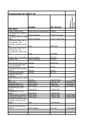


![Download in English [pdf 2Mb] - Foundation for Innovative New ...](https://img.yumpu.com/49580359/1/184x260/download-in-english-pdf-2mb-foundation-for-innovative-new-.jpg?quality=85)


![New laboratory diagnostic tools for tuberculosis control [.pdf]](https://img.yumpu.com/43339906/1/190x135/new-laboratory-diagnostic-tools-for-tuberculosis-control-pdf.jpg?quality=85)