Advanced Trauma Life Support ATLS Student Course Manual 2018
INITIAL PATIENT ASSESSMENT 45 Preload, the volume of venous blood return to the left and right sides of the heart, is determined by venous capacitance, volume status, and the difference between mean venous systemic pressure and right atrial pressure. This pressure differential determines venous flow. The venous system can be considered a reservoir, or capacitance, system in which the volume of blood is divided into two components: 1. The first component represents the volume of blood that would remain in this capacitance circuit if the pressure in the system were zero. This component does not contribute to the mean systemic venous pressure. 2. The second component represents the venous volume that contributes to the mean systemic venous pressure. Nearly 70% of the body’s total blood volume is estimated to be located in the venous circuit. Compliance of the venous system involves a relationship between venous volume and venous pressure. This pressure gradient drives venous flow and therefore the volume of venous return to the heart. Blood loss depletes this component of venous volume and reduces the pressure gradient; consequently, venous return is reduced. The volume of venous blood returned to the heart determines myocardial muscle fiber length after ventricular filling at the end of diastole. According to Starling’s law, muscle fiber length is related to the contractile properties of myocardial muscle. Myocardial contractility is the pump that drives the system. Afterload, also known as peripheral vascular resistance, is systemic. Simply stated, afterload is resistance to the forward flow of blood. Blood Loss Pathophysiology Early circulatory responses to blood loss are compensatory and include progressive vasoconstriction of cutaneous, muscular, and visceral circulation to preserve blood flow to the kidneys, heart, and brain. The usual response to acute circulating volume depletion is an increase in heart rate in an attempt to preserve cardiac output. In most cases, tachycardia is the earliest measurable circulatory sign of shock. The release of endogenous catecholamines increases peripheral vascular resistance, which in turn increases diastolic blood pressure and reduces pulse pressure. However, this increase in pressure does little to increase organ perfusion and tissue oxygenation. For patients in early hemorrhagic shock, venous return is preserved to some degree by the compensatory mechanism of contraction of the volume of blood in the venous system. This compensatory mechanism is limited. The most effective method of restoring adequate cardiac output, end-organ perfusion, and tissue oxygenation is to restore venous return to normal by locating and stopping the source of bleeding. Volume repletion will allow recovery from the shock state only when the bleeding has stopped. At the cellular level, inadequately perfused and poorly oxygenated cells are deprived of essential substrates for normal aerobic metabolism and energy production. Initially, compensation occurs by shifting to anaerobic metabolism, resulting in the formation of lactic acid and development of metabolic acidosis. If shock is prolonged, subsequent end-organ damage and multiple organ dysfunction may result. Administration of an appropriate quantity of isotonic electrolyte solutions, blood, and blood products helps combat this process. Treatment must focus on reversing the shock state by stopping the bleeding and providing adequate oxygenation, ventilation, and appropriate fluid resuscitation. Rapid intravenous access must be obtained. Definitive control of hemorrhage and restoration of adequate circulating volume are the goals of treating hemorrhagic shock. Vasopressors are contraindicated as a first-line treatment of hemorrhagic shock because they worsen tissue perfusion. Frequently monitor the patient’s indices of perfusion to detect any deterioration in the patient’s condition as early as possible so it can be reversed. Monitoring also allows for evaluation of the patient’s response to therapy. Reassessment helps clinicians identify patients in compensated shock and those who are unable to mount a compensatory response before cardiovascular collapse occurs. Most injured patients who are in hemorrhagic shock require early surgical intervention or angioembolization to reverse the shock state. The presence of shock in a trauma patient warrants the immediate involvement of a surgeon. Strongly consider arranging for early transfer of these patients to a trauma center when they present to hospitals that are not equipped to manage their injuries. Initial Patient Assessment Optimally, clinicians recognize the shock state during the initial patient assessment. To do so, they must be familiar with the clinical differentiation of causes of shock—chiefly, hemorrhagic and non-hemorrhagic shock. n BACK TO TABLE OF CONTENTS
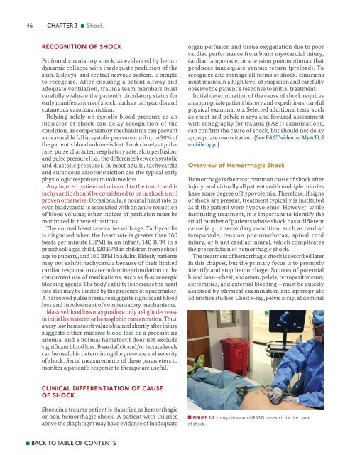
46 CHAPTER 3 n Shock Recognition of Shock Profound circulatory shock, as evidenced by hemodynamic collapse with inadequate perfusion of the skin, kidneys, and central nervous system, is simple to recognize. After ensuring a patent airway and adequate ventilation, trauma team members must carefully evaluate the patient’s circulatory status for early manifestations of shock, such as tachycardia and cutaneous vasoconstriction. Relying solely on systolic blood pressure as an indicator of shock can delay recognition of the condition, as compensatory mechanisms can prevent a measurable fall in systolic pressure until up to 30% of the patient’s blood volume is lost. Look closely at pulse rate, pulse character, respiratory rate, skin perfusion, and pulse pressure (i.e., the difference between systolic and diastolic pressure). In most adults, tachycardia and cutaneous vasoconstriction are the typical early physiologic responses to volume loss. Any injured patient who is cool to the touch and is tachycardic should be considered to be in shock until proven otherwise. Occasionally, a normal heart rate or even bradycardia is associated with an acute reduction of blood volume; other indices of perfusion must be monitored in these situations. The normal heart rate varies with age. Tachycardia is diagnosed when the heart rate is greater than 160 beats per minute (BPM) in an infant, 140 BPM in a preschool-aged child, 120 BPM in children from school age to puberty, and 100 BPM in adults. Elderly patients may not exhibit tachycardia because of their limited cardiac response to catecholamine stimulation or the concurrent use of medications, such as ß-adrenergic blocking agents. The body’s ability to increase the heart rate also may be limited by the presence of a pacemaker. A narrowed pulse pressure suggests significant blood loss and involvement of compensatory mechanisms. Massive blood loss may produce only a slight decrease in initial hematocrit or hemoglobin concentration. Thus, a very low hematocrit value obtained shortly after injury suggests either massive blood loss or a preexisting anemia, and a normal hematocrit does not exclude significant blood loss. Base deficit and/or lactate levels can be useful in determining the presence and severity of shock. Serial measurements of these parameters to monitor a patient’s response to therapy are useful. organ perfusion and tissue oxygenation due to poor cardiac performance from blunt myocardial injury, cardiac tamponade, or a tension pneumothorax that produces inadequate venous return (preload). To recognize and manage all forms of shock, clinicians must maintain a high level of suspicion and carefully observe the patient’s response to initial treatment. Initial determination of the cause of shock requires an appropriate patient history and expeditious, careful physical examination. Selected additional tests, such as chest and pelvic x-rays and focused assessment with sonography for trauma (FAST) examinations, can confirm the cause of shock, but should not delay appropriate resuscitation. (See FAST video on MyATLS mobile app.) Overview of Hemorrhagic Shock Hemorrhage is the most common cause of shock after injury, and virtually all patients with multiple injuries have some degree of hypovolemia. Therefore, if signs of shock are present, treatment typically is instituted as if the patient were hypovolemic. However, while instituting treatment, it is important to identify the small number of patients whose shock has a different cause (e.g., a secondary condition, such as cardiac tamponade, tension pneumothorax, spinal cord injury, or blunt cardiac injury), which complicates the presentation of hemorrhagic shock. The treatment of hemorrhagic shock is described later in this chapter, but the primary focus is to promptly identify and stop hemorrhage. Sources of potential blood loss—chest, abdomen, pelvis, retroperitoneum, extremities, and external bleeding—must be quickly assessed by physical examination and appropriate adjunctive studies. Chest x-ray, pelvic x-ray, abdominal Clinical Differentiation of Cause of Shock Shock in a trauma patient is classified as hemorrhagic or non-hemorrhagic shock. A patient with injuries above the diaphragm may have evidence of inadequate n FIGURE 3-2 Using ultrasound (FAST) to search for the cause of shock. n BACK TO TABLE OF CONTENTS
- Page 49 and 50: xlviii DETAILED CONTENTS Teamwork 5
- Page 51: l DETAILED CONTENTS Introduction 21
- Page 55 and 56: 1 INITIAL ASSESSMENT AND MANAGEMENT
- Page 57 and 58: 4 CHAPTER 1 n Initial Assessment an
- Page 59 and 60: 6 CHAPTER 1 n Initial Assessment an
- Page 61 and 62: 8 CHAPTER 1 n Initial Assessment an
- Page 63 and 64: 10 CHAPTER 1 n Initial Assessment a
- Page 65 and 66: 12 CHAPTER 1 n Initial Assessment a
- Page 67 and 68: 14 CHAPTER 1 n Initial Assessment a
- Page 69 and 70: 16 CHAPTER 1 n Initial Assessment a
- Page 71 and 72: 18 CHAPTER 1 n Initial Assessment a
- Page 73 and 74: 20 CHAPTER 1 n Initial Assessment a
- Page 75 and 76: 2 AIRWAY AND VENTILATORY MANAGEMENT
- Page 77 and 78: 24 CHAPTER 2 n Airway and Ventilato
- Page 79 and 80: 26 CHAPTER 2 n Airway and Ventilato
- Page 81 and 82: 28 CHAPTER 2 n Airway and Ventilato
- Page 83 and 84: 30 CHAPTER 2 n Airway and Ventilato
- Page 85 and 86: 32 CHAPTER 2 n Airway and Ventilato
- Page 87 and 88: 34 CHAPTER 2 n Airway and Ventilato
- Page 89 and 90: 36 CHAPTER 2 n Airway and Ventilato
- Page 91 and 92: 38 CHAPTER 2 n Airway and Ventilato
- Page 93 and 94: 40 CHAPTER 2 n Airway and Ventilato
- Page 95 and 96: 3 SHOCK The first step in the initi
- Page 97: 44 CHAPTER 3 n Shock The first step
- Page 101 and 102: 48 CHAPTER 3 n Shock However, the a
- Page 103 and 104: 50 CHAPTER 3 n Shock Class II Hemor
- Page 105 and 106: 52 CHAPTER 3 n Shock hypothermia, a
- Page 107 and 108: 54 CHAPTER 3 n Shock replacement du
- Page 109 and 110: 56 CHAPTER 3 n Shock aggregation an
- Page 111 and 112: 58 CHAPTER 3 n Shock Presence of Pa
- Page 113 and 114: 60 CHAPTER 3 n Shock rate can be ac
- Page 115 and 116: 4 THORACIC TRAUMA Thoracic injury i
- Page 117 and 118: 64 CHAPTER 4 n Thoracic Trauma Thor
- Page 119 and 120: 66 CHAPTER 4 n Thoracic Trauma shoc
- Page 121 and 122: 68 CHAPTER 4 n Thoracic Trauma comp
- Page 123 and 124: 70 CHAPTER 4 n Thoracic Trauma Norm
- Page 125 and 126: 72 CHAPTER 4 n Thoracic Trauma Seco
- Page 127 and 128: 74 CHAPTER 4 n Thoracic Trauma A B
- Page 129 and 130: 76 CHAPTER 4 n Thoracic Trauma Spec
- Page 131 and 132: 78 CHAPTER 4 n Thoracic Trauma temp
- Page 133 and 134: 80 CHAPTER 4 n Thoracic Trauma 18.
- Page 135 and 136: ABDOMINAL AND 5 PELVIC TRAUMA When
- Page 137 and 138: 84 CHAPTER 5 n Abdominal and Pelvic
- Page 139 and 140: 86 CHAPTER 5 n Abdominal and Pelvic
- Page 141 and 142: 88 CHAPTER 5 n Abdominal and Pelvic
- Page 143 and 144: 90 CHAPTER 5 n Abdominal and Pelvic
- Page 145 and 146: 92 CHAPTER 5 n Abdominal and Pelvic
- Page 147 and 148: 94 CHAPTER 5 n Abdominal and Pelvic
46<br />
CHAPTER 3 n Shock<br />
Recognition of Shock<br />
Profound circulatory shock, as evidenced by hemodynamic<br />
collapse with inadequate perfusion of the<br />
skin, kidneys, and central nervous system, is simple<br />
to recognize. After ensuring a patent airway and<br />
adequate ventilation, trauma team members must<br />
carefully evaluate the patient’s circulatory status for<br />
early manifestations of shock, such as tachycardia and<br />
cutaneous vasoconstriction.<br />
Relying solely on systolic blood pressure as an<br />
indicator of shock can delay recognition of the<br />
condition, as compensatory mechanisms can prevent<br />
a measurable fall in systolic pressure until up to 30% of<br />
the patient’s blood volume is lost. Look closely at pulse<br />
rate, pulse character, respiratory rate, skin perfusion,<br />
and pulse pressure (i.e., the difference between systolic<br />
and diastolic pressure). In most adults, tachycardia<br />
and cutaneous vasoconstriction are the typical early<br />
physiologic responses to volume loss.<br />
Any injured patient who is cool to the touch and is<br />
tachycardic should be considered to be in shock until<br />
proven otherwise. Occasionally, a normal heart rate or<br />
even bradycardia is associated with an acute reduction<br />
of blood volume; other indices of perfusion must be<br />
monitored in these situations.<br />
The normal heart rate varies with age. Tachycardia<br />
is diagnosed when the heart rate is greater than 160<br />
beats per minute (BPM) in an infant, 140 BPM in a<br />
preschool-aged child, 120 BPM in children from school<br />
age to puberty, and 100 BPM in adults. Elderly patients<br />
may not exhibit tachycardia because of their limited<br />
cardiac response to catecholamine stimulation or the<br />
concurrent use of medications, such as ß-adrenergic<br />
blocking agents. The body’s ability to increase the heart<br />
rate also may be limited by the presence of a pacemaker.<br />
A narrowed pulse pressure suggests significant blood<br />
loss and involvement of compensatory mechanisms.<br />
Massive blood loss may produce only a slight decrease<br />
in initial hematocrit or hemoglobin concentration. Thus,<br />
a very low hematocrit value obtained shortly after injury<br />
suggests either massive blood loss or a preexisting<br />
anemia, and a normal hematocrit does not exclude<br />
significant blood loss. Base deficit and/or lactate levels<br />
can be useful in determining the presence and severity<br />
of shock. Serial measurements of these parameters to<br />
monitor a patient’s response to therapy are useful.<br />
organ perfusion and tissue oxygenation due to poor<br />
cardiac performance from blunt myocardial injury,<br />
cardiac tamponade, or a tension pneumothorax that<br />
produces inadequate venous return (preload). To<br />
recognize and manage all forms of shock, clinicians<br />
must maintain a high level of suspicion and carefully<br />
observe the patient’s response to initial treatment.<br />
Initial determination of the cause of shock requires<br />
an appropriate patient history and expeditious, careful<br />
physical examination. Selected additional tests, such<br />
as chest and pelvic x-rays and focused assessment<br />
with sonography for trauma (FAST) examinations,<br />
can confirm the cause of shock, but should not delay<br />
appropriate resuscitation. (See FAST video on My<strong>ATLS</strong><br />
mobile app.)<br />
Overview of Hemorrhagic Shock<br />
Hemorrhage is the most common cause of shock after<br />
injury, and virtually all patients with multiple injuries<br />
have some degree of hypovolemia. Therefore, if signs<br />
of shock are present, treatment typically is instituted<br />
as if the patient were hypovolemic. However, while<br />
instituting treatment, it is important to identify the<br />
small number of patients whose shock has a different<br />
cause (e.g., a secondary condition, such as cardiac<br />
tamponade, tension pneumothorax, spinal cord<br />
injury, or blunt cardiac injury), which complicates<br />
the presentation of hemorrhagic shock.<br />
The treatment of hemorrhagic shock is described later<br />
in this chapter, but the primary focus is to promptly<br />
identify and stop hemorrhage. Sources of potential<br />
blood loss—chest, abdomen, pelvis, retroperitoneum,<br />
extremities, and external bleeding—must be quickly<br />
assessed by physical examination and appropriate<br />
adjunctive studies. Chest x-ray, pelvic x-ray, abdominal<br />
Clinical Differentiation of Cause<br />
of Shock<br />
Shock in a trauma patient is classified as hemorrhagic<br />
or non-hemorrhagic shock. A patient with injuries<br />
above the diaphragm may have evidence of inadequate<br />
n FIGURE 3-2 Using ultrasound (FAST) to search for the cause<br />
of shock.<br />
n BACK TO TABLE OF CONTENTS


