

Advanced Trauma Life Support ATLS Student Course Manual 2018

Create successful ePaper yourself
Turn your PDF publications into a flip-book with our unique Google optimized e-Paper software.
68<br />
CHAPTER 4 n Thoracic <strong>Trauma</strong><br />
compromise respiratory efforts by compressing the<br />
lung and preventing adequate oxygenation and ventilation.<br />
Insert a chest tube to improve ventilation and<br />
oxygenation, request emergent surgical consultation,<br />
and begin appropriate resuscitation. Massive<br />
acute accumulation of blood produces hypotension<br />
and shock and will be discussed further in the<br />
section below.<br />
n TABLE 4-1 outlines the different presentations of<br />
tension pneumothorax and massive hemothorax.<br />
Circulation Problems<br />
Major thoracic injuries that affect circulation and<br />
should be recognized and addressed during the primary<br />
survey are massive hemothorax, cardiac tamponade,<br />
and traumatic circulatory arrest.<br />
Pulseless electrical activity (PEA) is manifested by<br />
an electrocardiogram (ECG) that shows a rhythm<br />
while the patient has no identifiable pulse. This<br />
dysrhythmia can be present with cardiac tamponade,<br />
tension pneumothorax, or profound hypovolemia.<br />
Severe blunt injury can result in blunt rupture<br />
of the atria or the ventricles, and the only manifestation<br />
may be PEA arrest. Other causes of PEA<br />
arrest include hypovolemia, hypoxia, hydrogen<br />
ion (acidosis), hypokalemia/ hyperkalemia, hypoglycemia,<br />
hypothermia, toxins, cardiac tamponade,<br />
tension pneumothorax, and thrombosis (coronary<br />
or pulmonary).<br />
Inspect the skin for mottling, cyanosis, and pallor.<br />
Neck veins should be assessed for distention, although<br />
they may not be distended in patients with concomitant<br />
hypovolemia. Listen for the regularity and quality of<br />
the heartbeat. Assess a central pulse for quality, rate,<br />
and regularity. In patients with hypovolemia, the distal<br />
pulses may be absent because of volume depletion.<br />
Palpate the skin to assess its temperature and determine<br />
whether it is dry or sweaty.<br />
Measure blood pressure and pulse pressure, and<br />
monitor the patient with electrocardiography and<br />
pulse oximetry. Patients with blunt chest injury are at<br />
risk for myocardial dysfunction, which is increased by<br />
the presence of hypoxia and acidosis. Dysrhythmias<br />
should be managed according to standard protocols.<br />
Massive Hemothorax<br />
Massive hemothorax results from the rapid<br />
accumulation of more than 1500 mL of blood or onethird<br />
or more of the patient’s blood volume in the chest<br />
cavity (n FIGURE 4-5). It is most commonly caused by a<br />
penetrating wound that disrupts the systemic or hilar<br />
vessels, although massive hemothorax can also result<br />
from blunt trauma.<br />
In patients with massive hemothorax, the neck<br />
veins may be flat due to severe hypovolemia, or they<br />
may be distended if there is an associated tension<br />
pneumothorax. Rarely will the mechanical effects of<br />
massive intrathoracic blood shift the mediastinum<br />
enough to cause distended neck veins. A massive<br />
hemothorax is suggested when shock is associated with<br />
the absence of breath sounds or dullness to percussion<br />
on one side of the chest.<br />
Massive hemothorax is initially managed by<br />
simultaneously restoring blood volume and<br />
decompressing the chest cavity. Establish largecaliber<br />
intravenous lines, infuse crystalloid, and begin<br />
transfusion of uncrossmatched or type-specific blood<br />
as soon as possible. When appropriate, blood from<br />
the chest tube can be collected in a device suitable for<br />
autotransfusion. A single chest tube (28-32 French)<br />
is inserted, usually at the fifth intercostal space, just<br />
anterior to the midaxillary line, and rapid restoration<br />
of volume continues as decompression of the chest<br />
cavity is completed. The immediate return of 1500<br />
mL or more of blood generally indicates the need for<br />
urgent thoracotomy.<br />
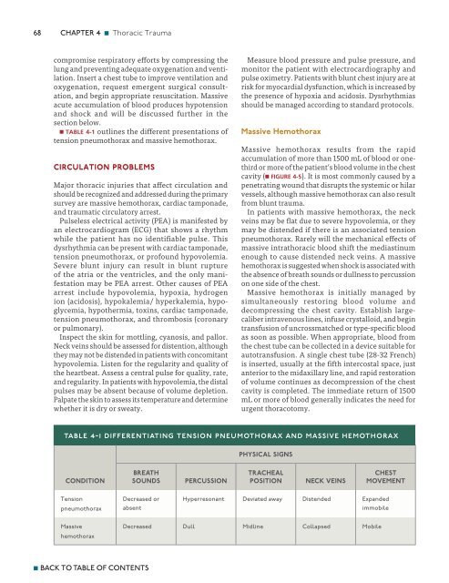
table 4-1 differentiating tension pneumothorax and massive hemothorax<br />
PHYSICAL SIGNS<br />
CONDITION<br />
BREATH<br />
SOUNDS<br />
PERCUSSION<br />
TRACHEAL<br />
POSITION<br />
NECK VEINS<br />
CHEST<br />
MOVEMENT<br />
Tension<br />
pneumothorax<br />
Decreased or<br />
absent<br />
Hyperresonant Deviated away Distended Expanded<br />
immobile<br />
Massive<br />
hemothorax<br />
Decreased Dull Midline Collapsed Mobile<br />
n BACK TO TABLE OF CONTENTS


