- Page 1 and 2:
TENTH EDITION ATLS ® Advanced Trau
- Page 3 and 4:
Chair of Committee on Trauma: Ronal
- Page 6:
FOREWORD My first exposure to Advan
- Page 9 and 10:
viii PREFACE MyATLS Mobile Applicat
- Page 11 and 12:
x PREFACE Gary A. Vercruysse, MD, F
- Page 13 and 14:
xii PREFACE Jacqueline Bustraan, MS
- Page 16 and 17:
ACKNOWLEDGMENTS It is clear that ma
- Page 18 and 19:
xvii ACKNOWLEDGMENTS Bertil Bouillo
- Page 20 and 21:
xix ACKNOWLEDGMENTS Oscar Guillamon
- Page 22 and 23:
xxi ACKNOWLEDGMENTS Mahesh Misra, M
- Page 24 and 25:
xxiii ACKNOWLEDGMENTS James Vosswin
- Page 26 and 27:
xxv ACKNOWLEDGMENTS James A. Geilin
- Page 28:
xxvii ACKNOWLEDGMENTS Tone Slåke R
- Page 31 and 32:
xxx COURSE OVERVIEW m. Protection o
- Page 33 and 34:
xxxii COURSE OVERVIEW Atls and Trau
- Page 35 and 36:
xxxiv COURSE OVERVIEW a systematize
- Page 37 and 38:
xxxvi COURSE OVERVIEW 69. Switzerla
- Page 39 and 40:
xxxviii COURSE OVERVIEW United Stat
- Page 41 and 42:
xl COURSE OVERVIEW 67. Hendrickson
- Page 43 and 44:
xlii COURSE OVERVIEW 122. Palusci V
- Page 46:
BRIEF CONTENTS Foreword Preface Ack
- Page 49 and 50:
xlviii DETAILED CONTENTS Teamwork 5
- Page 51:
l DETAILED CONTENTS Introduction 21
- Page 55 and 56:
1 INITIAL ASSESSMENT AND MANAGEMENT
- Page 57 and 58:
4 CHAPTER 1 n Initial Assessment an
- Page 59 and 60:
6 CHAPTER 1 n Initial Assessment an
- Page 61 and 62:
8 CHAPTER 1 n Initial Assessment an
- Page 63 and 64:
10 CHAPTER 1 n Initial Assessment a
- Page 65 and 66:
12 CHAPTER 1 n Initial Assessment a
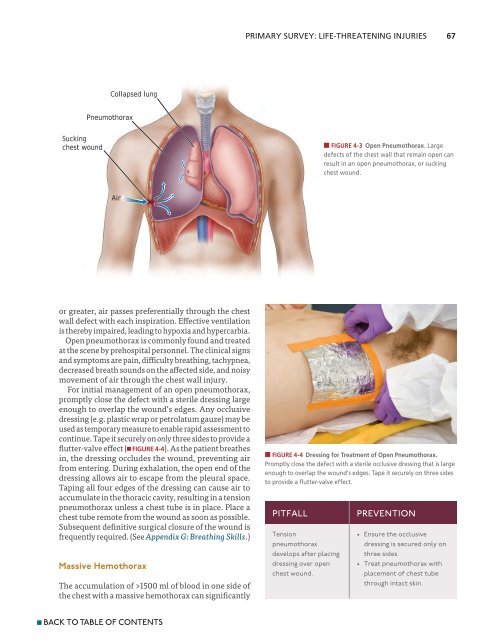
- Page 67 and 68:
14 CHAPTER 1 n Initial Assessment a
- Page 69 and 70: 16 CHAPTER 1 n Initial Assessment a
- Page 71 and 72: 18 CHAPTER 1 n Initial Assessment a
- Page 73 and 74: 20 CHAPTER 1 n Initial Assessment a
- Page 75 and 76: 2 AIRWAY AND VENTILATORY MANAGEMENT
- Page 77 and 78: 24 CHAPTER 2 n Airway and Ventilato
- Page 79 and 80: 26 CHAPTER 2 n Airway and Ventilato
- Page 81 and 82: 28 CHAPTER 2 n Airway and Ventilato
- Page 83 and 84: 30 CHAPTER 2 n Airway and Ventilato
- Page 85 and 86: 32 CHAPTER 2 n Airway and Ventilato
- Page 87 and 88: 34 CHAPTER 2 n Airway and Ventilato
- Page 89 and 90: 36 CHAPTER 2 n Airway and Ventilato
- Page 91 and 92: 38 CHAPTER 2 n Airway and Ventilato
- Page 93 and 94: 40 CHAPTER 2 n Airway and Ventilato
- Page 95 and 96: 3 SHOCK The first step in the initi
- Page 97 and 98: 44 CHAPTER 3 n Shock The first step
- Page 99 and 100: 46 CHAPTER 3 n Shock Recognition of
- Page 101 and 102: 48 CHAPTER 3 n Shock However, the a
- Page 103 and 104: 50 CHAPTER 3 n Shock Class II Hemor
- Page 105 and 106: 52 CHAPTER 3 n Shock hypothermia, a
- Page 107 and 108: 54 CHAPTER 3 n Shock replacement du
- Page 109 and 110: 56 CHAPTER 3 n Shock aggregation an
- Page 111 and 112: 58 CHAPTER 3 n Shock Presence of Pa
- Page 113 and 114: 60 CHAPTER 3 n Shock rate can be ac
- Page 115 and 116: 4 THORACIC TRAUMA Thoracic injury i
- Page 117 and 118: 64 CHAPTER 4 n Thoracic Trauma Thor
- Page 119: 66 CHAPTER 4 n Thoracic Trauma shoc
- Page 123 and 124: 70 CHAPTER 4 n Thoracic Trauma Norm
- Page 125 and 126: 72 CHAPTER 4 n Thoracic Trauma Seco
- Page 127 and 128: 74 CHAPTER 4 n Thoracic Trauma A B
- Page 129 and 130: 76 CHAPTER 4 n Thoracic Trauma Spec
- Page 131 and 132: 78 CHAPTER 4 n Thoracic Trauma temp
- Page 133 and 134: 80 CHAPTER 4 n Thoracic Trauma 18.
- Page 135 and 136: ABDOMINAL AND 5 PELVIC TRAUMA When
- Page 137 and 138: 84 CHAPTER 5 n Abdominal and Pelvic
- Page 139 and 140: 86 CHAPTER 5 n Abdominal and Pelvic
- Page 141 and 142: 88 CHAPTER 5 n Abdominal and Pelvic
- Page 143 and 144: 90 CHAPTER 5 n Abdominal and Pelvic
- Page 145 and 146: 92 CHAPTER 5 n Abdominal and Pelvic
- Page 147 and 148: 94 CHAPTER 5 n Abdominal and Pelvic
- Page 149 and 150: 96 CHAPTER 5 n Abdominal and Pelvic
- Page 151 and 152: 98 CHAPTER 5 n Abdominal and Pelvic
- Page 153 and 154: 100 CHAPTER 5 n Abdominal and Pelvi
- Page 155 and 156: 6 HEAD TRAUMA The primary goal of t
- Page 157 and 158: 104 CHAPTER 6 n Head Trauma Head in
- Page 159 and 160: 106 CHAPTER 6 n Head Trauma fibrous
- Page 161 and 162: 108 CHAPTER 6 n Head Trauma n FIGUR
- Page 163 and 164: 110 CHAPTER 6 n Head Trauma table 6
- Page 165 and 166: 112 CHAPTER 6 n Head Trauma covery.
- Page 167 and 168: 114 CHAPTER 6 n Head Trauma n FIGUR
- Page 169 and 170: 116 CHAPTER 6 n Head Trauma n FIGUR
- Page 171 and 172:
118 CHAPTER 6 n Head Trauma n FIGUR
- Page 173 and 174:
120 CHAPTER 6 n Head Trauma necessa
- Page 175 and 176:
122 CHAPTER 6 n Head Trauma Use 0.2
- Page 177 and 178:
124 CHAPTER 6 n Head Trauma not rea
- Page 179:
126 CHAPTER 6 n Head Trauma 18. Mar
- Page 182 and 183:
CHAPTER 7 Outline Objectives iNtrod
- Page 184 and 185:
ANATOMY AND PHYSIOLOGY 131 B A n FI
- Page 186 and 187:
RIGHT INTERNATIONAL STANDARDS FOR N
- Page 188 and 189:
DOCUMENTATION OF SPINAL CORD INJURI
- Page 190 and 191:
SPECIFIC TYPES OF SPINAL INJURIES 1
- Page 192 and 193:
RADIOGRAPHIC EVALUATION 139 Penetra
- Page 194 and 195:
GENERAL MANAGEMENT 141 When the low
- Page 196 and 197:
GENERAL MANAGEMENT 143 them to the
- Page 198 and 199:
BIBLIOGRAPHY 145 Bibliography 1. Bi
- Page 201 and 202:
8 MUSCULOSKELETAL TRAUMA Injuries t
- Page 203 and 204:
150 CHAPTER 8 n Musculoskeletal Tra
- Page 205 and 206:
152 CHAPTER 8 n Musculoskeletal Tra
- Page 207 and 208:
154 CHAPTER 8 n Musculoskeletal Tra
- Page 209 and 210:
156 CHAPTER 8 n Musculoskeletal Tra
- Page 211 and 212:
158 CHAPTER 8 n Musculoskeletal Tra
- Page 213 and 214:
160 CHAPTER 8 n Musculoskeletal Tra
- Page 215 and 216:
162 CHAPTER 8 n Musculoskeletal Tra
- Page 217 and 218:
164 CHAPTER 8 n Musculoskeletal Tra
- Page 219 and 220:
166 CHAPTER 8 n Musculoskeletal Tra
- Page 221 and 222:
9 THERMAL INJURIES The most signifi
- Page 223 and 224:
170 CHAPTER 9 n Thermal Injuries Th
- Page 225 and 226:
172 CHAPTER 9 n Thermal Injuries Ch
- Page 227 and 228:
174 CHAPTER 9 n Thermal Injuries Pi
- Page 229 and 230:
176 CHAPTER 9 n Thermal Injuries th
- Page 231 and 232:
178 CHAPTER 9 n Thermal Injuries ju
- Page 233 and 234:
180 CHAPTER 9 n Thermal Injuries Im
- Page 235 and 236:
182 CHAPTER 9 n Thermal Injuries lo
- Page 237 and 238:
184 CHAPTER 9 n Thermal Injuries 4.
- Page 239 and 240:
10 PEDIATRIC TRAUMA Injury remains
- Page 241 and 242:
188 CHAPTER 10 n Pediatric Trauma I
- Page 243 and 244:
190 CHAPTER 10 n Pediatric Trauma c
- Page 245 and 246:
192 CHAPTER 10 n Pediatric Trauma A
- Page 247 and 248:
194 CHAPTER 10 n Pediatric Trauma b
- Page 249 and 250:
196 CHAPTER 10 n Pediatric Trauma b
- Page 251 and 252:
198 CHAPTER 10 n Pediatric Trauma t
- Page 253 and 254:
200 CHAPTER 10 n Pediatric Trauma f
- Page 255 and 256:
202 CHAPTER 10 n Pediatric Trauma t
- Page 257 and 258:
204 CHAPTER 10 n Pediatric Trauma G
- Page 259 and 260:
206 CHAPTER 10 n Pediatric Trauma C
- Page 261 and 262:
208 CHAPTER 10 n Pediatric Trauma
- Page 263 and 264:
210 CHAPTER 10 n Pediatric Trauma 1
- Page 265:
212 CHAPTER 10 n Pediatric Trauma 6
- Page 268 and 269:
CHAPTER 11 Outline Objectives iNtro
- Page 270 and 271:
PRIMARY SURVEY WITH RESUSCITATION 2
- Page 272 and 273:
PRIMARY SURVEY WITH RESUSCITATION 2
- Page 274 and 275:
SPECIFIC INJURIES 221 table 11-6 ph
- Page 276 and 277:
BIBLIOGRAPHY 223 comprise only 12%
- Page 279 and 280:
12 TRAUMA IN PREGNANCY AND INTIMATE
- Page 281 and 282:
228 CHAPTER 12 n Trauma in Pregnanc
- Page 283 and 284:
230 CHAPTER 12 n Trauma in Pregnanc
- Page 285 and 286:
232 CHAPTER 12 n Trauma in Pregnanc
- Page 287 and 288:
234 CHAPTER 12 n Trauma in Pregnanc
- Page 289 and 290:
236 CHAPTER 12 n Trauma in Pregnanc
- Page 291 and 292:
238 CHAPTER 12 n Trauma in Pregnanc
- Page 293 and 294:
240 CHAPTER 13 n Transfer to Defini
- Page 295 and 296:
242 CHAPTER 13 n Transfer to Defini
- Page 297 and 298:
244 CHAPTER 13 n Transfer to Defini
- Page 299 and 300:
246 CHAPTER 13 n Transfer to Defini
- Page 301 and 302:
248 CHAPTER 13 n Transfer to Defini
- Page 303 and 304:
250 CHAPTER 13 n Transfer to Defini
- Page 305:
252 CHAPTER 13 n Transfer to Defini
- Page 310 and 311:
Appendix A OCULAR TRAUMA OBJECTIVES
- Page 312 and 313:
259 APPENDIX A n Ocular Trauma In c
- Page 314 and 315:
261 APPENDIX A n Ocular Trauma to t
- Page 318 and 319:
Appendix B HYPOTHERMIA AND HEAT INJ
- Page 320 and 321:
267 APPENDIX B n Hypothermia and He
- Page 322 and 323:
269 APPENDIX B n Hypothermia and He
- Page 324 and 325:
271 APPENDIX B n Hypothermia and He
- Page 328 and 329:
Appendix C TRAUMA CARE IN MASS-CASU
- Page 330 and 331:
277 APPENDIX C n Trauma Care in Mas
- Page 332 and 333:
279 APPENDIX C n Trauma Care in Mas
- Page 334 and 335:
281 APPENDIX C n Trauma Care in Mas
- Page 336 and 337:
283 APPENDIX C n Trauma Care in Mas
- Page 338 and 339:
285 APPENDIX C n Trauma Care in Mas
- Page 342 and 343:
Appendix D DISASTER PREPAREDNESS AN
- Page 344 and 345:
291 APPENDIX D n Disaster Preparedn
- Page 346 and 347:
293 APPENDIX D n Disaster Preparedn
- Page 348 and 349:
295 APPENDIX D n Disaster Preparedn
- Page 350 and 351:
297 APPENDIX D n Disaster Preparedn
- Page 352 and 353:
Pitfall Inadequate security Failed
- Page 356 and 357:
Appendix E ATLS AND TRAUMA TEAM RES
- Page 358 and 359:
305 APPENDIX E n ATLS and Trauma Te
- Page 360 and 361:
307 APPENDIX E n ATLS and Trauma Te
- Page 362 and 363:
309 APPENDIX E n ATLS and Trauma Te
- Page 364 and 365:
311 APPENDIX E n ATLS and Trauma Te
- Page 366 and 367:
313 APPENDIX E n ATLS and Trauma Te
- Page 370 and 371:
Appendix F TRIAGE SCENARIOS OBJECTI
- Page 372 and 373:
319 APPENDIX F n Triage Scenarios T
- Page 374 and 375:
321 APPENDIX F n Triage Scenarios T
- Page 376 and 377:
323 APPENDIX F n Triage Scenarios T
- Page 378 and 379:
325 APPENDIX F n Triage Scenarios T
- Page 380 and 381:
327 APPENDIX F n Triage Scenarios T
- Page 382 and 383:
329 APPENDIX F n Triage Scenarios T
- Page 384 and 385:
331 APPENDIX F n Triage Scenarios T
- Page 386:
333 APPENDIX F n Triage Scenarios 4
- Page 390 and 391:
Skill Station A AIRWAY Part 1: Basi
- Page 392 and 393:
339 APPENDIX G n Skills One-Person
- Page 394 and 395:
341 APPENDIX G n Skills STEP 5. Con
- Page 396:
343 APPENDIX G n Skills STEP 13. Co
- Page 399 and 400:
346 APPENDIX G n Skills STEP 1. D
- Page 402 and 403:
Skill Station C CIRCULATION LEARNIN
- Page 404 and 405:
351 APPENDIX G n Skills manual trac
- Page 406 and 407:
353 APPENDIX G n Skills STEP 3. Aft
- Page 408:
355 APPENDIX G n Skills STEP 2. If
- Page 411 and 412:
358 APPENDIX G n Skills A. Note fac
- Page 413 and 414:
360 APPENDIX G n Skills •• Flex
- Page 415:
362 APPENDIX G n Skills Utilization
- Page 419 and 420:
366 APPENDIX G n Skills n FIGURE G-
- Page 421:
368 APPENDIX G n Skills less than 1
- Page 425 and 426:
372 APPENDIX G n Skills G. Inspect
- Page 427:
374 APPENDIX G n Skills in the inju
- Page 431 and 432:
378 INDEX LTA for, 31-32, 32f Malla
- Page 433 and 434:
380 INDEX atlanto-occipital disloca
- Page 435 and 436:
382 INDEX Frostbite, 181-183, 182f
- Page 437 and 438:
384 INDEX Kussmaul’s sign, 69 Lac
- Page 439 and 440:
386 INDEX PEA. See Pulseless electr
- Page 441 and 442:
388 INDEX for musculoskeletal traum
- Page 443 and 444:
390 INDEX Tibial fractures, 163 Tou
- Page 445 and 446:
TRAUMA SCORES Correct triage is ess
- Page 447 and 448:
394 TRAUMA SCORES on Field Triage,
- Page 449 and 450:
396 INJURY PREVENTION Safety Admini
- Page 451 and 452:
398 INJURY PREVENTION providers to
- Page 453 and 454:
BIOMECHANICS OF INJURY Injuries occ
- Page 455 and 456:
402 BIOMECHANICS OF INJURY Ejection
- Page 457 and 458:
404 BIOMECHANICS OF INJURY •• A
- Page 459 and 460:
406 BIOMECHANICS OF INJURY in a 30-
- Page 461 and 462:
408 TETANUS IMMUNIZATION •• Wou
- Page 463 and 464:
410 TETANUS IMMUNIZATION Summary Gu
- Page 465 and 466:
SAMPLE TRAUMA FLOW SHEET Page 1 of
- Page 467 and 468:
414 SAMPLE TRAUMA FLOW SHEET Page 3
- Page 469 and 470:
416 SAMPLE TRAUMA FLOW SHEET Page 5
- Page 471 and 472:
418 SAMPLE TRAUMA FLOW SHEET Page 7
- Page 473 and 474:
420 SAMPLE TRAUMA FLOW SHEET Some h