

International Research Update September 2017 (6)

You also want an ePaper? Increase the reach of your titles
YUMPU automatically turns print PDFs into web optimized ePapers that Google loves.
MND<br />
ADVANCE<br />
AUSTRALIA<br />
ADVANCE<br />
INTERNATIONAL RESEARCH UPDATE<br />
Isabella Lambert-Smith, Australian Rotary Health PhD Scholar, University of Wollongong<br />
SEPTEMBER <strong>2017</strong><br />
MND research heats up<br />
As the weather warms up with the start of spring in Australia, so do the latest studies by<br />
motor neurone disease (MND) researchers around the world. Recently, researchers have<br />
solved a few mysteries about how some of the MND-causing genes contribute to motor<br />
neurone (MN) degeneration and discovered similarities between different diseases that give<br />
us clues on how to treat MND.<br />
FUS and SMN interact in gene splicing<br />
Although MND is commonly referred to as a single disease, it is actually a group of<br />
diseases that all involve the degeneration of MNs. Amyotrophic lateral sclerosis (ALS) is the<br />
most common form of MND. Spinal muscular atrophy (SMA) is a genetic disease that most<br />
often affects children. Like adult MND, SMA affects the MNs of the spinal cord, causing<br />
muscle weakness and wasting. SMA is caused when there is a deficiency of the MN protein<br />
called SMN. Changes in the gene encoding a protein called FUS are responsible for a<br />
significant fraction of ALS cases, and in many cases abnormal forms of the protein are<br />
found clumped together with other proteins in diseased MNs. FUS normally regulates a<br />
biological mechanism called gene splicing. In our DNA, about 40-60% of our 30,000 genes<br />
undergo splicing. This mechanism is important for cutting out regions of the DNA sequence<br />
that don’t actually code for proteins (see box). Abnormal FUS proteins are unable to<br />
regulate splicing, and also capture other components of the machinery that carry out<br />
splicing. This is strikingly similar to what happens to the SMN protein in SMA patients,<br />
suggesting that disrupted gene splicing in MNs may be a common theme in both diseases.<br />
Alessia Mirra and her colleagues in Rome, Italy, followed this lead and tested the relevance<br />
of FUS and SMN in a mouse model of FUS- associated ALS. They found that<br />
these FUS-ALS mice shared crucial<br />
molecular features that characterise<br />
mouse models of SMA, including defective<br />
splicing of genes that are vital for MNs. Of<br />
note, when Alessia’s team altered SMN<br />
levels in the MNs of these mice, the<br />
degeneration caused by FUS<br />
abnormalities stopped. These findings<br />
strongly suggest that a complex interplay<br />
exists between FUS and SMN in the<br />
regulation of gene splicing in MNs. Further<br />
research focus should therefore be<br />
directed towards strategies that target this<br />
pathway of gene splicing.<br />
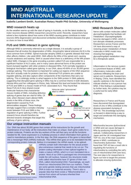
Our unique instruction manuals<br />
Our DNA has about 30,000 genes that contain all the instructions for the growth and<br />
maintenance of our body; all the instructions that make us unique. Each gene codes for a<br />
protein molecule. Proteins are the tiny workers of the cell that carry out all the functions<br />
needed for life. Gene expression is a multistep process in which information from genes is<br />
used to manufacture proteins. In the first step, the DNA sequence of the gene is transcribed<br />
into a messenger RNA (mRNA) molecule. This mRNA then goes on to be translated into the<br />
corresponding protein molecule. Before it is translated, the mRNA needs to be spliced.<br />
mRNAs contain both information for the actual protein<br />
plus extraneous material that needs to be cut out. The<br />
protein-coding information is then spliced together to<br />
make a mature mRNA, which is in turn translated into a<br />
protein. Most of our DNA is packaged into chromosomes<br />
inside the cell nucleus, however tiny structures inside<br />
cells called mitochondria have 37 genes of their own<br />
DNA, called mitochondrial DNA. Fascinatingly, we each<br />
inherit our mitochondrial DNA from our mothers. There<br />
are some genetic testing services that use mitochondrial<br />
DNA to trace our maternal ancestry. There’s also a<br />
popular idea that there exists a “Mitochondrial Eve", a<br />
woman from whom all humans inherited their<br />
mitochondrial DNA.<br />
By National Human Genome <strong>Research</strong> Institute<br />
[Public domain], via Wikimedia Commons<br />
MND <strong>Research</strong> Shorts<br />
Nerve cells contain molecules called<br />
glycosphingolipids that facilitate cell<br />
metabolism. Glycosphingolipids<br />
become damaged in MND, which in<br />
turn disrupts metabolic processes.<br />
<strong>Research</strong>ers across France and the<br />
UK have discovered a way of<br />
restoring proper metabolism of these<br />
molecules in MND model mice,<br />
showing that targeting<br />
glycosphingolipid metabolism may<br />
be a therapeutic option.<br />
Inflammation in the nervous system<br />
is a prominent feature of MND, with<br />
inflammatory molecules called<br />
cytokines infiltrating the brain and<br />
spinal cord in patients. <strong>Research</strong>ers<br />
in Beijing, China, have identified the<br />
specific cytokines in the blood that<br />
indicate whether or not a person has<br />
MND. If this finding is substantiated<br />
by further tests, the cytokine may be<br />
a useful tool for early MND<br />
diagnosis.<br />
<strong>Research</strong>ers in France and Russia<br />
have discovered that dysregulated<br />
levels of zinc in MNs contribute to the<br />
disease-associated clumping of<br />
TDP-43. This knowledge launches<br />
us forward in determining a way to<br />
target and ameliorate this damaging<br />
mechanism.<br />
Mitochondria become dysfunctional<br />
in MND and can cause damage in<br />
MNs by producing harmful free<br />
radicals. Collaborating researchers in<br />
Japan and the UK investigated how<br />
TDP-43, one of the main causative<br />
genes in MND, contributes to<br />
mitochondria dysfunction. They<br />
found that TDP-43 has a novel role in<br />
regulating the functions of<br />
mitochondria by controlling the cell’s<br />
use of mitochondrial DNA (see box).<br />
The next step is for researchers to<br />
figure out how to target this process<br />
to alleviate ensuing dysfunction.
New molecular tool kit to decipher<br />
calcium imbalance in MND<br />
All of the neurones in our body rely on the fine-tuned<br />
coordination of thousands of different molecules and metal<br />
ions (atoms with electric charge). One of the most important<br />
ions is calcium. Calcium is needed by nerves to transmit<br />
signals. It needs to be kept at precise concentrations in<br />
different compartments inside neurones in order for signals<br />
to be transmitted correctly. MNs are particularly sensitive to<br />
harmful cell overloads of calcium as they possess low levels<br />
of the proteins that regulate calcium concentration. Calcium<br />
overloads in neurones cause overstimulation and the<br />
generation of free radicals and other toxins that cause cell<br />
damage and death. This phenomenon is referred to as<br />
excitotoxicity and it is one of the main contributors to MN<br />
injury in MND. Rosa Pia Norante and her fellow researchers<br />
in Padova, Italy are working to understand the calciumrelated<br />
routes of MN dysfunction and damage. They<br />
generated a molecular tool kit of probes that target calcium<br />
in different compartments specifically inside MNs. These<br />
calcium probes will allow researchers to measure calcium<br />
responses and movements in cells in response to different<br />
stimuli for the first time. This will enable researchers to<br />
identify exactly where the calcium disturbances occur that<br />
cause excitotoxicity and MN degeneration.<br />
Potential strategy to repair the<br />
blood-spinal cord barrier<br />
The circulatory system is separated from the brain and spinal<br />
cord by a membranous barrier called the blood-central<br />
nervous system barrier (B-CNS-B). It protects the CNS from<br />
toxins that may be present in the blood and thus is vital for a<br />
healthy and functional CNS. The B-CNS-B is made up of<br />
specialised cells called endothelial cells (ECs) that regulate<br />
the flow of substances into and out of the CNS. In MND the<br />
ECs degenerate and the B-CNS-B loses its ability to defend<br />
the CNS. To combat this B-CNS-B breakdown in MND,<br />
Svitlana Garbuzova-Davis and her colleagues in Florida,<br />
USA investigated the potential of bone marrow. Bone<br />
marrow is a primary source of stem cells that produce ECs<br />
as well as other cell types. Stem cells that specifically<br />
produce ECs have a signal on their surface known as CD34.<br />
Svitlana’s team wanted to determine the potential of human<br />
bone marrow CD34 cells transplanted into symptomatic<br />
SOD1-MND mice to repair the B-CNS-B. Remarkably, the<br />
transplanted cells integrated into numerous capillaries of the<br />
B-CNS-B. This strengthened the B-CNS-B and protected the<br />
cells that surround and support MNs. While further study is<br />
needed to confirm these findings and further explore the<br />
effects of bone marrow CD34 cell transplantation, it indicates<br />
that this approach could be developed and combined with<br />
other therapeutic options to develop an effective treatment<br />
strategy.<br />
Chains of TDP-43 prevent<br />
MND-associated protein clumping<br />
In the vast majority of people with MND and a type of<br />
dementia called Frontotemporal dementia (FTD), their<br />
affected neurones contain clumps of different proteins<br />
mixed up with one very noticeable protein, called TDP-43.<br />
In addition to its presence in disease-associated protein<br />
clumps, TDP-43 is genetically associated with both of these<br />
diseases. This has led many researchers to investigate the<br />
exact role of TDP-43 in MND and FTD, a challenging task<br />
as detailed understanding of TDP-43’s 3D structure has<br />
been near impossible to study. That is, until Tariq Afroz and<br />
fellow researchers in Zurich, Switzerland, figured out how to<br />
examine it in intricate detail in diseased human tissue.<br />
Using a suite of ingenious biochemical techniques, they<br />
demonstrated that TDP-43 in its normal, physiological state<br />
in cells exists as short chains of several TDP-43 molecules<br />
joined together head-to-tail. This precise head-to-tail<br />
arrangement actually blocks the parts of the protein that can<br />
become sticky and cause clumping. Tariq’s work excitingly<br />
indicates that stabilising these functional TDP-43 chains<br />
may have therapeutic potential by preventing diseaseassociated<br />
clumping and restoring the normal cellular<br />
activity of TDP-43.<br />
A protein army to clear TDP-43 clumping<br />
After a protein is produced in a cell, its structure often needs<br />
to be modified by folding so that it assumes the correct<br />
shape to perform its function; a process similar to origami.<br />
Throughout a protein’s lifetime in the cell – which varies<br />
from minutes to years depending on the specific protein – it<br />
can also undergo abnormal modifications that change its<br />
shape into a non-functional, sticky form. Ping Wang and his<br />
colleagues in North Carolina, USA, have discovered that<br />
when a molecular structure called an acetyl is attached to<br />
TDP-43 it unfolds and becomes sticky. This causes TDP-43<br />
to clump together with other proteins in MNs. Cells contain<br />
an arsenal of proteins, called chaperones that fold newly<br />
made proteins and refold old proteins, which have been<br />
unfolded and become sticky. Ping and his group tested<br />
whether or not they could make the chaperone army come<br />
to TDP-43’s rescue and refold it back into its functional<br />
shape. In a cell culture MND model, they activated a protein<br />
called HSF1, essentially the “General” of the chaperone<br />
army, which coordinated a chaperone response that was<br />
able to refold TDP-43 molecules and clear away protein<br />
clumps. This group’s work has highlighted the potential of<br />
folding and refolding mechanisms as therapeutic targets to<br />
alleviate MN dysfunction associated with TDP-43 protein<br />
clumps.<br />
Structure of<br />
TDP-43 protein<br />
By Emw (Own work) [CC BY-SA 3.0<br />
(http://creativecommons.org/licenses/by-sa/3.0)<br />
MND <strong>Research</strong> Institute of Australia – the research arm of MND Australia<br />
PO Box 430, North Sydney, NSW 2059<br />
Ph: 61 2 8287 4989 email: research@mndaustralia.org.au website: www.mndresearch.org.au<br />
The MND Australia <strong>International</strong> <strong>Research</strong> <strong>Update</strong> is generously supported by MND Victoria


