Ellis-Van Creveld syndrome
Ellis-Van Creveld syndrome
Ellis-Van Creveld syndrome

Create successful ePaper yourself
Turn your PDF publications into a flip-book with our unique Google optimized e-Paper software.
Orphanet Journal of Rare Diseases<br />
Review<br />
<strong>Ellis</strong>-<strong>Van</strong> <strong>Creveld</strong> <strong>syndrome</strong><br />
Geneviève Baujat* and Martine Le Merrer<br />
BioMed Central<br />
Open Access<br />
Address: Centre de Référence des Maladies Osseuses Constitutionnelles, Hôpital Necker-Enfants Malades, 149 rue de Sèvres 75743, Paris Cedex<br />
15, France<br />
Email: Geneviève Baujat* - genevieve.baujat@nck.aphp.fr; Martine Le Merrer - lemerrer@necker.fr<br />
* Corresponding author<br />
Published: 4 June 2007<br />
Orphanet Journal of Rare Diseases 2007, 2:27 doi:10.1186/1750-1172-2-27<br />
This article is available from: http://www.OJRD.com/content/2/1/27<br />
Abstract<br />
<strong>Ellis</strong>-van <strong>Creveld</strong> <strong>syndrome</strong> (EVC) is a chondral and ectodermal dysplasia characterized by short<br />
ribs, polydactyly, growth retardation, and ectodermal and heart defects. It is a rare disease with<br />
approximately 150 cases reported worldwide. The exact prevalence is unknown, but the <strong>syndrome</strong><br />
seems more common among the Amish community. Prenatal abnormalities (that may be detected<br />
by ultrasound examination) include narrow thorax, shortening of long bones, hexadactyly and<br />
cardiac defects. After birth, cardinal features are short stature, short ribs, polydactyly, and<br />
dysplastic fingernails and teeth. Heart defects, especially abnormalities of atrial septation, occur in<br />
about 60% of cases. Cognitive and motor development is normal. This rare condition is inherited<br />
as an autosomal recessive trait with variable expression. Mutations of the EVC1 and EVC2 genes,<br />
located in a head to head configuration on chromosome 4p16, have been identified as causative.<br />
EVC belongs to the short rib-polydactyly group (SRP) and these SRPs, especially type III (Verma-<br />
Naumoff <strong>syndrome</strong>), are discussed in the prenatal differential diagnosis. Postnatally, the essential<br />
differential diagnoses include Jeune dystrophy, McKusick-Kaufman <strong>syndrome</strong> and Weyers<br />
<strong>syndrome</strong>. The management of EVC is multidisciplinary. Management during the neonatal period is<br />
mostly symptomatic, involving treatment of the respiratory distress due to narrow chest and heart<br />
failure. Orthopedic follow-up is required to manage the bones deformities. Professional dental care<br />
should be considered for management of the oral manifestations. Prognosis is linked to the<br />
respiratory difficulties in the first months of life due to thoracic narrowness and possible heart<br />
defects. Prognosis of the final body height is difficult to predict.<br />
Disease name<br />
<strong>Ellis</strong>-van <strong>Creveld</strong> <strong>syndrome</strong> (MIM 225500)<br />
Chondroectodermal dysplasia<br />
Mesoectodermal dysplasia<br />
Definition<br />
<strong>Ellis</strong> van <strong>Creveld</strong> <strong>syndrome</strong> (EVC) is an autosomal recessive<br />
skeletal dysplasia, with inter- and intra-familial varia-<br />
Received: 23 January 2007<br />
Accepted: 4 June 2007<br />
© 2007 Baujat and Le Merrer; licensee BioMed Central Ltd.<br />
This is an Open Access article distributed under the terms of the Creative Commons Attribution License (http://creativecommons.org/licenses/by/2.0),<br />
which permits unrestricted use, distribution, and reproduction in any medium, provided the original work is properly cited.<br />
bility, characterized by short ribs, short limbs, postaxial<br />
polydactyly, and dysplastic teeth and nails. Congenital<br />
heart defects occur in 60% of the individuals. Mutations<br />
in the EVC1 and EVC2 genes are associated with this <strong>syndrome</strong>.<br />
Epidemiology<br />
EVC is a rare disease. The exact prevalence remains<br />
unknown. About 100 cases have been reported between<br />
the first full description of the <strong>syndrome</strong> in 1940, by R.<br />
Page 1 of 5<br />
(page number not for citation purposes)
Orphanet Journal of Rare Diseases 2007, 2:27 http://www.OJRD.com/content/2/1/27<br />
<strong>Ellis</strong> and S. <strong>Van</strong> Crefeld [1], and 1968 [2]. Since 1968,<br />
approximately 50 other cases have been reported in the<br />
literature. EVC is found with an increased frequency<br />
among the Amish community in Lancaster Country,<br />
Pennsylvania, US, where the largest pedigree has been<br />
described: 52 cases in 30 sib ships [3].<br />
Clinical description<br />
EVC phenotype is variable and affects multiple organs.<br />
General detailed descriptions of the clinical manifestations<br />
in limited series or single reports are available [3-<br />
10].<br />
Prenatal abnormalities may be early discovered, after the<br />
18 th gestation week; they include narrow thorax, marked<br />
shortening of the long bones, hexadactyly of hands and<br />
feet, and cardiac defect [11,12], leading to discussing the<br />
diagnosis of short rib-polydactyly <strong>syndrome</strong>s (cf. below).<br />
Increased first-trimester fetal nuchal translucency thickness<br />
in association with ECV has been described at 13 th<br />
week of gestation [12].<br />
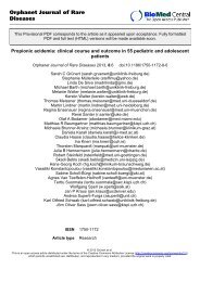
After birth, the cardinal features usually present are: 1)<br />
disproportionate small stature whith increasing severity<br />
from the proximal to distal portions of the limbs, and<br />
shortening of the middle and distal phalanges (Figure 1);<br />
2) polydactyly affecting hands (uni [exceptional: [13]] –<br />
or, usual, bilateral) and, occasionally, the feet (Figure 2);<br />
3) hidrotic ectodermal dysplasia mainly affecting the<br />
nails, hair and teeth; 4) congenital heart malformations<br />
occurring in about 50–60% of cases and comprising of<br />
single atrium, defects of the mitral and tricuspid valves,<br />
patent ductus, ventricular septal defect, atrial septal defect<br />
and hypoplastic left heart <strong>syndrome</strong>. The presence of congenital<br />
heart disease may support the diagnosis of the EVC<br />
<strong>syndrome</strong> and appears to be the main determinant of longevity<br />
[6,14].<br />
The oral manifestations spectrum is wide, including<br />
malocclusion, labiogingival adherences and gingival<br />
hypertrophy, labiogingival frenulum hypertrophy, accessory<br />
labiogingival frenula, serrated incisal margins, dental<br />
transposition, diastrema, conical teeth, enamel hypoplasia<br />
and hypodontia (Figure 3). Teeth may be prematurely<br />
erupted, at birth, or exfolliate prematurely [15].<br />
Several inconstant additional clinical findings are<br />
described, including strabismus, epi- and hypospadias,<br />
cryptorchidism [3], and thoracic wall and pulmonary<br />
malformations [16]. Renal abnormalities are found in<br />
very rare cases with agenesis, dysplasia, megaureter and<br />
nephrocalcinosis. Lethal nephronophthisis has been<br />
reported only once, in a patient with short limbs, short<br />
ribs, abnormal teeth and polydactyly (considered as EVC)<br />
[17], but this diagnosis may be discussed. Hematologic<br />
Patient shortness Figure at 1 the of the age limbs of 5 years showing long narrow chest and<br />
Patient at the age of 5 years showing long narrow chest and<br />
shortness of the limbs.<br />
Bilateral Figure 2polydactyly<br />
with short fingers in EVC patient<br />
Bilateral polydactyly with short fingers in EVC patient.<br />
Page 2 of 5<br />
(page number not for citation purposes)
Orphanet Journal of Rare Diseases 2007, 2:27 http://www.OJRD.com/content/2/1/27<br />
Anterior of Figure upper 3incisors<br />
view of the and mouth conical of lower EVC incisors patient showing absence<br />
Anterior view of the mouth of EVC patient showing absence<br />
of upper incisors and conical lower incisors.<br />
abnormalities have also been rarely reported: one case<br />
with dyserythropoiesis [18] and another associated with<br />
perinatal myeloblastic leukemia [19]. Head circumference<br />
and mental developement in EVC are normal.<br />
Because of the overlap between EVC and other short ribschondrodysplasias,<br />
and because molecular studies are<br />
only recently available and non-constantly performed,<br />
some old reports of EVC associated with exceptional features<br />
should be carefully read, since they may be mixed<br />
with publications that evidently do not deal with the EVC<br />
<strong>syndrome</strong>.<br />
The possibility of manifestations in heterozygous carriers<br />
has been discussed for a long time. From the observation<br />
of the large Amish kindred, McKusick concluded that<br />
there is no heterozygous manifestation of EVC [3]. However,<br />
other authors described polydactyly in relatives of<br />
four unrelated EVC families [20,21]. Spranger and Tariverdian<br />
[22] described a father of an EVC patient with fingers<br />
and teeth abnormalities, and then reviewed other<br />
reports of possible heterozygous manifestations. The<br />
Weyers acrofacial dysostosis, an autosomal dominant disorder<br />
described in 1952, is characterized by variable<br />
extremities and facial features. This condition has been<br />
found to be associated with EVC and EVC2 mutations<br />
that, finally, confirmed that Weyers dysostosis represents<br />
the heterozygous expression of the mutation, which, in<br />
homozygous form, causes the autosomal recessive disorder<br />
EVC [23,24].<br />
Skeletal features<br />
A variety of radiological skeletal features may be observed,<br />
including retarded bone maturation, fusion of the hamate<br />
and capitate bones of the wrist, defect of the lateral aspect<br />
of the proximal part of the tibia (knock-knees), cubitus<br />
valgus, hypoplastic cubitus, supernumerary carpal bone<br />
centre, clinodactyly of the 5 th finger, fusion of the 5 th and<br />
6 th metacarpals, disturbance in bone modeling of the metacarpals<br />
and/or phalanges. Bone age is usually retarded.<br />
Etiology<br />
The EVC gene was previously localized by linkage analysis<br />
to the distal short arm of chromosome 4 [25,26], in an<br />
area proximal to other chondrodystrophias. Mutations in<br />
this gene have been identified in EVC individuals from the<br />
Amish cases and from other pedigrees (Mexico, Equador<br />
and Brazil) [23]. But the screening of the 21 EVC coding<br />
exons in 58 patients with EVC permitted to identify only<br />
13 patients with homozygous mutations. Among the<br />
remaining 45 cases, no mutation in one or two allele was<br />
identified [27]. The cDNA analysis of fibroblast RNA from<br />
three of these patients was also normal. These observations<br />
had raised the possibility of genetic heterogeneity. A<br />
second gene, EVC2, was identified in an Ashkenasi child,<br />
immediately adjacent to EVC (named EVC1 or EVC), in a<br />
head to head configuration [28]. Expression of the gene is<br />
found in heart, placenta, lung, liver and skeletal muscle.<br />
EVC2 spans 166.4 kb and shares a common promoter<br />
region with EVC. The transcriptional start sites of EVC and<br />
EVC2 are separated by only 1643 bp. There is no significant<br />
sequence homology between EVC and EVC2 at either<br />
protein or nucleic levels. The EVC2 gene encodes a protein<br />
predicted to have one transmembrane segment, three<br />
coiled-coil regions, and one RhoGEF domain (SMART).<br />
The EVC2 protein has significant sequence homology<br />
with the tail domains of class IX non muscle myosins<br />
(BLAST). Mutations in the LIMBIN gene, a bovine orthologue<br />
of EVC2, are associated with bovine chondrodysplastic<br />
dwarfism [28]. Affected EVC individuals with<br />
mutations in EVC or EVC2 are phenotypically indistinguishable<br />
[27]. An interesting recent work with sequencing<br />
EVC and EVC2 in a series of 65 EVC patients identified<br />
EVC mutations in 20 families and EVC2 mutations in 25<br />
families [29]. No mutation has been identified in 20/65<br />
families but as none of these individuals had consanguineous<br />
parents (even if it was possible that some of these<br />
cases were misdiagnosed), this has lead to evidence for a<br />
genetic heterogeneity in EVC <strong>syndrome</strong>. As previously<br />
mentioned, heterozygous mutations in the EVC or EVC2<br />
genes also cause Weyers acrofacial dysostosis, an allelic<br />
disorder, showing autosomal dominant inheritance.<br />
Diagnostic methods<br />
The clinical diagnosis is based on observation of the<br />
symptoms and manifestations described above and sup-<br />
Page 3 of 5<br />
(page number not for citation purposes)
Orphanet Journal of Rare Diseases 2007, 2:27 http://www.OJRD.com/content/2/1/27<br />
ported by the skeletal survey. The definitive diagnosis is<br />
molecular, based on homozygosity for a mutation in the<br />
EVC and EVC2 genes by direct sequencing.<br />
Differential diagnosis<br />
EVC belongs to the short rib-polydactyly group (SRP).<br />
These SRPs are all autosomal recessive disorders that have<br />
been classified into types (Saldino-Noonan <strong>syndrome</strong>,<br />
type I; Majewski <strong>syndrome</strong>, type II; Verma-Naumoff <strong>syndrome</strong>,<br />
type III; Beemer-Langer <strong>syndrome</strong>, type IV; and<br />
Jeune Dystrophy). They are characterized by hypoplastic<br />
thorax due to short ribs, short limbs, frequent polydactyly<br />
and visceral abnormalities, and are discussed prenatally.<br />
Radiographically and histologically, SRP III (Verma-Naumoff<br />
<strong>syndrome</strong>, OMIM 263510) most resembles some<br />
forms of EVC [30,31]. The question of SRP being due to<br />
mutation in the EVC1 gene was excluded by Takamine et<br />
al. [32].<br />
Postnatally, the essential differential diagnoses include<br />
Jeune dystrophy, McKusick-Kaufman <strong>syndrome</strong> and Weyers<br />
<strong>syndrome</strong>. Jeune dystrophy (MIM 208500) is characterized<br />
by thoracic dystrophy, shortening of the<br />
extremities and generalized bone dysplasia. Similarities<br />
and differences of patients with EVC and Jeune dystrophy<br />
have been stressed [33,34]. There are no specific constant<br />
features to confirm the diagnosis of presumptive EVC but<br />
some features, including congenital heart disease, supernumerary<br />
digits and ectodermal dysplasia will mostly<br />
support the diagnosis of EVC <strong>syndrome</strong> than Jeune dystrophy.<br />
EVC and McKusick-Kaufman <strong>syndrome</strong> (MKK,<br />
MIM 236700), both recessively inherited disorders, share<br />
postaxial polydactyly and congenital heart defect. Distinguishing<br />
characteristics are the osteochondrodysplasia<br />
and ectodermal anomalies in EVC <strong>syndrome</strong>, and hydro<br />
metrocolpos in MKK <strong>syndrome</strong>. MKK is caused by mutations<br />
in a gene on chromosome 20p12, encoding a protein<br />
similar to members of the chaperonin family.<br />
Mutation in the same gene causes Bardet-Biedl <strong>syndrome</strong>-<br />
6 [35]. Weyers acrodental dysostosis (OMIM 193530) is,<br />
as mentioned above, the heterozygotous manifestation of<br />
the EVC gene. Disproportionate dwarfism, heart defect<br />
and thoracic dysplasia are not present in this autosomal<br />
dominant condition.<br />
Genetic counseling<br />
EVC <strong>syndrome</strong> is an autosomal recessive disorder, with a<br />
mendelian risk of 25% for subsequent pregnancies.<br />
Prenatal diagnosis<br />
EVC may be detected prenatally by ultrasound examination.<br />
The association of several structural fetal defects in<br />
the late first trimester, including narrow thorax, short and<br />
bowed long bones, rounded metaphyses, postaxial polydactyly,<br />
and cardiac defect may suggest the diagnosis ECV.<br />
Increased nuchal translucency thickness has been<br />
described associated with ECV [12]. In case of potential<br />
recurrence, prenatal diagnosis using molecular genetic<br />
techniques is, in theory, feasible, on DNA extracted from<br />
chorionic villus samples.<br />
Management<br />
The management of EVC is multidisciplinary. Symptomatic<br />
management is mostly required in the neonatal<br />
period, including treatment of the respiratory distress due<br />
both to narrow chest and heart failure. Neonatal teeth<br />
should be removed because they may impair the feeding.<br />
In infancy and early adulthood, general and specialized<br />
pediatric follow-up are also required: the short stature is<br />
considered resulting of chondrodysplasia of the legs and<br />
the possible treatment with growth hormone is considered<br />
ineffective. It is important to notice, however, that<br />
the association of growth hormone deficiency and ECV<br />
has been reported in one patient and, in this case, the<br />
growth hormone treatment had a favorable effect on<br />
growth [36]. The possibility of bones deformity, especially<br />
knee valgus with depression of the lateral tibial plateau<br />
and dislocation of the patella [37], needs regular orthopedic<br />
follow-up. Dentists play an important role in control<br />
of dental and oral manifestations. Dental treatment<br />
must be performed under prophylactic antibiotic coverage<br />
with consideration for the high incidence of cardiac<br />
defects in EVC patients. In addition, as in other chondrodysplasias,<br />
rare narrowing of the spinal canal can occur<br />
and justify a regular follow-up during adult life.<br />
Prognosis<br />
Although there is no systematic follow-up EVC series<br />
reported, the prognosis is linked to the respiratory difficulties<br />
in the first months of life and these difficulties are<br />
related to thoracic narrowness and possible heart defect.<br />
We insist on the fact that the cognitive development in<br />
EVC <strong>syndrome</strong> is normal. Prognosis of the final body<br />
height in individual patients with EVC is difficult to predict,<br />
as the rare publications of adult EVC cases report a<br />
variable final stature, from 119 cm [8] to 161 cm [4].<br />
Unresolved questions<br />
Clinical features<br />
The clinical spectrum of EVC <strong>syndrome</strong> is not well delineated<br />
at present. There are no specific constant features in<br />
EVC <strong>syndrome</strong>: some of them are usually present but their<br />
absence does not exclude the diagnosis. Depending on the<br />
diagnostic criteria, some patients may be recognized as<br />
EVC and other patients not. In the literature, there are<br />
many cases described as EVC <strong>syndrome</strong> in which the diagnosis<br />
is uncertain, and some of them definitely represent<br />
other entities. The reported data are limited to small<br />
series. Careful collection of case history, genotype-pheno-<br />
Page 4 of 5<br />
(page number not for citation purposes)
Orphanet Journal of Rare Diseases 2007, 2:27 http://www.OJRD.com/content/2/1/27<br />
type correlation with the two known genes and patient<br />
follow-up will provide supplementary information and<br />
further delineation of the EVC <strong>syndrome</strong>, and distinction<br />
from the other short rib-polydactyly <strong>syndrome</strong>s.<br />
Molecular mechanism<br />
ECV is associated with a genetic heterogeneity and EVC1<br />
and EVC2 do not account for the totality of EVC cases.<br />
Further studies are needed to elucidate other genes<br />
involved in EVC manifestations. They could also contribute<br />
to unraveling specific molecular processes that lead to<br />
the phenotypic manifestations of ECV.<br />
References<br />
1. <strong>Ellis</strong> RW, van Crefeld S: A <strong>syndrome</strong> characterized by ectodermaldysplasia,<br />
polydactyly, chondrodysplasia and congenital<br />
morbus cardia. Arch Dis Child 1940, 15:65.<br />
2. Lynch JI, Perry LW, Takakvwa T, Scott LP: Congenital heart disease<br />
and chondroextodermal dysplasia. Am J Dis Child 1968,<br />
115:80-87.<br />
3. Mac Kusick V: <strong>Ellis</strong>-van Crefeld <strong>syndrome</strong> and the Amish.<br />
Nature Genet 2000, 24:203-204.<br />
4. Oliveira da Siva E, Janovitz D, Cavalcanti de Albuquerque S: <strong>Ellis</strong>-van<br />
<strong>Creveld</strong> <strong>syndrome</strong>: report of 15 cases in an inbred kindred. J<br />
Med Genet 1980, 17:349-356.<br />
5. Waldrigues A, Grohmann L, Takahashi T, Reis H: <strong>Ellis</strong> van <strong>Creveld</strong>.<br />
An inbred kindred with 5 cases. Rev Bras de Pesquisas Med e Biol<br />
1977, 10:193-198.<br />
6. Blackburn M, Belliveau R: <strong>Ellis</strong>-van<strong>Creveld</strong> <strong>syndrome</strong>: a report<br />
of previously undescribed anomalies in two siblings. Am J Dis<br />
Child 1971, 122:267-270.<br />
7. Prabhu S, Daftary D, Dholakia H: Chondroectodermal dysplasia<br />
(<strong>Ellis</strong>-van <strong>Creveld</strong> <strong>syndrome</strong>): report of 2 cases. J Oral Surgery<br />
1978, 36:631-638.<br />
8. Renier JC, Larget-Piet L, Boasson M, Berthelot J, Fouillet L: Dysplasie<br />
chondroépidermique d'<strong>Ellis</strong>-van <strong>Creveld</strong>: deux cas dans une<br />
même fratrie. Revue du rhumatisme 1975, 42:417-422.<br />
9. Goor M, Farriaux J, Dupuis C, François P, Fontaine G: Le <strong>syndrome</strong><br />
d'<strong>Ellis</strong>-van <strong>Creveld</strong>: étude d'une observation familiale. La<br />
revue de Pédiatrie 1970, 4:233-236.<br />
10. Kushnick T, Paya K, Namunes P: Chondroectodermal Dysplasia.<br />
Am J Dis Child 1962, 103:77-80.<br />
11. Horigome H, Hamada H, Sohda S, Oyake Y, Kurosaki Y: Prenatal<br />
ultrasonic diagnosis of a case of <strong>Ellis</strong>-van <strong>Creveld</strong> <strong>syndrome</strong><br />
with a single atrium. Pediatr Radiol 1997, 27:942-944.<br />
12. Venkat-Raman N, Sebire N, Murphy K: Increased first-trimester<br />
fetal nuchal translucenty thickness in association with chondroectodermal<br />
dysplasia (<strong>Ellis</strong>-van <strong>Creveld</strong>). Ultrasound Obstet<br />
Gynecol 2005, 25:412-414.<br />
13. Engle M, Ehlers K: <strong>Ellis</strong>-van <strong>Creveld</strong> <strong>syndrome</strong> with asymmetric<br />
polydactyly and successful surgical correction of common<br />
atrium. Birth defects Orig Art Ser 1969, V:65-67.<br />
14. Digilio M, Marino B, Ammirati A, Borgaza U, Giannotti A, Dallapiccola<br />
B: Cardiac malformations in patients with oral-facial-skeletal<br />
<strong>syndrome</strong>s: clinical similarities with heterotaxia. Am J Med<br />
Genet 1999, 84:350-356.<br />
15. Winter G, Geddes M: Oral manifestations of chondroectodermal<br />
dysplasia (<strong>Ellis</strong>-<strong>Van</strong> <strong>Creveld</strong> Syndrome). Report of a<br />
case. Br Dent J 1967, 122:103-107.<br />
16. Moore T: Chondroectodermal dysplasia (<strong>Ellis</strong>-van <strong>Creveld</strong><br />
<strong>syndrome</strong>) with bronchial malformation and neonatal tension<br />
lobar emphysema. J Thoracic and Cardiovas Surg 1963,<br />
46:1-10.<br />
17. Moudgil A, Bagga A, Kamil ES, Rimoin DL, Lachman RS, Cohen AH,<br />
Jordan SC: Nephronophtisis associated with <strong>Ellis</strong>-van <strong>Creveld</strong><br />
<strong>syndrome</strong>. Pediatr Nephrol 1998, 12:20-22.<br />
18. Scurlock D, Ostler D, Nguyen A, Wahed A: <strong>Ellis</strong>-van <strong>Creveld</strong> <strong>syndrome</strong><br />
and dyserythropoiesis. Arch Pathol Lab Med 2005,<br />
129:680-682.<br />
19. Miller D, Newstead G, Young L: Perinatal leukemia with a possible<br />
variant of the <strong>Ellis</strong>-van Crefeld. J Pediatr 1969, 74:300-303.<br />
20. Fryns JP: Postaxial polydactyly as heterozygote manifestation<br />
in <strong>Ellis</strong>-van <strong>Creveld</strong> <strong>syndrome</strong>? [letter]. Am J Med Genet 1991,<br />
39:500.<br />
21. Goldblatt J, Minutillo C, Pemberton P, Hurst J: <strong>Ellis</strong>-van <strong>Creveld</strong><br />
<strong>syndrome</strong> in a western Australian Aboriginal community:<br />
postaxial polydactyly as a heterozygous manifestation? Med<br />
J Aust 1992, 157:271.<br />
22. Spranger S, Tariverdian G: Symtomatic heterozygozygosity in<br />
the <strong>Ellis</strong>-van <strong>Creveld</strong> <strong>syndrome</strong>? Clin Genet 1995, 47:217-220.<br />
23. Ruiz-Perez VL, Ide SE, Strom TM, Lorenz B, Wilson D, Woods K,<br />
King L, Francomano C, Freisinger P, Spranger S, Marino B, Dallapiccola<br />
B, Wright M, Meitinger T, Polymeropoulos MH, Goodship J:<br />
Mutations in a new gene in <strong>Ellis</strong>-van <strong>Creveld</strong> <strong>syndrome</strong> and<br />
Weyers acrodental dysostosis. Nature Genet 2000, 24:283-286.<br />
24. Ye X, Song G, Fan M, Shi L, Jabs EW, Huang S, Guo R, Bian Z: A novel<br />
heterozygous deletion in the EVC2 gene causes Weyers<br />
acrofacial dysostosis. Hum Genet 2006, 119:199-205.<br />
25. Francomano C, Ortez deLuna R, Ide S, Pyeritz R, Wright M, Polymeropoulos<br />
M: The gene for the <strong>Ellis</strong>-van <strong>Creveld</strong> <strong>syndrome</strong><br />
maps to chromosome 4p16. Am J Hum Genet 1995,<br />
57(suppl):A191.<br />
26. Polymeropoulos M, Ide S, Wright M, Goodship J, Weissenbach J, Pyeritz<br />
R, Da Silva E, Ortiz De Luna R, Francomano CA: The gene for<br />
the <strong>Ellis</strong>-van <strong>Creveld</strong> <strong>syndrome</strong> is located on chromosome<br />
4p16. Genomics 1996, 35:1-5.<br />
27. Ruiz-Perez V, Tompson S, Blair H, Espinoza-Valdez C, Lapunzina P,<br />
Silva E, Hamel B, Gibbs J, Young I, Wright M, Goodship J: Mutations<br />
in two nonhomologous genes in a head-to-head configuration<br />
cause <strong>Ellis</strong>-van <strong>Creveld</strong> <strong>syndrome</strong>. Am J Hum Genet 2003,<br />
72:728-732.<br />
28. Galdzicka M, Patnala S, Hirshman M, Cai J, Nitowsky , Egeland J, Ginns<br />
E: A new gene, EVC2, is mutated in <strong>Ellis</strong>-van <strong>Creveld</strong> <strong>syndrome</strong>.<br />
Molec Genet Metab 2002, 77:291-295.<br />
29. Tompson SW, Ruiz-Perez VL, Blair HJ, Barton S, Navarro V, Robson<br />
JL, Wright MJ, Goodship JA: Sequencing EVC and EVC2 identifies<br />
mutations in two-thirds of <strong>Ellis</strong>-van <strong>Creveld</strong> <strong>syndrome</strong><br />
patients. Hum Genet 2007, 120:663-670.<br />
30. Yang S, Langer L, Cacciarelli A, Dahms B, Unger E, Roskamp J: Three<br />
conditions in neonatal asphyxiating thoracic dysplasia<br />
(Jeune) and short rib-polydactyly <strong>syndrome</strong> spectrum: a clinicopathologic<br />
study. Am J Med Genet 1987, 3(suppl):191-207.<br />
31. Elcioglu N, Hall C: Diagnostic dilemmas in the short rib-polydactyly<br />
<strong>syndrome</strong> group. Am J Med Genet 2002, 111:392-400.<br />
32. Takamine Y, Krejci P, Wilcox W: Mutations in the EVC1 gene<br />
are not a common finding in the <strong>Ellis</strong>-van <strong>Creveld</strong> and Short-<br />
Rib-polydactyly type III <strong>syndrome</strong>s. Am J Med Genet 2004,<br />
130A:96-97.<br />
33. Maroteaux P, Savart P: La dystrophie thoracique asphyxiante.<br />
Etude radiologique et rapports avec le <strong>syndrome</strong> d'<strong>Ellis</strong>-van<br />
<strong>Creveld</strong>. Ann Radiol 1964, 7:332-338.<br />
34. Kolowski K, Szmgiel C, Barylak A, Stopyrowa M: Difficulties in differentiation<br />
between chondroectodermal dysplasia (<strong>Ellis</strong>van<br />
<strong>Creveld</strong> <strong>syndrome</strong>) and Asphyxiating Thoracic Dystrophy.<br />
Aust Radiol 1972, 16:401-410.<br />
35. Stone D, Slavotinek A, Bouffard G, Banerjee-Basu S, Baxevanis A, Barr<br />
M, Biesecker L: Mutation of a gene encoding a putative chaperonin<br />
causes McKusick-Kaufman <strong>syndrome</strong>. Nature Genet<br />
2000, 25:79-82.<br />
36. Pirazzoli P, Mazzanti L, Mandini M, Cau M, Ravagli L, Cacciari E: GHdeficiency<br />
in <strong>Ellis</strong>-van-Crefeld Syndrome: response to remplacement<br />
therapy. Growth Abnormalities 1989, 56:391-394.<br />
37. Shibata T, Kawabata H, Yasui N, Nakahara H, Hirabayashi S, Nakase<br />
T, et al.: Correction of knee deformity in patients with <strong>Ellis</strong>van<br />
<strong>Creveld</strong> <strong>syndrome</strong>. J Pediatr Orthop B 1999, 8:282-284.<br />
Page 5 of 5<br />
(page number not for citation purposes)