
You also want an ePaper? Increase the reach of your titles
YUMPU automatically turns print PDFs into web optimized ePapers that Google loves.
<strong>AOPT</strong><br />
TABLE OF CONTENTS<br />
Association for Ocular<br />
Pharmacology and Therapeutics<br />
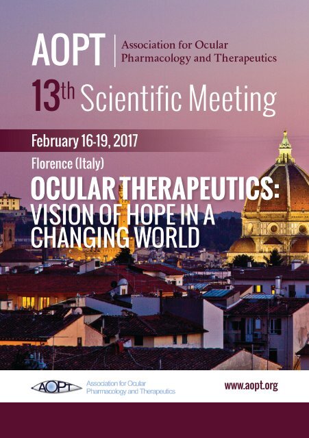
13 th <strong>Scientific</strong> <strong>Meeting</strong><br />
February 16-19, 2017<br />
Florence (Italy)<br />
OCULAR THERAPEUTICS:<br />
VISION OF HOPE IN A<br />
CHANGING WORLD<br />
www.aopt.org
INDEX<br />
THANKS TO OUR SPONSOR........................................................ pag. 4<br />
WELCOME MESSAGE.................................................................. pag. 7<br />
<strong>AOPT</strong> MEMBERSHIP INFORMATION.............................................. pag. 8<br />
<strong>AOPT</strong> PAST MEETINGS................................................................ pag. 9<br />
YOUR <strong>AOPT</strong> BOARD.................................................................... pag. 10<br />
YOUR LOCAL ORGANIZING COMMITTEE....................................... pag. 12<br />
TRAVEL AWARD WINNERS........................................................... pag. 13<br />
GENERAL INFORMATION............................................................. pag. 14<br />
INFORMATION FOR PRESENTERS................................................. pag. 15<br />
PALAZZ0 DEGLI AFFARI PLAN....................................................... pag. 16<br />
PROGRAM AT-A-GLANCE............................................................. pag. 17<br />
KEYNOTE SPEAKER.................................................................... pag. 18<br />
SCIENTIFIC PROGRAM............................................................... pag. 19<br />
IT-ARVO PROGRAM.................................................................... pag. 27<br />
PLATFORM ABSTRACT................................................................ pag. 29<br />
POSTER SESSION....................................................................... pag. 89
PANTONE<br />
3015 C<br />
THANKS TO OUR SPONSORS<br />
TITLE LEVEL<br />
PLATINUM LEVEL<br />
GOLD LEVEL<br />
SILVER LEVEL
THANKS TO OUR SPONSORS<br />
BRONZE LEVEL<br />
OTHER<br />
YOUNG INVESTIGATOR TRAVEL AWARDS<br />
UNTHSC/Elena and Tom Yorio
WELCOME MESSAGE<br />
Welcome to the 13 th <strong>Scientific</strong> <strong>Meeting</strong> of the<br />
Association for Ocular Pharmacology and<br />
Therapeutics (<strong>AOPT</strong>). The topic of this year’s<br />
conference is Ocular Therapeutics: Vision of<br />
hope in a changing world.<br />
We are thankful for our Italian hosts who have<br />
put together an exciting meeting at a great<br />
venue at the Firenze Fiera Congress Center<br />
here in Florence Italy. The meeting agenda is packed with exciting<br />
new information set in thirteen sessions as well as a special treat with<br />
a visit to the Uffizi Museum.<br />
The Association for Ocular Pharmacology and Therapeutics is a<br />
global not-for-profit organization for scientists and individuals from<br />
all disciplines related to ocular pharmacology and its therapeutic<br />
applications. <strong>AOPT</strong>- has a diverse, multi-national membership<br />
composed of preclinical and clinical scientists, students, and healthcare<br />
professionals. Members are from academic institutions, pharma and<br />
biotech industries, device companies, clinics and private practice.<br />
<strong>AOPT</strong>’s mission is to serve as a global forum and network for<br />
the publication, dissemination and exchange of information and<br />
knowledge on treatments of eye diseases, from basic and clinical<br />
ocular pharmacology and therapeutics to related disciplines such as<br />
pharmacokinetics and dynamics, metabolism, translational research,<br />
safety, drug delivery, and pharmaceutics.<br />
This conference brings together those individuals at the forefront<br />
of scientific advancement related to ocular pharmacology and<br />
therapeutics. The agenda is rich in provocative presentations that we<br />
hope stimulate productive discussions.<br />
What better place to set a new vision for ocular therapeutics than in<br />
Florence the “Cradle of the Renaissance”.<br />
Tom Yorio, PhD, FARVO<br />
President, <strong>AOPT</strong><br />
7
<strong>AOPT</strong> MEMBERSHIP INFORMATION<br />
There are four classes of membership in <strong>AOPT</strong>: Regular Members,<br />
Associate Members, Contributing Members, and Emeritus Members.<br />
REGULAR MEMBERS<br />
The Regular Membership represent individuals demonstrating<br />
a genuine interest in or making significant contribution to<br />
ocular pharmacology and therapeutics. This may be evidenced<br />
by a) scientific publications; b) attendance at pharmacological,<br />
ophthalmological, optometric, or visual science meetings; c) direct<br />
involvement in research. A candidate for membership completes the<br />
online membership form and pays the appropriate membership dues.<br />
Membership is for two years. A subscription to the Journal of Ocular<br />
Pharmacology and Therapeutics is optional.<br />
ASSOCIATE MEMBERS<br />
Associate Membership is for predoctoral and postdoctoral students.<br />
A candidate for this membership must have a pre-doctoral, or postdoctoral<br />
student status, and must complete the online membership<br />
form and pay the appropriate membership dues.<br />
CONTRIBUTING MEMBERS<br />
Contributing Membership is restricted to corporations, associations,<br />
and individuals who support the objectives of <strong>AOPT</strong> but do not satisfy<br />
the requirements of Regular Membership or individuals elected<br />
to membership in any class who voluntarily choose to become<br />
Contributing Members. A candidate for contributing membership<br />
completes the online membership form and pays the appropriate<br />
membership dues.<br />
EMERITUS MEMBERS<br />
Any Regular Member may make a written request to the Treasurer<br />
that his/her membership be transferred to that of an Emeritus<br />
Member. The request is subject to approval of the membership<br />
committee. Emeritus Members have all the rights and privileges of<br />
Regular Members, except those of voting and holding elective office.<br />
8
<strong>AOPT</strong> PAST MEETINGS<br />
MEETING DATE LOCATION ORGANIZER<br />
TWELFTH MEETING<br />
ELEVENTH MEETING<br />
FEB 26 - MAR 1, 2015<br />
FEBRUARY 7-10, 2013<br />
CHARLESTON, SC<br />
ALICANTE, SPAIN<br />
DAN STAMER<br />
JUANA GALLAR MARTINEZ<br />
TENTH MEETING<br />
FEBRUARY 17-20, 2011<br />
FT. WORTH, TX<br />
TOM YORIO / ABBOT CLARK<br />
NINTH MEETING<br />
FEBRUARY 18-21, 2009<br />
SALZBURG, AUSTRIA<br />
HERBERT REITSAMER<br />
EIGHTH MEETING<br />
FEBRUARY 9-11, 2007<br />
SAN DIEGO, CA<br />
JOHN LIU / ACHIM KRAUSS<br />
SEVENTH MEETING<br />
FEBRUARY 3-5, 2005<br />
CATANIA, SICILY, ITALY<br />
FILIPPO DRAGO<br />
SIXTH MEETING<br />
FEBRUARY 1-4, 2003<br />
KONA, HI<br />
PETER KADOR<br />
FIFTH MEETING<br />
NOVEMBER 2-5, 2000<br />
BIRMINGHAM, AL<br />
JIMMY BARTLETT<br />
FOURTH MEETING<br />
JANUARY 28-31, 1999<br />
IRVINE, CA<br />
ACHIM KRAUSS<br />
THIRD MEETING<br />
OCTOBER 22-24, 1997<br />
BETHESDA, MD<br />
PETER KADOR<br />
SECOND MEETING<br />
AUGUST 15-17, 1996<br />
LOS ANGELES, CA<br />
DAVID LEE<br />
FIRST MEETING<br />
OCULAR<br />
PHARMACOLOGY<br />
SYMPOSIUM<br />
JANUARY 26-29, 1995<br />
AUGUST 8-10, 1993<br />
NEW ORLEANS, LA<br />
NOVI, MI<br />
HERB KAUFMAN<br />
HITOSHI SHICHI<br />
9
YOUR <strong>AOPT</strong> BOARD<br />
PRESIDENT<br />
DR. THOMAS YORIO<br />
VICE-PRESIDENT<br />
PRESIDENT-ELECT<br />
DR. FILIPPO DRAGO<br />
IMMEDIATE-PAST-PRESIDENT<br />
DR. ACHIM KRAUSS<br />
TRUSTEE<br />
DR. MALINDA FITZGERALD<br />
TRUSTEE<br />
DR. ASH JAYAGOPAL<br />
TRUSTEE<br />
DR. UDAY KOMPELLA<br />
TRUSTEE<br />
DR. GANESH PRASANNA<br />
TRUSTEE<br />
DR. SHUSHENG WANG<br />
10
YOUR <strong>AOPT</strong> BOARD<br />
TRUSTEE<br />
DR. CHRISTINE WILDSOET<br />
TREASURER<br />
DR. PETER KADOR<br />
SECRETARY<br />
DR. CAROL TORIS<br />
TRUSTEE<br />
DR. CAMERON MILLAR<br />
TRUSTEE<br />
DR. IOK-HOU PANG<br />
11
YOUR LOCAL ORGANIZING COMMITTEE YOUR <strong>AOPT</strong> BOARD<br />
FILIPPO DRAGO<br />
CLAUDIO BUCOLO<br />
CATERINA GAGLIANO<br />
ALESSANDRO MUGELLI<br />
ENRICA STRETTOI<br />
MARINA ZICHE<br />
12
TRAVEL AWARD WINNERS<br />
Biagioni Martina Tuscan Doctorate School in Neuroscience,<br />
CNR Neuroscience Institute, Pisa, University of Florence, Italy<br />
Cupri Sarha Department of Drug Sciences, University of Catania, Italy<br />
Daszynski Damian Department of Pharmaceutical Sciences,<br />
University of Nebraska, Omaha, Nebraska, USA<br />
Fidilio Annamaria Department of Biomedical and Biothecnological Sciences,<br />
University of Catania, Italy<br />
Harper Angelica Department of Cell Biology,<br />
University of Oklahoma Health Sciences Center, Oklahoma City, Oklahoma, USA<br />
Johnson William Department of Ophthalmology, Duke University, Durham,<br />
North Carolina, USA<br />
Joubert Fanny Institut de la Vision, Equipe S12,<br />
UMR_S968 INSERM, UMR_7210 CNRS, UPMC, Paris<br />
Kelley Ryan Skaggs School of Pharmacy and Pharmaceutical Sciences,<br />
University of Colorado Anschutz Medical Center, Aurora, Colorado, USA<br />
Landowski Michael Department of Ophthalmology, Duke University, Durham,<br />
North Carolina, USA<br />
Lazzara Francesca Department of Biomedical and Biotechnological Sciences,<br />
University of Catania, Catania Italy<br />
Liu Yang North Texas Eye Research Institute,<br />
University of North Texas Health Science Center,Fort Worth Texas, USA<br />
Mishra Manish Kresge Eye Institute, Wayne State<br />
University School of Medicine, Detroit, Michigan, USA<br />
Mody Avani North Texas Eye Research Institute,<br />
University of North Texas Health Science Center,Fort Worth Texas, USA<br />
Patel Gaurang North Texas Eye Research Institute,<br />
University of North Texas Health Science Center,Fort Worth Texas, USA<br />
Pattabiraman Padmanabhan Department of Ophthalmology,Case Western Reserve<br />
University, Cleveland, Ohio, USA<br />
Perdices Lorena Institute for Health Research of Aragón (IIS Aragón), Zaragoza, Spain<br />
Platania Chiara B. M. BIOMETEC University of Catania,Italy<br />
Roubeix Christophe Department of Ophthalmology,<br />
Charite University Medicine Berlin, Germany<br />
Schmitt Heather Department of Ophthalmology,<br />
University of Wisconsin-Madison, Charter St. Madison, USA<br />
Smedowski Adrian Department of Physiology,<br />
Medical University of Silesia, Katowice, Poland<br />
Stefanov Antonia Institute of Neuroscience, National Research Council, Pisa, Italy<br />
Toro Mario Department of Ophthalmology, University of Catania, Italy<br />
Webber Hannah North Texas Eye Research Institute,<br />
University of North Texas Health Science Center,Fort Worth Texas, USA<br />
Yu Bo Tulane University, New Orleans, Lousiana LA, USA<br />
13
GENERAL INFORMATION<br />
Onsite registration desk<br />
Thursday february 16 th , 15.00-17:15<br />
Friday february 17 th , 08.30-10:30<br />
Fees:<br />
Regular Members: € 425,00<br />
Non-Members: € 525,00<br />
Students, Post Doc: € 250,00<br />
Name dadges<br />
All participants must wear their name badges during the meeting.<br />
Badges allow admission to all sessions, breaks, lunches, receptions and the banquet.<br />
Accompanying persons<br />
You can register as many accompanying persons as you want.<br />
Fees:<br />
Welcome reception and banquet: € 120,00<br />
Banquet only: € 80,00<br />
Accompanying persons are not allowed to attend scientific sessions and exhibition area.<br />
Welcome reception<br />
The welcome reception will be held on thursday february 16 th from 18.40 to 20.00<br />
in the exhibit area, first floor.<br />
<strong>AOPT</strong> business meeting<br />
The <strong>AOPT</strong> business meeting will be held on friday february 17 th from 16.40 to 17.40<br />
All <strong>AOPT</strong> members are encouraged to attend.<br />
<strong>AOPT</strong> banquet<br />
The <strong>AOPT</strong> banquet open to all regitered participants will be held on saturday 18th<br />
from 19.00 in the Basilica da Basso.<br />
Clothing<br />
Clothing in business casual for all occasion.<br />
Liability and personal insurance<br />
The <strong>AOPT</strong> 2017 Organizers can not accept liability for personal accidents or<br />
loss of or damage to private property of participants and accompanying persons.<br />
Safety and security<br />
We kindly request you not to leave bags, suitcases or backpacks unattended<br />
at any time during the meeting.<br />
14
INFORMATION FOR PRESENTERS<br />
Language<br />
The official language of the <strong>AOPT</strong> 2017 <strong>Meeting</strong> is english.<br />
Oral presentation<br />
Presenters using a powerpoint presentation should bring it on memory stick (usb) and<br />
load in the preview room near the conference hall, between 08,15-08,30 for morning<br />
sessions during the luch for afternoon sessions. Presenters with powerpoint and video<br />
are requested to check their presentation to be sure they work properly. Macintosh users<br />
must convert their files to powerpoint in order to be used on the pc, otherwise must<br />
bring their own computer and VGA adaptor.<br />
Poster presentation<br />
Posters (70 base x 100 height) need to be assembled by 08,00 on friday 17th and rimane<br />
on display until sunday afternoon. During the posters session, presenters must stand at<br />
their posters to present and discuss about their work. Posters left up on sunday evening<br />
will removed and discarded.<br />
Recording policy<br />
Recording any presentation or poster is prohibited, except by <strong>AOPT</strong> agent, or by authors.<br />
15
30<br />
E<br />
wc<br />
32<br />
170<br />
210<br />
wc wc wc wc<br />
122<br />
I/11<br />
24<br />
150<br />
265<br />
150<br />
265<br />
150<br />
265<br />
150<br />
265<br />
25<br />
150<br />
265<br />
36<br />
170<br />
210<br />
saletta<br />
mq:20,71<br />
H:285<br />
PALAZZO DEGLI AFFARI PLAN<br />
GROUND FLOOR<br />
1st floor<br />
Registration<br />
Exhibitors<br />
Coffee and lunch<br />
Poster area<br />
E<br />
Main entrance<br />
Conference room<br />
Preview room<br />
E<br />
FIRST FLOOR<br />
Bar<br />
Poster area<br />
E<br />
B5 SIFI<br />
JOPT<br />
B2 Allergan<br />
Registration<br />
desk<br />
F.C.B.<br />
Exhibition - Coffee and lunch area<br />
Medici<br />
senza<br />
Frontiere<br />
Nicox Santen Alfa Intes Chiesi Experimentica Angelini<br />
Cloakroom<br />
16
13:30 - 15:00<br />
15:00 - 16:30<br />
JOPT Board <strong>Meeting</strong><br />
<strong>AOPT</strong> Board <strong>Meeting</strong><br />
PROGRAM AT-A-GLANCE<br />
Thurdsay, February 16<br />
Friday, February 17 Saturday, February 18<br />
Sunday, February 19<br />
09:20 - 10:40<br />
SESSION 2<br />
NEW INSIGHTS IN THE THERAPEUTIC APPROACHES<br />
TO AMD: IS THERE A HOPE FOR AN AFFORDABLE<br />
TREATMENT?<br />
08:40-10:20<br />
SESSION 6<br />
ADVANCES IN OCULAR DRUG DELIVERY AND ITS ROLE<br />
IN PATIENT'S COMPLIANCE<br />
08:40-10:20<br />
SESSION 10<br />
GENETIC AND ORPHAN EYE DISEASES<br />
10:40-10:50 Break & Exhibits 10:20-10:40 Break & Exhibits 10:20-10:40 Break & Exhibits<br />
10:50-12:10<br />
SESSION 3<br />
DIABETIC RETINOPATHY, A DISEASE OF INCREASING<br />
CONCERN<br />
10:40-12:00<br />
SESSION 7<br />
GLAUCOMA: RESEARCH AND DRUG DISCOVERY IN THE<br />
XXI CENTURY<br />
10:40-12:00<br />
SESSION 11<br />
OCULAR BLOOD FLOW: AN ISSUE FOR DIAGNOSIS AND<br />
THERAPY<br />
12:10-13:30<br />
IT-ARVO sponsored symposium<br />
12:00-13:20<br />
Lunch-n-Learn<br />
12:00-13:00<br />
Lunch-Panel<br />
13:30 - 14:50<br />
SESSION 4<br />
REGULATORY ISSUES GOVERNING OPHTHALMIC<br />
DRUGS: PHARMACOECONOMICS AND DRUG<br />
DEVELOPMENT IN A CHANGING WORLD<br />
13:20- 14:40<br />
SESSION 8<br />
NEUROPROTECTION IN OPHTHALMOLOGY: DO WE<br />
STILL NEED IT?<br />
13:00-14:40<br />
SESSION 12<br />
TREATMENT OF REFRACTIVE ERROR DEFECTS: WHAT IS<br />
THE ROLE OF OCULAR PHARMACOLOGY?<br />
14:50 - 15:10 Break & Exhibits 14:40-15:00 Break & Exhibits 14:40-15:00 Break & Exhibits<br />
15:00 - 17:15<br />
Registration<br />
15:10 - 16:40<br />
SESSION 5<br />
YOUNG INVESTIGATOR<br />
15:00-16:20<br />
SESSION 9<br />
OCULAR IMMUNE DISORDERS IN THE TIME OF<br />
GLOBALIZED MEDICINE<br />
15:00-16:40<br />
SESSION 13<br />
OCULAR SURFACE DISEASES: AN EMERGING CONCERN<br />
IN OPHTHALMOLOGY<br />
17:15 - 17:20 Opening remarks<br />
16:40 - 17:40<br />
<strong>AOPT</strong> general business meeting<br />
16:20-17:30<br />
PANEL DISCUSSION<br />
THE ENDOVITREAL INSERTS TO REDUCE THE BURDEN<br />
OF ENDOVITREAL INJECTIONS IN AMD AND DME<br />
16:40-17:00<br />
Closing remarks<br />
17:20 - 18:40<br />
SESSION 1<br />
BIOMARKERS AND TECHNOLOGY FOR SUBTYPING EYE<br />
DISEASE: EBABLERS OF TRANSLATIONAL MEDICINE<br />
AND TARGETED THERAPIES<br />
17:40 - 19:00<br />
POSTER SESSION<br />
BREAK AND EXHIBITS<br />
17:30-18:40<br />
KEYNOTE ADDRESS<br />
FUNGAL KERATITIS, A MAJOR CAUSE OF BLINDNESS<br />
AND VISUAL IMPAIRMENT WORLDWIDE, ESPECIALLY IN<br />
DEVELOPING COUNTRIES<br />
18:40 - 20:00<br />
Welcome reception<br />
19:00-21:30<br />
BANQUET DINNER<br />
BASILICA DA BASSO<br />
17
KEYNOTE SPEAKER<br />
ERIC PEARLMAN PhD.<br />
Director of Institute for Immunology<br />
University of California, Irvine<br />
Prof. Pearlman started his studies at University of Glasgow, he earned his PhD<br />
in Microbiology at University of Texas (Health Sciences Center – San Antonio).<br />
He started its academic research studying “river blindness” – onchocerciasis.<br />
Eric Pearlman was part of the research group that found out that a bacterium,<br />
living inside the worm Onchocerca volvulus, was the real cause of inflammation<br />
and blindness, rather than the worm itself.<br />
These results have been published on Science in 2002.<br />
In 2005-2006, there was an outbreak of fungal keratitis in USA, northern Europe<br />
and UK; those corneal infections were difficult to treat and were related to contamination<br />
by Fusarium solani of a lens care product. Furthermore, this fungal<br />
keratitis is a major cause of blindness in developing world, because spores (conidia)<br />
are common in the soil and in the air of rural areas.<br />
Currently, Pearlman’s research is focused on Fusarium keratitis and his research<br />
group developed an animal model of this disease, in order to identify the biochemical<br />
mechanism of this infection and best pharmacological targets, to further<br />
develop new therapies.<br />
18
SCIENTIFIC<br />
PROGRAM<br />
under the auspices of
THURSDAY, FEBRUARY 16<br />
15:00-17:15 Registration<br />
17:15-17:20 Opening remarks<br />
SESSION 1<br />
BIOMARKERS AND TECHNOLOGY FOR SUBTYPING EYE DISEASE:<br />
ENABLERS OF TRANSLATIONAL MEDICINE AND TARGETED THERAPIES<br />
Moderators: Oliver Zeitz, Dan Stamer<br />
17:20-17:40 Outside-ophthalmology perspective: State-of-the-art<br />
for biomarkers in oncology<br />
Friedhelm Bladt<br />
17:40-18:00 Mining retinal imaging data for biomarkers of disease<br />
and therapy<br />
Sebastian Waldstein<br />
18:00-18:20 The potential of ocular exosomal biomarkers as<br />
therapeutic targets, and diagnostic and<br />
prognostic indicators<br />
Mikael Klingeborn<br />
18:20-18:40 Tearfilm biomarkers<br />
Leopold Schmetterer<br />
18:40-20:00 WELCOME RECEPTION<br />
FRIDAY, FEBRUARY 17<br />
SESSION 2<br />
NEW INSIGHTS IN THE THERAPEUTIC APPROACHES TO AMD:<br />
IS THERE A HOPE FOR AN AFFORDABLE TREATMENT?<br />
Moderators: Cathy Bowes Rickman, Chiara Eandi<br />
09:20-09:40 From AMD-associated polymorphisms to drug target identification<br />
Florian Sennlaub<br />
09:40-10:00 Complement signaling ar the RPE: Implications for AMD<br />
Olaf Strauss<br />
10:00-10:20 Placental growth factor: An additional therapeutic<br />
target for AMD<br />
Sandro De Falco<br />
10.20-10.40 Effects of Anti-C5a therapy on early and wet models of AMD<br />
Cathy Bowes Rickman<br />
10:40-10:50 BREAK AND EXHIBITS<br />
20
SESSION 3<br />
DIABETIC RETINOPATHY, A DISEASE OF INCREASING CONCERN<br />
Moderators: Marina Ziche, Ash Jayagopal<br />
10:50-11:10 iPSCs as a novel strategy for vascular repair in the retina<br />
Maria Grant<br />
11:10-11:30 Angiogenic/inflammatory activity of humor vitreous in<br />
proliferative diabetic retinopathy<br />
Marco Presta<br />
11:30-11:50 B2R signaling in neo-angiogenesis<br />
Sandra Donnini<br />
11:50-12:10 Imaging vascular dysfunction in diabetic retinopathy<br />
Ash Jayagopal<br />
12:10-13:30 LUNCH PANEL<br />
IT-ARVO sponsored symposium<br />
SESSION 4<br />
REGULATORY ISSUES GOVERNING OPHTHALMIC DRUGS:<br />
PHARMACOECONOMICS AND DRUG DEVELOPMENT IN A CHANGING WORLD<br />
Moderators: Filippo Drago, Peter Kador<br />
13:30-13:50 Challenges to the economic evaluation of interventions<br />
for retinal conditions: A review of NICE technology<br />
appraisals in retinal vein occlusion and diabetic macular edema<br />
Chrissy Almond<br />
13:50:14:10 Combination therapies in retinal diseases: Anticipated hurdles<br />
for regulatory approval and health technology assessment.<br />
Is there a risk for patient access to new innovative<br />
medicine in ophthalmology?<br />
Jean Claude Castanier<br />
14:10-14:30 Off-label drug use in ocular pharmacology<br />
Lucia Gozzo<br />
14:30-14:50 Clinical prevention of cataracts in diabetic dogs by kinostat<br />
Peter Kador<br />
14:50-15:10 BREAK AND EXHIBITS<br />
SESSION 5<br />
YOUNG INVESTIGATOR SESSION<br />
Moderators: Malinda Fitzgerald, Alessia Pascale<br />
15:10-15:25 Treatment of HDAC3 selective inhibitor prevents retinal<br />
ganglion cell nuclear atrophy and apoptosis after acute<br />
and chronic optic nerve injury<br />
Heather Schmitt<br />
21
15:25-15:40 Neuroprotection and neuroregeneration of Retinal Ganglion<br />
Cells using products of peripheral nerves predegeneration<br />
Adrian Smedowski<br />
15:40-15:55 Epigenetics as a code for mitochondrial DNA mismatch<br />
and its dysfunction in diabetic retinopathy<br />
Manish Mishra<br />
15:55-16:10 P2X7 receptor as a pharmacological target in<br />
diabetic retinopathy<br />
Chiara Platania<br />
16:10-16:25 Spleen derived monocytes in subretinal inflammation<br />
Christophe Roubeix<br />
16:25-16:40 The role of canonical Wnt signaling and K-cadherin in<br />
the maintenance of intraocular pressure<br />
Hannah Webber<br />
<strong>AOPT</strong> GENERAL BUSINESS MEETING<br />
16:40-17:40<br />
POSTER SESSION<br />
Moderators: Shusheng Wang, Julie Crider<br />
17:40-19:00<br />
SATURDAY, FEBRUARY 18<br />
SESSION 6<br />
ADVANCES IN OCULAR DRUG DELIVERY AND ITS ROLE IN PATIENT'S COMPLIANCE<br />
Moderators: Uday B. Kompella, Claudio Bucolo<br />
08:40-09:00 Biodegradable implants for sustained drug release:<br />
Manufacturing considerations, drug stability,<br />
and drug release<br />
Uday B. Kompella<br />
09:00-09:20 What delivery strategy for poorly soluble drugs?<br />
Robert Gurny<br />
09:20-09:40 Sustained release microtechnologies for the treatment of<br />
neurodegenerative diseases of the posterior segment<br />
Maria Del Rocio Herrero Vanrell<br />
09:40-10:00 Melanin binding and active transport in the RPE:<br />
Impact on ocular drug delivery and<br />
pharmacokinetics<br />
Arto Urtti<br />
22
10.00-10:20 Recent advances in the application of<br />
lipid-based nanocarriers to ocular drug delivery<br />
Rosario Pignatello<br />
10:20-10:40 BREAK AND EXHIBITS<br />
SESSION 7<br />
GLAUCOMA: RESEARCH AND DRUG DISCOVERY IN THE XXI CENTURY<br />
Moderators: Carol Toris, Padmanabhan Pattabiraman<br />
10:40-11:00 NCX 667, a lead nitric oxide (NO)-donating compound for a<br />
new class of ocular hypotensive agents<br />
Francesco Impagnatiello<br />
11:00-11:20 New glaucoma drainage device designs for lowering of IOP<br />
Carol Toris<br />
11:20-11:40 New highly effective and long-acting anti-glaucoma drug,<br />
new periorbital delivery method<br />
David Woodward<br />
11:40-12:00 Glycosylation status of clusterin, a secretory chaperone<br />
protein, regulates phagocytic activity and apoptosis in<br />
trabecular meshwork cells<br />
Padmanabhan Pattabiraman<br />
12:00-13:20 LUNCH-N-LEARN<br />
Keynote speaker Eric Pearlman<br />
Lab managing&career negotiation Carol Toris & Thomas Yorio<br />
Working in Industry Achim Krauss<br />
Starting a company/entrepreneurship Peter Kador & Robert Gurny<br />
The balancing act-Research, teaching...life in general Malinda Fitzgerald<br />
Peer Review Dan Stamer<br />
SESSION 8<br />
NEUROPROTECTION IN OPHTHALMOLOGY: DO WE STILL NEED IT?<br />
Moderators: Neville Osborne, Iok-HouPang<br />
13:20-13:40 Enhancement of mitochondrial function non-invasively as a<br />
means to provide neuroprotectionin ophthalmology?<br />
Neville Osborne<br />
13:40-14:00 The challenges in conducting neuroprotection studies<br />
Iok-Hou Pang<br />
14:00-14:20 Modulating autophagy to achieve retinal neuroprotection<br />
Rossella Russo<br />
14:20-14:40 Melatonin prevents photoreceptors death during aging<br />
Gianluca Tosini<br />
14:40-15:00 BREAK AND EXHIBITS<br />
23
SESSION 9<br />
OCULAR IMMUNE DISORDERS IN THE TIME OF GLOBALIZED MEDICINE<br />
Moderators: Pedram Hamrah, Juana Gallar<br />
15:00-15:20 Immunological basis for ocular graft versus host disease and<br />
novel therapeutic targets<br />
Sabrina N. Copsel<br />
15:20-15:40 Targeting inflammation: Pathogenesis and novel<br />
treatments for dry eye<br />
Chiara Bonzano<br />
15:40- 16:00 Rationale and mechanisms of neuro-regenerative therapy in<br />
patients with ocular surface disease<br />
Pedram Hamrah<br />
16:00- 16:20 Neuroanatomical, behavioral and electrophysiological<br />
data in a mouse model of dry eye<br />
Fanny Joubert<br />
PANEL DISCUSSION<br />
Moderators: Achim Krauss, Teresio Avitabile<br />
16:20-17:30 The endovitreal inserts to reduce the burden of endovitreal<br />
injections in AMD and DME<br />
KEYNOTE ADDRESS<br />
Moderator: Thomas Yorio<br />
17:30-18:40 Fungal keratitis, a major cause of blindness and visual<br />
impairment worldwide, especially in developing countries<br />
Eric Pearlman<br />
19:00-21:30 BANQUET DINNER<br />
SUNDAY, FEBRUARY 19<br />
SESSION 10<br />
GENETIC AND ORPHAN EYE DISEASES<br />
Moderators: Cheryl Rowe-Rendleman, Santi Spampinato<br />
08:40-09:00 Focus on retinitis pigmentosa (RP):<br />
translatability of success in animal models of orphan<br />
and genetic diseases of the eye<br />
Claire Gelfman<br />
24
09:00-09:20 Progesterone analogs as neuroprotectants in animal models<br />
of retinitis pigmentosa<br />
Tom Cotter<br />
09:20-09:40 Neuroprotection in inherited retinal degenerations:<br />
Role of antioxidants and neurotrophins to preserve or<br />
rescue cone function<br />
Benedetto Falsini<br />
09:40-10:00 The metabolic and redox signaling controlled by the<br />
rod-derived cone viability gene NXNL1<br />
Thierry Léveillard<br />
10:00-10.20 Development of investigational gene therapy for<br />
RPE65-mediated inherited retinal disease<br />
Daniel Chung<br />
10:20-10:40 BREAK AND EXHIBITS<br />
SESSION 11<br />
OCULAR BLOOD FLOW: AN ISSUE FOR DIAGNOSIS AND THERAPY<br />
Moderators: Jeffrey Kiel, Leopold Schmetterer<br />
10:40-11:00 Regional differences in blood flow as the basis for<br />
understanding retinal vascular disease<br />
Toke Bek<br />
11.00-11:20 Novel treatment for diabetic retinopathy by drug<br />
repositioning<br />
Taiji Nagaoka<br />
11:20-11:40 Retinal and choroidal vascular responses to electrical<br />
brain stem stimulation in rats<br />
Clemens Strohmaier<br />
11.40-12:00 Retinal oxygen extraction in diabetes and glaucoma<br />
Doreen Schmidl<br />
12:00-13:00 LUNCH PANEL<br />
SESSION 12<br />
TREATMENT OF REFRACTIVE ERROR DEFECTS: WHAT IS THE ROLE OF<br />
OCULAR PHARMACOLOGY?<br />
Moderators: Christine F. Wildsoet, Caterina Gagliano<br />
13:00-13:20 Can drug delivery help solve the problem of myopia?<br />
Heather Sheardown<br />
13:20 -13:40 Efficacy of atropine for progressive myopia<br />
in Europeans: two year results and comparison<br />
with results from East Asia<br />
Jan-Roelof Polling<br />
25
13:40-14:00 Orthokeratology combined with long-term instillations of<br />
very small atropine concentrations: A pre-evaluation of the<br />
myopia stabilizing effect<br />
Elena Tarutta<br />
14:00-14:20 Design, synthesis, and characterization of a selective inhibitor<br />
for retinaldehyde dehydrogenase (ALDH1A) enzymes<br />
Angelica Harper<br />
14.20-14:40 Medication crosslinking of the sclera: An experimental<br />
implementation of a technology of sclera strengthening<br />
treatment of myopia<br />
Elena Iomdina<br />
14:40-15:00 BREAK AND EXHIBITS<br />
SESSION 13<br />
OCULAR SURFACE DISEASES: AN EMERGING CONCERN IN OPHTHALMOLOGY<br />
Moderators: David Goldblum, Christophe Baudouin<br />
15:00-15:20 Corneal gene therapy: Beyond viral vectors<br />
Alexander V. Ljubimov<br />
15:20-15:40 Severe ocular allergies: From pathophysiology to future therapies<br />
Andrea Leonardi<br />
15:40-16:00 Gabapentin eye drops for the treatment of ophthalmic pain and<br />
ocular surface inflammation<br />
Dario Rusciano<br />
16:00-16:20 Altered electrical activity of corneal sensory receptor fibers<br />
during regeneration after corneal microkeratome lesion<br />
in the guinea-pig<br />
Juana Gallar<br />
16:20-16:40 Unique hydrogel technology in vitro model representing<br />
corneal layers<br />
Agne Žiniauskaitė<br />
16:40-17:00 Closing remarks<br />
26
IT-ARVO CHAPTER MEETING<br />
Florence (Italy) Palazzo degli Affari<br />
Friday 17 February - 12:10/13:30<br />
Horizon AMD: the drugs<br />
Moderators: Claudio Bucolo, Chiara Eandi<br />
12:10-12:30 Wet AMD: update on pharmacological therapy<br />
Chiara Eandi<br />
University of Torino, Eye Clinic, Torino, Italy<br />
12:30-12:50 Atrophic AMD: a panorama of new molecules with realistic future<br />
Monica Jablonski<br />
University of Tennessee Health Science Center, Hamilton Eye Institute, Memphis, TN, USA<br />
12:50-13:10 Pharmacological strategy for atrophic AMD<br />
Konstantin Petrukhin<br />
Columbia University, Eye Institute Research Annex, New York, NY, USA<br />
13:10-13:30 Liver X receptor ligands: new candidates to treat dry AMD?<br />
Goldis Malek<br />
Duke University, Albert Eye Research Institute, Durham, NC, USA<br />
Segreteria Organizzativa<br />
Medeacom s.r.l - info@medeacom.org - www.medeacom.org
PLATFORM<br />
ABSTRACT
OCULAR THERAPEUTICS: VISION OF HOPE IN A CHANGING WORLD SESSION 1<br />
OUTSIDE-OPHTHALMOLOGY PERSPECTIVE: STATE-OF-THE-ART<br />
FOR BIOMARKERS IN ONCOLOGY<br />
FRIEDHELM BLADT<br />
Director, Oncology Biomarker Strategists, Berlin, Germany<br />
Cancer treatment has made big advances in the past decade with the introduction<br />
of so-called targeted therapies. These small molecule inhibitors or antibodies<br />
against distinct molecular features of cancer cells should specifically increase<br />
antitumor efficacy and reduce unwanted adverse drug reactions, thus increasing<br />
the benefit for patients. Successful examples are e.g. imatinib for the treatment of<br />
chronic myeloic leukemia or crizotinib for the treatment of ALK-mutated lung<br />
cancer. These drugs were usually accompanied by programs to identify patients<br />
harboring these alterations, usually so far on the genetic or genomic level. The<br />
success of such biomarker enriched drug development programs has changed the<br />
perception and understanding of how precision medicine programs should be developed<br />
in oncology. To optimize success of new drugs, key factors include the<br />
rational selection of preclinical model systems, selection of (clinically suitable)<br />
biomarkers for patient selection and also the careful development of pharmacodynamic<br />
and safety biomarkers. Emerging technologies like RNA based selection,<br />
next generation sequencing and the use of liquid biopsies and more sensitive<br />
methods allowing to analyze circulating tumor (stem) cells or free DNA/RNA/<br />
miRNA might further change the diagnostic landscape in the coming years. The<br />
current approaches and limitations of biomarker programs in oncology will be<br />
highlighted here.<br />
30
OCULAR THERAPEUTICS: VISION OF HOPE IN A CHANGING WORLD SESSION 1<br />
MINING RETINAL IMAGING DATA FOR BIOMARKERS OF<br />
DISEASE AND THERAPY<br />
SEBASTIAN WALDSTEIN<br />
Department of Ophthalmology<br />
Medical University of Vienna, Austria<br />
The introduction of high-resolution in-vivo imaging has revolutionized diagnosis<br />
and management of retinal diseases. However, modern imaging generates a<br />
prohibitively large amount of data that remains untapped by human observers.<br />
At the same time, research in computational image analysis is advancing rapidly.<br />
Machine learning and artificial intelligence methods are starting to open a<br />
window of opportunity to capture the wealth of biomarkers provided by modern<br />
imaging: Sophisticated segmentation algorithms are capable of detecting several<br />
known retinal and choroidal layers as well as pathognomonic lesions. Moreover,<br />
discovery of hitherto unknown biomarkers with massive image datasets (“Big<br />
Data”) by unsupervised machine learning are beginning to be realized.<br />
This contribution will provide an overview of current developments in the area<br />
of computational analysis of retinal imaging biomarkers, as well as a perspective<br />
of future applications in clinical practice and research.<br />
It will include a brief overview of relevant known imaging biomarkers, summarize<br />
developments in biomarker discovery and discuss the role of machine learning<br />
in the establishment of future clinical trial endpoints.<br />
31
OCULAR THERAPEUTICS: VISION OF HOPE IN A CHANGING WORLD SESSION 1<br />
THE POTENTIAL OF OCULAR EXOSOMAL BIOMARKERS AS THERAPEUTIC<br />
TARGETS, AND DIAGNOSTIC AND PROGNOSTIC INDICATORS<br />
MIKAEL KLINGEBORN<br />
Department of Ophthalmology<br />
Duke University Medical Center, Durham, USA<br />
Interest in utilizing 30 -150 nanometer sized exosomes and other extracellular<br />
vesicles (EVs) as biomarkers of disease has increased exponentially in recent years.<br />
EVs (including exosomes) have several unique features that define ideal biomarkers:<br />
(i) a lipid bilayer provides protection for their RNA, DNA, and proteins cargo;<br />
(ii) they contain tissue-, cell-, or disease-specific proteins and nucleic acids; and<br />
(iii) their hardiness enables a wide range of methods for isolation and enrichment<br />
from a range of body fluids (e.g. plasma, serum, urine, aqueous humor, tears and<br />
vitreous).<br />
To identify biomarkers for retinal disease, we defined the proteome of exosomes<br />
from the retinal pigmented epithelium (RPE), which forms the outer blood-retinal<br />
barrier in the eye. The RPE is a highly polarized barrier, leading to the directional<br />
secretion of proteins, lipoprotein particles and EVs. Such a division dictates<br />
directed interactions between RPE and the systemic circulation (basolateral side)<br />
and the retina (apical side). As a model, we used primary cultures of differentiated<br />
porcine RPE monolayers on permeable supports.<br />
EVs were isolated from conditioned medium bathing either apical or basolateral<br />
RPE surfaces, from which exosomes were purified and processed for proteomic<br />
profiling. In parallel, EV size distribution and concentration were determined.<br />
Using protein correlation profiling mass spectrometry, a total of 556 proteins<br />
were identified in exosome preparations, 465 of which were uniquely released<br />
apically, and 16 uniquely released from the basolateral side. Basolaterally released<br />
exosomes and EVs from RPE cells theoretically enter the systemic circulation<br />
and thus basolateral-RPE specific exosomal proteins that we identified, such as<br />
Bestrophin-1, represent targets for immunoisolation of RPE-derived exosomes<br />
from blood.<br />
These data serve as a foundation for comparative studies aimed at elucidating the<br />
molecular pathophysiology of retinal diseases and to help identify potential therapeutic<br />
targets and systemic biomarkers for such diseases.<br />
32
OCULAR THERAPEUTICS: VISION OF HOPE IN A CHANGING WORLD SESSION 1<br />
TEARFILM BIOMARKERS<br />
LEOPOLD SCHMETTERER<br />
Singapore Eye Research Institute<br />
Dry eye disease (DED) is a multifactorial disease affecting the ocular surface.<br />
The prevalence of the disease is high and multiple risk factors including age, female-gender<br />
and environmental factors have been described. A problem in patients<br />
with DED is that signs and symptoms correlate poorly.<br />
This is a problem for clinical care and treatment monitoring as well as for approval<br />
of novel treatments, because regulatory authorities request superiority versus<br />
vehicle in both symptoms and one sign. Classical signs include tear film break<br />
up time and Schirmer test. Both techniques share problems in terms of reproducibility<br />
and subjectiveness. In the present talk novel biomarkers for DED will<br />
be discussed. An overview of pros and cons of imaging parameters, molecular<br />
parameters and tear osmolarity parameters will be provided.<br />
33
OCULAR THERAPEUTICS: VISION OF HOPE IN A CHANGING WORLD SESSION 2<br />
FROM AMD-ASSOCIATED POLYMORPHISMS TO DRUG<br />
TARGET IDENTIFICATION<br />
FLORIAN SENNLAUB<br />
Institut de la Vision, Sorbonne Universités<br />
UPMC Univ Paris 06, INSERM, CNRS<br />
Age-related macular degeneration (AMD) is a highly heritable major cause of<br />
blindness characterized by subretinal inflammation. Of all genetic factors, variants<br />
of Complement factor H (CFH) are associated with greatest linkage to AMD.<br />
Using loss of function genetics and orthologous models of AMD, we provide<br />
mechanistic evidence that deficiency in CFH completely prevents pathogenic<br />
subretinal accumulation of mononuclear phagocytes (MP) and accelerates resolution<br />
of inflammation. We show that MP-persistence arises secondary to binding<br />
of CFH to CD11b/CD18, which obstructs physiologically-occurring thrombospsondin-1<br />
(TSP-1)-CD47-mediated elimination of MPs from the subretinal space.<br />
The AMD-associated CFH402H isoform markedly increased this inhibitory effect<br />
on microglial cells, indicating a causal link to disease etiology.<br />
Pharmacological activation of CD47 accelerated resolution of both subretinal and<br />
peritoneal inflammation, which may be exploited in the therapy for chronic inflammatory<br />
diseases, including AMD.<br />
34
OCULAR THERAPEUTICS: VISION OF HOPE IN A CHANGING WORLD SESSION 2<br />
COMPLEMENT SIGNALING AR THE RPE: IMPLICATIONS FOR AMD<br />
OLAF STRAUSS 1 , GERHILD WILDNER 2 , CHRISTIAN HUBER 1 , CATHARINA BUSCH 1 , BÄRBEL ROHRER 3<br />
1 Experimental Ophthalmology, Dept. Ophthalmology Charite University Medicine Berlin Germany<br />
2<br />
Dept. Ophthalmology Ludwigs-Maximilians University Munich, Germany<br />
3<br />
Dept. Ophthalmology Medical University of South Carolina, Charleston SC, USA<br />
Purpose: Age-related macular degeneration involves functional changes or degeneration<br />
(AMD) of the retinal pigment epithelium (RPE). Polymorphisms in<br />
complement genes are associated with the AMD-risk. These polymorphisms lead<br />
to a less efficiently controlled alternative pathway of the complement cascade and<br />
to accumulation of active complement compounds in the outer retina. This has<br />
led so far to research about the effects of the terminal complement complex on<br />
the RPE. Thus the purpose of the study is to investigate the effects of the anaphylatoxins<br />
C3a and C5a on RPE cells.<br />
Methods: Increases in intracellular free Ca2+ in response to anaphylatoxins were<br />
investigated by Ca2+-imaging in ARPE-19 cells using fura-2 as Ca2+-sensitive fluorescence<br />
probe. Down-stream signaling was investigated by western-blot analysis<br />
of phosphorylated proteins, qPCR and multiplex analysis of secreted proteins.<br />
Results: ARPE-19 cells but also native human RPE cells express the anaphylatoxin<br />
receptors C3aR, C5aR as well as the C3 and C5. C3a and C5a led to increases<br />
of intracellular free Ca2+. Using double stimulation we detected interactive<br />
signaling between C3aR and C5aR where C5a appeared as a dominating factor.<br />
The downstream Akt-kinase, PI3-kinase are activated and the the transcription<br />
factors FOXO1 and FoxP3 are phosphorylated. The stimulation by C3a or C5a or<br />
the combination of both changed the secretion of chemokines/cytokines.<br />
Discussion: It appears that the RPE is an active player in the local regulation of<br />
complement activity. The RPE reacts to TCC but also to the anaphylatoxins and<br />
can thus react with secretion of immune modulatory factors upon complement<br />
activity.<br />
35
OCULAR THERAPEUTICS: VISION OF HOPE IN A CHANGING WORLD SESSION 2<br />
PLACENTAL GROWTH FACTOR: AN ADDITIONAL<br />
THERAPEUTIC TARGET FOR AMD<br />
SANDRO DE FALCO<br />
Angiogenesis Lab, Institute of Genetics and Biophysics<br />
National Research Council, Napoli, Italy<br />
Placental Growth Factor (PlGF), the second member of Vascular Endothelial<br />
Growth Factor (VEGF) family discovered, is redundant in physiological process<br />
but undoubtedly involved in pathological angiogenesis. It specifically binds VEGF<br />
receptor 1 that is expressed in endothelial cells but also in many other cellular<br />
types, among which pericytes and the inflammatory cells.<br />
Indeed, PlGF/VEGFR1 axis mediates both neovessels formation and stabilization<br />
as well as the inflammation associated to pathological angiogenesis. In preclinical<br />
models of pathological angiogenesis, the genetic ablation or the biochemical<br />
inhibition of PlGF strongly impair neovessels formation. Recent evidences<br />
corroborating the view that the inhibition of PlGF function may be considered<br />
for therapeutic treatments in AMD and other ocular neovascular diseases will be<br />
presented.<br />
36
OCULAR THERAPEUTICS: VISION OF HOPE IN A CHANGING WORLD SESSION 2<br />
EFFECTS OF ANTI-C5A THERAPY ON EARLY AND WET MODELS OF AMD<br />
CATHERINE BOWES RICKMAN 1,2, CHRISTOPHER B. TOOMEY 1,2, MICHAEL LANDOWSKI 1,<br />
HOLLY DONG 3 , MIKAEL KLINGEBORNE 1 , UNA KELLY 1 , ONS HARRABI 3 , JOHN LIN 3 , AND DANIEL R. SABAN 1,4<br />
1<br />
Department of Ophthalmology, 2 Department of Cell Biology, 4 Department of Immunology Duke<br />
University Medical Center, Durham, USA; 3 Rinat, Pfizer Inc., South San Francisco, USA<br />
Purpose: Patients with age-related macular degeneration (AMD) are grouped into<br />
three major categories: early, late “dry” and “wet” AMD. Complement activation<br />
has been strongly implicated in the AMD disease process. However, the mechanism<br />
by which chronic complement activation leads to the chorioretinal pathology<br />
seen in AMD and how to best target complement dysregulation pharmacologically<br />
remains unclear. We tested the impact of pharmacologically targeting<br />
C5a, a product of complement activation that is an immune cell chemoattractant<br />
and pro-inflammatory mediator in two mouse models of AMD.<br />
Methods: Early “dry” AMD-like pathology (sub-RPE deposit formation, RPE<br />
damage and vision loss) was quantified in aged (90 weeks) complement factor H<br />
heterozygous (Cfh+/-) mice fed a high fat, cholesterol-enriched (HFC) diet for 8<br />
weeks (Cfh+/-~HFC) ± 30 mg/kg anti-C5a therapy administered 1x/week by intraperitoneal<br />
injection over 8 weeks. Choroidal mononuclear phagocyte (MNP)<br />
populations were quantitatively assessed by intra- and extravascular flow cytometry.<br />
“Wet” AMD-like pathology was quantified using the laser choroidal neovascularization<br />
(CNV) model in C57BL/6J mice ± anti-C5a therapy administered one<br />
day prior to CNV induction and at day 5 and 11 post laser treatment.<br />
Results: Systemic anti-C5a therapy blocks MNP recruitment into the RPE/choroid,<br />
but does not appear to protect Cfh+/-~HFC mice from the early AMD-like<br />
pathology (RPE damage and visual function loss), which develops over the 8<br />
weeks of HFC diet. In contrast, anti-C5a treatment reduces CNV lesion size in the<br />
“wet” AMD-like pathology seen in the laser CNV model.<br />
Conclusions: These findings establish a role of C5a in choroidal monocyte recruitment<br />
and suggests that blockade of C5a may be a viable monotherapy for<br />
CNV in AMD.<br />
37
OCULAR THERAPEUTICS: VISION OF HOPE IN A CHANGING WORLD SESSION 3<br />
IPSCS AS A NOVEL STRATEGY FOR VASCULAR REPAIR IN THE RETINA<br />
MARIA GRANT<br />
professor of Ophthalmology at Indiana<br />
University Indianapolis, Indiana<br />
Vascular complications due to diabetes mellitus (DM) are the result of sustained<br />
vascular injury with insufficient vascular repair. In chronic diabetes, vascular<br />
reparative mechanism can be lost resulting in development of microvascular<br />
complications (MVC), such as diabetic retinopathy (DR). We assessed the reparative<br />
function of progenitor cells that circulate in the peripheral blood of diabetic<br />
individuals and found that the vascular wall-derived progenitor cells, endothelial<br />
colony forming cells (ECFCs), were depleted in diabetics with MVC. Bone marrow-derived<br />
progenitor cells, CD45+CD34+ were dysfunctional in diabetics with<br />
MVC. We found that human inducible pluripotent stem cells (hiPSCs)-derived<br />
ECFCs displayed the ability to form functional and durable blood vessels in vivo<br />
and conferred therapeutic revascularization by connecting with and remaining<br />
integrated with host rodent vessels long term. We characterized a mesoderm<br />
subset (SSEA5-KNA+ cells) generated from hiPSCs that gives rise to ECFCs. Finally,<br />
we used hiPSCs to generate CD34+CD45+ cells and tested the impact of<br />
co-administration of these cells with ECFCs within the vitreous. The addition of<br />
CD34+CD45+ cells with ECFCs resulted in the enhanced survival, function and<br />
reparative ability of the ECFCs. This beneficial effect was mediated by reducing<br />
retinal oxidative stress and inflammation. In summary, current interventions to<br />
foster normal vascular remodeling and restoration of blood flow to the ischemic<br />
and injured retina are limited. Our findings would support that hiPSC represent<br />
a novel tool to facilitate retinal vascular restoration and that combinations of vascular<br />
progenitors work synergistically to optimize repair.<br />
38
OCULAR THERAPEUTICS: VISION OF HOPE IN A CHANGING WORLD SESSION 3<br />
ANGIOGENIC/INFLAMMATORY ACTIVITY OF HUMOR VITREOUS IN<br />
PROLIFERATIVE DIABETIC RETINOPATHY<br />
MARCO PRESTA<br />
Department of Molecular and Translational Medicine<br />
University of Brescia, Italy<br />
Diabetic retinopathy (DR), a major complication of diabetes mellitus,is the leading<br />
cause of visual impairment in the working-age population. It begins as non-proliferative<br />
retinal abnormalities and progresses to moderate and severe proliferative<br />
diabetic retinopathy (PDR) characterized by neovascularization and a persistent<br />
grade of inflammation. Even though laser photocoagulation represents the gold<br />
standard therapy for PDR, anti-angiogenic vascular endothelial growth factor<br />
(VEGF) inhibitors are widely used. However, several limitations to anti-VEGF interventions<br />
exist, including local and systemic adverse effects and poor response<br />
in a significant percentage of patients. Furthermore, production of other angiogenic<br />
factors and pro-inflammatory mediators may nullify and/or cause resistance<br />
to anti-VEGF therapies. Indeed, angiogenesis and inflammation are closely<br />
related processes that play a pivotal role in ocular diseases associated with retinal<br />
neovascularization. Thus, a tight cross talk may exist between angiogenesis and<br />
inflammation in PDR, inflammatory responses contributing to neovessel formation<br />
and vice versa.<br />
Starting from the observation that diabetic patients treated with salicylates for<br />
rheumatoid arthritis showed a lower incidence of DR, the effect of intravitreal<br />
administration of anti-inflammatory corticosteroids (e.g. triamcinolone acetonide)<br />
has been investigated. However, beneficial effects can be transient and associated<br />
with steroid-related adverse events.<br />
This calls for a better understanding of the cross talk between angiogenesis and<br />
inflammation in PDR in order to identify novel anti-inflammatory approaches<br />
able to suppress retinal neovascularization.<br />
To this aim, the study of the biological effects exerted by PDR vitreous on endothelial<br />
cellsmay represent a useful tool to investigate the relationship between<br />
neovascular and inflammatory responses in preclinical in vitro and in vivo experimental<br />
models.<br />
39
OCULAR THERAPEUTICS: VISION OF HOPE IN A CHANGING WORLD SESSION 3<br />
B2R SIGNALING IN NEO-ANGIOGENESIS<br />
SANDRA DONNINI<br />
Department of Life Sciences,<br />
University of Siena, Italy<br />
Purpose: Abnormal retinal vascular permeability is the leading cause of vision<br />
loss in diseases such as diabetic retinopathy, exudative macular degeneration, retinal<br />
vascular occlusions, and others. The main cytokine involved in ocular vascular<br />
permeability is vascular endothelial growth factor (VEGF). VEGF antagonists<br />
have been successfully used as new treatment for diabetic retinopathy, however,<br />
local side effects and systemic complications have been reported.<br />
New therapeutic approaches to selectively block VEGF angiogenic and permeabilizing<br />
actions, while sparing VEGF protective and trophic actions are needed.<br />
Kinins, such as bradykinin (BK) and kallidin, play a primary role in the development<br />
of diabetic retinopathy by enhancing vascular permeability, leukocytes<br />
infiltration, and other inflammatory mechanisms. These deleterious effects are<br />
mediated by kinin B1 and B2 receptors (B1R and B2R), which are expressed in<br />
diabetic human and rodent retina. In this study we assessed the contribution of<br />
B2R signaling in angiogenesis.<br />
Methods and Results: We demonstrated that BK, through the activation of its<br />
B2R, enhances vascular permeability and promotes angiogenesis in in vitro and in<br />
vivo models, which are significantly inhibited by the B2R antagonist, Fasitibant.<br />
In endothelial and circulating pro-angiogenic cells, B2R stimulation elicited NFκB<br />
activation, leading to COX-2 overexpression, PGE-2 production and VEGF<br />
output. B2R antagonist prevented the BK/NF-κB axis and the ensuing amplification<br />
of inflammatory/angiogenic responses.<br />
Conclusion: Based on our findings, BK/B2R system appears to be involved in the<br />
control of angiogenesis, and Fasitibant has the properties to be further studied as<br />
an alternative drug in treatment of diabetic retinopathy and macular degeneration.<br />
40
OCULAR THERAPEUTICS: VISION OF HOPE IN A CHANGING WORLD SESSION 3<br />
IMAGING VASCULAR DYSFUNCTION IN DIABETIC RETINOPATHY<br />
ASHWATH JAYAGOPAL<br />
F. Hoffman-La Roche, Ltd.<br />
Basel, Switzerland<br />
Vascular inflammation and barrier inetgrity damage are associated with initiation<br />
and progression of diabetic retinopathy. Imaging strategies are continously being<br />
developed to improve clinical management of this disease, by enabling early<br />
detection, staging of disease, and assessment of therapeutic response in patients.<br />
In this presentation, emerging strategies for imaging diabetic retinal vasculature<br />
in the clinic will be presented, including instrumentation, contrast agents, and<br />
image processing technologies. Applications of these approaches in imaging hypoxia,<br />
blood-retinal barrier dysfunction, and vascular inflammation will be discussed<br />
as important examples.<br />
41
OCULAR THERAPEUTICS: VISION OF HOPE IN A CHANGING WORLD SESSION 4<br />
CHALLENGES TO THE ECONOMIC EVALUATION OF INTERVENTIONS FOR<br />
RETINAL CONDITIONS: A REVIEW OF NICE TECHNOLOGY APPRAISALS IN<br />
RETINAL VEIN OCCLUSION AND DIABETIC MACULAR EDEMA<br />
CHRISSY A. ALMOND 1 , KARL W. PATTERSON 1 , NIC J. BRERETON 1<br />
1<br />
BresMed, Sheffield, UK<br />
Purpose: To identify the main challenges to the economic evaluation of interventions<br />
for retinal conditions as part of health technology assessment.<br />
Methods: Review ofUK National Institute for Health and Care Excellence technology<br />
appraisals for the treatment of macular oedema due to retinal vein occlusion<br />
(3) and diabetic macular oedema (4).<br />
Results: The main challenge identified was adequately to capture the quality of<br />
life (QoL) improvement provided by treatment. Patients can be treated in their<br />
best-seeing eye, worse-seeing eye or both with implications for visual acuity, and<br />
hence quality-of-life benefit. Clinical trial data are often collected for the treated<br />
eye only, or separately to the non-treated eye, whereas in practice it is the impact<br />
on the whole person that is important. Other issues included appropriatenessof<br />
valuation of QoL and the extrapolation of data beyond clinical trials.<br />
Conclusions: Over time, advances have been made in the economic evaluation<br />
of these treatments in response to feedback from previous technology appraisals.<br />
However, further advances could be made as long-term efficacy data become<br />
available and with changes to the way data are collected in clinical trials.<br />
42
OCULAR THERAPEUTICS: VISION OF HOPE IN A CHANGING WORLD SESSION 4<br />
COMBINATION THERAPIES IN RETINAL DISEASES: ANTICIPATED HURDLES FOR<br />
REGULATORY APPROVAL AND HEALTH TECHNOLOGY ASSESSMENT IN FRANCE<br />
JEAN CLAUDE CASTANIER 1 , OLIVIER WONG 2<br />
1Independent Consultant in Pricing and Reimbursement, Bandol France<br />
2<br />
Mediqualité, Paris<br />
The French HTA process assess the value of all products irrespective of their indication<br />
or mode of action. A committee of 21 members decides for 66 million of<br />
French inhabitants and focus mainly on clinical effectiveness. There is no regional<br />
regulation or funding. The country is highly centralized.<br />
The HTA body (HAS) uses several tools to advise the payer organization and the<br />
Ministry of Health on value.<br />
These tools are:<br />
• The global medical value (SMR) which drives the reimbursement rate from<br />
0%, 15%, 30%, 65% to 100%. This decision is mainly driven by the ratio safety/<br />
efficacy and the willingness to fund from a solidarity and ethical point of view.<br />
• The additional medical benefit (ASMR) from 1 to 5 impacts heavily the price<br />
negotiation.<br />
• The target of the eligible population for setting a price volume agreement, if<br />
applicable<br />
• The conditions for prescriptions such as restrictions to specialists or hospital<br />
or prior request of multi-team assessment or prior authorization from the<br />
sick fund or a special formulary called “médicament d’exception”.<br />
The economic value is assessed by HAS only for very innovative drugs through<br />
the QALY/ICER system however the genuine French payers are more interested<br />
on the budget impact modelling.<br />
The economic committee (CEPS) negotiates the price, the volume and the funding<br />
on top of the DRG (if applicable), depending on the value assessment delivered<br />
by the French HTA body, namely HAS.<br />
In ophthalmology care, for future innovative compounds access are likely to be<br />
heavily challenged. The reason is that the HTA body focus mainly on the primary<br />
end-point. This HTA has set the bar for a substantial improvement at 10<br />
letters (2 lines) improvement, on top of the best optimized SOC. Thus it will be<br />
important to be successful in France to focus rather on refractory patients, fast<br />
progressers or increase the length the trial, to capture the relevant benefit.<br />
For dry MD and geographic atrophy an important proportion of avoidance of<br />
fovea involvement will be a must have. Non inferiority is always challenged by<br />
HAS (French HTA body) and this has been translated in no launch in France of<br />
several products in Keratitis or chronic glaucoma.<br />
Ultimately to successfully launch a product or a technology in France, there is<br />
more need for early pipeline review and more interaction between “trialists” and<br />
researchers on the one hand, and HTA and payer experts, on the other.<br />
43
OCULAR THERAPEUTICS: VISION OF HOPE IN A CHANGING WORLD SESSION 4<br />
OFF-LABEL DRUG USE IN OCULAR PHARMACOLOGY<br />
LUCIA GOZZO 1 , LAURA LONGO 1 , SILVANA MANSUETO 1 , FILIPPO DRAGO 1,2<br />
1<br />
Regional Pharmacovigilance Centre/Clinical Pharmacology <strong>Program</strong>, University Hospital of<br />
Catania, Italy; 2 Department of Biomedical and Biotechnological Sciences, University of Catania, Italy<br />
According to the EMA, off-label use “relates to situations where the medicinal<br />
product is intentionally used for a medical purpose not in accordance with the<br />
authorised product information.” Off-label prescribing is not currently regulated<br />
at European level but some Countries adopted specific rules. From 2014, Italy<br />
permits off-label use of less costly safe and effective drugs even in presence<br />
of authorized alternatives with higher cost. Then, bevacizumab was re-listed as<br />
therapeutic option for AMD.<br />
We analyzed data from the Registries and the Pharmacovigilance Network between<br />
2014 and 2016, in order to evaluate the use of bevacizumab, ranibizumab<br />
and aflibercept and ADRs reports.<br />
We found in Sicilian Registries 122 treatments with bevacizumab for AMD, 5,542<br />
with ranibizumab (2,498 for AMD), 2,365 with aflibercept (1,839 for AMD). In<br />
Sicily, 63 ADRs were reported with ranibizumab (43 non-serious, 7 serious with<br />
1 death, 13 undefined), 5 ADRs were reported with aflibercept (3 non-serious regarding<br />
treatment failure, 2 serious with 1 death) and no ADRs were reported for<br />
bevacizumab. At national level, 153 ADRs were reported with ranibizumab (69<br />
non-serious, 65 serious with 3 deaths, 19 undefined), 26 ADRs were reported with<br />
aflibercept (9 non-serious, 17 serious with 2 deaths) and 25 ADRs were reported<br />
for bevacizumab (4 non-serious, 19 serious with 1 death, 2 undefined).<br />
A discussion regarding off-label use of intravitreal bevacizumab is still in place<br />
for the putative increased risk for patient safety and for economic consideration.<br />
An European harmonized approach would be of great value to improve off-label<br />
drug use.<br />
44
OCULAR THERAPEUTICS: VISION OF HOPE IN A CHANGING WORLD SESSION 4<br />
CLINICAL PREVENTION OF CATARACTS IN DIABETIC DOGS BY KINOSTAT<br />
PETER F. KADOR 1,2,3 , M. WYMAN 1 , M. PAULOS 1 , LYNETTE M. SMITH 4 , KAREN BLESSING 1,2<br />
AND THE KINOSTAT TRIAL STUDY GROUP<br />
1<br />
Therapeutic Vision, Inc. Omaha, 2 College of Pharmacy, 3 Department of Ophthalmology, 4 College of<br />
Public Health, 4 Department of Ophthalmology, University of Nebraska Medical Center,<br />
Omaha, Nebraska, USA.<br />
Purpose: Bilateral cataracts develop in a majority of diabetic dogs within the first<br />
year of diabetes. A 9-month randomized, masked, multicenter, placebo controlled<br />
Proof of Efficacy Clinical Trial was conducted to determine whether the topical<br />
aldose reductase inhibitor Kinostat® can significantly reduce the clinical development<br />
of blinding cataracts.<br />
Methods: Newly diabetic dogs of all sizes, breeds, and sex with only equatorial<br />
vacuoles of less than 360o present and no other ocular disease were recruited at 11<br />
centers in the United States and evaluated by board certified veterinary ophthalmologists<br />
at the time of enrollment and then at 1, 2, 3, 6 and 9 months. The dog’s<br />
owners administered the topical formulations TID. Dogs not developing cortical<br />
cataracts during the 9-month period are then given Kinostat® with ophthalmic<br />
evaluations required at 6-month intervals.<br />
Results: Of the 179 dogs recruited, 127 successfully completed the 9 month study<br />
and were analyzed for efficacy. The results confirm that the daily administration<br />
of Kinostat® to diabetic dogs significantly (p=0.0169) prevents cataract formation<br />
with the placebo group being 2.18 times more likely to develop cataracts. Longterm<br />
administration showed prevention up to 6 years. A required toxicology<br />
study found that daily application of Kinostat® at doses of up to 5x the recommended<br />
doses did not induce any direct local or systemic toxic effects in any of<br />
the tissues examined.<br />
Conclusion: Kinostat® is the first drug to significantly reduce the clinical development<br />
of diabetic cataracts and represents an alternate treatment paradigm that<br />
reduces the need for cataract surgery.<br />
45
OCULAR THERAPEUTICS: VISION OF HOPE IN A CHANGING WORLD SESSION 5 - YOUNG INVESTIGATOR<br />
TREATMENT OF HDAC3 SELECTIVE INHIBITOR PREVENTS RETINAL<br />
GANGLION CELL NUCLEAR ATROPHY AND APOPTOSIS AFTER ACUTE AND<br />
CHRONIC OPTIC NERVE INJURY<br />
HEATHER M. SCHMITT 1,2,4 , GUOJUN CHEN 3,4 , YUYUAN WANG 3,4 , CASSANDRA L. SCHLAMP 1,4 ,<br />
SHAOQUIN GONG 3,4 , ROBERT W. NICKELLS 1,4<br />
1<br />
Ophthalmology and Visual Sciences, University of Wisconsin-Madison, Madison, WI 2 Cellular<br />
and Molecular Pathology, University of Wisconsin-Madison, Madison, WI 3 BIONATES Theme,<br />
Wisconsin Institute for Discovery, Madison, WI 4 McPherson Eye Research Institute, University of<br />
Wisconsin-Madison, Madison, WI<br />
Purpose: In retinal ganglion cells (RGCs) affected by optic nerve crush (ONC),<br />
HDAC3 regulates nuclear atrophy as an early response to axonal injury. Conditional<br />
knockout of Hdac3 and HDAC3 selective inhibition with RGFP966 prevent<br />
nuclear atrophy post ONC. Systemic dosing of RGFP966, which crosses the<br />
blood brain barrier, is necessary however for application to chronic models of<br />
optic nerve injury.<br />
Methods: Investigation of an intravitreal injection of RGFP966 was done to assess<br />
optimal dosing for prevention of nuclear atrophy and apoptosis up to 14 days after<br />
ONC. Intraperitoneal (IP) doses (range of 0-10mg/kg) were given and assessed by<br />
mass spectrometry and immunofluorescence for histone deacetylation and RGC<br />
survival after ONC. DBA/2J mice, which develop glaucoma, were treated IP between<br />
6-10 months with the most effective dose; 2mg/kg every 3 days. Sustained<br />
release of RGFP966 was investigated using intravitreal injection of drug mixed<br />
with microparticles and subcutaneous injection of drug mixed with hydrogel.<br />
Results: A 2µM intravitreal injection of RGFP966 provided transient protection<br />
after ONC, and a 2mg/kg intraperitoneal injection of RGFP966 every 3 days was<br />
optimal for protection against cell loss up to 4 weeks after ONC. Importantly, inhibition<br />
of HDAC3 activity with repeated systemic dosing of RGFP966 protected<br />
against RGC loss in aged DBA/2J mice. Preliminary results indicate that sustained<br />
release of RGFP966 from intravitreally injected microparticles or subcutaneously<br />
injected hydrogel protected against histone deacetylation induced 4 weeks later.<br />
Conclusion: Extended release of HDAC3 inhibitor RGFP966 may serve as a therapeutic<br />
for chronic neurodegenerative diseases such as glaucoma.<br />
46
OCULAR THERAPEUTICS: VISION OF HOPE IN A CHANGING WORLD SESSION 5 - YOUNG INVESTIGATOR<br />
NEUROPROTECTION AND NEUROREGENERATION OF RETINAL GANGLION<br />
CELLS USING PRODUCTS OF PERIPHERAL NERVES PREDEGENERATION<br />
ADRIAN SMEDOWSKI 1,2 , MARITA PIETRUCHA-DUTCZAK 1 , JOANNA LEWIN-KOWALIK 1<br />
1<br />
Department of Physiology, School of Medicine in Katowice, Medical University of Silesia, Katowice,<br />
Poland; 2 Clinical Department of Ophthalmology, School of Medicine with Division of Dentistry in<br />
Zabrze, Medical University of Silesia, Katowice, Poland<br />
Purpose: To investigate neuroprotective effects of intravitreal therapy using peripheral<br />
nerve predegeneration products-sciatic nerve homogenate and activated<br />
Schwann cells-towards Retinal Ganglion Cells (RGC) in rat glaucoma model.<br />
Methods: Experimental glaucoma was induced in Wistar rats unilaterally using<br />
“the Bead Model”. The right eye served as a healthy control. Animals received<br />
either intravitreal injection of Schwann cells-isolated from injured sciatic nerveon<br />
2nd day after glaucoma induction, either sciatic nerve homogenate-on day<br />
2nd, 7th or 14th after glaucoma induction. PBS injection was used as a negative<br />
control. Animals were bred up to 6 weeks and intraocular pressure was monitored<br />
using laboratory tonometer. After 6 weeks, animals were sacrificed, eyes<br />
with optic nerves were enucleated and processed for histology and immunohistochemistry.<br />
RGC survival was compared by counting RGC bodies and optic<br />
nerve axons from control and treated eyes.<br />
Results: In group treated with sciatic nerve homogenate, injection performed on<br />
14th day following glaucoma induction was correlated with the highest RGC survival<br />
(28% RGC loss in treated group vs 40% RGC loss in control group; p
OCULAR THERAPEUTICS: VISION OF HOPE IN A CHANGING WORLD SESSION 5 - YOUNG INVESTIGATOR<br />
EPIGENETICS AS A CODE FOR MITOCHONDRIAL DNA MISMATCH AND ITS<br />
DYSFUNCTION IN DIABETIC RETINOPATHY<br />
MANISH MISHRA 1 , RENU A. KOWLURU 1<br />
1<br />
Kresge Eye Institute, Wayne State University School of Medicine, 4717 St Antoine St, Detroit,<br />
Michigan 48201, USA<br />
Purpose: Mitochondrial dysfunction plays a significant role in the development<br />
of diabetic retinopathy, and its DNA (mtDNA) is damaged with increased mtD-<br />
NA-mismatch, fueling into a futile cycle of free radicals. Compared to the other<br />
regions, the damage is more at the displacement loop (D-loop) of the mtDNA,<br />
a non-coding region important for mtDNA transcription and replication. DNA<br />
methyltransferases (Dnmts), enzymes that methylate cytosine-base forming<br />
5-methylcytosine (5mC), are activated and 5mC can be spontaneously deaminated<br />
to thymine, causing DNA-mismatch. Our aim is to understand the role of<br />
mtDNA methylation in mitochondrial DNA-mismatch and dysfunction in the<br />
development of diabetic retinopathy.<br />
Methods: Human retinal endothelial cells incubated in high glucose, with or<br />
without Dnmt inhibitor (5-Aza-2'-deoxycytidine, 5-Aza; 1μM) were analyzed for<br />
mtDNA methylation and mismatch using methylated-DNA immunoprecipitation<br />
and surveyor-nuclease digestion kits respectively. In same cell preparations,<br />
mitochondrial function was evaluated by measuring mtDNA encoded Cytochrome<br />
b (Cytb) gene transcription and electron transport chain complex-III<br />
activity.<br />
Results: High glucose increased mtDNA-mismatch at the D-loop compared to<br />
the other mtDNA regions. At the D-loop region, DNA methylation was also<br />
increased by ~2.5-fold. Regulation of Dnmt activity by 5-Aza ameliorated glucose-induced<br />
increase in mtDNA methylation and prevented mtDNA-mismatch.<br />
In same cell preparations, 5-Aza prevented glucose-induced decrease in<br />
Cytb expression, and the activity of complex-III.<br />
Conclusions: Dnmt inhibitors, via regulating the levels of mtDNA methylation,<br />
ameliorate mtDNA-mismatch, and prevent mitochondrial dysfunction.<br />
Thus, identification of pathways leading to aberrations of mtDNA sequence<br />
could help identify novel therapeutic targets to inhibit the development of diabetic<br />
retinopathy.<br />
48
OCULAR THERAPEUTICS: VISION OF HOPE IN A CHANGING WORLD SESSION 5 - YOUNG INVESTIGATOR<br />
P2X7 RECEPTOR AS PHARMACOLOGICAL TARGET IN<br />
DIABETIC RETINOPATHY<br />
CHIARA BIANCA MARIA PLATANIA, GIOVANNI GIURDANELLA 1 , LUISA DI PAOLA 2 ,<br />
GIAN MARCO LEGGIO 1 , SALVATORE SALOMONE 1 , FILIPPO DRAGO 1 , CLAUDIO BUCOLO 1<br />
1<br />
Section of Pharmacology, Department of Biomedical and Biotechnological Sciences,<br />
School of Medicine, University of Catania, Catania, Italy<br />
2<br />
School of Engineering, University Campus BioMedico, Roma, Italy<br />
Purpose: To build and validate an in-silico/in-vitro approach for discovery of new<br />
anti-inflammatory ligands (P2X7 inhibitors) to be used for treatment of diabetic<br />
retinopathy.<br />
Methods: Homology modeling, protein contact network analysis, molecular<br />
docking, MM-GBSA calculations were carried out in order to built and validate an<br />
in-silico approach aimed in finding new ligands, selective toward the P2X7 receptor.<br />
Several P2X7 ligands have been studied by the in-silico approach described,<br />
the most promising P2X7 inhibitor has been tested on human retinal pericytes,<br />
cultured with high glucose levels (25 mM). The effects of the P2X7 inhibitor have<br />
been assessed by cell viability, LDH and IL-1β levels.<br />
Results: We have generated and validated an in-silico/in-vitro platform to discover<br />
novel P2X7 receptor inhibitors. The in-silico platform is able to identify selective<br />
P2X7 inhibitors, with high true positive rate. The P2X7 inhibitor with best<br />
predicted ADME properties has shown anti-inflammatory and protective activity<br />
in the described in-vitro model.<br />
Conclusions: Our data suggested that P2X7 receptor can be an interesting pharmacological<br />
target for diabetic retinopathy. Furthermore, our in-silico/in-vitro<br />
screening platform is suitable for discovery of new and effective P2X7 inhibitors<br />
to be used for treatment of diabetic retinopathy.<br />
49
OCULAR THERAPEUTICS: VISION OF HOPE IN A CHANGING WORLD SESSION 5 - YOUNG INVESTIGATOR<br />
SPLEEN DERIVED MONOCYTES IN SUBRETINAL INFLAMMATION<br />
CHRISTOPHER ROUBEIX 1,3 ,SÉBASTIEN AUGUSTIN 2 ,SERGIO CRESPO-GARCIA 1<br />
SOPHIE LAVALETTE 2 , NADINE REICHHART 1 , XAVIER GUILLONNEAU 2 , OLAF STRAUβ 1,3 , FLORIAN SENNLAUB 2,3<br />
1Department of Ophthalmology, Charite University Medicine Berlin, Berlin Germany<br />
2Institut de la Vision, UMRS 968, UPMC, Paris France<br />
3<br />
Berlin Institute of Health (BIH), Berlin, Germany<br />
Purpose: Angiotensin II type 1 receptor expressing mononuclear phagocytes<br />
(AT1+MPs) originating from the spleen have been shown to play an important<br />
role in the recruitment of circulating inflammatory CCR2+Mo in ischemic myocardial<br />
inflammation. The involvement of CCR2+Mo in Age Related Macular<br />
Degeneration (AMD) progression is well established.<br />
We examined here the role of splenic AT1+MPs in subretinal inflammation and<br />
choroidal neovascularization (CNV).<br />
Methods: Subretinal inflammation and choroidal neovascularisation (CNV) was<br />
induced by laser-injury (450mW; 250um; 50ms) in eyes of C57Bl6 or CCR2 KO<br />
mice daily treated or not by intraperitoneal AT1 antagonist Losartan (125mg/kg/<br />
day), and carrying or not subcutaneous osmotic pumps releasing systemic Angiotensin<br />
II (1ug/kg/min) with or without splenectomy. 7 days after the laser<br />
impacts IBA1+ and AT1+ subretinal MPs and CD102+CNV were quantified on<br />
immune-stained retinal and RPE/choroidal flatmounts.<br />
Results: Our immunostaining revealed 2 distinct sub-populations of subretinal<br />
MPs, Iba1+AT1--and Iba1+AT1+-MPs. Pharmacological antagonism of AT1 by<br />
losartan significantly decreased whereas AngII osmotic pumps exacerbated the<br />
number of both subretinal MP types and CNV. Interestingly, splenectomies significantly<br />
decreased subretinal MP accumulation and CNV and prevented the<br />
proinflammatory effect of systemic AngII. The deletion of CCR2 did not affect<br />
the recruitment of the AT1+MPs.<br />
Conclusion: Our study shows that Iba1+AT1+-MPs participate in subretinal inflammation,<br />
their infiltration of the subretinal space is independent to CCR2/<br />
CCL2 pathway, but strongly favored by systemic AngII.<br />
The observation that splenectomy prevented this effect suggests that splenic<br />
AT1+MPs participate importantly in the process. Our study might help explain<br />
why hypertension confers a risk to develop AMD.<br />
50
OCULAR THERAPEUTICS: VISION OF HOPE IN A CHANGING WORLD SESSION 5 - YOUNG INVESTIGATOR<br />
THE ROLE OF CANONICAL WNT SIGNALING AND K-CADHERIN IN<br />
THE MAINTENANCE OF INTRAOCULAR PRESSURE<br />
HANNAH C. WEBBER, JACLYN Y. BERMUDEZ, CAMERON J. MILLAR, WEIMING MAO, ABBOT F. CLARK<br />
North Texas Eye Research Institute, University of North Texas Health Sciences Center,<br />
Fort Worth, Texas, USA<br />
Purpose: Primary open angle glaucoma is associated with increased intraocular<br />
pressure (IOP) and pathological changes in the trabecular meshwork (TM). Inhibition<br />
of canonical Wnt signaling in the TM raises IOP, though underlying<br />
mechanisms behind this remain unknown. We hypothesize that canonical Wnt<br />
signaling in the TM regulates IOP via cadherins junctions.<br />
Methods: NTM cells (gift from Novartis) were treated with or without 100ng/<br />
ml recombinant Wnt3a or 1ug/ml sFRP-1 for 4-48 hours. Membrane fractions or<br />
whole cell lysates were isolated for western immunoblotting (WB) and probed<br />
for cadherins and β-catenin. NTM cells were also immunostained for cadherins<br />
or β-catenin. RNA was extracted from NTM cells for cDNA synthesis and qPCR<br />
analysis of cadherins. Ad5.CMV recombinant adenoviruses encoding K-cadherin<br />
and/or sFRP-1 were injected into eyes of 4-6 month old female BALB/cJ mice<br />
(n=6/group).<br />
Conscious IOP was measured for 35 days. NTM cells were plated for cellular impedance<br />
assays using the Acea iCelligence system and transfected with 0.5nM<br />
non-targeting or K-cadherin siRNA.<br />
Results: WB showed that Wnt3a increased β-catenin and K-cadherin expression,<br />
which was inhibited with addition of sFRP-1.<br />
Immunostaining showed Wnt3a induced β-catenin accumulation on the cell<br />
membrane. qPCR showed Wnt3a significantly increased K-cadherin expression<br />
(n=3, p
OCULAR THERAPEUTICS: VISION OF HOPE IN A CHANGING WORLD SESSION 6<br />
BIODEGRADABLE IMPLANTS FOR SUSTAINED DRUG RELEASE:<br />
MANUFACTURING CONSIDERATIONS, DRUG STABILITY AND DRUG RELEASE<br />
UDAY B. KOMPELLA<br />
University of Colorado Anschutz Medical Campus<br />
A biodegradable implant based on poly(lactic-co-glycolic) acid (PLGA) is approved<br />
by the US FDA for sustained delivery of a corticosteroid in treating back of the<br />
eye diseases. Any competing generic product for such an implant typically requires<br />
a human clinical study comparing the generic product with the reference,<br />
brand product. Such a study, while critical, is expected to be expensive and cumbersome.<br />
The long term goal of this study is to understand differences in implant<br />
physicochemical and drug release properties based on manufacturing methods.<br />
Further, it is our goal to assess batch to batch and within batch variations in the<br />
manufactured implants. This presentation will summarize our experience to date<br />
with dexamethasone-PLGA implants manufactured using melt-compression and<br />
hot-melt extrusion methods. We discovered that dexamethasone cumulative release<br />
when monitored over several weeks exhibits a rise and a fall behavior, indicative<br />
of drug degradation in the release medium.<br />
Using an LC-MS method, we identified over 10 major degradation products of<br />
dexamethasone during in vitro release studies. The extent of degradation was<br />
incorporated into the release profiles in order to estimate to actual drug release<br />
patterns. The observed degradation in vitro may not be relevant to in vivo release<br />
in the vitreous humor, where prolonged retention of either drug or degradation<br />
product in the vitreous humor is unlikely.<br />
52
OCULAR THERAPEUTICS: VISION OF HOPE IN A CHANGING WORLD SESSION 6<br />
WHAT DELIVERY STRATEGY FOR POORLY SOLUBLE DRUGS?<br />
ROBERT GURNY 1,2 , THIBAULT MUGNIER 1 , NAOUAL DAHMANA 2 , VERENA SANTER 2 ,<br />
YOGESHWAR KALIA 2 , DORIS GABRIEL 1<br />
1<br />
Apidel SA, 29, Quai du Mont Blanc, CH 1201-Geneva, Switzerland,<br />
2<br />
University of Geneva, University of Lausanne, Switzerland, 1, Michel Servet,<br />
CH 1211-Geneva 4, Switzerland<br />
The development of aqueous eye drop formulations is challenging, because of<br />
poor water solubility and low corneal bioavailability of numerous drugs (APIs).<br />
For example natamycin (Natacyn®, Alcon) is on the market since many years as<br />
a suspension-based formulation, which contains 50 mg/mL of the drug in form<br />
of micrometer-sized particles. Cyclosporine (Ikervis®, Santen) is commercialized<br />
as an emulsion and fusidic acid (Fusithalmic®, Leo) and prednisolone acetate (Pred<br />
Mild®, Allegan) are also presented as suspensions.<br />
We will a present a novel innovative formulation approach for several poorly<br />
soluble APIs in form of a nanocarrier based system (PEGylated fruit acids, methoxy<br />
poly(ethylene) hexyl-substituted poly (lactic acid) (mPEGhexPLA)). Using<br />
this strategy, we successfully reduced the particle size of the existing product by<br />
a factor of up to 500 times with a particle size in the lower nano-range (approx.<br />
10-20nm). The formulations developed are perfectly clear solutions allowing<br />
smooth transport of the drug into corneal structures without transient blurring<br />
and/or local irritation often reported after application of suspension-based preparations.<br />
In vitro activity of mPEGhexPLA nanocarriers was found comparable<br />
or superior to free, suspended particulate formulations. Corneal penetration of<br />
the novel nanocarrier based formulations was significantly increased compared<br />
to suspension-based formulations and allowed to obtain comparable tissue levels.<br />
For example, a 100x lower formulation strength of natamycin (0.05%w/w nanocarrier<br />
based formulation) showed after 6 hrs in the pig cornea comparable tissue<br />
levels as Natacyn® (5% w/w) suspension.<br />
Given the localized nature of the infection a topical treatment is particularly attractive<br />
with lower doses. Formulation optimization of numerous suspended eye<br />
products should be envisioned in many cases in order to increase the comfort of<br />
the patient and decrease the amount of active in the formulation and therefore<br />
avoiding side effects from systemic absorption trough the nasal mucosa.<br />
53
OCULAR THERAPEUTICS: VISION OF HOPE IN A CHANGING WORLD SESSION 6<br />
SUSTAINED RELEASE MICROTECHNOLOGIES FOR THE TREATMENT OF<br />
NEURODEGENERATIVE DISEASES OF THE POSTERIOR SEGMENT<br />
MARIA DEL ROCIO HERRERO VANRELL 1,2 , ALICIA ARRANZ-ROMERA 1,2<br />
CRISTINA GARCÍA- -CABALLERO 1 , SERGIO ESTEBAN- PÉREZ 1,2 , IRENE.T. MOLINA-MARTÍNEZ 1,2<br />
IRENE BRAVO-OSUNA 1,2<br />
1<br />
Department of Pharmacy and Pharmaceutical Technology (Research Group 920415), Faculty of<br />
Pharmacy, Complutense University, 28040 Madrid, Spain; 2 Red Temática de Investigación<br />
Cooperativa Sanitaria en Enfermedades Oculares (Oftared) e<br />
Instituto de Investigación Sanitaria del Hospital Clínico San Carlos (IdISSC), Madrid, Spain<br />
Purpose: Pathologies affecting the optic nerve and the retina are the major causes<br />
of irreversible blindness in elderly population. Most of them are chronic and multifactorial<br />
and requiere mantained concentrations of the active substance in the<br />
site of action during long periods of time. In some cases, frequent injections are<br />
needed to control the disease. Administration of neuroprotective, antiapoptotic<br />
and antioxidant substances, has demonstrated to delay the degeneration. Drug<br />
Delivery Systems emerge as therapeutic tools to avoid succesive administration.<br />
Among them, microparticulate systems has gained a lot of interest as they are<br />
employed for long term delivery and multiloading purposes. The main advantage<br />
of these formulations is that they do not need surgery procedures for their administration<br />
and can be injected as a conventional injection. Furthermore, they<br />
disappear from the site of administration once the drug has been released.<br />
Methods: Application of different microtechnologies to load particles with several<br />
active substances. Characterization of the microparticles and evaluation of the<br />
efficacy in animal models of neurodegenerative diseases of the posterior segment.<br />
Results: Microparticles are effectively able to co-encapsulate biotechnological<br />
products and low molecular weight molecules. The particles release the therapeutic<br />
agents during long term. Therapeutic efficacy was demonstrated after MPs<br />
administration (intravitreal and periocular routes).<br />
Conclusions: Microparticles effectively co-encapsulate biotechnological products<br />
and low molecular weight molecules. Particles release the therapeutic agents<br />
for several months. The efficacy of the formulations have been demonstrated in<br />
animals models of optic nerve and retinal degeneration.<br />
54
OCULAR THERAPEUTICS: VISION OF HOPE IN A CHANGING WORLD SESSION 6<br />
MELANIN BINDING AND ACTIVE TRANSPORT IN THE RPE:<br />
IMPACT ON OCULAR DRUG DELIVERY AND PHARMACOKINETICS<br />
ARTO URTTI<br />
Faculty of Pharmacy, University of Helsinki, Helsinki,<br />
Finland; School of Pharmacy, University of Eastern Finland, Kuopio, Finland<br />
Retinal pigment epithelium (RPE) is a key tissue in blood retina barrier. The RPE<br />
cells are highly pigmented and, therefore, capable of binding many drugs that<br />
bind to melanin. Drug transport in the RPE might be affected by the transporter<br />
protein, but the expression of these proteins has not been quantitated in the RPE<br />
cells. We investigated melanin binding and transporter expression in the RPE,<br />
and simulated potential interplay of these factors.<br />
Melanin was isolated from the porcine RPE and binding of more than 20 compounds<br />
to melanin was investigated.<br />
The results indicate very broad range in melanin binding. We investigated expression<br />
of drug transporters in the RPE cells, and quantitated expression of 16<br />
transporters, while 25 transporters were below the quantitation limit.<br />
We carried out also cell binding studies to the isolated RPE cells and modelled<br />
the cellular kinetics. The simulations suggest that there is a significant interplay<br />
between the melanin binding and permeability of drug in the plasma membranes.<br />
This indicates that melanin binding and active drug transport together are affecting<br />
drug distribution and accumulation to the RPE cells.<br />
55
OCULAR THERAPEUTICS: VISION OF HOPE IN A CHANGING WORLD SESSION 6<br />
RECENT ADVANCES IN THE APPLICATION OF LIPID-BASED<br />
NANOCARRIERS TO OCULAR DRUG DELIVERY<br />
ROSARIO PIGNATELLO<br />
NANO-i – Research Center for Ocular Nanotechnology<br />
Department of Drug Sciences, University of Catania, Catania, Italy<br />
Controlled release of drugs to the eye tissues is still an important, though challenging<br />
topic of research. The eye is an organ highly protected from extraneous<br />
compounds by anatomical, functional and biochemical mechanisms. Such<br />
defense tools often limit the time of contact of the therapeutic formulation with<br />
the eye surface and lead to an insufficient bioavailability of the applied drugs, especially<br />
at the level of the posterior segment.<br />
Many nanotechnology strategies have been exploited for the diagnosis and cure<br />
of ocular diseases. Nanosized ocular drug delivery systems have given important<br />
results in the last years, both as topical applications on the eye surface or after<br />
intraocular administration. These colloidal carriers can be suitably engineered<br />
to overcome corneal and retinal barriers to drug penetration, protect the encapsulated<br />
drug, enhance compliance and safety of ophthalmic drugs, and prolong<br />
their activity by a controlled and/or prolonged site-specific release profile.<br />
Although the basic research on ophthalmic delivery systems supplies many technological<br />
approaches, very few of them have been able to reach a clinical relevance<br />
or to be translated into pre-industrial or industrial applications.<br />
The main reason lies in the complexity and specificity of the formulation parameters<br />
that ophthalmic products always require to tackle the high sensitivity of<br />
ocular tissues.<br />
The lecture will survey some of the more recent papers and patents regarding<br />
nanotechnology applications to ophthalmic controlled and targeted drug and<br />
gene delivery, with a specific attention to lipid-based nanocarriers, such as solid<br />
lipid nanoparticles (NLC), nanostructured lipid vectors (NLC), liposomal systems,<br />
micelles, etc.<br />
56
OCULAR THERAPEUTICS: VISION OF HOPE IN A CHANGING WORLD SESSION 6<br />
NCX 667, A LEAD NITRIC OXIDE (NO)-DONATING COMPOUND FOR A NEW<br />
CLASS OF OCULAR HYPOTENSIVE AGENTS<br />
FRANCESCO IMPAGNATIELLO 1 , E. BASTIA 1 , N. ALMIRANTE 1 , C. TORIS 2 , C: LANZI 3 , E. ONGINI 1 ,<br />
E. MASINI 3 ,M.V.W BERGAMINI 4<br />
1<br />
Nicox Research Institute, Milan, Italy; 2 Department of Ophthalmology, Case Western Reserve<br />
University, Cleveland, OH; 3 Department of NEUROFARBA, University of Florence, Florence, Italy;<br />
4<br />
Nicox Ophthalmics, Inc., Fort Worth, TX, USA<br />
Primary open-angle glaucoma (POAG) is a common ocular disorder affecting ˜2%<br />
of the adult population and is the second-leading cause of blindness worldwide.<br />
The predominant risk factor for glaucoma progression is an increase in intraocular<br />
pressure (IOP), mediated via a reduction in aqueous humor outflow facility<br />
through the conventional (trabecular meshwork and Schlemm’s canal) outflow<br />
pathway.<br />
Current IOP lowering pharmacological strategies target aqueous humor production<br />
(i.e. β-blockers, carbonic anhydrase inhibitors) or drainage via the uveoscleral,<br />
nonconventional, outflow pathway (i.e. PGF2alpha agonists). Therapies<br />
targeting primarily the conventional pathway consist of older cholinomimetics<br />
and a rho kinase inhibitor recently approved in Japan.<br />
Data from a variety of experimental animal models coupled with recent clinical<br />
studies strongly support an important role of nitric oxide (NO) in lowering IOP<br />
by enhancing the facility of aqueous humor drainage via the conventional outflow<br />
route.<br />
NCX 667, a novel NO donor synthesized by Nicox, lowers IOP in rabbit and<br />
non-human primate models of ocular hypertension following single and repeated<br />
treatment schedules.<br />
As a consequence of NO donation, NCX 667 lowers IOP by 20% or more regardless<br />
of the specific model and animal species used. Furthermore, repeated acute<br />
dosing with NCX 667 elicits sustained IOP-lowering activity over time with no<br />
signs of tachyphylaxis or ocular discomfort.<br />
NCX 667 is a lead compound for further development to lower IOP in POAG<br />
patients.<br />
57
OCULAR THERAPEUTICS: VISION OF HOPE IN A CHANGING WORLD SESSION 7<br />
NEW GLAUCOMA DRAINAGE DEVICE DESIGNS FOR LOWERING OF IOP<br />
CAROL TORIS<br />
Case Western Reserve University, Cleveland Ohio<br />
Over the past 10 years many drugs have come on the market that lower intraocular<br />
pressure (IOP) to treat glaucoma. These drugs need to be given topically<br />
one to 4 times daily. For an elderly presbyopic, arthritic patient, this can pose a<br />
challenge. Interest has turned to glaucoma drainage devices that are designed to<br />
improve aqueous humor drainage via numerous pathways. It is hoped that these<br />
devices would eliminate or reduce the need for topical drops and the compliance<br />
issues associated with their application.<br />
This presentation will describe numerous devices that are approved for human<br />
use and some designs that are in development. Devices can be categorized into<br />
snorkles that traverse the trabecular meshwork to provide direct communication<br />
between the anterior chamber and Schlemm’s canal, scaffolds and tubes that<br />
dilate Schlemm’s canal, tubes inserted into the suprachoroidal space to improve<br />
uveoscleral drainage and tubes that provide communication from the anterior<br />
chamber directly to the ocular surface or subconjunctival space.<br />
These devices reduce IOP by improving outflow facility or possibly uveoscleral<br />
outflow, or creating alternative routes that bypass the areas of greatest resistance<br />
to then allow drainage to the ocular surface. The cause of failures of these devices<br />
is predominantly clogging, erosion, or dislodging.<br />
In summary, glaucoma drainage devices (MIGS) may be the treatment of the future<br />
for glaucoma provided side effects can be avoided. These devices would not<br />
only benefit elderly patients but patients with little to no access to a pharmacy or<br />
routine doctor visits. While research continues on understanding signaling molecules<br />
and drainage pathways and developing treatments that may eventually<br />
cure glaucoma, the MIGS are proving to be important options for IOP lowering.<br />
58
OCULAR THERAPEUTICS: VISION OF HOPE IN A CHANGING WORLD SESSION 7<br />
NEW HIGHLY EFFECTIVE AND LONG-ACTING ANTI-GLAUCOMA DRUG,<br />
NEW PERIORBITAL DELIVERY METHOD<br />
DAVID WOODWARD<br />
Dept of Bioengineering, Imperial College London, London, England<br />
Purpose: Two features define the future of glaucoma therapeutics: (1) greatly improved<br />
ocular hypotensive efficacy (2) a delivery method that improves patient<br />
convenience and compliance. These studies were intended determine whether<br />
dermal periorbital delivery of an exceptionally efficacious and potent ocular<br />
hypotensive agent 3-[(3’–fluoro-4-fluorobiphenyl-3-carbonyl) amino] phenoxyaceticacid<br />
isopropyl esterwould fulfil the required criteria for a next generation<br />
anti-glaucoma drug.<br />
Methods:Intraocular pressure was measured in ocular hypertensive and normotensive<br />
eyes of conscious monkeys, trained to accept pneumatonometry when<br />
under gentle restraint. For periorbital application the compound was formulated<br />
in polyethylene glycol and applied radially by using as roller ball device connected<br />
to a cylindrical reservoir.<br />
Results: A single 0.006% dose of3-[(3’–fluoro-4-fluorobiphenyl-3-carbonyl) amino]<br />
phenoxyaceticacid isopropyl ester,given as an eye drop, produced a profound<br />
decrease in intraocular pressure in “glaucomatous” monkeys that persisted for<br />
one-two weeks. It was not uncommon for a single 0.006% or 0.01% eyedrop to<br />
reduce intraocular pressure to 6-7 mmHg. Application to the periorbital dermis<br />
of a 0.1% dose to ocular normotensive monkeys produced a similarly profound<br />
reduction in intraocular pressure, which was well maintained.<br />
Conclusions: The compound 3-[(3’–fluoro-4-fluorobiphenyl-3-carbonyl) amino]<br />
phenoxyaceticacid isopropyl ester possesses the efficacy and duration of action<br />
properties to be considered as representative of the next generation of anti-glaucoma<br />
agents. Moreover, application to the periorbital skin using a roller ball device<br />
would be a more convenient method of ophthalmic drug delivery than eye<br />
drops and is non-invasive in contrast to other “dropless” technologies.<br />
59
OCULAR THERAPEUTICS: VISION OF HOPE IN A CHANGING WORLD SESSION 7<br />
GLYCOSYLATION STATUS OF CLUSTERIN, A SECRETORY CHAPERONE<br />
PROTEIN, REGULATES PHAGOCYTIC ACTIVITY AND APOPTOSIS IN<br />
TRABECULAR MESHWORK CELLS<br />
PADMANABHAN PATTABIRAMAN, CAROL B. TORIS<br />
Department of Ophthalmology and Visual Sciences,<br />
Case Western Reserve University, Cleveland, Ohio<br />
Purpose: Clusterin, an N-glycosylated secretory molecular chaperone, requires<br />
glycosylation for its secretion, chaperone activity and its role in autophagy.<br />
Clusterin is expressed in trabecular meshwork(TM), found in aqueous humor,<br />
and its mRNA levels are decreased in TM of primary open angle glaucoma<br />
(POAG). Because very little is known about its functions in TM, we investigated<br />
the regulation of clusterin expression, glycosylation, secretion, and its functional<br />
role in TM.<br />
Method: Using immunoblotting and immunofluorescence analyses, we assessed-a)<br />
expression and secretion of clusterin in-primary human TM (HTM)<br />
cells, glaucomatous (GTM) and normal (NTM) TM lines, b) effects of stressors<br />
including TGFβ2 and elevated pressure (2X) on clusterin expression, c) role of<br />
clusterin glycosylation in HTM by expressing wild type secretory clusterin or<br />
mutant clusterin lacking glycosylation on–i) phagocytic activity by challenging<br />
HTM cells with pHRodo-labeled E.coli, ii) apoptosis using annexin-V-FITC and<br />
Akt signaling. All experiments had N>6.<br />
Results: Immunoblotting revealed the presence of fully glycosylated and nascent<br />
unglycosylated clusterin in HTM cells. Immunofluorescence for clusterin<br />
showed punctate staining in cytoplasm. Significant (p
OCULAR THERAPEUTICS: VISION OF HOPE IN A CHANGING WORLD SESSION 8<br />
ENHANCEMENT OF MITOCHONDRIAL FUNCTION NON-INVASIVELY AS A<br />
MEANS TO PROVIDE NEUROPROTECTION IN OPHTHALMOLOGY?<br />
NEVILLE OSBORNE<br />
Nuffield Department of Clinical Neurosciences, University of Oxford, UK<br />
&Fundación de InvestigaciónOftalmológica,Oviedo, Spain<br />
The term neuroprotection in ophthalmology implies the use of pharmacological<br />
agents to slow-down insults to tissues like the retina and as a consequence<br />
preserve vision. A successful example of neuroprotection in ophthalmology is<br />
the use of VEGF antagonists in the treatment of age-related macular degeneration<br />
(AMD). However, challenges remain in providing credibility for the view<br />
that neuroprotection is a possibility for the successful treatment of diseases like<br />
diabetic retinopathy or glaucoma. For such diseases, laboratory studies suggest<br />
that if retinal mitochondrial functions can bepreserved this is likely to result in<br />
neuroprotection.<br />
However, for this idea to be tested agents will have to be delivered to the retina<br />
regularly with a prerequisite of having minimum side effects. Significantly, red<br />
light at wavelengths between 600 to 1000 nm isabsorbed by the mitochondrial<br />
photoacceptor moleculecytochrome c oxidase and in the process improvesmitochondrial<br />
energy metabolism thereby decreasing inflammation and enhancingcell<br />
survival. Studies on animal models with defined retinal injury, as well as<br />
retinal and optic nerve disease that mimic AMD, retinitis pigmentosa and glaucoma<br />
have now demonstrated that red light therapy attenuates cell death, protects<br />
retinal function and exerts anti-inflammatory actions.<br />
Such studies strongly suggest that neuroprotection for various retinal diseases are<br />
achievable by use of red light as a non-invasive methodology.<br />
Clinical trials are now being undertaken to determine ways of delivery of an<br />
appropriate amount of red light to the human retina for the treatment of both<br />
chronic and acute retinal diseases. One exciting possibility is to devise spectacles<br />
that increase the intensity of red light (in the range of 800-1000nm) specifically<br />
but nevertheless and have no effect on the normal wavelengths of the visual<br />
spectrum reaching the retina.<br />
61
OCULAR THERAPEUTICS: VISION OF HOPE IN A CHANGING WORLD SESSION 8<br />
THE CHALLENGES IN CONDUCTING NEUROPROTECTION STUDIES<br />
IOK-HOU PANG<br />
Iok-Hou Pang, PhD, FARVO<br />
Professor & Chair<br />
Pharmaceutical Sciences, College of Pharmacy<br />
North Texas Eye Research Institute<br />
University of North Texas Health Science Center<br />
A critical unmet medical need in many retinal diseases, such as glaucoma, is the<br />
development of neuroprotective treatment. Unfortunately, many obstacles and<br />
challenges make this effort very difficult and currently unsuccessful.<br />
This presentation will discuss some of these challenges and propose potential<br />
solutions to reduce risk and lower budgetary hurdle for development of new<br />
therapies.<br />
62
OCULAR THERAPEUTICS: VISION OF HOPE IN A CHANGING WORLD SESSION 8<br />
MODULATING AUTOPHAGY TO ACHIEVE RETINAL NEUROPROTECTION<br />
ROSSELLA RUSSO 1 , GIUSEPPE PASQUALE VARANO 1 , ANNAGRAZIA ADORNETTO 1 , FRANCESCA<br />
NAZIO 3 , LUIGI ANTONIO MORRONE 1,2 , MARIA TIZIANA CORASANITI 4 , FRANCESCO CECCONI 3 ,<br />
CARLO NUCCI 5 , GIACINTO BAGETTA 1,2<br />
1<br />
Department of Pharmacy, Health and Nutritional Sciences, Section of Preclinical and Translational<br />
Pharmacology, and 2 University Consortium for Adaptive Disorders and Head Pain (UCADH),<br />
Section of Neuropharmacology of Normal and Pathological Neuronal Plasticity,<br />
University of Calabria, Arcavacata di Rende, Italy; 3 Department of Biology,<br />
University of Rome Tor Vergata, Rome, Italy; 4 Department of Health Sciences,<br />
University “Magna Graecia” of Catanzaro, Catanzaro, Italy; 5 Ophthalmology Unit, Department of<br />
Experimental Medicine and Surgery, University of Rome Tor Vergata Rome, Italy<br />
Purpose: Autophagy, the cellular process responsible for degradation and recycling<br />
of cytoplasmic components through the autophagosomal-lysosomal compartment,<br />
has been implicated in acute and chronic diseases. At variance with<br />
the latter, the role of autophagic modulation in the neurodegenerative process<br />
occurring in retinal ganglion cells (RGCs) exposed to glaucoma-related stressor<br />
stimuli is still debated.<br />
In an attempt to define autophagy efficiency as a determinant for RGC survival<br />
here we analyzed the autophagic response and the upstream regulatory mechanisms<br />
in retinas exposed to an ischemic insult.<br />
Methods: Retinal ischemia was induced in adult wild type C57BL/6J or GFP-LC3<br />
transgenic mice by transient elevation of intraocular pressure. Expression of<br />
autophagy related proteins (Atg) and upstream regulators (mTOR, AMPK) was<br />
studied by western blotting and immunofluorescence. RGCs were labeled by<br />
fluorogold and survival was assessed in AMBRA1+/- mice and upon rapamycin<br />
treatment or caloric restriction.<br />
Results: The expression of the autophagosomal-associated form of Atg8 (LC3II),<br />
was significantly reduced by ischemia, while the protein accumulated in the ganglion<br />
cell layer after 6 hours of reperfusion. A biphasic modulation of the autophagic<br />
substrate p62, characterized by a significant build up during the late phase<br />
of reperfusion that followed an earlier reduction, was also reported.<br />
Increased RGC death was observed in autophagy-deficient Ambra+/- mice subjected<br />
to retinal ischemia, while autophagy induction by rapamycin or caloric<br />
restriction improved RGC survival.<br />
Conclusion: Our results suggest that ischemic insult induces a dynamic modulation<br />
of autophagy in the retina and identify in the catabolic pathway an important<br />
endogenous neuroprotective mechanism that can be targeted to achieve<br />
neuroprotection.<br />
63
OCULAR THERAPEUTICS: VISION OF HOPE IN A CHANGING WORLD SESSION 8<br />
MELATONIN PREVENTS PHOTORECEPTORS DEATH DURING AGING<br />
GIANLUCA TOSINI 1 , AIDA SANCHEZ-BRETANO 1 , ILARIA PIANO 1,2 , CLAUDIA GARGINI 2 ,KENKICHI BABA 1<br />
1<br />
Department of Pharmacology and Neuroscience Institute, Morehouse School of Medicine,<br />
Atlanta, USA; 2 Dipartimento di Farmacia, Universita di Pisa, Pisa, Italy<br />
Purpose: Several studies have shown that melatonin synthesis in the retina primarily<br />
occurs during the night and its levels are low during the day. Melatonin<br />
exerts its influence by binding to G protein-coupled receptors named melatonin<br />
receptor type 1 (MT1) and type 2 (MT2). MT1 and MT2 receptors activate a wide<br />
variety of signaling pathways and both receptors are present in the vertebrate<br />
photoreceptors where they form MT1/MT2 heteromers. Previous studies have<br />
shown that melatonin signaling is involved in the modulation of photoreceptor<br />
viability during aging and other studies have implicated melatonin in the pathogenesis<br />
of age-related macular degeneration.<br />
Methods: Melatonin-proficient mice (C3H-f+/+) and melatonin-proficient mice<br />
lacking melatonin receptors (MT1 or MT2) were used for the in vivo studies.<br />
Photoreceptor-like cells (661 W) were used for the in vitro. Melatonin signaling<br />
was investigated with western blotting, immunocytochemistry and Q-PCR.<br />
Results: Melatonin receptors knock-out mice showed a decrease in the number<br />
of cone photoreceptors during aging. Mice lacking melatonin receptors also<br />
showed an alteration in the daily activation of the AKT-FOXO1 cell survival<br />
pathway.<br />
In 661W melatonin activated pathways similar to what observed in rods and<br />
cones. In addition, melatonin prevented 661W cells death induced by 2 hours of<br />
exposure to H2O2 by preventing the activation of the Fas pathway.<br />
Conclusions: We believe that melatonin signaling via MT1/MT2 heteromers<br />
protects photoreceptors during aging. The neuroprotective effects of melatonin<br />
on photoreceptors cells (and possibly other retinal cells) are exerted by suppressing<br />
the activation of Fas pathway during night-time.<br />
64
OCULAR THERAPEUTICS: VISION OF HOPE IN A CHANGING WORLD SESSION 9<br />
IMMUNOLOGICAL BASIS FOR OCULAR GRAFT VERSUS HOST<br />
DISEASE AND NOVEL THERAPEUTIC TARGETS<br />
SABRINA N. COPSEL<br />
Post Doctoral Associate , Laboratory of Robert Levy PhD<br />
Department of Microbiology and Immunology, University of Miami Miller School of Medicine<br />
Graft-versus-host disease (GVHD) after allogeneic hematopoietic stem cell transplantation<br />
(aHSCT) is a multiorgan disorder resulting from inflammatory cytokines<br />
and donor T cells which damage skin, liver, gastrointestinal tract, and<br />
the eye surface. Ocular GVHD (oGVHD) occurs in 60-90% of chronic GVHD<br />
patients and is characterized by inflammation, dry eye, Meibomian gland dysfunction,<br />
conjunctiva damage, punctate keratopathy, corneal ulceration and perforation.<br />
Our group has developed a novel pre-clinical matchedunrelated donor<br />
HSCT model that results in systemic and ocular GVHD with onset kinetics similar<br />
to what is clinically observed, enabling dissection ofoGVHD immune mechanisms.We<br />
demonstrated that the presence of donor Tcells in ocular tissue orchestrate<br />
therecruitment of inflammatory macrophages in this compartmentthat<br />
contribute to the ocular damageand recently, developed a scoring index for the<br />
ocular surface and adnexa.Therefore, regulating inflammatory cells recruited to<br />
ocular tissueis proposed as a strategy to ameliorate oGVHD. A recent approach to<br />
diminish inflammatory cytokines is targeting bromodomain and extra-terminal<br />
proteins (BET). We examined the ability of a new BET inhibitor (BETi) EP313<br />
in comparison with other BETi (JQ1or IBET-151) to regulate inflammatory cytokines<br />
using the macrophage RAW-264 cell line after LPS stimulation. EP313<br />
significantly decreased TNFα and IL-6 levels. To study the capacity of BETi to inhibit<br />
ocular inflammation, we utilized an in vivo model of corneal inflammation<br />
induced by topical LPS.Our data shows that BETi administered first systemically<br />
and then locally reduced corneal opacification and decreasedinflammatory cytokine<br />
expression.Because ocular GVHD is promoted by inflammation and donor<br />
T cells, we reason regulating both may effectively ameliorate this disorder.<br />
Transfer of expandedregulatory T cell (Tregs) is a promising therapy to suppress<br />
donor T cells and subsequently regulate GHVD. We therefore asked if BET inhibitors<br />
(BETi) could be combined with Tregs in vivowithout interfering with their<br />
phenotype/function/expansion. Strikingly, Tregs undergoing marked proliferationwith<br />
a novelprotocol (TNFRSF25 /CD25 stimulation) were not impaired in<br />
theirexpansion in the presence of EP313 and, in fact,exhibited stronger suppressive<br />
function vs Tregs expanded without EP313. Finally, we performed an aHSCT<br />
to examine the combined effect of in vivoexpanded Tregs and EP313. Importantly,<br />
the clinical GVHD score of mice receiving the combination strategy of Treg expansion+EP313<br />
was superior to all other groups and recipients of this regimen<br />
did not show ocular complications.In total,BETi treatment provides important<br />
advantages in regulating inflammation in the ocular compartment due to direct<br />
regulation of inflammatory cell function and promotion of Treg cell function.<br />
Therefore, we anticipate its combination with Treg cellular therapy willresult in<br />
blockingalloreactive cells and subsequent recruitment -and function of - infiltrating<br />
cells in ocular tissue thereby ameliorating oGVHD.<br />
65
OCULAR THERAPEUTICS: VISION OF HOPE IN A CHANGING WORLD SESSION 9<br />
TARGETING INFLAMMATION: PATHOGENESIS AND NOVEL TREATMENTS<br />
FOR DRY EYE<br />
CHIARA BONZANO<br />
Clinica Oculistica, Università di Genova, Genoa, Italy<br />
The tear film, lacrimal gland, corneal and conjunctival epithelia and Meibomian<br />
glands work together as a functional unit to provide an efficient system recognized<br />
as the ocular surface. The integrity of this unit is necessary for the health<br />
and normal function of the eye and visual system.<br />
Recent studies show that immunological mechanisms also play a pivotal role in<br />
regulating the ocular surface environment. Our studies demonstrate how anti-inflammatory<br />
factors such as the expression of vascular endothelial growth<br />
factor receptor-3 (VEGFR-3) in corneal cells, immature corneal resident antigen-presenting<br />
cells, and regulatory T cells play an active role in protecting the<br />
ocular surface.<br />
Dry eye disease (DED) affects millions of people worldwide and negatively influences<br />
the quality of life for patients. In its most severe forms, DED may lead to<br />
blindness. The etiology and pathogenesis of DED remains largely unclear.<br />
The aim of this presentation is to summarize the role of the disruption of afferent<br />
and efferent immunoregulatory mechanisms that are responsible for the chronicity<br />
of the disease, its symptoms, and its clinical signs and to illustrate current<br />
anti-inflammatory treatments for DED.<br />
66
OCULAR THERAPEUTICS: VISION OF HOPE IN A CHANGING WORLD SESSION 9<br />
RATIONALE AND MECHANISMS OF NEURO-REGENERATIVE THERAPY IN<br />
PATIENTS WITH OCULAR SURFACE DISEASE<br />
PEDRAM HAMRAH<br />
Cornea Service, Tufts New England Eye Center, Boston, MACenter for Translational<br />
Ocular Immunology, Department of Ophthalmology, Tufts Medical Center<br />
Tufts University School of Medicine, Boston, MA<br />
Corneal nerves, which may be involved in the pathogenesis of dry eye disease<br />
(DED), play a significant role in ocular surface health and function and are involved<br />
in corneal epithelial maintenance, tear secretion, and blinking. In vivo<br />
confocal microscopy (IVCM) studies have observed a significantly reduced subbasal<br />
nerve density in patients with DED, correlating to clinical severity and corneal<br />
sensation in these patients. Further, significant improvements in dry eye<br />
signs and symptoms after DED treatment were evident only in a subgroup with<br />
near-normal corneal SNFL, potentially explaining the variability of patients’ response<br />
to DED therapy. In addition, abnormal morphological changes of corneal<br />
nerves by IVCM have been observed in subsets of patients with DED with more<br />
severe symptoms, suggesting an underlying attempt of corneal nerves to regenerate,<br />
presumably subsequent to the nerve degeneration. Injured nerves are known<br />
to develop hypersensitivity (hyperalgesia), or become the source of spontaneous<br />
discharge (allodynia), explaining the hyperalgesia of some patients with DED. Regenerative<br />
activity is manifested by sprouting from endbulbs and the formation<br />
of microneuromas, seen as abrupt swelling of injured nerve endings and neurite<br />
sprouting. Recent evidence has demonstrated that the treatment of patients with<br />
DED and corneal neuropathic symptoms with autologous serum tears, showed<br />
restoration of nerve topography through nerve regeneration, correlating with<br />
improvement in symptoms. This supports the notion that corneal nerve damage<br />
results in alterations in afferent trigeminal pathways to, at least in part, result<br />
in patient symptoms. Thus, given the significant overlap of DED with corneal<br />
neuropathic disease, therapeutic strategies resulting in regeneration of damaged<br />
nerves may help alleviate patient signs and symptoms.<br />
regeneration of damaged nerves may help alleviate patient signs and symptoms.<br />
67
OCULAR THERAPEUTICS: VISION OF HOPE IN A CHANGING WORLD SESSION 9<br />
NEUROANATOMICAL, BEHAVIORAL AND ELECTROPHYSIOLOGICAL<br />
DATA IN A MOUSE MODEL OF DRY EYE<br />
FANNY JOUBERT 1,2,3 , LAURENCE BODINEAU 5 , ELODIE REBOUSSIN 1,2,3 , PIERRE-SERGE LAUNAY 1,2,3 ,<br />
KARIMA KESSAL 1,2,3 , JOSÉ SAHEL 1,2,3,4 , CHRISTOPHE BAUDOUIN 1,2,3,4 , STÉPHANE MÉLIK PARSADANIANTZ 1,2,3<br />
AND ANNABELLE RÉAUX LE GOAZIGO 1,2,3<br />
1<br />
INSERM, U968, Paris, F-75012, France, 2 Sorbonne Universités, UPMC, Paris 06, UM 80, Institut<br />
de la Vision, 75012 Paris, France, 3 CNRS, UMR 7210, Paris, F-75012, France, 4 Centre Hospitalier<br />
National d’Ophtalmologie des Quinze-Vingts, Paris, F-75012, France, 5 Sorbonne Universités, UPMC<br />
Univ Paris 06, INSERM, UMR_S1158 Neurophysiologie respiratoire expérimentale et clinique, Paris,<br />
F-75013, France<br />
Purpose: Ocular surface diseases (OSDs) are among the most frequent ocular<br />
pathologies, with prevalence ranging 20% of the general population. The most<br />
frequent OSDs are dry eye disease associated with chronic ocular pain. Here we<br />
investigated the peripheral and central neuroinflammation and the ciliary nerve<br />
activity in response to dry eye induced ocular pain.<br />
Methods: RTqPCR and immunohistochemistry were used to measure the peripheral<br />
and central neuroinflammation and wiping test was used for measuring<br />
ocular sensitivity in adult male mice topically treated for 7 days with 0.2%<br />
benzalkonium chloride (BAK). For electrophysiological experiments, the mouse<br />
corneal epithelium was injured using a trephine (1.5 mm). After 24, 48 and 72<br />
hours, eye was placed in the two-compartment chamber and extracellular spontaneous<br />
impulse activity of the ciliary nerve was recorded.<br />
Results: BAK-treated animals developed severe dry eye, characterized by corneal<br />
inflammation and higher corneal sensitivity. We showed increased ATF3, FOS<br />
and Iba1 immunoreactivity and higher IL-6 and TNF-α mRNA levels in the trigeminal<br />
ganglion. Interestingly ocular inflammation induced higher FOS and<br />
Iba1 positive-cells in the sensory trigeminal complex in BAK animals.<br />
Futhermore, preliminary data showed altered basal activity of the ciliary nerve<br />
and modified response after mechanical stimulation in injured cornea.<br />
Conclusions: These works demonstrate that corneal inflammation/injury increases<br />
the corneal nociceptors activity and induced central neuroinflammatory<br />
process. Thus, altered activity in intracellular signaling might play a priming<br />
role in the central sensitization of ocular related brainstem circuits, which represents<br />
a significant factor in dry eye pain development.<br />
68
OCULAR THERAPEUTICS: VISION OF HOPE IN A CHANGING WORLD SESSION 9<br />
FUNGAL KERATITIS, A MAJOR CAUSE OF BLINDNESS AND VISUAL<br />
IMPAIRMENT WORLDWIDE, ESPECIALLY IN DEVELOPING COUNTRIES<br />
ERIC PEARLMAN<br />
Director Of Institute For Immunology<br />
University of California, Irvine<br />
The World Health Organization estimates that 1.8 million people in developing<br />
nations are blinded annually from corneal ulcers; furthermore, in developing nations<br />
in Asia and Africa, up to 65% of total corneal ulcers are caused by fungal infection.<br />
In the USA and in industrialized nations, contact lenses are the major risk<br />
factor, and an ineffective contact lens care solution was the cause of an outbreak<br />
of Fusarium keratitis in 2005/2006.<br />
The vast majority of corneal infections are caused by filamentous molds of the<br />
Fusarium and Aspergillus species, which can also penetrate the vitreous and<br />
cause endophthalmitis.<br />
Current regimens for fungal keratitis are often ineffective, with up to 60% of<br />
fungal keratitis cases requiring corneal transplantation.<br />
Given that much of the disease manifestations leading to vision loss occur as a<br />
result of the host inflammatory response to fungal hyphae in the corneal stroma,<br />
therapies are directed not only at killing the pathogens, but also in curtailing the<br />
host immune response. Current and novel approaches to anti-fungal and anti-inflammatory<br />
therapy will be discussed.<br />
69
OCULAR THERAPEUTICS: VISION OF HOPE IN A CHANGING WORLD SESSION 10<br />
FOCUS ON RETINITIS PIGMENTOSA (RP): TRANSLATABILITY OF SUCCESS<br />
IN ANIMAL MODELS OF ORPHAN AND GENETIC DISEASES OF THE EYE<br />
CLAIRE GELFMAN (ORA, INC), ANDY WHITLOCK (ORA, INC), MICHAEL YOUNG (RENEURON),<br />
ANTHONY VUGLER (UCL-INSTITUTE OF OPHTHALMOLOGY), SARA PATEL (RENEURON)<br />
Senior Director, Pre-Clinical and Translational Services at Ora Ophthalmic Research Associates<br />
A well-defined preclinical path forward into the clinic is every drug developer’s<br />
dream. The reality though is that even when a path has been carved, preclinical<br />
success does not always translate into success in the clinic.<br />
The issue is even more complex when the indication under consideration is a<br />
rare disease, when the disease pathology is less well-understood, the number of<br />
patients world-wide is not so prevalent, and funding is not as accessible for preclinical<br />
and clinical POC studies. Retinitis pigmentosa (RP) represents a rare ocular<br />
disorder with genetic origins. It is a heterogeneous group of diseases with<br />
a variety of mutations affecting photoreceptor function and can result in blindness.<br />
Common symptoms include difficulty with night vision as well as a loss of<br />
peripheral vision. While many animal models of RP are available resulting in a<br />
better understanding of the disease pathology, there is still no cure.<br />
This presentation will focus on animal models of RP and the types of therapeutic<br />
strategies that have been evaluated in terms of both safety and early efficacy.<br />
The information gleaned from such studies can be used to guide clinical decisions<br />
around dosing and patient population recruitment.<br />
A case study will be presented outlining recent data obtained using human retinal<br />
progenitor cells (hRPC) in rodent models of RP, and how that information was<br />
used to support a clinical study currently in progress.<br />
70
OCULAR THERAPEUTICS: VISION OF HOPE IN A CHANGING WORLD SESSION 10<br />
PROGESTERONE ANALOGS AS NEUROPROTECTANTS IN ANIMAL MODELS<br />
OF RETINITIS PIGMENTOSA<br />
THOMAS G. COTTER 1 , SARAH ROCHE 1 , ASHLEIGH BYRNE1,<br />
ANI RUIZ-LOPEZ 1 VIOLETA GOMEZ 2 AND NICOLAS CUENCA 2<br />
1<br />
School of Biochemistry & Cell Biology, Biosciences Institute, University College Cork,<br />
Ireland and 2 University of Alicante Spain<br />
Purpose: Retinitis Pigmentosa (RP) is a condition where loss of both rod and subsequently<br />
cone photoreceptor cells leads to blindness. This research looks at the<br />
therapeutic potential of a progesterone analog (Norgestrel) as a very promising<br />
neuroprotectant molecule. Norgestrel is found in some forms of the contraceptive<br />
pill.<br />
Methods: The rd10 and light induced mouse models of RP were both used in this<br />
study with similar results. Methods used include, ergs, optomotor tests, immunohistochemistry<br />
and several other standard laboratory methods.<br />
Results: The progesterone analog Norgestrel preserves retinal morphology out to<br />
day 40 well beyond the peak of cell death at day 15 in the untreated rd10 animals.<br />
Visual acuity at day 40, in the treated animals, was remarkably the same as control<br />
C57 animals, ergs were also markedly improved.<br />
The neuroprotective properties of Norgestrel appear to operate through its effects<br />
on both photoreceptor and microglia cells. In the former it up-regulates key<br />
cell survival pathways and also induces the production of bFGF which acts as an<br />
autocrine survival factor for photoreceptors. In addition it also stimulates photoreceptors<br />
to produce and release fractalkine which prevents microglial cells from<br />
entering the photoreceptor layer. In untreated animals microglia are responsible<br />
for the destruction of photoreceptors and Norgestrel prevents this.<br />
Lastly, Norgestrel also acts directly on microglia to dampen the inflammatory<br />
phenotype of these cells.<br />
Conclusions: The progesterone analog Norgestrel is a strong neuroprotectant of<br />
photoreceptors in the rd10 and other animal models of RP.<br />
71
OCULAR THERAPEUTICS: VISION OF HOPE IN A CHANGING WORLD SESSION 10<br />
NEUROPROTECTION IN INHERITED RETINAL DEGENERATIONS:<br />
ROLE OF ANTIOXIDANTS AND NEUROTROPHINS TO PRESERVE OR<br />
RESCUE CONE FUNCTION<br />
BENEDETTO FALSINI<br />
Institute of Ophthalmology, Fondazione Policlinico Gemelli,<br />
Universita Cattolica del S. Cuore, Rome, Italy<br />
Purpose: An important goal of neuroprotection in inherited retinal degenerations<br />
(IRD) is to preserve or rescue cone-mediated function, responsible of common<br />
daylight visual functions. Oxidative damage and loss of neurotrophic factors<br />
are major mechanisms that have been implicated in IRD-associated cone system<br />
dysfunction/degeneration. We report the results of pilot clinical trials on antioxidants<br />
and neurotrophic factors (performed at Fondazione Policlinico Gemelli,<br />
Universita Cattolica, Rome) aimed at preserving or rescuing cone function in<br />
IRDs.<br />
Methods: Single-center, IRB approved, Phase IIa clinical trials were performed to<br />
evaluate potential efficacy of 1. oral saffron antioxidant (20 mg/day, double-blind,<br />
placebo-controlled) on central cone function (acuity, focal macular electroretinogram,<br />
fERG) in ABCA4-related Stargardt’s macular dystrophy (STARSAF02,<br />
clinicaltrials.gov NCT01278277) 2. nerve growth factor (NGF) topical eye-drops<br />
(total dose of 1 mg in 5 ml of saline solution, open label, single-arm study) on<br />
central and peripheral cone function (acuity, fERG, Goldman visual field) in advanced<br />
RP patients (RP01, EudraCT n. 2008-004561-26).<br />
Results: 1. STARSAF02: fERG stabilization was found after 6 months of saffron<br />
but not placebo administration; no change in visual acuity was observed, 2. RP01:<br />
fERG amplitude improvements (above the 95% limits of test-retest variability)<br />
were found 30 days after NGF treatment in 3/9 patients. These changes were<br />
associated with Goldman isopter V/4e size improvements (10-40 degrees on the<br />
major axis), no change in acuity but improved subjective vision.<br />
Conclusions: Antioxidants and NGF show promise in the clinic as potential therapeutic<br />
strategies to preserve or rescue cone-mediated function in selected subtypes<br />
or stages of IRDs.<br />
72
OCULAR THERAPEUTICS: VISION OF HOPE IN A CHANGING WORLD SESSION 10<br />
THE METABOLIC AND REDOX SIGNALING CONTROLLED BY<br />
THE ROD - DERIVED CONE VIABILITY GENE NXNL1<br />
THIERRY LÉVEILLARD 1 EMMANUELLE CLÉRIN 1 , NAJATE AÏT-ALI 1 , YING YANG 1 , GÉRALDINE<br />
MILLET-PUEL 1 , ERIKA T. CAMACHO 2 , DENIZ DALKARA 1 , ALAIN VAN DORSSELAER 3 , JOSÉ-ALAIN SAHEL 1<br />
1<br />
INSERM, U968, Paris, F-75012, France; UPMC Univ Paris 06, UMR_S 968, Institut de la Vision,<br />
Paris, F-75012, France; 3 CNRS, UMR_7210, Paris, F-75012, France<br />
2<br />
School of Mathematical & Natural Sciences, Arizona State University, USA<br />
3<br />
BioOrganic Mass Spectrometry Laboratory (LSMBO), IPHC, Université de Strasbourg,<br />
67087 Strasbourg, France<br />
Retinitis pigmentosa is an inherited retinal degeneration that processes from the<br />
death of rods followed by dysfunction and degeneration of cones. The nucleoredoxin-like<br />
1 gene (NXNL1) encodes by alternative splicing for the trophic factor<br />
RdCVF that stimulates aerobic glycolysis in cones by interaction with its cell surface<br />
receptor basigin-1 that is linked to the glucose transporter GLUT1. RdCVF<br />
accelerates glucose uptake by cones to sustain cone outer segment renewal. Rd-<br />
CVF prevents secondary cone degeneration in recessive and dominant animal<br />
models of retinitis pigmentosa. The second product of the NXNL1 gene, the thioredoxin<br />
RdCVFL protects the cones against hyperoxia. The administration of<br />
RdCVF or RdCVFL prevents visual loss in the rd10 mouse, a mouse model of recessive<br />
retinitis pigmentosa, using a non cell- and a cell-autonomous mechanism<br />
respectively. In order to translate this promising therapy towards the clinic, we<br />
have evaluated the therapeutic benefit of delivering both products of the NXNL1<br />
gene by subretinal injection with an AAV vector targeting retinal pigmented epithelial<br />
cells and cones.<br />
We measured the visual acuity of the rd10 mice, using optometry after subretinal<br />
injection of an AAV serotype 2 encoding for RdCVF and RdCVFL as compared to<br />
AAV2-RdCVF, AAV2-GFP and sham controls. The kinetics of the loss of visual<br />
acuity was statistically significantly retarded after injection of AAV-RdCVF-Rd-<br />
CVFL and very significantly considering the medical objective.<br />
Our results demonstrate that this metabolic and redox treatment will likely be<br />
successful in preserving central vision in patients suffering of retinitis pigmentosa<br />
independently of causative mutations.<br />
73
OCULAR THERAPEUTICS: VISION OF HOPE IN A CHANGING WORLD SESSION 10<br />
DEVELOPMENT OF INVESTIGATIONAL GENE THERAPY FOR RPE65-MEDIATED<br />
INHERITED RETINAL DISEASE<br />
DANIEL CHUNG<br />
Clinical Ophthalmic Lead, Spark Therapeutics, Inc. Philadelphia<br />
Purpose: Several early-phase human trials provided preliminary evidence of the potential<br />
safety and efficacy of adeno-associated virus-mediated human RPE65 augmentation<br />
for RPE65-mutation-associated inherited retinal dystrophies. We report the<br />
latest results from a Phase 3, open-label, randomized, controlled trial that concluded<br />
in 2015 at Children’s Hospital of Philadelphia and the University of Iowa evaluating<br />
the safety and efficacy of AAV2-hRPE65v2 (SPK-RPE65) to treat RPE65-mediated inherited<br />
retinal dystrophies (NCT00999609).<br />
Methods: Thirty-one subjects with disease-causing biallelic RPE65 mutations were<br />
randomized 2:1 to intervention or control. Eligibility criteria included age ≥3 yearsold;<br />
bilateral visual acuity worse than 20/60 and/or visual field less than 20 degrees<br />
in any meridian; evidence of sufficient viable retinal cells by fundus photography<br />
and optical coherence tomography; ability to be evaluated on mobility testing; and<br />
willingness to provide consent or parental permission and assent, where appropriate.<br />
Subjects in the intervention group received subretinal injections of AAV2-hRPE65v2<br />
sequentially to each eye within an 18-day window. Control subjects did not receive<br />
AAV2-hRPE65v2 for at least 1 year from baseline, but completed the same testing regiment<br />
as those in the intervention arm. Using a standardized subretinal delivery procedure<br />
and under general anesthesia, 1.5E11 vector genomes/eye were delivered in a total<br />
volume of 300 µl. Standardized mobility testing under different luminance conditions<br />
was the primary efficacy endpoint, with secondary endpoints including full field light<br />
sensitivity testing, assigned first eye mobility change score and visual acuity.<br />
Results: All subjects completed Year 1 follow-up testing. Phase 3 study results include<br />
demographics, safety information, and mobility testing change score (performance at<br />
1 year compared with baseline), and secondary endpoints of full field tight sensitivity<br />
testing, assigned first eye mobility change score and visual acuity. A separate study<br />
analyzing mobility test data in untreated normal and retinal dystrophy cohorts was<br />
used to validate the mobility test’s ability to distinguish low vision from normal-sighted<br />
populations, differentiate a range of performance in low vision subjects, and confirm<br />
changes in functional vision over time. The trial of 31 subjects met with statistical<br />
significance its primary endpoint, the bilateral mobility test change score (p =<br />
0.001), as well as the first two of three secondary endpoints, specifically full-field light<br />
sensitivity threshold testing, or FST (p < 0.001), and the assigned first eye mobility<br />
test change score (p = 0.001). Statistical significance was not achieved for the third<br />
secondary endpoint, visual acuity (p = 0.17). No serious adverse events (SAEs) and no<br />
deleterious immune responses related to SPK-RPE65 were reported.<br />
Conclusions: Results of this study, the first Phase 3 gene therapy study completed for<br />
a retinal dystrophy, provides additional evidence regarding the potential efficacy and<br />
safety of gene therapy intervention by surgical subretinal administration of AAV2-<br />
hRPE65v2 (SPK-RPE65) as measured by the primary endpoint of mobility testing, and<br />
2 of the 3 secondary endpoints.<br />
74
OCULAR THERAPEUTICS: VISION OF HOPE IN A CHANGING WORLD SESSION 11<br />
REGIONAL DIFFERENCES IN BLOOD FLOW AS THE BASIS FOR<br />
UNDERSTANDING RETINAL VASCULAR DISEASE<br />
TOKE BEK<br />
Toke Bek. Department of Ophthalmology, Aarhus University Hospital,<br />
DK-8000 Aarhus C, DENMARK<br />
The neurosensory structure of the retina shows distinct regional variations<br />
with derived effects on vascular structure. The foveal area which consists of the<br />
photoreceptor layer only, is nourished entirely from the choriocapillaris. The<br />
extrafoveal retina extending to the vascular arcades and thereby delimiting the<br />
macular area has a rich content of metabolically active cells and therefore needs<br />
three capillary layers. These layers are supplemented with a fourth capillary layer<br />
supplying the retinal nerve fiber layer in the arcuate areas where this layer is<br />
thick, peripheral from which the vascular density decreases towards the retinal<br />
periphery.<br />
The branching pattern of retinal vessels is also regionally varying with a dichotomous<br />
branching pattern in the macular area and small vessels leaving at right<br />
angle to larger irradiating vessels in the retinal midperipery.<br />
It is likely that the regional distribution of retinal vascular lesions reflects regional<br />
variations in vascular structure. Therefore, the distribution of retinal vascular<br />
lesions is a gate for understanding the pathophysiology of retinal vascular disease.<br />
Examples of the information value in regional variations in vascular lesions will<br />
be presented and suggestions for future studies of vascular function aimed at elucidating<br />
the pathophysiology of retinal vascular disease will be proposed.<br />
75
OCULAR THERAPEUTICS: VISION OF HOPE IN A CHANGING WORLD SESSION 11<br />
NOVEL TREATMENT FOR DIABETIC RETINOPATHY BY<br />
DRUG REPOSITIONING<br />
TAIJI NAGAOKA<br />
Novel Treatment for Diabetic Retinopathy by Drug Repositioning<br />
Although diabetic retinopathy is a leading cause of blindness in Western countries,<br />
the causes of its vascular and visual pathologies are not fully understood. We<br />
have reported that the RBF may decrease in type 2 diabetics before development<br />
of retinopathy or mild retinopathy (IOVS, 2010), suggesting that the improvement<br />
of the impaired retinal blood flow may be a target for a novel treatment of<br />
diabetic retinopathy. We have examined whether any drugs that have been generally<br />
used for systemic disorders (i.e., diabetes, hyperlipidemia, hypertension)<br />
may be repositioned to treat diabetic retinopathy.<br />
For this purpose, porcine retinal arterioles (50 µm to 100 µm in diameter) were<br />
isolated and pressurized without flow for in vitro study.<br />
We found that some of these drugs can dilate retinal arterioles, which may contribute<br />
to improve retinal blood flow in patients with diabetes. My talk will focus<br />
on the possibility that “drug repositioning” may be a novel strategy for the treatment<br />
of diabetic retinopathy.<br />
76
OCULAR THERAPEUTICS: VISION OF HOPE IN A CHANGING WORLD SESSION 11<br />
RETINAL AND CHOROIDAL VASCULAR RESPONSES TO ELECTRICAL BRAIN<br />
STEM STIMULATION IN RATS<br />
CLEMENS STROHMAIER 1,2 , KAROLINA MOTLOCH 1 , CHRISTIAN RUNGE 1 ,<br />
JEFFREY W. KIEL 2 , HERBERT A. REITSAMER 1<br />
1<br />
Ophthalmology, Paracelsus Medical University, SALK, Müllner Hauptsrasse 48,<br />
5020 Salzburg, Austria, 2 Ophthalmology, UTHSCSA, 7703 Floyd Curl Drive, 78229 San Antonio, TX<br />
Purpose: Electrical brain stem stimulation at the coordinates of the nucleus salivatorius<br />
superior (SSN) is known to increase choroidal blood flow, but not retinal<br />
blood flow. The present study investigates the retinal and choroidal vascular responses<br />
to SSN stimulation. Furthermore, data on possible neurotransmitters is<br />
presented.<br />
Methods: Sprague Dawley rats (n= 17) were anesthetized using pentobarbital<br />
sodium and paralyzed with gallamine triethiodide. Choroidal blood flow was<br />
measured using Laser Doppler flowmetry. Retinal vessel diameters were measurement<br />
with a fundus camera customized for rats. Stimulations at the SSN coordinates<br />
were performed at 20Hz, 9 µA, 1 ms pulse duration and 200 pulses. After<br />
baseline measurements with subsequent SSN stimulations, L-NAME (10 mg/<br />
kg) was applied intravenously and the stimulation protocol was repeated.<br />
Results: Stimulation at the SSN coordinates increased choroidal blood flow from<br />
248.17 ± 46.92 arbitrary units (a.u.) to 347.30 ± 60.44 a.u. (p ≤ 0.05). Stimulation<br />
at the SSN coordinates increased the retinal arterial diameter by 6.41 ± 1.65 % and<br />
the venous diameter by 3.48 ± 1.93 % (both p < 0.05). L-NAME application reduced<br />
the arterial response significantly to 2.93 ± 0.91 %.<br />
Conclusion: Electrical stimulation at the SSN coordinates yielded a significant increase<br />
in choroidal blood flow and induced retinal vasodilation, the application of<br />
L-NAME did not block the stimulation effect and thus indicates that NO is not the<br />
sole neurotransmitter.<br />
77
OCULAR THERAPEUTICS: VISION OF HOPE IN A CHANGING WORLD SESSION 11<br />
RETINAL OXYGEN EXTRACTION IN DIABETES AND GLAUCOMA<br />
DOREEN SCHMIDL 1 , LEOPOLD SCHMETTERER 1,2,3,4 ,RENE WERKMEISTER 2 ,<br />
KLEMENS FONDI 1 , AHMED BATA 1 , GERHARD GARHÖFER 1<br />
1<br />
Department of Clinical Pharmacology, Medical University of Vienna, 2 Center of Medical Physics<br />
and Biomedical Engineering, Medical University of Vienna, 3 Singapore Eye Research Institute,<br />
4<br />
Nanyang Technological University<br />
Purpose: We have recently presented a method to measure oxygen extraction of<br />
the human retina. This technique combines measurement of total retinal blood<br />
flow using Doppler Optical Coherence Tomography (OCT) with measurement<br />
of oxygen saturation using spectroscopic reflectometry. In the present studies we<br />
investigated whether retinal oxygen extraction is altered in early diabetes and<br />
glaucoma.<br />
Methods: A total of 24 subjects with type 1 diabetes without diabetic retinopathy<br />
and 40 patients with primary open angle glaucoma (POAG) were included<br />
in these studies. In addition, we included a total of 64 healthy subjects who were<br />
age- and sex-matched to the patient groups. Retinal blood flow was measured by<br />
bi-directional Doppler OCT. Oxygen saturation was measured using reflectometry<br />
and oxygen extraction was calculated.<br />
Results: In patients with diabetes we observed increased retinal blood flow and<br />
decreased retinal oxygen extraction as compared to healthy subjects (p < 0.05<br />
each). Retinal nerve fiber layer thickness and multifocal electroretinography parameters<br />
were not different between the two groups. In patients with POAG we<br />
observed decreased retinal blood flow and decreased oxygen extraction (p < 0.05<br />
each). The decrease in oxygen extraction was correlated with visual field defect<br />
(p < 0.01).<br />
Conclusions: In type 1 diabetes we observed increased retinal blood flow and<br />
decreased retinal oxygen extraction at an early stage of the disease at which no<br />
changes in retinal function or retinal nerve fiber layer thickness could be detected.<br />
In POAG a reduction in retinal blood flow and retinal oxygen extraction was<br />
observed that correlated with the amount of visual field defect. Whether this is a<br />
cause or a consequence of the disease is unknown.<br />
78
OCULAR THERAPEUTICS: VISION OF HOPE IN A CHANGING WORLD SESSION 12<br />
CAN DRUG DELIVERY HELP SOLVE THE PROBLEM OF MYOPIA?<br />
HEATHER SHEARDOWN<br />
McMaster University, Department of Chemical Engineering<br />
Natural barriers, including rapid tear turnover, a highly impermeable epithelial<br />
layer and isolation of delicate tissues, significantly limit the amount of drug that<br />
can be delivered to the eye.<br />
There is a need for alternative delivery methods to enhance therapies which<br />
have the potential to treat of host of ocular conditions including myopia. An understanding<br />
of the mechanisms used to control the release of the pharmacological<br />
active as well as the novel methods to enhance residence time will be discussed<br />
with a focus on the novel delivery systems developed for anterior segment<br />
drug delivery and degradable systems which have the potential to be used in the<br />
posterior eye.<br />
79
OCULAR THERAPEUTICS: VISION OF HOPE IN A CHANGING WORLD SESSION 12<br />
EFFICACY OF ATROPINE FOR PROGRESSIVE MYOPIA IN EUROPEANS: TWO<br />
YEAR RESULTS AND COMPARISON WITH RESULTS FROM EAST ASIA<br />
JAN ROELOF POLLING 1-3 , ASTRID VAN DER SCHANS 1 , J. WILLEM L. TIDEMAN 1,3 ,<br />
CAROLINE C.W. KLAVER 1,3<br />
1<br />
Department of Ophthalmology, Erasmus MC, university medical center Rotterdam, the Netherlands;<br />
2 Department of Optometry & Orthoptics, Faculty of Health, University of Applied Sciences,<br />
Utrecht, the Netherlands; 3 Department of Epidemiology, Erasmus MC, university medical center<br />
Rotterdam, the Netherlands<br />
Purpose: Randomized controlled trials have shown the efficacy of atropine for<br />
progressive myopia, and this treatment has become the preferred practice pattern<br />
for this condition in many Asian countries. This study explores the two<br />
year effectiveness of atropine 0.5% treatment for progressive high myopia and<br />
adherence to therapy in a non-Asian country compared with studies from Asian<br />
countries.<br />
Methods: We performed an effectiveness study of atropine eye drops for progressive<br />
myopia in Rotterdam, the Netherlands. We included 205 children (mean age<br />
9.8 yrs. ± 3.3) of European (n=138; 67.3%), Asian (n=51; 24.9%) and African (n=16;<br />
7.8%) descent, performed a standardized eye examination including cycloplegic<br />
refraction and axial length at baseline, prescribed atropine eye drops 0.5% daily,<br />
and examined the children every 6 months at follow up. For the comparison with<br />
our data randomized controlled trials in Asian cohorts comparing atropine high<br />
dose (0.5-1.0%) with placebo were searched in PubMed. Primary outcome was<br />
progression of myopia in a 1 and 2 year period under atropine high dose regime.<br />
Results: Mean spherical equivalent (SE) at baseline was -6.15D (±3.59); mean annual<br />
progression before treatment -1.0D/yr; and the proportion of high myopes<br />
(≤-6.0D) was 40,5%. Median follow up was 23,6 months. The mean progression<br />
of SE diminished substantially during the first year -0.24D ± 1.1 and the second<br />
year -0.51D ±0.64. We included 4 Asian studies in our analysis who had a mean<br />
difference of -0.55D (CI 0.46-0.64) in the 2 year treatment period.<br />
Conclusion: Our study confirms that in every day patients in the western world<br />
myopia progression can be halted by atropine 0.5%. Racial differences in response<br />
to treatment could not be detected in our study neither by literature comparison.<br />
80
OCULAR THERAPEUTICS: VISION OF HOPE IN A CHANGING WORLD SESSION 12<br />
ORTHOKERATOLOGY COMBINED WITH LONG-TERMINSTILLATIONS OF VERY<br />
SMALL ATROPINE CONCENTRATIONS: A PRE-EVALUATION OF<br />
THE STABILIZING EFFECT<br />
ELENA TARUTTA, TATYANA YU. VERZHANSKAYA<br />
Helmholtz Research Institute of Eye Diseases, Moscow<br />
Orthokeratology lenses (OKL) and long-term atropine instillations are currently<br />
considered the most effective methods of inhibiting progressive myopia.<br />
Purpose: First evaluation of effectiveness and safety of myopia inhibition by combining<br />
OKL with instillations of atropine microdoses.<br />
Material and methods: 13 children aged 7 to 11.5 with acquired low (8 eyes), moderate<br />
(8) and high myopia (10), found to have progressive myopia after a 1-2-year<br />
OKL usage (6-zone DL-ESA), daily received two drops of 0.01% atropine 3 hours<br />
before wearing OKL. Beside standard examinations, we measured axial length<br />
(AL, IOL–Master, Zeiss) relative accommodation reserves (RAR), objective accommodative<br />
response (OAR, Grand Seiko WR5100K) before adding atropine and<br />
6 months after it. Progression rate was assessed by AL increase (mm/year), also<br />
recalculated in diopters/year.<br />
Results: Before atropine instillations, average progression rate was 0.68 D/year<br />
(0.22 mm/year): respectively, 0.74, 0.8 and 0.49 D/year (0.24, 0.26 and 0.16 mm/<br />
year) for low, moderate and high myopia. 6 months after, the figures were respectively<br />
0.54, 0.69, 0.33 and 0.63 D/year (0.18, 0.23, 0.11, 0.21 mm/year). As<br />
observations were few, all differences are statistically insignificant. Moderate<br />
myopia showed the best inhibiting trend. Contrariwise, high myopia tended to<br />
progress faster with atropine. RAR were high: 4.0 D before atropine and -4.0 D<br />
6 months later, which is related to pseudoaccommodation due to aberration increase<br />
in OKL users. OAR decreased slightly (from -2.7 to -2.5 D).<br />
Conclusion: Although pre-evaluation cannot absolutely confirm the effectiveness<br />
of long-term atropine support of OKL, the observed results are positive and<br />
validate further research.<br />
81
OCULAR THERAPEUTICS: VISION OF HOPE IN A CHANGING WORLD SESSION 12<br />
DESIGN, SYNTHESIS, AND CHARACTERIZATION OF A SELECTIVE INHIBITOR<br />
FOR RETINALDEHYDE DEHYDROGENASE (ALDH1A) ENZYMES<br />
ANGELICA HARPER 1 , ANH LE 2 , TIM MATHER 3 , ANTHONY BURGETT 2 , JODY A. SUMMERS 1<br />
1<br />
Department of Cell Biology, University of Oklahoma Health Sciences Center, Oklahoma City, OK,<br />
United States of America; 2 Department of Chemistry and Biochemistry, University of Oklahoma,<br />
Norman, OK, United States of America; 3 Oklahoma Medical Research Foundation, Oklahoma City,<br />
OK, United States of America<br />
Purpose: Retinaldehyde dehydrogenase 2 (RALDH2) has been identified as a potential<br />
therapeutic target for the control of postnatal ocular growth.<br />
The objective in this study was to use an intelligent-drug design approach to develop<br />
a RALDH2-selective inhibitor to further examine the role of RALDH2 in<br />
myopia.<br />
Methods: MoleGro software was used to dock the structure of dichloro-all-trans-retinone<br />
(DAR) into models of chick RALDH2 and human<br />
ALDH2. DAR was synthesized by a modified dihalomethyllithium approach.<br />
Selectivity and mechanism of inhibition was determined in vitro using NADH<br />
assays with recombinant RALDH2. The effect of DAR on retinoic acid (RA) synthesis<br />
was determined in 1) cells overexpressing RALDH2, 2) choroidal cell lysates,<br />
and 3) choroid tissue by an in vitro RA synthesis assay. Toxicity on scleral<br />
tissue was measured with a proteoglycan synthesis assay.<br />
Results: Docking suggested selectivity of DAR to RALDH2 compared to hu-AL-<br />
DH2 (MolDock score: -71.92±6.83 vs. 14.41±17.98). In vitro assays indicated that<br />
DAR inhibits RALDH2 in an irreversible and dose dependent manner (IC50=52.2<br />
nM, 20 min pre-incubation), with no significant inhibitory effect on hu-ALDH2.<br />
DAR successfully inhibited RA synthesis in 1) cells overexpressing RALDH2<br />
(p
OCULAR THERAPEUTICS: VISION OF HOPE IN A CHANGING WORLD SESSION 12<br />
MEDICATION CROSSLINKING OF THE SCLERA:<br />
AN EXPERIMENTAL IMPLEMENTATION OF A TECHNOLOGY OF<br />
SCLERA STRENGTHENING TREATMENT OF MYOPIA<br />
ELENA IOMDINA 1 , ELENA TARUTTA 1 , ALISA SIANOSYAN 1 , INNA KHOROSHILOVA-MASLOVA 1 ,<br />
ALEXANDER KORIGODSKY 2 , IVAN ZAKHAROV 2 , NATALYA IGNATIEVA 3<br />
1<br />
Helmholtz Research Institute of Eye Diseases, Moscow, 2 OOO HiBiTech, Moscow, 3 Lomonosov<br />
Moscow State University, Russia<br />
Purpose: Experimental implementation of scleral collagen crosslinking by<br />
sub-Tenon’s capsule injections of a biologically active composition, Scleratex, in<br />
the equatorial and posterior pole areas of the eye.<br />
Material and Methods: A placebo-controlled study into the safety and effectiveness<br />
of sub-Tenon’s capsule injections of Scleratex (a solution of the basic amino<br />
acid salts in the form of succinates) was performed on 47 Chinchilla rabbits (94<br />
eyes). 0.1 ml of Scleratex or placebo solution was injected once a week under the<br />
Tenon’s capsule of the experimental and the fellow eyes, respectively. The first<br />
series (4 injections) lasted 1 month, and the second series (12 injections) took 3<br />
months. After the course of injections, all structures of 22 enucleated eyes, including<br />
retina, were studied morphologically using light microscopy, while<br />
scleral samples from the remaining 72 eyes were used to determine the elasticity<br />
modulus (on the testing machine Autograph AGS-H, SHIMADZU, Japan) and the<br />
level of collagen crosslinking (by denaturation temperature Td using differential<br />
scanning calorimetry on the calorimeter, Phoenix DSC 204, Netzsch, Germany).<br />
Results: The weekly injections of Scleratex performed for 1 month or 3 months<br />
showed no clinical or morphological signs of local irritation, damage, or toxicity.<br />
A 15 to 20% increase in collagen crosslinking and a 1.8-fold increase in the elasticity<br />
modulus of the sclera with respect to the fellow eye were detected. This increase<br />
was accompanied by an increase in the number of cells, formation of new<br />
connective tissue on the scleral surface, and appearance of additional vessels. In<br />
total, this is an evidence of an effective trophic and sclera strengthening influence<br />
of Scleratex.<br />
Conclusion: The outcome of experimental implementation of a minimally invasive<br />
technology for scleral collagen crosslinking shows it to be a plausible method<br />
of scleral strengthening and antidystrophic treatment of progressive myopia.<br />
83
OCULAR THERAPEUTICS: VISION OF HOPE IN A CHANGING WORLD SESSION 13<br />
CORNEAL GENE THERAPY: BEYOND VIRAL VECTORS<br />
ALEXANDER V. LJUBIMOV<br />
Eye <strong>Program</strong>, Board of Governors Regenerative Medicine Institute and<br />
Department of Biomedical Sciences, Cedars-Sinai Medical Center, Los Angeles, USA<br />
Purpose: Cornea is ideal for gene therapy with easy accessibility, immune privilege,<br />
easy transgene expression monitoring, and topical drug application. Viral<br />
vehicles (adeno-associated virus, adenovirus, herpes simplex virus type 1, and<br />
lentivirus) allowing for lasting effects, high efficiency and time-controlled action<br />
have been used to successfully transfect corneal cells.<br />
However, as viruses may induce immune reactions, uncontrolled integration<br />
into the host genome, and are toxic for stem cells, new and safer non-viral vectors<br />
are being developed, with more focus on nanoparticles (NP).<br />
They are fairly easy to synthesize with low costs, can accommodate large vectors,<br />
are mostly non-inflammatory, do not cause genomic modifications, and are<br />
amenable to cell targeting. NP including poly (lactide-co-glycolide) NP loaded<br />
with antifibrotic pirfenidone, inorganically-coated all-trans retinoic acid NP,<br />
elastin-like polypeptide-based NP bearing a mitogenic protein lacritin, gold NP<br />
with BMP7 gene, cationic NP with TGF-β and CTGF siRNAs, polymeric micelles<br />
with bcl-xL gene were used to promote corneal wound healing, reduce stromal<br />
haze after photorefractive keratectomy, and cell apoptosis. NP with shRNA to<br />
VEGF-A inhibited corneal neovascularization upon alkaline burns.<br />
Methods: For corneal gene therapy we have used new nanobioconjugates (NBC)<br />
based on polymalic acid that do not have NP drawbacks: passive cellular uptake<br />
and cargo leakage leading to side effects. Diabetic corneas have wound healing<br />
alterations and impaired corneal epithelial stem cell (CESC) functions. AV gene<br />
therapy proved to be rather toxic for cultured CESC, prompting the use of nano<br />
vehicles.<br />
Results: Non-toxic NBC were engineered to increase the expression of diabetes-downregulated<br />
c-met gene and decrease diabetes-upregulated MMP-10 and<br />
cathepsin F genes. Cell-targeted NBC increased diabetes-impaired CESC wound<br />
healing and the expression of diabetes-suppressed stem cell markers.<br />
Conclusions: NBC and NP allowing customizable use of various drugs provide<br />
promising versatile nano vehicles for next-generation preclinical and clinical applications<br />
of corneal gene therapy.<br />
84
OCULAR THERAPEUTICS: VISION OF HOPE IN A CHANGING WORLD SESSION 13<br />
SEVERE OCULAR ALLERGIES: FROM PATHOPHYSIOLOGY TO<br />
FUTURE THERAPIES<br />
ANDREA LEONARDI<br />
Department of Neuroscience, Ophthalmology Unit, University of Padova, Italy<br />
Allergic conjunctivitis is often considered an easy-to treat and self limiting allergic<br />
inflammation of the conjunctiva without long term complications and potential<br />
damage for the visual function. However, ocular allergic (OA) includes<br />
a variety of inflammatory diseases of the ocular surface affecting lids, cornea,<br />
lachrymal gland and tear film, at different levels of severity. The inflammatory<br />
mechanism of seasonal (SAC) or occasional allergic conjunctivitis is typically<br />
type I hypersensitivity IgE-mediated, whereas in chronic allergic disorders, such<br />
as vernal keratoconjunctivitis (VKC) or atopic keratoconjunctivitis (AKC), the<br />
mechanisms are more complex and probably involve both IgE and T cell-mediated<br />
responses. Nevertheless, acute and chronic diseases have in common: 1)<br />
the possible sensitization to environmental allergens; 2) the IgE-mast cell activation<br />
with subsequent mediator cascade; 3) the conjunctival inflammation with a<br />
prevalence of eosinophils; 4) the presence of lymphocytes with a Th2, Th9 and<br />
Th17 profiles of cytokine production; 5) a mucosal hyper-reactivity. Corneal involvement<br />
is common in VKC and AKC associated with neuro-inflammation, tissue<br />
remodelling and fibrosis, resulting in potential corneal damage and scarring.<br />
Therefore, multiple mediators, cytokines, chemokines, growth factors, proteases<br />
and enzymes are over-expressed in severe OA.<br />
Interestingly, transcriptomic, proteomic and gycomic techniques, reveal the<br />
presence of low abundant and high abundant proteins and glycoproteins with<br />
pro- and anti-inflammatory properties, which may represent either disease biomarker<br />
or target for new treatments. Understanding inflammation in ocular allergy<br />
may provide indication for a rational treatment of these diseases and future<br />
potential therapeutic approaches including immune-modulators, such as cyclosporine<br />
(CsA) and tacrolimus.<br />
In particular, CsA can be considered for treatment of moderate to severe VKC<br />
and AKC. It decreases signs and symptoms, and the need for steroids. Corneal<br />
complications should be carefully monitored and anti-inflammatory therapy adjusted;<br />
in these cases, steroids must be used since the pathogenesis of the ulcer<br />
is strictly immune-mediate. Corticosteroids are preferred over CsA, since they<br />
are more effective in inhibiting the inflammatory component of corneal damage<br />
(i.e., eosinophil- and neutrophil-liberated epithelial toxic mediators). If a systemic<br />
hypersensitivity to identified allergens exists, specific immunotherapy may be<br />
considered. The indications for of anti-IgE therapies are still unclear.<br />
85
OCULAR THERAPEUTICS: VISION OF HOPE IN A CHANGING WORLD SESSION 13<br />
GABAPENTIN EYE DROPS FOR THE TREATMENT OF OPHTHALMIC<br />
PAIN AND OCULAR SURFACE INFLAMMATION<br />
DARIO RUSCIANO 1 , CLAUDIO BUCOLO 2 , GABRIELLA LUPO 2 , DANIELA C. ANFUSO 2 ,<br />
MELANIA OLIVIERI 1 , MARTINA CRISTALDI 1 , SALVATORE PEZZINO 3 , FILIPPO DRAGO 2<br />
1<br />
Sooft Italia, Catania, Italy; 2 Department of Biomedical and Biotechnology Sciences,<br />
University of Catania, Italy; 3 Bioos srl, Catania, Italy<br />
Purpose: Gabapentin is a synthetic molecule that targets voltage-dependent calcium<br />
channels and purinergic adenosine A1 receptors, thus eliciting analgesic,<br />
anti-convulsive and antinflammatory responses. To date, gabapentin has been<br />
successfully used for the treatment of neuropathic pain and epileptic convulsions.<br />
However, in most of the published studies systemic gabapentin was not adequate<br />
to control corneal neuropathic pain. Therefore, aim of this study has been to<br />
study the efficacy of gabapentin eye drops to control corneal pain and ocular surface<br />
inflammation after topical instillation.<br />
Methods: The effect of gabapentin eye drops was investigated on the inflammatory<br />
response of lipopolysaccharide (LPS)‐stimulated rabbit corneal cells (SIRC) and<br />
on endotoxin-induced uveitis (EIU) in rats. Further, a rat model of corneal pain<br />
was carried out using formaldehyde as insult.<br />
Results: A topical formulation of gabapentin was capable of reducing corneal pain<br />
and attenuate the ocular inflammatory reaction both in vitro and in vivo.<br />
Topical treatment with gabapentin significantly (p
OCULAR THERAPEUTICS: VISION OF HOPE IN A CHANGING WORLD SESSION 13<br />
ALTERED ELECTRICAL ACTIVITY OF CORNEAL SENSORY RECEPTOR FIBERS<br />
DURING REGENERATION AFTER CORNEAL MICROKERATOME<br />
LESION IN THE GUINEA-PIG<br />
JUANA GALLAR, CAROLINA LUNA, SUSANA QUIRCE, LAURA RINCÓN-FRUTOS,<br />
CARLOS BELMONTE, M. CARMEN ACOSTA<br />
Instituto de Neurociencias, Universidad Miguel Hernández-CSIC, San Juan de Alicante, Spain<br />
Purpose: To characterize neural activity of corneal sensory receptors in the guinea-pig<br />
cornea at different times after corneal surgical lesion.<br />
Methods: Electrical activity of corneal sensory receptor fibers was recorded 1-60<br />
days after performing a mid-stromal surgical lesion (4mm-diameter incomplete<br />
circular flap) with a custom-made microkeratome in anesthetized guinea-pigs<br />
of both sexes. Nerve terminal impulse activity was recorded in vitro from the<br />
excised cornea and from single ciliary nerve fibers. Responses to thermal stimulation<br />
(changing bath temperature from 34ºC -basal temperature- to 20ºC -cooling<br />
ramp- or 50ºC -heating ramp-), mechanical (von Frey hairs) and chemical<br />
stimulation (30s-duration gas jets of 98% CO2 in air) were analyzed in intact and<br />
lesioned eyes.<br />
Results: Except peripheral to the lesion or at the flap hinge, no activity was recorded<br />
within the lesion area, suggesting postsurgical functional denervation.<br />
1-3 days after microkeratome-lesion, responses of polymodal nociceptors to chemical<br />
and heat stimulation were transiently increased and mechanical threshold<br />
decreased. Spontaneous activity and mechanical threshold of mechanonociceptors<br />
were not significantly modified after surgery.<br />
Ongoing activity at basal temperature and response to cooling ramps of cold thermoreceptors<br />
were transiently increased 7-14 days after lesion and tend control<br />
values afterwards.<br />
Conclusions: Corneal nerve fibers regenerating in lesioned corneas maintain<br />
their electrophysiological characteristics and responses to natural stimuli, albeit<br />
firing at higher frequencies the first days after injury.<br />
This may be due to sensitization induced by inflammatory mediators and/or to<br />
the well documented altered expression of sodium and potassium channels produced<br />
after nerve lesion.<br />
87
OCULAR THERAPEUTICS: VISION OF HOPE IN A CHANGING WORLD SESSION 13<br />
UNIQUE HYDROGEL TECHNOLOGY - IN VITRO MODEL<br />
REPRESENTING CORNEAL LAYERS<br />
AGNĖ ŽINIAUSKAITĖ 1 •<br />
, VYTAUTAS CEPLA 2 , RAMŪNAS VALIOKAS 3 , GIEDRIUS KALESNYKAS 1 , AND<br />
JENNI J. HAKKARAINEN 1<br />
1<br />
Experimentica Ltd., Kuopio, Finland<br />
2<br />
UAB Ferentis, Vilnius, Lithuania<br />
3<br />
Department of Nanoengineering, Center for Physical Sciences and Technology, Vilnius, Lithuania<br />
Purpose: The cornea is an effective penetration barrier to drugs applied topically<br />
onto the eye. In early drug development, it is important to evaluate the potency of<br />
a drug candidate for its ability to permeate through the cornea. The purpose of this<br />
study was to develop an in vitro model representing the corneal component layers<br />
(epithelium and stroma) using a unique hydrogel technology and human corneal<br />
epithelial cells (HCE-T).<br />
Methods: Different hydrogel components and cross-linking techniques were used.<br />
The apparent permeability coefficient (Papp) values of low and high permeability<br />
marker molecules across the blank hydrogels and hydrogels with HCE-T cells on<br />
top of hydrogels were measured. Expression and localization of tight junction proteins,<br />
ZO-1 and occludin, were assessed using immunocytochemistry.<br />
Results: Papp values were significantly lower for hydrogels with HCE-T cells cultured<br />
on top of the hydrogel than the Papp values for blank hydrogels. However,<br />
there were differences in the expression and localization of tight junction proteins<br />
depending on the hydrogel type where the cells were grown.<br />
Conclusions: The use of hydrogel technology is a promising model for the corneal<br />
stroma. However, additional development is needed to obtain an in vitro model<br />
comprising all functional corneal layers and possessing adequate epithelial barrier<br />
function.<br />
88
POSTER<br />
SESSION
POSTER BOARD<br />
TITLE AND AUTHOR<br />
1 IN VIVO COMPARISON OF THE RESIDENCE TIME OF CROSS-LINKED COMPARED<br />
TO LINEAR HYALURONIC ACID IN RABBIT EYE<br />
MIRKO MUZZI 1 ; RITA MENCUCCI 2<br />
1<br />
Department of Health Sciences, Section of Clinical Pharmacology and Oncology,<br />
University of Florence, Florence, Italy; 2 Ophthalmology Unit, Careggi Hospital, Florence, Italy<br />
2 PLACENTA GROWTH FACTOR PLAYS A ROLE IN IMMUNE RESPONSE<br />
ASSOCIATED WITH CHOROIDAL NEOVASCULARIZATION<br />
SERGIO CRESPO-GARCIA 1 , CAITLIN CORKHILL 1 , CHRISTOPHE ROUBEIX 1,2 ,<br />
NOR BERT KOCIOK 1 , OLAF STRAUSS 1 , ANTONIA M. JOUSSEN 1 , NADINE REICHHART 1<br />
1<br />
Department of Ophthalmology, Charité Universitätsmedizin Berlin, Berlin, Germany<br />
2<br />
Einstein Foundation, Berlin, Germany<br />
3 AUTOREGULATION OF RETINALGANGLIONCELLFUNCTION TO<br />
METABOLICCHALLENGE IN GLAUCOMA<br />
GIOVANNI LUCA ROMANO 1 , CHOU TSUNG HANG 2 ,CLAUDIO BUCOLO 1<br />
FILIPPO DRAGO 1 , VITTORIO PORCIATTI 2<br />
1<br />
Department of BiomedicalBiotechnologicalSciences (BIOMETEC), University of Catania<br />
2<br />
Bascom Palmer EyeInstitute,University of Miami, Miller School of Medicine<br />
Miami, FL, UnitedStates<br />
4 CANNABINOIDS IN OCULAR PATHOPHYSIOLOGY<br />
ANNA-MARIA SZCZESNIAK, ALEX STRAIKER<br />
Department of Pharmacology, Dalhousie University Halifax, NS., Canada<br />
5 IN SILICO PREDICTIONOF CONJUNCTIVAL DRUG PERMEABILITY<br />
EVA M. DEL AMO 1 , EVA RAMSAY 1,2 , THEO PICARDET 1 , SEPPO AURIOLA 1 ,<br />
ELISA TOROPAINEN 1 , MARIKA RUPONEN 1 , ARTO URTTI 1,2<br />
1<br />
School of Pharmacy, University of Eastern Finland, Kuopio, Finland<br />
2<br />
Faculty of Pharmacy, University of Helsinki, Finland<br />
6 INTRAVITREAL NA3 IS SUPPORTS RETINAL STRUCTURE AND<br />
FUNCTION IN THE RCS RAT<br />
MONICA M. JABLONSKI, XIANGDI WANG, YUNFENG SHI, AND SUMANA CHINTALAPUDI<br />
University of Tennessee Health Science Center, Memphis, TN 38163<br />
7 INTERACTION OF REACTIVATED ASTROCYTES AND RETINAL GANGLION CELLS<br />
FOLLOWING ENDOTHELIN ADMINISTRATION<br />
SHAOQING HE, HAI-YING MA, AND THOMAS YORIO<br />
North Texas Eye Research Institute, University of North Texas Health<br />
Science Center at Fort Worth, USA<br />
8 CHARACTERIZATION OF CALCIUM CHANNEL EXPRESSION IN PRIMARY<br />
OPTIC NERVE HEAD ASTROCYTES<br />
YULIYA NAUMCHUK 1 , VIDHYA R. RAO 1,2,3 , ALEXANDRA D. HEGEL 1,3 ,<br />
ALEXANDER ROCKWELL 1 , VICKI HUSAK 3 ,SIMON KAJA 1,2,3<br />
1<br />
Department of Molecular Pharmacology and Therapeutics, Loyola University Chicago,<br />
Stritch School of Medicine, Maywood, IL, USA<br />
2<br />
Department of Ophthalmology, Loyola University Chicago<br />
Stritch School of Medicine, Maywood, IL, USA<br />
3<br />
Research Service, Edward Hines Jr. VA Hospital, Hines, IL, USA<br />
90
POSTER BOARD<br />
TITLE AND AUTHOR<br />
9 TARGETING OPTIC NERVE HEAD ASTROCYTES IN DRUG DISCOVERY FOR<br />
PRIMARY OPEN ANGLE GLAUCOMA<br />
SIMON KAJA 1,2,3 ,VIDHYA R. RAO 1,2,3 ,ALEXANDRA D. HEGEL 1,2,3 ,<br />
ALEXANDERJAMIE C. FLOSS 2 , VICKI HUSAK 3 , EVAN B. STUBBS JR. 1,3<br />
1<br />
Department of Ophthalmology, Loyola University Chicago, Stritch School<br />
of Medicine, Maywood, IL, USA<br />
2<br />
Department of Molecular Pharmacology and Therapeutics, Loyola University Chicago<br />
Stritch School of Medicine, Maywood, IL, USA<br />
3<br />
Research Service, Edward Hines Jr. VA Hospital, Hines, IL, USA 4<strong>Program</strong> in<br />
Neuroscience, Loyola University Chicago, Stritch School of Medicine, Maywood, IL, USA<br />
10 ALDH2 REGULATES ANGIOGENESIS<br />
GINEVRA NANNELLI 1 , ERIKA TERZUOLI 1 , MARINA ZICHE 1 AND SANDRA DONNINI 1<br />
1<br />
Department of Life Sciences, University of Siena, Via Aldo Moro 2, 53100, Siena, Italy<br />
11 PURINERGIC RECEPTOR MEDIATED INDUCTION OF INTERLEUKIN-1β IN MÜLLER<br />
AND MICROGLIAL CELLS IN RELATION TO GLAUCOMA<br />
JULIE SANDERSON 1 , MATTHEW FELGATE 1 , SOFIA HABIB 1,2 , PHILLIP WRIGHT 1 ,<br />
LEANNE STOKES 1 , NUWAN NIYADURUPOLA 2 AND DAVID C BROADWAY 1,2<br />
1<br />
School of Pharmacy, University of East Anglia, Norwich, UK<br />
2<br />
Department of Ophthalmology, Norfolk and Norwich University Hospital, Norwich, UK<br />
12 AGE-RELATED DIFFERENCES AFTER BRIGHT LIGHT EXPOSUREIN BALB/C MICE<br />
SYMANTAS RAGAUSKAS 1,3 , TAMUNA BOLKVADZE 1 , AGNE ZINIAUSKAITE 1 ,<br />
HENRI O. LEINONEN 2,4 , HEIKKI TANILA 2 , GIEDRIUS KALESNYKAS 1<br />
1<br />
R&D, Experimentica Ltd, Kuopio, Finland,<br />
2<br />
Neurobiology, University of Eastern Finland, Kuopio, Finland<br />
3<br />
State Research Institute for Innovative Medicine, Vilnius, Lithuania<br />
4<br />
Department of Pharmacology, Case Western Reserve University, Cleveland, Ohio, USA<br />
13 CORNEAL SURFACE TEMPERATURE UNDER<br />
PERFLUOROHEXYLOCTANE EYE DROPS<br />
M. CARMEN ACOSTA, CAROLINA LUNA, SUSANA QUIRCE, ENRIQUE VELASCO,<br />
ADOLFO ARACIL, JUANA GALLAR<br />
Universidad Miguel Hernandez, Valencia, Spain<br />
14 INNER RETINAL CHANGE IN NORMAL-TENSION GLAUCOMA<br />
JIE HYUN KIM, HAE-YOUNG LOPILLY PARK, CHAN KEE PARK<br />
Department of Ophthalmology and Visual Science,<br />
College of Medicine, The Catholic University of Korea, Seoul, Korea<br />
15 ANTERIOR CHAMBER VERSUS POSTERIOR CHAMBER PERFUSION IN LIVING<br />
MICE DOES NOT INFLUENCE MEASUREMENT OF AQUEOUS OUTFLOW<br />
FACILITY BY CONSTANT FLOW INFUSION<br />
J. CAMERON MILLAR, NAVITA N. LOPEZ, GAURANG C. PATEL, TIEN N. PHAN AND ABBOT F. CLARK<br />
North Texas Eye Research Institute, University of North Texas Health Science Center, 3500<br />
Camp Bowie Boulevard, Fort Worth, TX USA<br />
16 NOVEL TARGETS OF δ-OPIOID RECEPTOR AGONIST FOR RGC NEUROPROTECTION<br />
SHAHID HUSAIN<br />
Medical University of South Carolina, USA<br />
91
POSTER BOARD<br />
TITLE AND AUTHOR<br />
17 MAPRACORAT, A NOVEL SELECTIVE GLUCOCORTICOID RECEPTOR AGONIST,<br />
INHIBITS CYTOKINE SECRETION IN HUMAN MAST CELLS<br />
MONICA BAIULA 1 , SANTI M. SPAMPINATO 1<br />
1<br />
Dept. of Pharmacy and Biotechnology, University of Bologna, Bologna, Italy<br />
18 CIPROXIFAN, AN H3 RECEPTOR INVERSE AGONIST, LOWERS IOP AND AMELIORATES<br />
OCULAR VASCULAR REACTIVITY IN NEW ZEALAND WHITE RABBIT MODELS OF GLAUCOMA<br />
CECILIA LANZI 1 , LAURA LUCARINI 1 , MARIA CONCETTA DURANTE 1 , ALESSANDRO PINI 2 ,<br />
FRANCESCO IMPAGNATIELLO 3 , ELENA BASTIA 3 , HOLGER STARK 4 AND EMANUELA MASINI 1<br />
1<br />
Department of NEUROFARBA, Section of Pharmacology, ; 2 Department of Experimental and<br />
Clinical Medicine, University of Florence, Italy; 3 Nicox Research Institute, Bresso, Milan, Italy<br />
4<br />
Heinrich-Heine Düsseldorf University, Institute of Medicinal Chemistry, Düsseldorf, Germany<br />
19 EFFICACY OF A NEW FOOD SUPPLEMENT IN A MURINE MODEL OF OPTICNEURITIS<br />
DARIO RUSCIANO 1 , MAURIZIO CAMMALLERI 2 , FILIPPO LOCRI 2 , MASSIMO DAL MONTE 2<br />
AND PAOLA BAGNOLI 2<br />
1<br />
Sooft Italia, Montegiorgio, Italy; 2 Department of Biology, University of Pisa, Italy<br />
20 THE WNT SIGNALING PATHWAY IN TRABECULAR MESHWORK CELLSCAN BE MODULATED<br />
BY EXOSOMES DERIVED FROM NON-PIGMENTED CILIARY EPITHELIAL CELLS<br />
ELIE BEIT-YANNAI 1 , SOFIA AVISSAR 1 , NATALIE LERNER 1<br />
1<br />
Clinical Biochemistry and Pharmacology, Ben-Gurion University, Beer-Sheva, Israel<br />
21 EXPRESSION OF OCULAR SURFACE MUCIN IN DRY EYE INDUCED MOUSE MODEL BY<br />
CURRENT DRY EYE TOPICAL MEDICATIONS<br />
INHEE MOON, YEO AREUM 1 , NOH HAEMI 1 , HYUN CHANG KIM 1,2 ,<br />
JONG SUK SONG 3 , HYUNG KEUN LEE1, 4<br />
Department of ophthalmology, Severance hospital, College of medicine, Yonsei University<br />
Seodaemun-gu, Seoul, Korea<br />
22 PKCβ INHIBITION IMPAIRS VEGF INDUCED OCULAR ANGIOGENESIS<br />
LUCIA MORBIDELLI, MARTINA MONTI, DARIA MOCHLY-ROSEN 1 , MARINA ZICHE<br />
Department of Life Sciences, University of Siena, Via A. Moro 2, 53100 Siena, Italy and<br />
1<br />
Department of Chemical and Systems Biology, Stanford University School of Medicine<br />
Stanford, CA 94305, USA<br />
23 ANTIANGIOGENIC AND ANTI-INFLAMMATORY ACTIVITY OF UPARANT IN THE RABBIT CORNEA<br />
VALERIO CICCONE, LORENZO BAZZANI, DARIO RUSCIANO 1 , VINCENZO PAVONE 2 , MARIO DE ROSA 3 ,<br />
MARINA ZICHE AND LUCIA MORBIDELLI<br />
Dept. Life Sciences, Univ. Siena, Italy; 1 Sooft Italia Spa, Montegiorgio, Italy<br />
2<br />
Department of Chemical Science, University of Naples “Federico II” via Cintia, 80126<br />
Napoli, Italy, 3 Department of Experimental Medicine, Second University of Naples, Napoli, Italy<br />
24 LIGHT-INDUCIBLE RHODOPSIN MUTANTS (TVRM4/+) MICE: CHARACTERIZATION AND<br />
THERAPEUTIC APPROACH<br />
ILARIA PIANO 1 , CLAUDIA GARGINI 1 , ELENA NOVELLI 2 , MARTINA BIAGIONI 2,4 , FABIOLA BONEZZI 3 ,<br />
GIUSEPPE CAMPISI 3 , RICCARDO GHIDONI 3 , CLAUDIA GARGINI 1 AND ENRICA STRETTOI 2 .<br />
1<br />
Department of Pharmacy, University of Pisa, Italy; 2 CNR Neuroscience Institute, Pisa, Italy<br />
3<br />
Biochemistry and Molecular Biology Laboratory, Health Sciences Department<br />
University of Milan, San Paolo Hospital Medical School, Milan, Italy<br />
4<br />
Tuscan Doctorate School in Neuroscience, University of Pisa<br />
92
POSTER BOARD<br />
TITLE AND AUTHOR<br />
25 TRPV4 STIMULATION INDUCED ARALKYLAMINE N-ACETYLTRANSFERASE (AANAT)<br />
PHOSPHORYLATION AND MELATONIN PRODUCTION VIA CA-CALMODULIN PATHWAY IN<br />
HUMAN CILIARY BODY EPITHELIAL CELLS<br />
JESÚS PINTOR, HANAN AWAD ALKOZI AND MARIA J. PEREZ DE LARA<br />
1<br />
Department of Biochemistry and Molecular Biology IV, Faculty of Optics and Optometry,<br />
Universidad Complutense de Madrid, Madrid, Spain<br />
26 INTRAPERITONEAL INJECTION OF AN ANTI-APOPTOTIC PEPTIDE INHIBITS RETINAL<br />
GANGLION CELL DEATH IN ANIMAL MODELS OF GLAUCOMA<br />
RAM H. NAGARAJ 1 , RAGHU R. KRISHNAMOORTHY 2 , SRUTHI SAMPATHKUMAR 3<br />
AND DOROTA L. STANKOWSKA 2<br />
1<br />
Department of Ophthalmology, University of Colorado School of Medicine, Aurora, CO 80045<br />
2<br />
North Texas Eye Research Institute, UNT Health Science Center, Fort Worth, TX 76107 and<br />
3<br />
Department of Ophthalmology and Visual Sciences, Case Western Reserve University<br />
Cleveland, OH 44106<br />
27 MICROARRAY TRANSCRIPTOME ANALYSIS OF CORNEA AND LACRIMAL GLAND OF<br />
IL-22 KNOCK-OUT AND LYMPHATIC HYPOPLASIA TRANSGENIC MOUSE MODELS<br />
EUN YOUNG CHOI, MD 1 , HYUN GOO KANG, MD 1 , AREUM YEO1, SO YI JUNG 2 , HYUNG KEUN LEE, MD 1 ,<br />
1<br />
Department of Ophthalmology, Yonsei University College of Medicine<br />
Seoul, Republic of Korea; 2 Macrogen Inc. Seoul, Korea<br />
28 REGULATION OF INTRAOCULAR PRESSURE BY MICRORNA CLUSTER MIR-143/145<br />
JING MA 1 , XINYU LI 2 , MEI, XIN 3 , PEDRO GONZALEZ 4 AND SHUSHENG WANG 1, 5<br />
1<br />
Department of Cell and Molecular Biology,<br />
2<br />
Department of Ophthalmology, Tongji Hospital, Tongji Medical College,<br />
Huazhong University of Science and Technology, 1095 Jiefang Road, Wuhan,<br />
Hubei 430030, People's Republic of China<br />
3<br />
Cincinnati Children's Hospital Medical Center, Department of Pediatrics,<br />
University of Cincinnati, Cincinnati 45247, OH, USA<br />
4<br />
Department of Ophthalmology, Duke University, Durham, North Carolina, USA<br />
5<br />
Department of Ophthalmology, Tulane University, New Orleans, LA, 70118, USA<br />
29 ADVANCED ANTAGONIST OF RETINOL-BINDING PROTEIN 4 FOR TREATMENT OF<br />
THE ATROPHIC FORM OF AGE-RELATED MACULAR DEGENERATION<br />
KONSTANTIN PETRUKHIN<br />
Department of Ophthalmology, Columbia University<br />
New York, NY 10032, USA<br />
30 UNIQUE HYDROGEL TECHNOLOGY - IN VITRO MODEL REPRESENTING CORNEAL LAYERS<br />
AGNĖ ŽINIAUSKAITĖ, VYTAUTAS CĖPLA, RAMŪNAS VALIOKAS, GIEDRIUS KALESNYKAS AND<br />
JENNI J. HAKKARAINE<br />
Experimentica Ltd, R&D department, Microkatu, Finland<br />
31 HSP27 ADDITIONINTENSIFIES AII AMACRINE CELL AND SYNAPSE DAMAGE INDUCED<br />
BY S100BIMMUNIZATION IN AN AUTOIMMUNE GLAUCOMA MODEL<br />
STEPHANIE C. JOACHIM, SABRINA REINEHR, SANDRA KUEHN, CHRISTINA CASOLA,<br />
DENNIS KOCH, GESA STUTE, H. BURKHARD DICK<br />
Experimental Eye Research Institute, University Eye Hospital,<br />
Ruhr-University Bochum, Bochum, Germany<br />
93
POSTER BOARD<br />
TITLE AND AUTHOR<br />
32 PEA-15 PHOSPHOPROTEIN MEDIATES OPTIC NERVE ASTROCYTE PHAGOCYTOSIS<br />
YANG LIU 1,2 , GULAB ZODE 1 , ABBOT F. CLARK 1 , IOK-HOU PANG 1,2<br />
1<br />
North Texas Eye Research Institute, 2 Department of Pharmaceutical Sciences<br />
University of North Texas Health Science Center, Fort Worth, TX 76107<br />
33 EPIGALLOCATEQUINGALLATE AND MELATONIN IN ORAL ADMINISTRATION<br />
IMPROVE VISUAL FUNCTION IN A RETINAL DEGENERATION MODEL, THE P23H RAT<br />
LORENA PERDICES 1 , ISABEL PINILLA 1,2 , LORENA FUENTES-BROTO 1,3 , FRANCISCO J. SEGURA 4 ,<br />
GEMA INSA SÁNCHEZ 2 , ELVIRA ORDUNA 2 , ANA ISABEL SÁNCHEZ-CANO 5,2 , NICOLÁS CUENCA 6<br />
1<br />
Institute for Health Research of Aragón (IIS Aragón), Zaragoza, Spain<br />
2<br />
Universitary Hospital Lozano Blesa, Zaragoza, Spain;<br />
3<br />
Department of Pharmacology and Physiology, University of Zaragoza, Zaragoza, Spain<br />
4<br />
Department of Surgery, University of Zaragoza, Zaragoza, Spain<br />
5<br />
Department of Applied Physics, Zaragoza University, Spain<br />
6<br />
Department of Physiology Genetics and Microbiology, Alicante University, Alicante, Spain<br />
34 OFF-LABEL DRUGS USE IN OCULAR PHARMACOLOGY<br />
LUCIA GOZZO 1 , LAURA LONGO 1 , SILVANA MANSUETO 1 , FILIPPO DRAGO 1,2<br />
1<br />
Regional Pharmacovigilance Centre/Clinical Pharmacology <strong>Program</strong>, University Hospital<br />
of Catania, Italy; 2 Department of Biomedical and Biotechnological Sciences<br />
University of Catania, Italy<br />
35 VITAMIN D IN SYSTEMIC SCLEROSIS PATIENTS WITH DRY EYE SYNDROME<br />
MIRIAM GALLO AFFLITTO, CARLO RAPISARDA 1 , ROBERTA AMATO 1,2 , SALVO FICILI 1,2 , DAVIDE SCOLLO 1<br />
GIOVANNI PANTA 1 , DANIELA ROCCA 1,2 , AGATA MESSINA 1,2 , ROSARIO FOTI 3 , TERESIO AVITABILE 1<br />
ELISA VISALLI 3 , CATERINA GAGLIANO 1,2<br />
1<br />
Eye Clinic, Catania University, Italy; 2 Neurovisual Science Technology (NEST), Italy<br />
3<br />
Operative Unit of Reumatolgy,A.O.U. Policlinico Vittorio Emanuele, Catania University, Italy<br />
36 DESIGN, SYNTHESIS, AND CHARACTERIZATION OF A SELECTIVE INHIBITOR FOR<br />
RETINALDEHYDE DEHYDROGENASE (ALDH1A) ENZYMES<br />
ANGELICA HARPER 1 , ANH LE 2 , TIM MATHER 3 , ANTHONY BURGETT 2 ,JODY A. SUMMERS 1<br />
1<br />
Department of Cell Biology, University of Oklahoma Health Sciences Center<br />
Oklahoma City, United States of America;<br />
2<br />
Department of Chemistry and Biochemistry<br />
University of Oklahoma, Norman, OK, United States of America<br />
3<br />
Oklahoma Medical Research Foundation, Oklahoma City, OK, United States of America<br />
37 NANOTECHNOLOGICAL SIRNA FORMULATIONS FOR THE TREATMENT OF<br />
DIABETIC RETINOPATHY<br />
SARHA CUPRI, MARIALAURA AMADIO, ALESSIA PASCALE, CECILIA OSERA, VELIA D'AGATA,<br />
AGATA GRAZIA D'AMICO, GIAN MARCO LEGGIO, BARBARA RUOZI, STEFANO GOVONI,<br />
FILIPPO DRAGO, CLAUDIO BUCOLO, ROSARIO PIGNATELLO<br />
Department of Drug Sciences, University of Catania, Italy<br />
38 PROTECTIVE EFFECT OF ID PROTEIN ON TGFβ2INDUCED FIBROSIS<br />
IN HUMAN TRABECULAR MESHWORK CELLS:<br />
IMPLICATION FOR DEVELOPING A GLAUCOMA THERAPY<br />
AVANI A. MODY, ROBERT J. WORDINGER, ABBOT F. CLARK<br />
North Texas Eye Research Institute<br />
University of North Texas Health Science Center, Fort Worth, TX USA<br />
94
POSTER BOARD<br />
TITLE AND AUTHOR<br />
39 ROLE OF GLUCOCORTICOID RECEPTOR GRβ IN GLUCOCORTICOID-INDUCED<br />
OCULAR HYPERTENSION AND GLAUCOMA IN MICE<br />
GAURANG C. PATEL, YANG LIU, J. CAMERON MILLAR, AND ABBOT F. CLARK<br />
North Texas Eye Research Institute, University of North Texas Health Science Center<br />
Fort Worth, TX-76107, USA<br />
40 HUMAN-SPECIFIC LONG NON-CODING RNAS REGULATEOCULAR ANGIOGENESIS<br />
BO YU 1 , QINBO ZHOU 1 , CHASTAIN ANDERSON 1 , JAKUB HANUS 1 , FANGKUN ZHAO 1 , JING MA 1<br />
KUN ZHANG 3 AND SHUSHENG WANG 1, 2<br />
1<br />
Department of Cell and Molecular Biology<br />
2<br />
Department of Ophthalmology, Tulane University New Orleans, LA, 70118, USA<br />
3<br />
Department of Computer Science, Xavier University, New Orleans, LA, 70125<br />
41 OCULAR TISSUE DISTRIBUTION OF ORALLY ACTIVE MULTIFUNCTIONAL ANTIOXIDANTS<br />
DAMIAN M. DASZYNSKI 1 , THEODOR A. WOOLMAN 1 , KAREN BLESSING1, AND PETER F. KADOR 1,2<br />
1College of Pharmacy, University of Nebraska Medical Center, Omaha, NE, USA<br />
2<br />
Department of Ophthalmology, University of Nebraska Medical Center, Omaha, NE, USA<br />
42 TARGETING INFLAMMATION TO DELAY PHOTORECEPTOR DEGENERATION IN AN ANIMAL<br />
MODEL OF RETINITIS PIGMENTOSA<br />
MARTINA BIAGIONI 1 , VIVIANA GUADAGNI 1 , ELENA NOVELLI 1 , ENRICA STRETTOI 1<br />
1<br />
CNR Neuroscience Institute, Pisa, Italy<br />
43 AGE-RELATED MACULAR DEGENERATION AND TGF-β1: PHARMACODYNAMIC<br />
AND PHARMACOKINETIC PROFILE<br />
FRANCESCA LAZZARA, CLAUDIO BUCOLO, LEGGIO GIAN MARCO, VINCENZO FISICHELLA,<br />
GIURDANELLA GIOVANNI, CHIARA BIANCA MARIA PLATANIA, ANNAMARIA FIDILIO,<br />
FEDERICA GERACI, FILIPPO DRAGO<br />
Department of Biomedical and Biotechnological Sciences, School of Medicine<br />
University of Catania, Catania, Italy<br />
44 ANTIOXIDANT AND OSMOPROTECTING ACTIVITY OF TAURINE IN DRY EYE MODELS<br />
ANNAMARIA FIDILIO, CLAUDIO BUCOLO, CHIARA BIANCA MARIA PLATANIA, FRANCESCA LAZZARA<br />
FEDERICA GERACI, FILIPPO DRAGO<br />
Department of Biomedical and Biotechnological Sciences, School of Medicine<br />
University of Catania, Catania, Italy<br />
45 IDENTIFICATION OF A GENE SIGNATURE REGULATED BY A LNCRNA ASSOCIATED<br />
WITH EXFOLIATION GLAUCOMA<br />
WILLIAM M. JOHNSON 1 , INAS F. ABOOBAKAR 1 , LAURA K. FINNEGAN 2 , R. RAND ALLINGHAM 1<br />
MICHAEL A. HAUSER 1,3,4 , W. DANIEL STAMER 1<br />
1<br />
Department of Ophthalmology, Duke University Medical Center, Durham, NC, USA<br />
2<br />
School of Genetics and Microbiology, Trinity College Dublin, Dublin, Ireland<br />
3<br />
Department of Medicine, Duke University Medical Center, Durham, NC, USA<br />
4<br />
Singapore Eye Research Institute, Singapore National Eye Center, Singapore, Singapore<br />
Duke, National University of Singapore, Singapore, Singapore<br />
46 MIRNAS IN VITREOUS HUMOR OF PATIENTS AFFECTED BY IDIOPATHIC EPIRETINAL<br />
MEMBRANE AND MACULAR HOLE<br />
MARIO D. TORO (PRESENTER), 1 ANDREA RUSSO 1 , MARCO RAGUSA 2 , ANTONIO LONGO 1<br />
TERESIO AVITABILE 1 , CINZIA DI PIETRO 2 , DAVIDE BARBAGALLO 2 , MICHELE REIBALDI 1<br />
1<br />
Department of Ophthalmology, University of Catania<br />
2<br />
Department of Biology, University of Catania<br />
95
POSTER BOARD<br />
TITLE AND AUTHOR<br />
47 INNER RETINAL REMODELING IN AN INDUCIBLE MOUSE<br />
MODEL OF RETINITIS PIGMENTOS<br />
ANTONIA STEFANOV 1 , ELENA NOVELLI 1 , ENRICA STRETTOI 1<br />
1<br />
CNR Neuroscience Institute, Pisa, Italy<br />
48 RETINAL PK PROFILE OF CURCUMIN AFTER ORAL ADMINISTRATION IN RABBITS<br />
1<br />
CLAUDIO BUCOLO, 1 ANNAMARIA FIDILIO, 1 CHIARA BIANCA MARIA PLATANIA,<br />
1<br />
FRANCESCA LAZZARA, 1 FEDERICA GERACI, 2 CATENO PIAZZA<br />
1<br />
SALVATORE SALOMONE, 1 FILIPPO DRAGO<br />
1<br />
Department of Biomedical and Biotechnological Sciences, School of Medicine<br />
University of Catania, Catania, Italy<br />
2<br />
Research Centre, Unifarm, Catania, Italy<br />
49 NANOPARTICLE INTERACTION WITH VITREOUS HUMOR AND ITS EFFECT ON RETINAL<br />
PIGMENT EPITHELIAL CELL UPTAKE<br />
RYAN A. KELLEY AND UDAY B. KOMPELLA<br />
Skaggs School of Pharmacy and Pharmaceutical Sciences<br />
University of Colorado Anschutz Medical Center, Aurora, CO, USA<br />
50 TRANSPALPEBRAL RHEOOPHTHALMOGRAPHYEVALUATION OF THE IMPACT OF<br />
PROSTAGLANDIN ANALOGS ON OCULAR HEMODYNAMICS INEARLY PRIMARY<br />
OPEN-ANGLE GLAUCOMA<br />
ELENA IOMDINA 2 , ALINA KLEYMAN 1 , OLGA KISELEVA 1 , ALEXANDER BESSMERTNY 1 ,<br />
PETER LUZHNOV 3 , DMITRY SHAMAEV 3<br />
1<br />
Department of Glaucoma, Moscow Helmholtz Research Institute of<br />
Eye Diseases, Moscow, Russia<br />
2<br />
Department of Refraction Pathology, Binocular Vision Anomalies and<br />
Ophthalmoergonomics, Moscow Helmholtz Research Institute of Eye Diseases, Moscow, Russia<br />
51 PRESENCE OF MELANOPSIN IN HUMAN CRYSTALLINE LENS EPITHELIAL<br />
CELLS AND ITS ROLE IN MELATONIN SYNTHESIS<br />
HANAN AWAD ALKOZI, XIAOYU WANG, MARIA JESUS PEREZ DE LARA AND JESUS PINTOR<br />
Department of Biochemistry and Molecular Biology IV, Faculty of Optics and Optometry<br />
Universidad Complutense de Madrid, Madrid, Spain<br />
52 IMMUNOHISTOCHEMICAL CHARACTERIZATION OF NEUROTRANSMITTERS IN THE<br />
EPISCLERAL CIRCULATION IN RATS<br />
ANJA LADEK 1 , ANDREA TROST 1 , CHRISTIAN RUNGE 1 , FALK SCHRÖDL 1<br />
CLEMENS A. STROHMAIER 1 , HERBERT A. REITSAMER 1<br />
1<br />
Ophthalmology, Paracelsus Medical University<br />
SALK, MüllnerHauptstrasse 48, 5020 Salzburg, Austria<br />
53 ROLE OF LACTOBIONIC ACID AND HYALURONIC ACID IN PROMOTION OF CORNEAL<br />
EPITHELIAL WOUND HEALING IN VITRO AND IN VIVO<br />
MELANIA OLIVIERI 1 , DARIO RUSCIANO 1 , SALVATORE PEZZINO 2 , C. DANIELA ANFUSO 3<br />
GABRIELLA LUPO 3 , MARTINA CRISTALDI 1<br />
1<br />
Sooft Italia, University of Catania, Catania, Italy<br />
2Bioos Italia, University of Catania, Catania, Italy<br />
3<br />
Department of Biomedical and Biotechnological Sciences<br />
University of Catania, Catania, Italy<br />
96
POSTER BOARD<br />
TITLE AND AUTHOR<br />
54 EFFICACY AND SAFETY ASSESSMENT OF LACTOBIONIC ACID FOR THE TREATMENT<br />
OF DRY EYE SYNDROME<br />
ALESSANDRA PIZZO 1 , GAGLIANO C. 1,2 , AMATO R. 1,2 , FICILI S. 1,2 , MALAGUARNERA G. 1,2<br />
1<br />
Ophthalmology Department, University of Catania, Eye Clinic, Catania (Italy)<br />
2<br />
Neurovisual Science Technology (NEST), Catania (Italy)<br />
55 VEGF EXACERBATESHIGH-GLUCOSE-INDUCED DAMAGE IN HUMAN RETINAL<br />
ENDOTHELIAL CELLS<br />
GIOVANNI GIURDANELLA, FRANCESCA LAZZARA, CLAUDIO BUCOLO<br />
SALVATORE SALOMONE, FILIPPO DRAGO<br />
Dept. of Biomedical and Biotechnological Sciences, School of Medicine<br />
University of Catania, Catania, Italy<br />
56 SULODEXIDE PREVENTS HIGH GLUCOSE DAMAGE IN HUMAN RETINAL<br />
ENDOTHELIAL CELLS<br />
GIOVANNI GIURDANELLA 1 , FRANCESCA LAZZARA 1 , NUNZIA CAPORARELLO 1 ,<br />
GIAN MARCO LEGGIO 1 , GABRIELLA LUPO 1 , CARMELINA DANIELA ANFUSO 1 ,<br />
CLAUDIO BUCOLO 1 , SALVATORE SALOMONE 1 , FILIPPO DRAGO 1<br />
1<br />
Dept. of Biomedical and Biotechnological Sciences, School of Medicine,<br />
University of Catania, Catania, Italy<br />
57 GABAPENTIN NANOCARRIERS TO MANAGE OCULAR PAIN<br />
ROSARIO PIGNATELLO 1 , SARHA CUPRI 1 , CLAUDIO BUCOLO 2 , FILIPPO DRAGO 2 ,<br />
DARIO RUSCIANO 3 , TERESA MUSUMECI 1 , GIOVANNI PUGLISI 1<br />
1<br />
NANO-i — Research Center on Ocular Nanotechnology,<br />
Department of Drug Sciences, University of Catania, Catania, Italy<br />
2<br />
Department of Biomedical and Biotechnological Sciences School of Medicine<br />
University of Catania, Catania, Italy<br />
3<br />
SOOFT Italia SpA, Montegiorgio (Fermo), Italy<br />
58 RECENT ADVANCES IN THE APPLICATION OF LIPID-BASED NANOCARRIERS<br />
TO OCULAR DRUG DELIVERY<br />
R. PIGNATELLO, C. CARBONE , T. MUSUMECI, S. CUPRI, V. PEPE, G. PUGLISI<br />
NANO-i — Research Center on Ocular Nanotechnology<br />
Department of Drug Sciences, University of Catania, Catania, Italy<br />
59 P2X7 RECEPTOR AS PHARMACOLOGICAL TARGET IN DIABETIC RETINOPATHY<br />
CHIARA BIANCA MARIA PLATANIA, GIOVANNI GIURDANELLA 1 , LUISA DI PAOLA 2<br />
GIAN MARCO LEGGIO 1 , SALVATORE SALOMONE A , FILIPPO DRAGO A , CLAUDIO BUCOLO A<br />
1<br />
Section of Pharmacology, Department of Biomedical and<br />
Biotechnological Sciences School of Medicine,<br />
University of Catania, Catania, Italy<br />
2<br />
School of Engineering, University Campus BioMedico, Roma, Italy<br />
60 STRUCTURAL DIFFERENCES BETWEEN P2X7 AND P2X4 RECEPTORS:<br />
A PROTEIN CONTACT NETWORK ANALYSIS<br />
CHIARA BIANCA MARIA PLATANIA 1 , LUISA DI PAOLA 2 , FILIPPO DRAGO 1 , CLAUDIO BUCOLO 1<br />
1<br />
Department of Biomedical and Biotechnological Sciences, School of Medicine<br />
University of Catania, Catania ,<br />
2<br />
Università-Campus Biomedico, Roma<br />
97
POSTER BOARD<br />
TITLE AND AUTHOR<br />
61 EFFECT OFDIETARY CHOLESTEROL ONOCULAR PATHOLOGIES IN AN AGE-RELATED<br />
MACULAR DEGENERATION MOUSE MODEL<br />
MICHAEL LANDOWSKI 1 , UNA KELLY 1 , MARYBETH GROELLE 1 , CATHERINE BOWES RICKMAN 1,2<br />
1<br />
Department of Ophthalmology, Duke University Medical Center,<br />
Durham, NC, USA<br />
2<br />
Department of Cell Biology, Duke University Medical Center,<br />
Durham, NC, USA<br />
62 TREATMENT WITH AN HDAC3 SELECTIVE INHIBITOR PREVENTS RETINAL GANGLION CELL<br />
NUCLEAR ATROPHY AND APOPTOSIS AFTER ACUTE AND CHRONIC OPTIC NERVE INJURY<br />
HEATHER M. SCHMITT 1 , 2,4 , GUOJUN CHEN 3,4 , YUYUANWANG 3,4 , CASSANDRA L. SCHLAMP 1,4<br />
SHAOQUIN GONG 3,4 , ROBERT W. NICKELLS 1,4<br />
1<br />
Ophthalmology and Visual Sciences, University of Wisconsin-Madison, Madison, WI<br />
2<br />
Cellular and Molecular Pathology, University of Wisconsin-Madison, Madison WI<br />
3<br />
BIONATES Theme, Wisconsin Institute for Discovery, Madison WI<br />
4<br />
McPherson Eye Research Institute, University of Wisconsin-Madison, Madison, WI<br />
63 B2R SIGNALINGIN NEO-ANGIOGENESIS<br />
SANDRA DONNINI, ERIKA TERZUOLI, MARINA ZICHE<br />
Department of Life Sciences<br />
University of Siena, Italy<br />
64 OCULAR PHARMACOKINETICS PROFILE OF PALMITOYLETHANOLAMIDE<br />
ENCAPSULATED IN NEW NANOSTRUCTURED LIPIDIC CARRIER<br />
FEDERICA GERACI 1 , CLAUDIO BUCOLO 1 , CHIARA BIANCA MARIA PLATANIA 1<br />
GIOVANNI LUCA ROMANO 1 , CARMELO PUGLIA 2 , ROSARIO PIGNATELLO 2 ,<br />
EDUARDO MARIA SOMMELLA 3 , PIETRO CAMPIGLIA 3 , CARMINE OSTACOLO 4 , FILIPPO DRAGO 1<br />
1<br />
Department of Biomedical and Biotechnological Sciences, School of Medicine<br />
University of Catania, Catania, Italy, 2 Section of Pharmaceutical Technology, Department of<br />
Drug Sciences, University of Catania Catania, Italy<br />
3<br />
Department of Pharmacy, University of Salerno, Fisciano (SA), Italy<br />
4<br />
Department of Pharmacy, School of Medicine, University Federico II of Naples, Naples, Italy<br />
98
POSTER BOARD N 1<br />
IN VIVO COMPARISON OF THE RESIDENCE TIME OF CROSS-LINKED<br />
COMPARED TO LINEAR HYALURONIC ACID IN RABBIT EYE<br />
MIRKO MUZZI 1 ; RITA MENCUCCI 2<br />
1<br />
Department of Health Sciences, Section of Clinical Pharmacology and Oncology<br />
University of Florence, Florence, Italy; 2 Ophthalmology Unit, Careggi Hospital, Florence, Italy<br />
Purpose: Dry Eye Disease is one of the most common ophthalmic disorders. Tear<br />
substitutes are commonly prescribed and hyaluronic acid is one of the most used<br />
components, requiring several administrations a day. Our aim is to evaluate the<br />
behaviour of a chemically modified and crosslinked derivative of hyaluronic acid<br />
in the attempt to find a tear substitute capable to have a longer<br />
residence time on the ocular surface<br />
Methods: Linear hyaluronic acid (HA) and cross-linked hyaluronic acid<br />
(CLHA) were derivatized by the fluorescent probe 5-dimethylamino-naphthalene-1-(2-amino-ethyl)-sulphonamide<br />
to obtain<br />
the respective fluorescent green compounds. HA and CLHA fluorescent solutions<br />
(50µl of 0.5%) were instilled onto the ocular surface of rabbit’s eyes whereas<br />
the controls received 50µl of fluorescent saline solution.<br />
The permanence of fluorescence and its intensity were evaluated using a<br />
software for images after 1,10, 30 and 60 minutes.<br />
Results: one minute after the instillation, the fluorescence was 60% higher with<br />
both HA and CLHA compared to controls.<br />
After 10 minutes, the intensity of the fluorescence signal was 33% higher for HA<br />
and 97% higher for CLHA respect to control. Notably, after further 20 minutes<br />
the fluorescence was still 64% higher than control only in CLHA treated eyes,<br />
while as for HA no difference respect to control was found.<br />
Conclusions: the present study of residence time kinetics in rabbit eye shows that<br />
CLHA has a longer residence time on the ocular surface compared to linear HA.<br />
99
POSTER BOARD N 2<br />
PLACENTA GROWTH FACTOR PLAYS A ROLE IN IMMUNE RESPONSE<br />
ASSOCIATED WITH CHOROIDAL NEOVASCULARIZATION<br />
SERGIO CRESPO-GARCIA 1 , CAITLIN CORKHILL 1 , CHRISTOPHE ROUBEIX 1,2,<br />
NOR BERT KOCIOK 1 , OLAF STRAUSS 1 , ANTONIA M. JOUSSEN 1 , NADINE REICHHART 1<br />
1<br />
Department of Ophthalmology, Charité Universitätsmedizin Berlin, Berlin, Germany<br />
2<br />
Einstein Foundation, Berlin, Germany<br />
Purpose: Inflammatory cells such as mononuclear phagocytes (MP) are crucial<br />
for choroidal neovascularization (CNV) progression. A switch from pure<br />
anti-VEGF-A intravitreal treatment to aflibercept, a drug with combined anti-VEGF-A<br />
and anti-placenta growth factor (PlGF) activity, has been reported to<br />
be beneficial for some patients who do not respond to anti-VEGF-A alone. Since<br />
MP express VEGFR1, we hypothesize that the interplay of PlGF/VEGFR1 in immune<br />
cells plays a critical role for CNV.<br />
Methods: Laser burns (50µm, 0.1s, 120 mW) induced CNV, and immune cells and<br />
neovascularization were analyzed both in vivo and ex vivo. Proteins were detected<br />
using immunohistochemistry. We studied differential expression of angiogenic<br />
factors and macrophage polarization markers by means of qPCR. Intravitreal<br />
injection of aflibercept or anti-PlGF was performed 1 day after laser.<br />
Results: Early after laser, Plgf but not Vegfa was significantly upregulated.<br />
VEGF-A up-regulation is limited to the scar, whereas PlGF is more widely distributed.<br />
Pro-inflammatory macrophage (M1) markers were upregulated in the early<br />
phase of CNV. However, pro-angiogenic (M2) markers showed no clear trend.<br />
Both aflibercept and anti-PlGF diminished the leakage reduction associated to<br />
CNV in vivo. Correlating these data, the overall amount of activated subretinal<br />
MPs, and especially, of those that expressed PlGF, was also reduced in the scar site.<br />
Aflibercept showed a stronger reduction in both parameters.<br />
Conclusions: The results hint at an interplay between PlGF/VEGFR1 and MPs<br />
that is important in the early inflammatory phase of CNV. Thus anti-PlGF might<br />
represent an interesting pharmacological approach in patients not responding to<br />
standard age-related macular degeneration therapies.<br />
100
POSTER BOARD N 3<br />
AUTOREGULATION OF RETINALGANGLIONCELLFUNCTION TO<br />
METABOLICCHALLENGE IN GLAUCOMA<br />
GIOVANNI LUCA ROMANO 1 , CHOU TSUNG HANG 2 ,CLAUDIO BUCOLO 1<br />
FILIPPO DRAGO 1 , VITTORIO PORCIATTI 2<br />
1<br />
Department of Biomedical Biotechnological Sciences (BIOMETEC), University of Catania<br />
2<br />
Bascom Palmer EyeInstitute,University of Miami, Miller School of Medicine, Miami, FL, UnitedStates<br />
Purpose: Failure of autoregulatory mechanisms is thought to trigger cell death in<br />
glaucoma. In a mouse model prone to glaucoma (DBA/2J) we propose to investigate<br />
the autoregulatory response of retinal ganglion cells (RGC) under flickering<br />
light to increase metabolic demand and cause vasodilation and compare it with<br />
the response of control mice that do not develop glaucoma (C57BL/6J) RGC response<br />
dynamics with and without flicker added will be assessed with pattern<br />
electroretinogram PERG, a sensitive measure of RGC function.<br />
Methods: As the PERG is the fundamental tool of this proposal, PERG methods<br />
was optimized to record robust responses simultaneously from both eyes using<br />
a common non-corneal electrode. Comparing to standard, this approach eliminates<br />
the need of corneal manipulation that may spuriously alter IOP and induce<br />
cataract. Finally, the use of a common non-corneal electrode minimizes interocular<br />
variability and test-retest variability.<br />
Results: When a 101 Hz flicker is superimposed to the PERG stimulus, a normal<br />
PERG signal is generated. During 11 Hz flicker, the PERG signal substantially decreases.<br />
Conclusions: Preliminary results indicate that while in control mice the flicker-PERG<br />
amplitude declines and the latency increases, the opposite effect is seen<br />
in DBA/2J mice. This indicates that while in control mice the flicker-induced<br />
metabolic unbalance results in an autoregulatory RGC response, this process is<br />
altered in DBA/2J mice. Results are significant as the flicker-PERG can disclose<br />
early RGC dysfunction and predict severity of glaucoma and indicates a clear autoregulatory<br />
PERG response to flickering light in healthy retina.<br />
101
POSTER BOARD N 4<br />
CANNABINOIDS IN OCULAR PATHOPHYSIOLOGY<br />
ANNA-MARIA SZCZESNIAK, ALEX STRAIKER<br />
Department of Pharmacology, Dalhousie University Halifax, NS., Canada<br />
Purpose: The pathology of glaucoma is characterized by optic nerve damage<br />
and retinal ganglion cell (RGCs) death. Intraocular pressure (IOP) is a risk factor<br />
associated with glaucoma. Components of the endocannabinoid system (ECS),<br />
including receptors, endocannabinoids, and their biosynthetic and degradative<br />
enzymes such as MAGL, are expressed in ocular tissues. Modulation of the ECS<br />
may be useful in the treatment of glaucoma. The objectives of this study were to:<br />
1. Determine the effects of exogenous and endogenous cannabinoids on IOP in a<br />
genetic ocular hypertensive model (nee mouse).<br />
2. Assess whether the modulation of the ECS is neuroprotective for RGC survival.<br />
Methods: IOP was measured by rebound tonometry in adult nee mice administered<br />
with either the MAGL enzyme inhibitor JZL184, the cannabinoid receptor<br />
agonist WIN55,212-2, or respective vehicles. RGC survival was evaluated by immunohistochemical<br />
staining with anti-Brn3a antibody following chronic cannabinoid<br />
treatments.<br />
Results: Inhibition of MAGL enzyme with JZL184 (which decreases degradation<br />
of the endogenous cannabinoid 2-AG), or treatment with WIN55,212-2, did not<br />
lower IOP acutely in nee mice. However, chronic treatment with JZL184, but<br />
not with WIN55,212-2, resulted in a significant neuroprotective effect on RGC<br />
survival.<br />
Conclusion: Unlike previous reports, ECS manipulation via WIN55,212-2 or<br />
JZL184 did not reduce IOP in nee mice, which may be a reflection in the difference<br />
between nee (angle-closure) vs other open-angle hypertensive models.<br />
Yet, the significant neuroprotection provided by chronic administration with<br />
ECS-modulating drugs suggest that they may be useful in the treatment of glaucoma,<br />
independent of an effect on IOP modulation.<br />
102
POSTER BOARD N 5<br />
IN SILICO PREDICTIONOF CONJUNCTIVAL DRUG PERMEABILITY<br />
EVA M. DEL AMO 1 , EVA RAMSAY 1,2 , THEO PICARDET 1 , SEPPO AURIOLA 1 , ELISA TOROPAINEN 1<br />
MARIKA RUPONEN 1 , ARTO URT TI 1,2<br />
1<br />
School of Pharmacy, University of Eastern Finland, Kuopio, Finland<br />
2<br />
Faculty of Pharmacy, University of Helsinki, Finland<br />
Purpose: To build a computational quantitative structure-property relationship<br />
(QSPR) model of conjunctival permeability.<br />
Methods: Generation of conjunctival permeability (Papp, conjunctiva) values from<br />
32 drugs: Ex vivo permeability of the 32 drugs-in one dose with fresh porcine conjunctiva<br />
was undertaken in Ussing/diffusion chambers and quantified with LC-<br />
MS/MS method.<br />
Generation of the physicochemical descriptors: 34 molecular descriptors were obtained<br />
using ACDlabs® software<br />
QSPR model: multivariate analysis methods were used to build the equation relating<br />
the Papp, conjunctiva with the relevant molecular descriptors.<br />
Principal component analysis (PCA) and linear partial least square (PLS) (Simca<br />
plus®) were the methods employed.<br />
Results: A predicting model for conjunctival permeability was obtained with a Q2<br />
value of 0.624<br />
Log P app, conjunctiva (cm/s)=<br />
- 4.1594 - 0.6121×LogPSA - 0.0792×HD + 3.2914×Halogen ratio<br />
The relevant descriptors were polar surface area (PSA), hydrogen bond donor (HD)<br />
capacity and halogen ratio. The model predicted accurately the internal and external<br />
test sets with a mean fold error of 1.58 and 1.30 respectively.<br />
Conclusions: The QSPR model can be used to predict the conjunctival permeation<br />
of ophthalmic topical drugs based in their chemical structure. Descriptors related to<br />
geometry of the molecule (such PSA) are more relevant than lipophilic ones.<br />
103
POSTER BOARD N 6<br />
INTRAVITREAL NA3 IS SUPPORTS RETINAL STRUCTURE AND<br />
FUNCTION IN THE RCS RAT<br />
MONICA M. JABLONSKI, XIANGDI WANG, YUNFENG SHI, AND SUMANA CHINTALAPUDI<br />
University of Tennessee Health Science Center, Memphis, TN 38163<br />
Purpose: AMD is a debilitating disease affecting as many as 25% of elderly individuals.<br />
The purpose of this investigation was to determine if asialo-triantennary<br />
(aka NA3) provides neuroprotective support to the retina of the well-characterized<br />
RCS rat, a proof of principal atrophic AMD model.<br />
Methods: P21 rats were dosed intravitreally in both eyes with NA3 in PBS either<br />
once or twice per week for two weeks. Control groups included PBS injections<br />
and no injections. ERG, OCT and visual acuity (V A) data were collected at three<br />
time points: baseline before the start of treatment; one week after first injection;<br />
and two weeks after first injection. Rats were sacrificed after two weeks of therapy<br />
and eyes were enucleated. One eye from each rat was embedded in plastic for<br />
histological analyses including measurement of retinal layer thickness.<br />
The fellow eye was prepared for GFAP immunochemistry.<br />
Results: The structure and function of the retina was positively affected by NA3.<br />
Compared to PBS-injected or no-injection control rats, the following parameters<br />
were positively affected by NA3-treatment: ERG amplitudes (a- and b-waves);<br />
VA measurements; outer nuclear layer thickness; photoreceptor outer segment<br />
organization; and GFAP immunoreactivity. Weekly NA3 injections were as or<br />
more efficacious than a twice weekly dosing schedule.<br />
Conclusion: Intravitreal NA3 treatment supports proper photoreceptor structure<br />
and retinal function in the RCS rat. It also preserved visual acuity. These data<br />
suggest that NA3 may be an effective therapy for atrophic AMD.<br />
The development of a topical extended release formulation is in progress.<br />
104
POSTER BOARD N 7<br />
INTERACTION OF REACTIVATED ASTROCYTES AND RETINAL GANGLION<br />
CELLS FOLLOWING ENDOTHELIN ADMINISTRATION<br />
SHAOQING HE, HAI-YING MA, AND THOMAS YORIO<br />
North Texas Eye Research Institute, University of North Texas Health<br />
Science Center at Fort Worth, USA<br />
Purpose: Endothelin-1(ET-1) and its receptors are involved in the etiology of glaucoma.<br />
However, ET-mediated activation of astrocytes (ASTs) and their effect on<br />
retinal ganglion cell (RGC) survival are largely unknown. This study aimed to<br />
studying the role of ETs in the interaction between RGCs and ASTs.<br />
Methods: Primary rat RGCs and ASTs isolated from rat pups using antibody-panning<br />
methods were treated with 100nM endothelin-1 or endothelin-3 for 24 hours<br />
and subsequent protein detection was performed using western blot and immunocytochemistry.<br />
ET-1-mediated intracellular calcium was monitored in RGCs,<br />
ASTs and in a co-culture of RGCs and ASTs using Fura-2 AM calcium imaging.<br />
Cell apoptosis and necrosis was detected using Annexin V and propidium iodide<br />
staining.<br />
Results: The treatment of ET-1 or ET-3 induced the upregulation of GFAP, NCAM,<br />
c-Jun, JNK and Ki67 in ASTs. Increases in ET-1 induced GFAP were attenuated by<br />
administration of SP600125, an inhibitor of JNK, but not BQ788, an antagonist of<br />
ETB receptor. In addition, ET-1 enhanced intracellular calcium ([Ca2+]i) in ASTs<br />
and RGCs respectively. Verapamil, an L-type calcium channel blocker, inhibited<br />
the influx of calcium in ASTs, but not in RGCs. ET-1-induced elevation of [Ca2+]i<br />
was significantly attenuated in co-culture, and accordingly less cell death was also<br />
observed in co-culture based on our preliminary data.<br />
Conclusions: ET-1 induced the activation of astrocytes through a mechanism that<br />
may be involved in the control of calcium-mediated signaling and JNK/c-Jun pathway.<br />
The reactivation of ASTs initially could be neuroprotective, however, longterm<br />
it could cause axon damage and RGC death.<br />
105
POSTER BOARD N 8<br />
CHARACTERIZATION OF CALCIUM CHANNEL EXPRESSION IN PRIMARY<br />
OPTIC NERVE HEAD ASTROCYTES<br />
YULIYA NAUMCHUK 1 , VIDHYA R. RAO 1,2,3 , ALEXANDRA D. HEGEL 1,3 ,<br />
ALEXANDER ROCKWELL 1 , VICKI HUSAK 3 ,SIMON KAJA 1,2,3<br />
1<br />
Department of Molecular Pharmacology and Therapeutics, Loyola University Chicago,<br />
Stritch School of Medicine, Maywood, IL, USA;<br />
2<br />
Department of Ophthalmology, Loyola University Chicago,<br />
Stritch School of Medicine, Maywood, IL, USA<br />
3<br />
Research Service, Edward Hines Jr. VA Hospital, Hines, IL, USA<br />
Purpose: Pathological changes in optic nerve head astrocyte (ONHA) structure<br />
and function, such as glial activation and extracellular matrix remodeling, are<br />
pathological hallmarks of primary open angle glaucoma (POAG).<br />
POAG is the most common form of glaucoma, characterized by increased intraocular<br />
pressure (IOP). Despite their critical role in the pathophysiology of glaucoma,<br />
surprisingly little is known regarding the mechanosensitive calcium signaling<br />
pathways in ONHAs. The goal of this study was, therefore, to determine<br />
the expression profile of mechanosensitive calcium channels.<br />
Methods: Immunocytochemistry was performed on primary adult rat ONHAs<br />
grown on poly-L-lysine coated glass coverslips, as described by us previously<br />
(Kaja et al., 2015 Exp Eye Res 138:159-66). Images were obtained using high resolution<br />
confocal and total internal reflection fluorescence microscopy (TIRFM).<br />
Results: ONHAs showed strong immunoreactivity for all three subtypes of the<br />
group of polycystin-2 (transient receptor potential P) calcium channels. In accordance<br />
with their function as intracellular calcium channels, fluorescence appeared<br />
as cytoplasmic punctate staining, which colocalized with the endoplasmic<br />
reticulum. Furthermore, we detected strong immunoreactivity for mechanosensitive<br />
Piezo-1 cation channels. Using TIRFM, we confirmed that Piezo-1-specific<br />
immunoreactivity was present exclusively near the plasma membrane.<br />
Conclusions: Extending our previous work that identified differential intracellular<br />
signaling of inositol-1,4,5-trisphosphate and ryanodine receptors, this novel<br />
data provide tentative evidence for the presence of complex mechanosensitive<br />
calcium signaling in ONHAs.<br />
The known interaction of Piezo-1 with TRPP channels in other organ systems<br />
allows us to speculate that Piezo-1 channels may mediate activation of intracellular<br />
signaling pathways evoked by elevated IOP in POAG.<br />
106
POSTER BOARD N 9<br />
TARGETING OPTIC NERVE HEAD ASTROCYTES IN DRUG DISCOVERY FOR<br />
PRIMARY OPEN ANGLE GLAUCOMA<br />
SIMON KAJA 1,2,3 ,VIDHYA R. RAO 1,2,3 ,ALEXANDRA D. HEGEL 1,2,3 ,<br />
ALEXANDERJAMIE C. FLOSS 2 , VICKI HUSAK 3 , EVAN B. STUBBS JR. 1,3<br />
1<br />
Department of Ophthalmology, Loyola University Chicago, Stritch School<br />
of Medicine, Maywood, IL, USA<br />
2<br />
Department of Molecular Pharmacology and Therapeutics, Loyola University Chicago<br />
Stritch School of Medicine, Maywood, IL, USA<br />
3<br />
Research Service, Edward Hines Jr. VA Hospital, Hines, IL, USA 4 <strong>Program</strong> in Neuroscience,<br />
Loyola University Chicago, Stritch School of Medicine, Maywood, IL, USA<br />
Purpose: To establish and validate a cell culture model system that assesses the cellular<br />
and molecular consequences of elevated intraocular pressure (IOP) on the<br />
efficacy of therapeutic drug candidates with glioprotective properties for the management<br />
of primary open angle glaucoma (POAG).<br />
Methods: Primary adult rat optic nerve head astrocytes (ONHAs) were exposed<br />
to control ambient pressure or elevated hydrostatic pressure (25-30 mm Hg above<br />
ambient pressure) for 16 hr using a custom-built cell culture pressure chamber.<br />
ONHAs were subsequently challenged with chemically-induced oxidative stress<br />
using tert-butylhydroperoxide (tBHP; 0-500 µM for 5h).<br />
For proof-of-concept experiments, some ONHAs cultures were pre-treated with<br />
the prototypic antioxidant Trolox (100 µM). Cell viability was measured using<br />
MTT and LDH assays; levels of oxidative stress were quantified using the fluorescent<br />
indicator dye, CellROX®.<br />
Results: Elevated hydrostatic pressure did not alter cell viability, but significantly<br />
increased sensitivity to subsequent exposure to oxidative stress (LD50 for tBHP<br />
were 179±2µM vs. 84±1µM; n=3; P
POSTER BOARD N 10<br />
ALDH2 REGULATES ANGIOGENESIS<br />
GINEVRA NANNELLI 1 , ERIKA TERZUOLI 1 , MARINA ZICHE 1 AND SANDRA DONNINI 1<br />
1<br />
Department of Life Sciences, University of Siena, Via Aldo Moro 2, 53100, Siena, Italy<br />
Purpose: Maintenance of endothelial function is essential for the prevention and<br />
control of many diseases associated with deregulation of angiogenesis.<br />
Despite their having relatively little dependence on oxidative phosphorylation<br />
for ATP production, endothelial cells contain mitochondria.<br />
However, endothelial mitochondria are centrally involved in maintaining the<br />
fine regulatory balance between mitochondrial calcium concentration, reactive<br />
oxygen species (ROS) production, and NO. Recent findings have shown a prominent<br />
role of mitochondria in angiogenesis. However, how mitochondria affect<br />
the angiogenic function of endothelial cells is unknown.<br />
Aldehyde dehydrogenases (ALDHs) are a family of NADP-dependent enzymes<br />
with common structural and functional features that catalyze the oxidation of a<br />
broad spectrum of aldehydes.<br />
Experimental data from literature demonstrate that mitochondrial ALDH2 plays<br />
a role in tubulogenesis.<br />
In this study the aim was to evaluate the role of mitochondrial ALDH2 activity<br />
on HUVEC proangiogenic functions.<br />
Materials and methods: To investigate the role of ALDH2 activity on HUVEC<br />
pro-angiogenic functions, the contribution of<br />
ALDH2 activity on HUVEC sprouting and proliferation were investigated.<br />
Cells were pretreated with Daidzin, a selective ALDH2 inhibitor, in the presence<br />
or absence of a positive stimulus, and they were tested for their ability to migrate<br />
and growth using scratch assay and cell proliferation assay.<br />
Results and conclusions: The results show that ALDH2 inhibition with Daidzin<br />
reduces cell proliferation. Similarly, Daidzin<br />
impairs cell ability to migrate. Together these data support our hypothesis that<br />
ALDH2 inhibition causes angiogenic dysfunction.<br />
Acknowledgment: This work was supported by Associazione Ricerca sul Cancro (AIRC IG 15443).<br />
108
POSTER BOARD N 11<br />
PURINERGIC RECEPTOR MEDIATED INDUCTION OF INTERLEUKIN-1β IN<br />
MÜLLER AND MICROGLIAL CELLS IN RELATION TO GLAUCOMA<br />
JULIE SANDERSON 1 , MATTHEW FELGATE 1 , SOFIA HABIB 1,2 , PHILLIP WRIGHT 1 ,<br />
LEANNE STOKES 1 , NUWAN NIYADURUPOLA 2 AND DAVID C BROADWAY 1,2<br />
1<br />
School of Pharmacy, University of East Anglia, Norwich, UK<br />
2<br />
Department of Ophthalmology, Norfolk and Norwich University Hospital, Norwich, UK<br />
Purpose: IL-1β is a target for development of neuroprotective strategies. Its regulation<br />
is associated with signalling via the P2X7 receptor and we have shown previously<br />
that, as well as mediating loss of retinal ganglion cells (RGCs), activation<br />
of the P2X7 receptor caused an upregulation and release of IL-1β in human retina.<br />
The purpose of this research was to investigate purinergic receptor-mediated induction<br />
of IL-1β in glial cells using Müller and microglial cell lines.<br />
Method: Two cell lines were used: MIO-M1 (human retinal Müller cells) and BV2<br />
(mouse brain microglial cells). Cell viability and death were evaluated using MTS<br />
and LDH assays respectively. Induction of IL-1β was evaluated using RT-PCR (expression)<br />
and ELISA (release).<br />
Results: BV2 cells exhibited a dose-dependent decrease in viability/increase in death<br />
following a 24h exposure to ATP (100μM – 3mM). The P2X7 receptor antagonist<br />
AZ10606120 (200nM) protected cells from cell death due to ATP (3mM). MIO-M1<br />
cells exhibited no significant change in cell viability or death (24h) at the ATP concentrations<br />
used. LPS (0.5µg/ml) stimulation produced a strong induction at 24h of<br />
IL-1β mRNA in BV2, but not MIO-M1 cells. In BV2 cells, ATP (300µM) increased<br />
IL-1β mRNA expression after 24h, although no significant increased in secretion of<br />
mature protein was seen. No changes in IL-1β expression or release were observed<br />
in MIO-M1 cells treated for 24h with ATP (300µM).<br />
Conclusion: Müller and microglial cells exhibited differential responses to ATP.<br />
Understanding the pathway of ATP-mediated IL-1β regulation in microglia may<br />
provide insight into the mechanisms of RGC death in glaucoma.<br />
109
POSTER BOARD N 12<br />
AGE-RELATED DIFFERENCES AFTER BRIGHT LIGHT<br />
EXPOSUREIN BALB/C MICE<br />
SYMANTAS RAGAUSKAS 1,3 , TAMUNA BOLKVADZE 1 , AGNE ZINIAUSKAITE 1 ,<br />
HENRI O. LEINONEN 2,4 , HEIKKI TANILA 2 , GIEDRIUS KALESNYKAS 1<br />
1<br />
R&D, Experimentica Ltd, Kuopio, Finland,<br />
2<br />
Neurobiology, University of Eastern Finland, Kuopio, Finland<br />
3<br />
State Research Institute for Innovative Medicine, Vilnius, Lithuania<br />
4<br />
Department of Pharmacology, Case Western Reserve University, Cleveland, Ohio, USA<br />
Purpose: Exposure of BALB/c mice to bright light is an established preclinical<br />
model for the dry form of age-related macular degeneration. Here we tested the<br />
hypothesis that aged BALB/c mice show differential functional deficits after light<br />
damage compared to young mice.<br />
Methods: Three and seven months old male BALB/c mice (n=6) were exposed to<br />
bright light (10,000 lux) for 14 hours. Age- and gender matched controls (n=6)<br />
were kept under normal light conditions. Retinal function was evaluated on day 1<br />
and on day 7 after exposure to bright light using flash electroretinogram (fERG).<br />
Outer nuclear layer (ONL) thickness was measured using in vivo optical coherence<br />
tomography (OCT; Envisu R2200 system, Bioptigen Inc., NC, USA).<br />
Results: Functional measurements of both young and aged BALB/c mice showed<br />
a decrease in the a-wave amplitude and a b-wave amplitude as compared to naïve<br />
controls on day 1 after exposure to bright light. On the follow-up day 7 retinal<br />
function fully recovered in young mice, whereas the aged mice showed only<br />
partial recovery. Similarly, the aged mice showed higher decrease of the outer<br />
nuclear layer (ONL) thickness than that in young mice.<br />
Conclusions: Aged BALB/c mice are more susceptible to functional deficits and<br />
morphological retinal damage after exposure to bright light compared with<br />
young BALB/c mice. Our data provide new evidence for differential regulation<br />
of cell death mechanisms during aging.<br />
110
POSTER BOARD N 13<br />
CORNEAL SURFACE TEMPERATURE UNDER PERFLUOROHEXYLOCTANE<br />
EYE DROPS<br />
M. CARMEN ACOSTA, CAROLINA LUNA, SUSANA QUIRCE, ENRIQUE VELASCO,<br />
ADOLFO ARACIL, JUANA GALLAR<br />
Universidad Miguel Hernandez, Valencia, Spain<br />
Purpose: To compare corneal surface temperature measured from infrared video<br />
images (IRVI) before and after topical instillation of a single 10µl-drop of perfluorohexyloctane<br />
(PFHO) in guinea pigs of both sexes.<br />
Methods: Temperature values at central cornea (CST) and temporal conjunctiva<br />
(CJST) were measured before, 2 and 10 min after topical PFHO. IRVI were recorded<br />
(IR-thermal video camera InfRec R300SR, Nippon Avionics) and analyzed using<br />
dedicated software. CST and CJST were measured immediately after eye opening<br />
(T0) and 5s (T5) and 10s (T10) afterwards. Slopes of temperature decay (T0/T5 and<br />
T0/T10) were calculated. Tearing and blinking rate were also measured.<br />
Results: PFHO evoked a transient increase of blinking and tearing rate, and a mild<br />
conjunctival hyperemia. Two min after PFHO, T0-CST (36.9±0.1ºC vs 35.2±0.3ºC,<br />
before/after, n=10, *p
POSTER BOARD N 14<br />
INNER RETINAL CHANGE IN NORMAL-TENSION GLAUCOMA<br />
JIE HYUN KIM, HAE-YOUNG LOPILLY PARK, CHAN KEE PARK<br />
Department of Ophthalmology and Visual Science,<br />
College of Medicine, The Catholic University of Korea, Seoul, Korea<br />
Purpose: Normal-tension glaucoma (NTG) is known as an optic neuropathy<br />
characterized by progressive retinal ganglion cell death and glaucomatous visual<br />
field loss. NTG is reported to be related with systemic hemodynamic factors, such<br />
as fluctuating blood pressure and systemic hypotension. However, the mechanism<br />
how these factors contribute to glaucomatous damage at the optic nerve<br />
head and retina is unknown. In this study, we investigated the role of cell death<br />
mechanism of the retina in NTG.<br />
Methods: NTG induced to have systemic hypotension. Apoptosis of RGCs were<br />
examined by Terminal deoxynucleotidyl transferase-mediated dUTP nick-end<br />
labeling (TUNEL). Expression of various markers related to RGC apoptosis and<br />
glial cell activation were analyzed by western blot analysis and immunohistochemical<br />
staining of the retina.<br />
Results: IOP elevation is not detected. We have analyzed the loss of Brn3a-positive<br />
RGCs and TUNEL-positive cells were detected in the ganglion cell layer.<br />
Expression of glial fibrillary acidic protein was increased throughout the retinal<br />
layer.<br />
Conclusions: These findings suggest that systemic hemodynamic factors may<br />
contribute to the changes of astrocyte and muller cells in retina cell death without<br />
elevated IOP. The role of this phenomenon needs further investigation.<br />
112
POSTER BOARD N 15<br />
ANTERIOR CHAMBER VERSUS POSTERIOR CHAMBER PERFUSION IN<br />
LIVING MICE DOES NOT INFLUENCE MEASUREMENT OF<br />
AQUEOUS OUTFLOW FACILITY BY CONSTANT FLOW INFUSION<br />
J. CAMERON MILLAR, NAVITA N. LOPEZ, GAURANG C. PATEL, TIEN N. PHAN AND ABBOT F. CLARK<br />
North Texas Eye Research Institute, University of North Texas Health Science Center, 3500<br />
Camp Bowie Boulevard, Fort Worth, TX USA<br />
Purpose: In ex-vivo mouse eyes, values for facility (C) are variable depending<br />
upon whether perfusate is introduced to the anterior chamber (AC) vs. the posterior<br />
chamber (PC). AC perfusion causes posterior iridial bowing, pupil block, and<br />
scleral spur traction, increasing C. PC perfusion does not yield this effect resulting<br />
in a lower value for C. We investigated if AC vs. PC perfusion of the living<br />
mouse eye similarly influences C.<br />
Methods: C57-BL/6J mice ( ) (20-24 weeks) were divided into 4 groups (4 animals/group).<br />
C was measured (constant flow infusion (OU)) from a 50 µL syringe.<br />
In Groups 1 and 2 a 30G needle was placed in the AC and PC, respectively.<br />
To investigate the effect of ciliary muscle (CM) and iridial tone on C, Groups 3 and<br />
4 were perfused (AC or PC, respectively) with tropicamide added to the perfusate<br />
(100 µM).<br />
Results: C in Groups 1 (AC) and 2 (PC) was 22.6 ± 2.4 vs. 23.1 ± 2.4 nL/min/mmHg,<br />
respectively (mean ± SEM, P = 0.869). Tropicamide induced cycloplegia (confirmed<br />
by histology) and mydriasis (confirmed visually). C in Group 3 (AC (tropicamide))<br />
was greater than C in Group 4 (PC (tropicamide)) (22.0 ± 4.0 vs. 13.6 ± 1.6<br />
nL/min/mmHg, respectively, P = 0.0341).<br />
Conclusions: C in living mice is not different when measured via AC or PC perfusion.<br />
However in cycloplegic/mydriatic mouse eyes C is greater when eyes are<br />
perfused via the AC. CM and possibly also iridial tone plays a role in establishment<br />
of C.<br />
113
POSTER BOARD N 16<br />
NOVEL TARGETS OF δ-OPIOID RECEPTOR AGONIST FOR RGC<br />
NEUROPROTECTION<br />
SHAHID HUSAIN<br />
Medical University of South Carolina, USA<br />
Purpose: This study is designed to determine the potential downstream targets of<br />
δ-opioid receptor that are involved in RGC neuroprotection against glaucomatous<br />
injury.<br />
Methods: Brown Norway rats were used to elevate intraocular pressure (IOP)<br />
by injecting 50 µL of 2M hypertonic saline into the circumferential limbal veins.<br />
IOP was recorded as the average of 6-8 consecutive measurements prior to surgery<br />
(baseline IOP) and weekly after treatment, using a calibrated Tonolab tonometer.<br />
Animals were treated with delta opioid-receptor agonist, SNC-121 (1<br />
mg/kg; i.p) daily for 7 days. Pattern electroretinograms (PERG), retinal ganglion<br />
cells (RGCs) in flat mount, and axons were counted 4-6 week post injury. The<br />
changes in the neurotrophins and cytokines were measured by 84-gene RT profilerTM<br />
PCR array kit. Additionally, the cAMP levels and phospho-CREB were<br />
measured by ELISA, Western blotting, and immunohistochemistry.<br />
Results: We have found that 7-days administration of a δ-opioid-receptor agonist<br />
resulted in significant long-term (42 days) neuroprotection in a chronic glaucoma<br />
model. This long-term neuroprotective response supports the idea that opioids<br />
induce epigenetic changes in the retina and optic nerve allowing RGCs to maintain<br />
their functional integrity under conditions that normally lead to progressive<br />
neuronal loss. We found that chronic δ-opioid administration increases the level<br />
of histone-H3 acetylation, stimulates cAMP/CREB signaling, and eventually increasing<br />
neurotrophic factors expression. The level of cAMP was decreased by<br />
32% in the ocular hypertensive eyes, but significantly increased in δ-opioid-receptor<br />
agonist (SNC-121) treated normal and ocular hypertensive eyes. It is important<br />
that chronic treatment with a δ-opioid agonist for 7 days increased the<br />
levels of cAMP at day 7 and cAMP production was further elevated significantly<br />
at days 14 and 42, while SNC-121 treatment had been stopped at day 7. The phosphorylation<br />
of CREB (a downstream target of cAMP) was also significantly reduced<br />
in ocular hypertensive eyes at day 42, which was also significantly (p
POSTER BOARD N 17<br />
MAPRACORAT, A NOVEL SELECTIVE GLUCOCORTICOID RECEPTOR<br />
AGONIST, INHIBITS CYTOKINE SECRETION IN HUMAN MAST CELLS<br />
MONICA BAIULA 1 , SANTI M. SPAMPINATO 1<br />
1<br />
Dept. of Pharmacy and Biotechnology, University of Bologna, Bologna, Italy<br />
Purpose: Mapracorat, a glucocorticoid receptor agonist (SEGRA), has been proposed<br />
for the topical treatment of inflammatory eye disease. Data from in vitro<br />
and in vivo studies suggest an improved side-effect profile of this compound<br />
compared to classical glucocorticoids. In previous studies we demonstrated that<br />
mapracorat increased eosinophil apoptosis and reduced eosinophil migration in a<br />
model of allergic conjunctivitis. To further explore the mechanism of action of<br />
mapracorat, this study assessed its effects, in comparison with dexamethasone, on<br />
cytokine secretion in mast cells as these can greatly influence eosinophil activity<br />
in inflamed ocular tissues. In fact, a vast number of cytokines synthesized by mast<br />
cells can influence inflammatory eye diseases as well as eosinophil biology.<br />
Methods: Luminex technology was used to determine the effect of mapracorat<br />
(0.1-1-10µM) on ionomycin-induced cytokine release levels in human mast cell<br />
line (HMC-1). Dexamethasone was used as comparison. We focused on mast cell<br />
cytokines that are responsible for initiation, orchestration and support of the allergic<br />
reaction.<br />
Results: Ionomycin induced multiple cytokine release in human mast cells. Mapracorat<br />
significantly reduced ionomycin-induced inflammatory cytokine release (including<br />
IL-6, IL-8, IL-1ra, IP-10, MCP-1, MIP1α, MIP1β and TNF-α) in a dose-dependent<br />
manner. Mapracorat showed efficacy superior to dexamethasone.<br />
Conclusions: Data from this in vitro model indicate that mapracorat is efficacious<br />
and potent in blocking the release of the majority of pro-inflammatory cytokines<br />
from mast cells. This effect together with its effects on eosinophils suggests that<br />
it may provide a new option for the treatment of ophthalmic conditions with an<br />
inflammatory component.<br />
115
POSTER BOARD N 18<br />
CIPROXIFAN, AN H3 RECEPTOR INVERSE AGONIST, LOWERS IOP AND<br />
AMELIORATES OCULAR VASCULAR REACTIVITY IN NEW ZEALAND WHITE<br />
RABBIT MODELS OF GLAUCOMA<br />
CECILIA LANZI 1 , LAURA LUCARINI 1 , MARIACONCETTA DURANTE 1 , ALESSANDRO PINI 2 ,<br />
FRANCESCO IMPAGNATIELLO 3 , ELENA BASTIA 3 , HOLGER STARK 4 AND EMANUELA MASINI 1<br />
1<br />
Department of NEUROFARBA, Section of Pharmacology,<br />
2<br />
Department of Experimental and Clinical Medicine, University of Florence, Italy<br />
3<br />
Nicox Research Institute, Bresso, Milan, Italy<br />
4<br />
Heinrich-Heine Düsseldorf University, Institute of Medicinal Chemistry, Düsseldorf, Germany<br />
Previous evidences suggest that H3R receptors are expressed in ocular structures.<br />
However, their involvement in the regulation of intraocular pressure (IOP) and<br />
ocular blood flow has only sporadically been investigated.<br />
We report on the IOP-lowering effects resulting from application of ciproxifan,<br />
a well known H3R receptor inverse agonist, at different concentrations (0.1, 0.3,<br />
0.5, 1%) in rabbits with transient or stable ocular hypertension induced by the<br />
injection of hypertonic saline (100 µl, 5% tOHT-rabbits) or carbomer (100 µl, 0.1%<br />
stable OHT-rabbits), respectively.<br />
Ecocolor Doppler was used to evaluate changes in retinal artery resistance index<br />
(RI) before and after repeated dosing. Finally, we investigated the expression<br />
of histamine receptors in naïve rabbit ciliary bodies, retinae and optic nerve by<br />
Western blot as well as by immunofluorescence staining.<br />
IOP rose from 16.4±3.2 mmHg to 39.4±4.8 mmHg in tOHT-rabbits and from<br />
14.2±5.3 to 36.8±5.6 in stable-OHT-rabbits starting four days after carbomer injection.<br />
Ciproxifan dose-dependently reduced IOP at 60min in tOHT rabbits.<br />
Similarly, IOP and RI of the retinic artery were significantly reduced in animal<br />
treated with 1% and 0.5% ciproxifan in stable OHT-rabbits. Western blot analysis<br />
as well as immunofluorescence staining demonstrated the presence of histamine<br />
H1 and H3 receptors in ciliary bodies, retinae and optic nerve of naïve rabbits.<br />
The expression of these receptors was also detected in cultured trabecular meshwork<br />
cells. Ciproxifan effectively lowers IOP and ameliorates the vascular performance<br />
of the retinic artery.<br />
116
POSTER BOARD N 19<br />
EFFICACY OF A NEW FOOD SUPPLEMENT IN A MURINE MODEL<br />
OF OPTICNEURITIS<br />
DARIO RUSCIANO 1 , MAURIZIO CAMMALLERI 2 , FILIPPO LOCRI 2 , MASSIMO DAL MONTE 2<br />
AND PAOLA BAGNOLI 2<br />
1<br />
Sooft Italia, Montegiorgio, Italy; 2Department of Biology, University of Pisa, Italy<br />
Purpose: Optic neuritis is an inflammatory, demyelinating condition that causes<br />
acute visual loss. Corticosteroidsare the standard treatment for optic neuritis. Aim<br />
of this work was to investigate in a murine model of optic neuritisthe efficacy<br />
of an innovative food supplement orally administered during the course of the<br />
disease.<br />
Methods: C57BL/6 mice were immunized with myelo-oligodendrocyte glycoprotein35-55(MOG<br />
35-55) to induce an experimental autoimmune optic neuritis.<br />
A mixture of fatty acids and lycopene diluted in sugar-water was given by daily<br />
gavage to animals fed with a standard diet, starting on the day of MOG(35-55)<br />
administration and continuing until their sacrifice, 16 days later. Control animals<br />
received gavage treatment with sugar-water only. Markers of inflammation and<br />
macrophage infiltration were investigated in the retina and the optic nerve by<br />
mRNA and protein expression.<br />
Results: Sixteen days after MOG(35-55) administration a significant increase in<br />
markers of inflammation (TNFα, IL1β, IL6, IL7, IL8, GFAP, ICAM1 and iNOS)<br />
and macrophage infiltration (CD68, F4/80 and oncomodulin) was measured in<br />
the retina. In the optic nerve TNFα, IL1β, IL6 and IL8 were undetectable while<br />
the other markers showed an increase after MOG(35-55)administration. Both in<br />
the retina and in the optic nerve food supplementation resulted in a significant<br />
reduction of the inflammatory markers but not of the markers of macrophage<br />
infiltration.<br />
Conclusions: Our data show that oral administration of food supplement is able to<br />
limit the production of inflammatory markers in a mouse model of optic neuritis<br />
thus suggesting this formulation a useful tool to slow down inflammatory processes<br />
associated to the demyelination of optic nerve.<br />
117
POSTER BOARD N 20<br />
THE WNT SIGNALING PATHWAY IN TRABECULAR MESHWORK CELLSCAN<br />
BE MODULATED BY EXOSOMES DERIVED FROM NON-PIGMENTED CILIARY<br />
EPITHELIAL CELLS<br />
ELIE BEIT-YANNAI 1 , SOFIA AVISSAR 1 , NATALIE LERNER 1<br />
1<br />
Clinical Biochemistry and Pharmacology, Ben-Gurion University, Beer-Sheva, Israel<br />
Purpose: Cross talk between the ocular drainage system tissues contributes to the<br />
intraocular pressure homeostasis in health and disease. The present study aims to<br />
uncover exosomes as signaling mediators in the system<br />
Methods: Exosomes extracted from non-pigmented ciliary epithelia cell line<br />
(ODM2) were characterized for size zeta potential, miRNA and protein content<br />
using TRPS, GS-MSMS, Western Blot, microarray methods and, Image stream,<br />
confocal and electron microscopy analysis. Normal trabecular meshwork cells<br />
line (NTM5) were incubated with ODM derived exosomes for various periods of<br />
time and the modulation of Wnt signaling pathway was analyzed.<br />
Results: ODM2 derived exosomes were purified and detected as small rounded<br />
50-140 nm membrane vesicles positive for classic exosome markers, FLOT1,<br />
ICAM, CD81, CD63, ANAXA5, TSG101 and Alix. Using confocal microscopy, we<br />
demonstrated time-dependent specific accumulation of ODM2-derived exosomes<br />
in NTM5 cells. ODM2 exosomes induced significant decreased phosphorylation<br />
of GKS3β and reduced β-catenin levels expression in the NTM5 cells.<br />
Endogenous expression of Wnt-regulated genes in NTM cells, Axin2 and Lif1,<br />
were significantly reduced at 2h. Furthermore, treatment of NTM5 cells with<br />
ODM2 derived exosomes resulted in a significant decrease in pan-Cadherin expression<br />
at 12h and 24h.<br />
Conclusions: The data suggest that ODM2 cells release exosome-like vesicles and<br />
that these nanoparticles affect canonical Wnt signaling in NTM5 cells. These<br />
findings might have therapeutic relevance since canonical Wnt pathway is involved<br />
in intra-ocular pressure regulation.<br />
118
POSTER BOARD N 21<br />
EXPRESSION OF OCULAR SURFACE MUCIN IN DRY EYE INDUCED MOUSE<br />
MODEL BY CURRENT DRY EYE TOPICAL MEDICATIONS<br />
INHEE MOON 1 ,YEO AREUM 1 , NOH HAEMI 1 , HYUN CHANG KIM1, 2 , JONG SUK SONG 3 ,<br />
HYUNG KEUN LEE1, 4<br />
1<br />
Institute of Vision Research, Department of Ophthalmology,<br />
Yonsei University College of Medicine, Seoul, Korea<br />
2<br />
Department of Preventive Medicine, Yonsei University College of Medicine, Seoul, Korea<br />
3<br />
Department of Ophthalmology, Korea University College of Medicine, Seoul, Korea<br />
4<br />
Institute of Corneal Dystrophy Research, Department of Ophthalmology,<br />
Yonsei University College of Medicine, Seoul, Korea<br />
Purpose: Mucin is a component of tear fluid and is essential to the wettability of<br />
the ocular surface. In this study, we aimed to evaluate the effects of 4 different<br />
dry eye medications on mucin subtype (MUC1, 4, 16, 5AC) levels in a dry eye-induced<br />
mouse model.<br />
Methods: C57BL/6 mice were separated into 6 groups: the control, dry eye-induced<br />
group, and four groups that respectively used cyclosporin A 0.05%, rebamipide<br />
2%, diquafusol 3%, prednisolone 1% twice a day. MUC 1, 4, 5ac, 16 and<br />
inflammatory cytokines were estimated through Q-PCR from the conjunctival<br />
cells and corneal epithelial cells. Through PAS staining, IHC, conjunctival goblet<br />
cells were analyzed. FACS staining was done for evaluating MUC1, 5ac, 16 level.<br />
Results: By Q-PCR, the expression of MUC1, 4 mRNA were significantly elevated<br />
in the diquafusol group(p
POSTER BOARD N 22<br />
PKCβ INHIBITION IMPAIRS VEGF INDUCED OCULAR ANGIOGENESIS<br />
LUCIA MORBIDELLI, MARTINA MONTI, DARIA MOCHLY-ROSEN 1 , MARINA ZICHE<br />
Department of Life Sciences, University of Siena, Via A. Moro 2, 53100 Siena, Italy<br />
1<br />
Department of Chemical and Systems Biology,<br />
Stanford University School of Medicine Stanford, CA 94305, USA<br />
Purpose: A crucial role for Protein Kinase C (PKC) β has been demonstrate in<br />
promoting angiogenesis, a process important for corneal neovascularization,<br />
age-related macular degeneration (AMD) and diabetic retinopathy. We aimed to<br />
evaluate the antiangiogenic potential of novel inhibitors of PKCβ1 and 2 isoforms<br />
on VEGF-induced neovascularization in vivo, in the rabbit cornea assay.<br />
Methods: VEGF at a fixed dose (200 ng) was enclosed in slow release pellets and<br />
implanted in the cornea of albino rabbits. Compounds to be tested were embedded<br />
together with VEGF in a single pellet (single pellet experiments) or separate adjacent<br />
pellets. In both experimental protocols, compounds were co-released with<br />
the angiogenic factor and delivered to the cornea simultaneously, or alternatively<br />
the inhibitor was given during neovascular growing. In vitro experiments<br />
were conducted on microvascular endothelial cells to evaluate the functional and<br />
intracellular effect of the inhibitors.<br />
Results: Our data demonstrate the efficacy of PKCβ inhibitors in reducing VEGF<br />
induced neovascularization. Both β1 and β2 antagonists were effective, being the<br />
antiangiogenic effect elicited by the β1 selective inhibitor stronger. Moreover,<br />
the addition of PKCβ inhibitor 5 days after the implant of VEGF bearing pellets<br />
was able to impair neovascular progression. Data on endothelial cell migration,<br />
proliferation and organization strengthened the antiangiogenic activity of the<br />
PKCβ1 selective inhibitor, though the impairment of VEGF induced Akt (but not<br />
ERK1/2) signalling.<br />
Conclusions: Based on these data, we propose the development of PKCβ inhibitors<br />
as antiangiogenic strategies in angiogenesis-dependent ocular diseases.<br />
120
POSTER BOARD N 23<br />
ANTIANGIOGENIC AND ANTI-INFLAMMATORY ACTIVITY OF UPARANT IN<br />
THE RABBIT CORNEA<br />
VALERIO CICCONE, LORENZO BAZZANI, DARIO RUSCIANO 1 , VINCENZO PAVONE 2 ,<br />
MARIO DE ROSA 3 , MARINA ZICHE AND LUCIA MORBIDELLI<br />
Dept. Life Sciences, Univ. Siena, Italy; 1 Sooft Italia Spa, Montegiorgio, Italy<br />
2<br />
Department of Chemical Science, University of Naples “Federico II” via Cintia, 80126<br />
Napoli, Italy, 3 Department of Experimental Medicine, Second University of Naples, Napoli, Italy<br />
Purpose: Diseases involving pathologic neovascularization of the eye are represented<br />
by proliferative retinopathies including retinopathy of prematurity, diabetic<br />
retinopathy, and age-related macular degeneration or corneal angiogenesis.<br />
The angiogenic phenotype is often associated with inflammation.<br />
During the neovascularization process the urokinase-type plasminogen activator<br />
(uPA)/uPA receptor (uPAR) system sustains the proteolytic degradation of the extracellular<br />
matrix by sprouting endothelial cells. The inhibitory activity of Ac-<br />
L-Arg-Aib-L-Arg-α(Me)Phe-NH2 (UPARANT), a modified peptide derived from<br />
uPAR and able to interfere with uPAR/integrin, has been reported on vascular<br />
endothelial growth factor (VEGF)-driven angiogenesis in different in vitro and<br />
in vivo models.<br />
The purpose of this study was to assess the antiangiogenic and anti-inflammatory<br />
activities of UPARANT in the rabbit cornea assay when given in the formulation<br />
of ocular drops.<br />
Methods: Angiogenesis was evaluated as neovascular growth induced by VEGF<br />
slow release in the cornea stroma, while an inflammatory response was induced<br />
by alkali burns. Ocular tissues were recovered to measure inflammatory markers.<br />
Results: The local administration of UPARANT significantly inhibited VEGF<br />
induced neovascularization and alkali burn associated inflammation of the conjunctiva,<br />
sclera and cornea tissues. At tissue level a reduction of cyclooxygenase-2<br />
expression and PGE2 production were associated to UPARANT treatment.<br />
Conclusions: Taken together, these data indicate that UPARANT may represent<br />
a promising therapeutic agent for local use in angiogenesis- and inflammation-dependent<br />
diseases of the eye.<br />
121
POSTER BOARD N 24<br />
LIGHT-INDUCIBLE RHODOPSIN MUTANTS (TVRM4/+) MICE:<br />
CHARACTERIZATION AND THERAPEUTIC APPROACH<br />
ILARIA PIANO 1 , CLAUDIA GARGINI 1 , ELENA NOVELLI 2 , MARTINA BIAGIONI 2,4 , FABIOLA BONEZZI 3 ,<br />
GIUSEPPE CAMPISI 3 , RICCARDO GHIDONI 3 , AND ENRICA STRET TOI 2<br />
1<br />
Department of Pharmacy, University of Pisa, Italy<br />
2<br />
CNR Neuroscience Institute, Pisa, Italy<br />
3<br />
Biochemistry and Molecular Biology Laboratory, Health Sciences Department,<br />
University of Milan, San Paolo Hospital Medical School, Milan, Italy<br />
4<br />
Tuscan Doctorate School in Neuroscience, University of Pisa<br />
Purpose: Rhodopsin (RHO) mutations are responsible for 25-40% of the dominant<br />
cases of Retinitis Pigmentosa (RP). Tvrm4/+ mice, heterozygous for a I307N<br />
dominant mutation of RHO, display a normal retinal phenotype when raised in<br />
ambient light conditions but undergo photoreceptor degeneration when briefly<br />
exposed to strong, white light.<br />
Here, we induce a retinal phenotype in Tvrm4/+ mice and treat the animals<br />
with intra-vitreal injections of Myriocin, a serine-palmitoyl-transferase inhibitor<br />
shown to delay retinal degeneration in rd10 mice. These mimick autosomal<br />
recessive RP caused by a phosphodiesterase mutation.<br />
Methods: Tvrm4/+ mice aged 2-4 months were pupil-dilated and exposed to<br />
white light pulses (12,000 lux) for 1 or 2 min. 24h post-induction, Myriocin (1.9<br />
mM) was injected intravitreally in one group of mice; controls were injected<br />
with vehicle alone. 48h after light-induction, mice were tested for scotopic and<br />
photopic ERGs, retinal morphology and LC/MS spectrometry assays on retinal<br />
samples.<br />
Results: Light-induction of Tvrm4/+ mice triggers a typical rod-cone degeneration<br />
in an area concentric to the optic disc. Rod outer segments shorten after<br />
48h. Cones degenerate more slowly. Myriocin injection a) decreased the number<br />
of apoptotic cells in the outer retina; b) lowered retinal levels of de-novo synthesized<br />
ceramide species (C18 and C21); c) improved scotopic ERG responses.<br />
Conclusions: Inhibition of ceramide de novo synthesis by ocular Myriocin is<br />
effective in counteracting photoreceptor degeneration also in a RHO dominant<br />
model of RP, disclosing mutation-independent perspectives for the treatment of<br />
this disease.<br />
Funded by: Macula Vision Research Foundation (USA) and Fondazione Roma (Italy).<br />
122
POSTER BOARD N 25<br />
TRPV4 STIMULATION INDUCED ARALKYLAMINE N-ACETYLTRANSFER-<br />
ASE (AANAT) PHOSPHORYLATION AND MELATONIN PRODUCTION VIA<br />
CA-CALMODULIN PATHWAY IN HUMAN CILIARY BODY EPITHELIAL CELLS<br />
JESÚS PINTOR, HANAN AWAD ALKOZI AND MARIA J. PEREZ DE LARA<br />
Department of Biochemistry and Molecular Biology IV, Faculty of Optics and Optometry,<br />
Universidad Complutense de Madrid, Madrid, Spain<br />
Purpose: To study the regulation by phosphorylation of aralkylamine N-acetyltransferase<br />
(AANAT) activating the TRPV4 channel present in human ciliary<br />
body epithelial cells, measuring also melatonin production.<br />
Methods: Non-pigmented ciliary epithelial cells were supplied by Dr. Coca-Prados.<br />
Cells were stimulated with TRPV4 agonists and antagonist (such as GS-<br />
K1016790A or RN-1734), and the expression of p-AANAT was measured by western-blot<br />
(ab3439, 1:1000, Abcam). Melatonin concentrations were quantified by<br />
HPLC equilibrating the system with 40% methanol, 60% H2O, measuring at a<br />
flow rate of 0.8 ml/min and at a wavelength of 244nm.<br />
Results: First it was determined the adequate time and dose of the TRPV4 agonist<br />
GSK1016790A to reach the maximal phosphorylation of AANAT. An increase<br />
of 36% was observed after 5 minutes incubation with 10nM GSK (**p
POSTER BOARD N 26<br />
INTRAPERITONEAL INJECTION OF AN ANTI-APOPTOTIC PEPTIDE INHIBITS<br />
RETINAL GANGLION CELL DEATH IN ANIMAL MODELS OF GLAUCOMA<br />
RAM H. NAGARAJ 1 , RAGHU R. KRISHNAMOORTHY 2 , SRUTHI SAMPATHKUMAR 3<br />
AND DOROTA L. STANKOWSKA 2<br />
1<br />
Department of Ophthalmology, University of Colorado School of Medicine, Aurora, CO 80045<br />
2<br />
North Texas Eye Research Institute, UNT Health Science Center, Fort Worth, TX 76107 and<br />
3<br />
Department of Ophthalmology and Visual Sciences, Case Western Reserve University<br />
Cleveland, OH 44106<br />
Purpose:Axonal degeneration and death of retinal ganglion cells (RGC) are primary<br />
contributors to vision loss in glaucoma. In animal models of glaucoma,several<br />
approacheshave been shown to prevent or delay RGC apoptosis, including<br />
pharmacological agents and gene therapy. The purpose of this study was to determine<br />
ifintraperitoneal administration of the core peptide derived from small heat<br />
shock protein αB-crystallin (ABCP) could inhibit RGC death in animal models of<br />
glaucoma.<br />
Methods:Brown Norway rats were retrogradely labeled (to detect RGCs) using<br />
Fluoro-gold and IOP was elevated (150 mmHg/days) in one eye using the Morrison’s<br />
method, while the contralateral eye served as control. The rats were intraperitoneally<br />
injected with 10μg of ABCP (n=3 animals per group) three times<br />
per week for five weeks. Surviving RGCs were counted in retinal flat mounts.<br />
In the model of ischemia reperfusion (I/R) injury,C57BL/6 micewere subjected<br />
to IOP elevation of 120 mmHg for 30 min, followed by rapid reperfusion. Intraperitoneal<br />
ABCP injections (10 μg/animal) were given 3h before and immediately<br />
after the procedure and then once daily post I/R injury for 14 days. RGC apoptosis<br />
was assessed using TUNEL kit according manufacturer protocol (n=2 animals per<br />
group).Intraperitoneal administration of scrambled peptide was used as negative<br />
control in the Morrison’s model.<br />
Results:Intraperitoneal injections of ABCP significantly (p
POSTER BOARD N 27<br />
MICROARRAY TRANSCRIPTOME ANALYSIS OF CORNEA AND LACRIMAL<br />
GLAND OF IL-22 KNOCK-OUT AND LYMPHATIC HYPOPLASIA TRANSGENIC<br />
MOUSE MODELS<br />
EUN YOUNG CHOI, MD 1 , HYUN GOO KANG, MD 1 , AREUM YEO1, SO YI JUNG 2 , HYUNG KEUN LEE, MD 1 ,<br />
1<br />
Department of Ophthalmology, Yonsei University College of Medicine<br />
Seoul, Republic of Korea; 2 Macrogen Inc. Seoul, Korea<br />
Purpose: This study investigated the gene expression of corneal and lacrimal<br />
gland tissues from transgenic models with knocked out IL-22 expression and induced<br />
lymphatic hypoplasia.<br />
Method: Tissue samples from cornea and lacrimal glands of ILL-22 knock-out-<br />
(IL-22 KO: IL22 Cre ) and delta-LV(∆LV: Lyve-1 wt/Cre ;Vegfr2 flox/flox ) transgenic mice<br />
were used to compare with wild type control mice. Microarray chips (Affymetrix<br />
GeneChip® Mouse Gene 2.0 ST Array) were used for initial analysis and the results<br />
were compared with Affymetrix® Expression Console Software to obtain<br />
gene expression levels with significant fold changes(FC) greater than twice the<br />
control. Genes that were found significant were then imputed into the Database<br />
for Annotation, Visualization and Integrated Discovery(DAVID) for functional<br />
annotation and gene-enrichment analysis.<br />
Results: Comparison of IL-22 KO and control mice revealed 12 genes related to<br />
oxidation, 23 genes related to ocular development and maturation, 86 genes related<br />
to lens formation from corneal tissues, and 231 genes related to the immune response<br />
in addition to 122 genes related to signal transduction from lacrimal gland<br />
samples (|FC|≥2, p
POSTER BOARD N 28<br />
REGULATION OF INTRAOCULAR PRESSURE BY MICRORNA<br />
CLUSTER MIR-143/145<br />
JING MA 1 , XINYU LI 2 , MEI XIN 3 , PEDRO GONZALEZ 4 AND SHUSHENG WANG 1, 5<br />
1<br />
Department of Cell and Molecular Biology,<br />
2<br />
Department of Ophthalmology, Tongji Hospital, Tongji Medical College, Huazhong University of<br />
Science and Technology, 1095 Jiefang Road, Wuhan, Hubei 430030, People's Republic of China<br />
3<br />
Cincinnati Children's Hospital Medical Center, Department of Pediatrics, University of Cincinnati,<br />
Cincinnati 45247, OH, USA<br />
4<br />
Department of Ophthalmology, Duke University, Durham, North Carolina, USA<br />
5<br />
Department of Ophthalmology, Tulane University, New Orleans, LA, 70118, USA<br />
Purpose: Elevated intraocular pressure (IOP), which causes optic nerve damage<br />
and retinal ganglion cell death, is a primary risk factor for blindness in glaucoma<br />
patients. IOP is controlled by the balance between aqueous humor secretion by<br />
the ciliary body (CB) and its drainage by trabecular meshwork (TM). The purpose<br />
of the project is to determine how microRNAs (miRNAs) regulate IOP and<br />
glaucoma in vivo.<br />
Methods: Reporter mice were used to determine the expression of miR-143/145 in<br />
the eye. IOP was measured in miR-143/145 knockout mice under normal and experimental<br />
glaucoma conditions. Moreover, the regulation of outflow facility and<br />
trabecular meshwork contractility by miR-143/145 was examined, and the target<br />
genes that potentially mediate the function of miR-143/145 were confirmed.<br />
Results: Here we provide genetic evidence that miRNAs are important IOP regulators<br />
in vivo. Specifically, we found that miR-143/145 are expressed in smooth<br />
muscle, pericytes and trabecular meshwork in the eye. miR-143/145 knockout<br />
mice showed significantly reduced IOP, consistent with ~2-fold increase in outflow<br />
facility. However, in an experimental glaucoma mouse model, the IOP increase<br />
in miR-143/145 knockout mice was similar to the wildtype control mice.<br />
Mechanistically, we found that miR-143/145 regulate the contractility of TM<br />
cells, consistent with its regulation of Actin-related protein 2/3 complex subunit<br />
2, 3 and 5 and Myosin light chain kinase (MLCK) in these cells.<br />
Conclusions: We found that miR-143/145 are important regulators of IOP by regulating<br />
the contractility of trabecular meshwork. Manipulating miR-143/145 levels<br />
may have important therapeutic implications in controlling IOP in glaucoma<br />
patients.<br />
126
POSTER BOARD N 29<br />
ADVANCED ANTAGONIST OF RETINOL-BINDING PROTEIN 4 FOR TREATMENT<br />
OF THE ATROPHIC FORM OF AGE-RELATED MACULAR DEGENERATION<br />
KONSTANTIN PETRUKHIN<br />
Department of Ophthalmology, Columbia University, New York, NY 10032, USA<br />
Purpose: Dry AMD is a multifactorial disorder with several pathogenic factors<br />
contributing to the disease progression. Here we report identification of the advanced<br />
non-retinoid RBP4 antagonist with the desired attributes of a potential<br />
clinical candidate and describe its characterization in relevant animal models<br />
mimicking important aspects of dry AMD.<br />
Methods: The effect of compound dosing on lipofuscin bisretinoid synthesis was<br />
assessed in the Abca4-/- mice as well as in the double knock-out Abca4-/-Rdh8-/-<br />
mouse model following 3 months of dosing. Normalization of complement system<br />
dysregulation was assessed in Abca4-/- mice after 3 months of compound<br />
administration using immunoblot analysis of retinal extracts as well as immun<br />
fluorescence analysis of retinal sections to determine expression levels for C3/<br />
C3b, CFH, CFD, MCP-1 as well as for C-reactive protein. The protective effect of<br />
compound dosing against combined bisretinoid and retinaldehyde toxicity was<br />
evaluated in the double knock-out Abca4-/-Rdh8-/- model by assessing ERG parameters<br />
and measuring anatomical preservation of photoreceptor cells.<br />
Results: Compound administration induced drastic inhibition of lipofuscin bisretinoid<br />
synthesis in eyecups of the Abca4-/- mice as well as in the retinas of<br />
double knock-out Abca4-/-Rdh8-/- mice. Compound administration inhibited<br />
photoreceptor degeneration and suppressed ERG attenuation in the Abca4-/-<br />
Rdh8-/- model. Compound dosing normalized the expression of dysregulated<br />
complement system components in the retinas of Abca4-/- mice.<br />
Conclusions: The in vivo efficacy profile of the advanced drug candidate indicates<br />
that the compound may be considered as a potential therapy for dry AMD<br />
and Stargardt disease.<br />
127
POSTER BOARD N 30<br />
UNIQUE HYDROGEL TECHNOLOGY - IN VITRO MODEL REPRESENTING<br />
CORNEAL LAYERS<br />
AGNĖ ŽINIAUSKAITĖ, VYTAUTAS CEPLA, RAMŪNAS VALIOKAS, GIEDRIUS KALESNYKAS<br />
AND JENNI J. HAKKARAINE<br />
Experimentica Ltd, R&D department, Microkatu, Finland<br />
Purpose: The cornea is an effective penetration barrier to drugs applied topically<br />
onto the eye. In early drug development, it is important to evaluate the potency<br />
of a drug candidate for its ability to permeate through the cornea. The purpose of<br />
this study was to develop an in vitro model representing the corneal component<br />
layers (epithelium and stroma) using a unique hydrogel technology and human<br />
corneal epithelial cells (HCE-T).<br />
Methods: Different hydrogel components and cross-linking techniques were used.<br />
The apparent permeability coefficient (Papp) values of low and high permeability<br />
marker molecules across the blank hydrogels and hydrogels with HCE-T cells on<br />
top of hydrogels were measured. Expression and localization of tight junction proteins,<br />
ZO-1 and occludin, were assessed using immunocytochemistry.<br />
Results: Papp values were significantly lower for hydrogels with HCE-T cells<br />
cultured on top of the hydrogel than the Papp values for blank hydrogels. However,<br />
there were differences in the expression and localization of tight junction<br />
proteins depending on the hydrogel type where the cells were grown.<br />
Conclusions: The use of hydrogel technology is a promising model for the corneal<br />
stroma. However, additional development is needed to obtain an in vitro<br />
model comprising all functional corneal layers and possessing adequate epithelial<br />
barrier function.<br />
128
POSTER BOARD N 31<br />
HSP27 ADDITIONINTENSIFIES AII AMACRINE CELL AND SYNAPSE DAMAGE<br />
INDUCED BY S100BIMMUNIZATION IN AN AUTOIMMUNE<br />
GLAUCOMA MODEL<br />
STEPHANIE C. JOACHIM, SABRINA REINEHR, SANDRA KUEHN, CHRISTINA CASOLA,<br />
DENNIS KOCH, GESA STUTE, H. BURKHARD DICK<br />
Experimental Eye Research Institute, University Eye Hospital,<br />
Ruhr-University Bochum Bochum, Germany<br />
Purpose: Previous studies revealed a loss of retinal ganglion cells (RGCs) and optic<br />
nerve fibers after immunization with the S100 protein. An addition of heat shock<br />
protein 27 (HSP27) lead also to a decrease of RGCs. In the current study, we aimed<br />
to analyze the effect on retinal cells.<br />
Methods: Rats were immunized with S100B or S100+HSP27, IOP was measured<br />
throughout the study. After 4 weeks, retinas were processed for immunohistology<br />
and Western Blot analysis. Retinal ganglion cells (Brn-3a), amacrine cells<br />
(ChAT), macroglia (GFAP, vimentin), and photoreceptors (rhodopsin, opsin)<br />
were quantified. Additionally, synapses were analyzed with Bassoon (presynaptic<br />
zone) and PSD95 (postsynapse).<br />
Results: No IOP alterations were noted in all groups. A 30% RGC loss was observed<br />
in both immunized groups at 4 weeks (S100 p=0.003; S100+HSP p=0.001).<br />
Cholinergic amacrine cells were also affected (S100 p=0.02; S100+HSP p=0.05),<br />
while photoreceptors remained intact. In regard to Bassoon, no changes were observable<br />
for S100, while less staining was noted in the S100+HSP group (p=0.02).<br />
Similar results were measured for PSD95, no alterations were observed in the<br />
S100 group, while a significant reduction of PSD95 signal was noted in S100+HSP<br />
animals (p=0.04). . An increase in astrocyte reactivity was noted (S100 p=0.05;<br />
S100+HSP p=0.04), while Müller glia remained unaltered.<br />
Conclusions: Findings from this study indicate that immunization with ocular<br />
antigens rather damages RGCs and amacrine cells than photoreceptors. However,<br />
the combination of S100 and HSP27 had a stronger additive effect of the<br />
retinal synapses and AII amacrine cells.<br />
129
POSTER BOARD N 32<br />
PEA-15 PHOSPHOPROTEIN MEDIATES OPTIC NERVE ASTROCYTE<br />
PHAGOCYTOSIS<br />
YANG LIU 1,2 , GULAB ZODE 1 , ABBOT F. CLARK 1 , IOK-HOU PANG 1,2<br />
1<br />
North Texas Eye Research Institute, 2 Department of Pharmaceutical Sciences<br />
University of North Texas Health Science Center, Fort Worth, TX 76107<br />
Purpose: Protein phosphorylation plays important roles in regulating cellular<br />
process. Using a phosphoproteomic approach, we detected a key phosphoprotein,<br />
phosphoprotein enriched in astrocytes (PEA-15), in retinal and optic nerve astrocytes.<br />
The role of PEA-15 in mediating astrocyte functions was further studied.<br />
Methods: Adult C57BL/6J mice were used in this study. Retinal degeneration<br />
was induced by intraorbital optic nerve crush (ONC) and ocular hypertension<br />
(OH). Dexamethasone acetate was periocularly injected weekly to induce OH<br />
and intraocular pressure was monitored using a TonoLab rebound tonometer.<br />
Phosphoproteomic study was performed using optic nerve crushed retinas.<br />
PATHER Classification System was used for bioinformatics analysis. Identified<br />
proteins were validated by western blotting and immunofluorescence staining in<br />
separate experiments (n ≥ 3). Phospho-site mutations were generated by site-directed<br />
mutagenesis. Flow cytometry-based phagocytosis assay was performed<br />
using primary cultured mouse optic nerve astrocytes.<br />
Results: ONC significantly increased phosphorylation of more than 100 retinal<br />
proteins. Among them, 53 proteins were identified. PANTHER analysis showed<br />
these proteins fall into several specific biological themes, such as metabolic processes,<br />
cellular component organization or biogenesis, anti-apoptosis and axon<br />
guidance. One of the identified proteins, PEA-15, showed 3.8 fold increase of<br />
phosphorylation in retinal and optic nerve head astrocytes at 12 hours after ONC.<br />
Similarly, ocular hypertension also increased astrocytic PEA-15 phosphorylation.<br />
PEA-15 knockdown significantly decreased optic nerve astrocyte phagocytosis by<br />
33%. Phosphomimetic mutations on S104 and S116 promoted astrocyte phagocytosis.<br />
Conclusions: PEA-15 phosphorylation mediates astrocyte phagocytosis, which<br />
likely affects retinal ganglion cell survival and regeneration after injury. This<br />
study provides potential therapeutic target for retinal neurodegeneration.<br />
130
POSTER BOARD N 33<br />
EPIGALLOCATEQUINGALLATE AND MELATONIN IN ORAL ADMINISTRATION<br />
IMPROVE VISUAL FUNCTION IN A RETINAL DEGENERATION MODEL,<br />
THE P23H RAT<br />
LORENA PERDICES 1 , ISABEL PINILLA 1,2 , LORENA FUENTES-BROTO 1,3 , FRANCISCO J. SEGURA 4 ,<br />
GEMA INSA SÁNCHEZ 2 , ELVIRA ORDUNA 2 , ANA ISABEL SÁNCHEZ-CANO 5,2 , NICOLÁS CUENCA 6<br />
1<br />
Institute for Health Research of Aragón (IIS Aragón), Zaragoza, Spain<br />
2<br />
Universitary Hospital Lozano Blesa, Zaragoza, Spain;<br />
3<br />
Department of Pharmacology and Physiology, University of Zaragoza, Zaragoza, Spain<br />
4<br />
Department of Surgery, University of Zaragoza, Zaragoza, Spain<br />
5<br />
Department of Applied Physics, Zaragoza University, Spain<br />
6<br />
Department of Physiology Genetics and Microbiology, Alicante University, Alicante, Spain<br />
Purpose: Retinitis Pigmentosa (RP) is the most frequent cause of retinal degeneration.<br />
P23H is one of the most common rhodopsin mutations, which cause rod<br />
degeneration and oxidative stress. Melatonin and epigallocatechin gallate (EGCG)<br />
have been reported to exhibit anti-apoptosis, antioxidant and neuroprotective effects.<br />
Thus, we evaluated the synergistic effects of melatonin and EGCG in an<br />
animal model of RP, the P23H rat.<br />
Methods: 20 heterozygotes P23H line 1 rats were used and compared to 20 SD<br />
crossed with LE rats, the reference group. Four different treatment groups were<br />
established: Vehicle, 10 mg/kg/day of Melatonin and/or EGCG, administered in<br />
the drinking water from 30 to 180 postnatal days. Visual function was evaluated<br />
by a monthly optomotor test that measures visual acuity and contrast sensitivity.<br />
Results: P23HxLE rats treated with melatonin or EGCG showed better visual acuity<br />
and contrast sensitivity than those treated with vehicle in all measurements<br />
done after 30 days of treatment, slowing the disease progression.<br />
SDxLE rats treated with melatonin or EGCG increased, after 60 days of treatment<br />
(90 days old), visual function parameters even higher than young animals.<br />
Conclusions: Oral treatment of melatonin or EGCG improved vision in wild type<br />
animals and delayed vision loss in P23H rats. Furthermore, combination of melatonin<br />
and EGCG had a better effect than any of those treatments alone suggesting<br />
that both drugs have different mechanisms of action and their effects improving<br />
visual function are synergistic.<br />
131
POSTER BOARD N 34<br />
OFF-LABEL DRUGS USE IN OCULAR PHARMACOLOGY<br />
LUCIA GOZZO 1 , LAURA LONGO 1 , SILVANA MANSUETO 1 , FILIPPO DRAGO 1,2<br />
1<br />
Regional Pharmacovigilance Centre/Clinical Pharmacology <strong>Program</strong>,<br />
University Hospital of Catania, Italy;<br />
2<br />
Department of Biomedical and Biotechnological Sciences, University of Catania, Italy<br />
In compliance with national laws, Sicilian Department of Health has regulated<br />
the formal procedures that professionals must follow for off-label drug prescription.<br />
In an hospital setting, prescribers must request authorization for off-label<br />
treatment to the Health Director.<br />
Regional Pharmacovigilance Centre of Catania, within the Clinical Pharmacology<br />
<strong>Program</strong> of the Policlinic-University Hospital of Catania, supports Health<br />
Director in the assessment, approval, management and follow-up of requests for<br />
off-label drug prescriptions according to L. 94/1998 and to L. 648/96.<br />
We used a database that collect all the requests for off-label use since Clinical<br />
Pharmacology <strong>Program</strong> has been activated in 2012.<br />
We focused our attention over off-label prescriptions from Ophthalmology and<br />
we found that 80% were applied for mitomycin (MMC). This drug is an antimetabolite<br />
used for decades in order to reduce postoperative scarring during<br />
glaucoma drainage surgery (trabeculectomy) through the inhibition of fibroblast<br />
activity. MMC can be applied intra-operatively on a sponge placed for one to five<br />
minutes between the conjunctiva and sclera at the start of the operation. Evidences<br />
supporting off-label use of MMC in glaucoma surgery were considered so<br />
strong that AIFA in 2016 granted reimbursement for this unauthorized indication<br />
according to L. 648/1996.<br />
The experience of Clinical Pharmacology <strong>Program</strong> of Policlinic-University Hospital<br />
of Catania, shows that monitoring off-label prescriptions in an hospital setting<br />
could allow to detect unmet medical needs and to identify drugs with favorable<br />
risk/benefit profile which could be included in the lists of L. 648/1996 and<br />
reimbursed by the National Health System (NHS).<br />
132
POSTER BOARD N 35<br />
VITAMIN D IN SYSTEMIC SCLEROSIS PATIENTS WITH DRY EYE SYNDROME<br />
MIRIAM GALLO AFLITTO, CARLO RAPISARDA 1 , ROBERTA AMATO 1,2 , SALVO FICILI 1,2 ,<br />
DAVIDE SCOLLO 1 , GIOVANNI PANTA 1 , DANIELA ROCCA 1,2 , AGATA MESSINA 1,2 , ROSARIO FOTI 3 ,<br />
TERESIO AVITABILE 1 , ELISA VISALLI 3 , CATERINA GAGLIANO 1,2<br />
1<br />
Eye Clinic, Catania University, Italy; 2 Neurovisual Science Technology (NEST), Italy<br />
3<br />
Operative Unit of Reumatolgy,A.O.U. Policlinico Vittorio Emanuele, Catania University, Italy<br />
Purpose: The purpose of this study was to explore the clinical and pathogenic significance<br />
of vitamin D (25-hydroxyvitamin D) in systemic sclerosis (SSc) patients<br />
with dry eye syndrome (DES). The relationship between the severity of DES, SSc<br />
disease activity and levels of 25-hydroxyvitamin D was investigated.<br />
Methods: In this cross-sectional study, 32 consecutive patients with SSc and DES<br />
were enrolled. We measured blood concentrations of 25-hydroxyvitamin D (25<br />
OH D) and correlated the results with SSc disease activity calculated with modified<br />
Rodman Skin Score (mRSS), Tear osmolarity measurements (TearLab system),<br />
Tear Break-Up Time (TBUT) test, and Schirmer´s tests (type I and II).<br />
Results: Levels of 25 OH D were decreased in all SSc patients with DES as compared<br />
to normal controls and the reduced levels of 25 OH D were stable over<br />
the observed period of 2 months. Levels of 25 OH D correlated inversely with<br />
mRSS score and tear osmolarity (r = 0.610, p < 0.001), positively correlated with<br />
Schirmer values, (r = -0.231, p = 0.045), and TBUT values (r = -0.325, p = 0.007)<br />
among all patients.<br />
Conclusions: Our study demonstrated a high relationship with SSc disease acitivity,<br />
severity of DES and low levels of 25-hydroxyvitamin D.<br />
133
POSTER BOARD N 36<br />
DESIGN, SYNTHESIS, AND CHARACTERIZATION OF A SELECTIVE INHIBI-<br />
TOR FOR RETINALDEHYDE DEHYDROGENASE (ALDH1A) ENZYMES<br />
ANGELICA HARPER 1 , ANH LE 2 , TIM MATHER 3 , ANTHONY BURGET T 2 ,JODY A. SUMMERS 1<br />
1<br />
Department of Cell Biology, University of Oklahoma Health Sciences Center<br />
Oklahoma City, United States of America; 2 Department of Chemistry and Biochemistry<br />
University of Oklahoma, Norman, OK, United States of America<br />
3<br />
Oklahoma Medical Research Foundation, Oklahoma City, OK, United States of America<br />
Purpose: Retinaldehyde dehydrogenase 2 (RALDH2) has been identified as a potential<br />
therapeutic target for the control of postnatal ocular growth. The objective<br />
in this study was to use an intelligent-drug design approach to develop a<br />
RALDH2-selective inhibitor to further examine the role of RALDH2 in myopia.<br />
Methods: MoleGro software was used to dock the structure of dichloro-all-trans-retinone<br />
(DAR) into models of chick RALDH2 and human<br />
ALDH2. DAR was synthesized by a modified dihalomethyllithium approach.<br />
Selectivity and mechanism of inhibition was determined in vitro using NADH<br />
assays with recombinant RALDH2. The effect of DAR on retinoic acid (RA) synthesis<br />
was determined in 1) cells overexpressing RALDH2, 2) choroidal cell lysates,<br />
and 3) choroid tissue by an in vitro RA synthesis assay. Toxicity on scleral<br />
tissue was measured with a proteoglycan synthesis assay.<br />
Results: Docking suggested selectivity of DAR to RALDH2 compared to hu-AL-<br />
DH2 (MolDock score: -71.92±6.83 vs. 14.41±17.98). In vitro assays indicated that<br />
DAR inhibits RALDH2 in an irreversible and dose dependent manner (IC50=52.2<br />
nM, 20 min pre-incubation), with no significant inhibitory effect on hu-ALDH2.<br />
DAR successfully inhibited RA synthesis in 1) cells overexpressing RALDH2<br />
(p
POSTER BOARD N 37<br />
NANOTECHNOLOGICAL SIRNA FORMULATIONS FOR THE TREATMENT OF<br />
DIABETIC RETINOPATHY<br />
SARHA CUPRI, MARIALAURA AMADIO, ALESSIA PASCALE, CECILIA OSERA, VELIA D'AGATA,<br />
AGATA GRAZIA D'AMICO, GIAN MARCO LEGGIO, BARBARA RUOZI, STEFANO GOVONI, FILIPPO DRAGO<br />
CLAUDIO BUCOLO, ROSARIO PIGNATELLO<br />
Department of Drug Sciences, University of Catania, Italy<br />
Purpose: This study assessed whether the strategy to targeting to human antigen<br />
R (HuR), a member of the ELAV protein family, may represent a new potential<br />
therapeutic strategy to manage diabetic retinopathy 1 . We evaluated the delivery<br />
of siRNA silencing HuR expression, by different nanocarriers, consisting on cationic<br />
SLN and liposomes (as lipoplexes).<br />
Methods: The SLN carriers were prepared by a modification of QESD technique<br />
2 . Liposomes were obtained by a combination of the TLE technique and<br />
extrusion by LiposoFast® 3 . The complexation ratio (N/P), was set at 4:1 and 10:1,<br />
with 2.5 µM HuR siRNA. They have been characterized by PCS for mean size,<br />
PDI, zeta potential. The morphology was determinated by atomic force microscopy<br />
(AFM) and scanning transmission electron microscopy (STEM). Nanocarriers<br />
were injected intravitreously on Male Sprague–Dawley rats after diabetes<br />
induction ( streptozotocin), to measure the effects of naked or complexed siRNA<br />
on HuR protein expression and VEGF production. Retinal HuR and VEGF levels<br />
were detected by Western blot and ELISA, respectively.<br />
Results: Retinal HuR and VEGF are significantly increased in STZ-rats (control)<br />
and are blunted by HuR siRNA treatment. Lipoplexes with a weak positive surface<br />
charge and with a 4:1 N/P (cationic lipid nitrogen to siRNA phosphate) ratio<br />
exert a better transfection efficiency, significantly dumping retinal HuR and<br />
VEGF levels.<br />
Conclusions: These findings are the proof-of-concept that HuR represents a novel<br />
pharmacological target useful to counteract pathologies implicating VEGF deregulation,<br />
such as DR, and can be efficaciously delivered by optimized cationic<br />
lipid-based nanocarriers.<br />
1<br />
Amadio, M. et al. The PKCbeta/HuR/VEGF pathway in diabetic retinopathy. Biochem. Pharmacol. 2010,<br />
80, 1230-1237.<br />
2<br />
Pignatello, R. et al. Optimization and validation of a new method for the production of lipid nanoparticles for ophthalmic<br />
application. Int. J. Med. Nano Res. 2014, 1:006.<br />
3<br />
Pignatello, R., Sarpietro, M.G. General experimental set-up of liposomal systems for DSC. In: Drug-biomembrane<br />
interaction studies. The application of calorimetric techniques (Pignatello, R., ed.). Elsevier/Woodhead Publ. Ltd.,<br />
Cambridge, UK; pp. 363-379 (2013).<br />
135
POSTER BOARD N 38<br />
PROTECTIVE EFFECT OF ID PROTEIN ON TGFβ2INDUCED FIBROSIS IN<br />
HUMAN TRABECULAR MESHWORK CELLS: IMPLICATION FOR DEVELOPING<br />
A GLAUCOMA THERAPY<br />
AVANI A. MODY, ROBERT J. WORDINGER, ABBOT F. CLARK<br />
North Texas Eye Research Institute<br />
University of North Texas Health Science Center, Fort Worth, TX USA<br />
Purpose: Elevated transforming growth factor β2 (TGFβ2) expression in the<br />
trabecular meshwork (TM) causes increased deposition of extracellular matrix<br />
(ECM) and prevents ECM turnover by increasing expression of plasminogen activator<br />
inhibitor-I (PAI-1), leading to elevated intraocular pressure (IOP) in glaucoma<br />
patients. In cardiovascular diseases, bone morphogenetic proteins (BMP)<br />
through induction of inhibitor of DNA binding proteins (ID1, ID3); transcription<br />
regulators known to suppress bHLH promoter activity, regulate TGFβ2 induced<br />
ECM production. However, in TM cells the underlying mechanism for BMP4<br />
inhibition of TGFβ2-induced fibrosis remains undetermined. Our study will determine<br />
whether ID1and ID3 proteins are downstream targets of BMP4, which<br />
attenuates TGFβ2 induction of ECM proteins in cultured human TM cells.<br />
Method: Primary human TM (HTM) cells were treated with BMP4 for 0-48hr,<br />
and ID1 and ID3 mRNA and protein expression was determined by Q-PCR and<br />
western immunoblotting. HTM cells were treated with a BMPR inhibitor to confirm<br />
that BMP4 signaling is necessary for induction of ID1 and ID3 protein expression.<br />
GTM3 cells were transfected with ID1 or ID3 vectors to determine their<br />
inhibitory effects on TGFβ2 induced fibronectin and PAI-1 protein expression.<br />
Results: BMP4 significantly induced early expression of ID1 and ID3 mRNA<br />
(p
POSTER BOARD N 39<br />
ROLE OF GLUCOCORTICOID RECEPTOR GRβ IN GLUCOCORTICOID-IN-<br />
DUCED OCULAR HYPERTENSION AND GLAUCOMA IN MICE<br />
GAURANG C. PATEL, YANG LIU, J. CAMERON MILLAR, AND ABBOT F. CLARK<br />
North Texas Eye Research Institute, University of North Texas Health Science Center<br />
Fort Worth, TX-76107, USA<br />
Purpose: Prolonged glucocorticoid (GC) therapy causes GC-induced ocular hypertension<br />
(OHT) that can lead to iatrogenic glaucoma and permanent vision<br />
loss. Alternatively spliced isoform of the glucocorticoid receptor GRβ acts as<br />
dominant negative regulator of GC activity. Overexpressing GRβ in human trabecular<br />
meshwork (TM) cells inhibits GC-induced glaucomatous changes and<br />
damage. The purpose of this study was to determine whether increased expression<br />
of GRβ prevented as well as reversed GC-OHT in mice.<br />
Methods: To generate GC-OHT, C57BL/6J mice received weekly periocular injections<br />
of dexamethasone acetate. Prior to or several weeks after DEX-Ac administration,<br />
mouse eyes were injected intravitreally with Ad5.null or Ad5.hGRβ<br />
expression vectors to transduce TM. Intraocular pressure (IOP) was measured<br />
using TonoLab tonometer, and outflow facilities were measured in living mice<br />
using constant flow infusion technique. Fibronectin and collagen I expression<br />
were evaluated using immunoblotting from mouse anterior segment tissues.<br />
Results: DEX-Ac significantly increased IOP compared to vehicle control eyes<br />
(p
POSTER BOARD N 40<br />
HUMAN-SPECIFIC LONG NON-CODING RNAS REGULATEOCULAR<br />
ANGIOGENESIS<br />
BO YU 1 , QINBO ZHOU 1 , CHASTAIN ANDERSON 1 , JAKUB HANUS 1 , FANGKUN ZHAO 1 , JING MA 1<br />
KUN ZHANG 3 AND SHUSHENG WANG 1, 2<br />
1Department of Cell and Molecular Biology<br />
2<br />
Department of Ophthalmology, Tulane University New Orleans, LA, 70118, USA<br />
3<br />
Department of Computer Science, Xavier University, New Orleans, LA, 70125<br />
Purpose: Many animal studies failed to translate into humans, which is true for<br />
angiogenesis. The mechanisms underlying the species-specific regulation of angiogenesis<br />
is still unclear. The purpose of this project is to identify EC-enriched<br />
human-specific long non-coding RNAs (lncRNA), and study their function using<br />
human angiogenesis models.<br />
Methods: We employed expression profiling in 5 different human cell lines (3<br />
endothelial cell (EC) lines, 2 non-EC lines) to screen for EC-enriched lncRNAs.<br />
Real time PCR and bioinformatics analysis were performed to confirm their expression.<br />
Knock-down, overexpression as well as CRISPR/Cas9-based knock-in<br />
approaches were adopted to study the function of candidate lncRNAs using in<br />
vitro and ex vivo models.<br />
Results: About 500 lncRNAs were identified to be enriched in ECs (2-fold cutoff).<br />
A list of the lncRNAs show a correlated expression profile with nearby coding<br />
mRNAs that are implicated in vascular development and disease. Specifically,<br />
we found that a novel human specific lncEGFL7OS is required for proper angiogenesis<br />
in human systems. Silencing of lncEGFL7OS represses EC proliferation<br />
and migration, and impairs vascular tube formation in both Matrigel and EC/fibroblast<br />
co-culture angiogenesis assays. Mechanistically, lncEGFl7OS regulates<br />
MAPK and AKT pathways to enhance angiogenesis. RNA immunoprecipitation<br />
assays established that lncEGFL7OS function by interacting with key transcription<br />
factors for angiogenesis.<br />
Conclusions: We identified a list human-specific EC-enriched lncRNAs. We<br />
found a novel lncRNA lncEGFL7OS is required for human angiogenesis by interacting<br />
with transcription factors to regulate MAPK and AKT pathways in ECs.<br />
lncRNAs may represent key regulators of human angiogenesis-related diseases,<br />
including age-related macular degeneration.<br />
138
POSTER BOARD N 41<br />
OCULAR TISSUE DISTRIBUTION OF ORALLY ACTIVE MULTIFUNCTIONAL<br />
ANTIOXIDANTS<br />
DAMIAN M. DASZYNSKI 1 , THEODOR A. WOOLMAN 1 , KAREN BLESSING1, AND PETER F. KADOR 1,2<br />
1College of Pharmacy, University of Nebraska Medical Center, Omaha, NE, USA<br />
2<br />
Department of Ophthalmology, University of Nebraska Medical Center, Omaha, NE, USA<br />
Purpose: To determine the ocular distribution of orally administered multifunctional<br />
antioxidants (MFAOs) along with their monofunctional and parent analogs<br />
in rats and compare this distribution to drug levels obtained in the brain,<br />
heart, liver, kidney, and sciatic nerve. The ultimate purpose of this study is to<br />
determine whether these drug levels can be correlated with various molecular<br />
parameters in order to predict the specific tissue targeting of these compounds.<br />
Methods: Twenty-four compounds consisting of 6 MFAOs, their monofunctional<br />
analogs and nonfunctional parent compounds were incorporated into standard<br />
rat chow (0.05%) and orally administered to Sprague Dawley rats. After 7 days<br />
of feeding, all rats were terminally perfused with PBS and the eyes along with<br />
the brain, heart, liver, kidney, and sciatic nerve were removed for subsequent<br />
quantification of drug levels by HPLC-MS. Eyes were further dissected to allow<br />
drug level analysis of the cornea, iris/ciliary processes, lens, neural retina, and<br />
retinal pigment epithelium attached to the remaining posterior segment. Drug<br />
levels were correlated with molecular parameters calculated using MOE which<br />
included atomic polarizabilities, molecular refractivity, logP, topological polar<br />
surface area, kappa shape indices, Balaban’s topological index, dipole moment,<br />
surface area, volume, shape, water accessible surface area, hydrophilic/hydrophobic<br />
volume, polar volume, critical packing parameter , hydrophilic-lipophilic<br />
descriptors, water accessible surface area of all hydrophobic/polar atoms, and<br />
positive/negative charge weighted surface areas.<br />
Results: Several correlations were observed between ocular drug concentrations<br />
versus calculated MOE molecular parameters. The HK-piperidine with<br />
R2 = methoxy and the HK-pyrrolidine with R2 = hydrogen MFAOs were generally<br />
shown to accumulate in greater quantities relative to their parent and<br />
monofunctional analogs. The JHX-series with R2 = methoxy or hydrogen, the<br />
HK-piperidine with R2 = hydrogen, and the HK-pyrrolidine with R2 = methoxy<br />
showed greater accumulation of either free radical scavenger or metal chelator<br />
monofunctional analog compared to the parent or multifunctional analogs.<br />
Conclusion: Results to date suggest QSAR studies that correlate the ocular levels<br />
of MFAOs and their analogs with in silico MOE calculated molecular parameters<br />
have merit in helping us understand the distribution of these drugs in the eye.<br />
139
POSTER BOARD N 42<br />
TARGETING INFLAMMATION TO DELAY PHOTORECEPTOR DEGENERATION<br />
IN AN ANIMAL MODEL OF RETINITIS PIGMENTOSA<br />
MARTINA BIAGIONI 1 , VIVIANA GUADAGNI 1 , ELENA NOVELLI 1 , ENRICA STRET TOI 1<br />
1<br />
CNR Neuroscience Institute, Pisa, Italy<br />
In Retinitis Pigmentosa (RP) a genetic defect causes the primary degeneration<br />
of rods, followed by the secondary death of cones, culminating into blindness.<br />
We previously showed that Environmental Enrichment (EE) effectively slows<br />
down photoreceptor degeneration in rd10 mutant mice, a well-established model<br />
of human RP. Searching for the molecular effectors of this protective response,<br />
we found that retinal degeneration results in a major inflammatory and immune<br />
response at retinal level, and that this response is effectively reduced by exposure<br />
to EE.<br />
To test the hypothesis that EE rescue effects could be mimicked by an anti-inflammatory<br />
treatment, we implemented a protocol of dexamethasone administration<br />
to rd10 mice.<br />
Groups of rd10 mice received daily Dexamethasone (Soldesam forte 4mg/ml)<br />
from P23 to P45, administered with a gavage needle; control mice received water.<br />
For histological studies, retinas were fixed in 4% PFA and stained with cell-specific<br />
antibodies for cone photoreceptors (cone opsins) and microglia (Iba1), then<br />
imaged by fluorescence and confocal microscopy for cells counts.<br />
Preliminary results demonstrate that Dexamethasone treated mice show an<br />
increased number of surviving cones compared to matched controls. The anti-inflammatory<br />
effect of the drug is confirmed by a decreased number of retinal<br />
microglial cells displaying an activated state. Molecular studies are ongoing to<br />
confirm a reduced pattern of retinal inflammation.<br />
These data open the perspective of slowing down retinal decay in human RP<br />
targeting the inflammatory response. Indeed, a sustained Dexamethason release<br />
device is already in use for macular edema and its repurposing for RP should be<br />
easily achievable.<br />
140
POSTER BOARD N 43<br />
AGE-RELATED MACULAR DEGENERATION AND TGF-β1:<br />
PHARMACODYNAMIC AND PHARMACOKINETIC PROFILE<br />
FRANCESCA LAZZARA, CLAUDIO BUCOLO, LEGGIO GIAN MARCO, VINCENZO FISICHELLA,<br />
GIURDANELLA GIOVANNI, CHIARA BIANCA MARIA PLATANIA, ANNAMARIA FIDILIO, FEDERICA GERACI,<br />
FILIPPO DRAGO<br />
Department of Biomedical and Biotechnological Sciences, School of Medicine<br />
University of Catania, Catania, Italy<br />
Purpose: To investigate the protective effect of TGF-β1 in an animal model of<br />
age-related macular degeneration (AMD) and to evaluate the ocular bioavailability<br />
of TGF-β1 formulation with lipidic system.<br />
Methods: Sprague-Dawley rats were used. Human Aβ1-42 oligomers were prepared<br />
and intravitreally injected (10µM) with and without recombinant human<br />
TGF-β1 (1ng/1 μl). After 48h, the animals were sacrificed and the eyes removed.<br />
The apoptotic markers Bax and Bcl-2 were assessed by Western Blot analyses.<br />
Small lipid unilamellar vesicles loaded with TGFβ1 were complemented by annexin<br />
V and Ca2+ prior topical administration to albino rabbits. TGFβ1 bioavailability<br />
was evaluated in the vitreous at different time points (30’, 60’, 120’, 180’,<br />
240’) by a commercial ELISA kit, after single topical administration of the new<br />
formulation. Ocular tolerability of TGFβ1 formulation was also assessed by a<br />
modified Draize’s test.<br />
Results: Treatment with Aβ oligomers induced a strong increase of Bax protein<br />
level (about 4fold; p
POSTER BOARD N 44<br />
ANTIOXIDANT AND OSMOPROTECTING ACTIVITY OF TAURINE<br />
IN DRY EYE MODELS<br />
ANNAMARIA FIDILIO, CLAUDIO BUCOLO, CHIARA BIANCA MARIA PLATANIA,<br />
FRANCESCA LAZZARA, FEDERICA GERACI, FILIPPO DRAGO<br />
Department of Biomedical and Biotechnological Sciences, School of Medicine<br />
University of Catania, Catania, Italy<br />
Purpose: To assess the efficacy of ophthalmic formulations composed of taurine<br />
(TAU) and sodium hyaluronate (SH) to manage ocular surface diseases.<br />
Methods: Rabbit corneal epithelial cells (SIRCs) were subjected to oxidative stress<br />
(1 mM H2O2) and treated with the following formulations: 0.2% SH, 0.4% SH,<br />
0.4% SH + 0.5% TAU. Reactive oxygen species (ROS) were evaluated by commercial<br />
kit (ab113851).<br />
Dry eye was induced in albino rabbits by topical application (4 times per day) of<br />
1% atropine eye drops and fifteen minutes after atropine instillation the eyes were<br />
treated with formulations (0.2% SH, 0.4% SH, 0.4% SH + 0.5% TAU). The following<br />
endpoints were evaluated: Ferning test, Schirmer’s test, tear breakup time<br />
(TBUT), tear osmolarity and MMP-9 expression in tear by Western Blotting.<br />
Results: Taurine significantly (p
POSTER BOARD N 45<br />
IDENTIFICATION OF A GENE SIGNATURE REGULATED BY A LNCRNA<br />
ASSOCIATED WITH EXFOLIATION GLAUCOMA<br />
WILLIAM M. JOHNSON 1 , INAS F. ABOOBAKAR 1 , LAURA K. FINNEGAN 2 , R. RAND ALLINGHAM 1<br />
MICHAEL A. HAUSER 1,3,4 , W. DANIEL STAMER 1<br />
1<br />
Department of Ophthalmology, Duke University Medical Center, Durham, NC, USA<br />
2<br />
School of Genetics and Microbiology, Trinity College Dublin, Dublin, Ireland<br />
3<br />
Department of Medicine, Duke University Medical Center, Durham, NC, USA<br />
4<br />
Singapore Eye Research Institute, Singapore National Eye Center, Singapore, Singapore<br />
Duke, National University of Singapore, Singapore, Singapore<br />
Purpose: Exfoliation glaucoma (XFG) is a clinically aggressive and genetically<br />
distinct form of glaucoma caused by extrafibullary material deposition in the trabecular<br />
meshwork, leading to increased intraocular pressure and death to retinal<br />
ganglion cells. Gene variants located in the promoter of a novel long non coding<br />
RNA (lncRNA), LOXL1-AS1 associate with XFG risk. Since lncRNAs can impact<br />
susceptibility to disease through alterations in gene expression, we investigated<br />
the targets of LOXL1-AS1.<br />
Methods: RNAseq was performed on cDNA derived from an immortalized lens<br />
epithelial cell line (B-3), where LOXL1-AS1 expression was knocked down. Incubation<br />
of LOXL1-AS1 or control RNA with nuclear lysates followed by SDS-<br />
PAGE, silver staining and mass spectrometry analysis, which revealed distinct<br />
LOXL1-AS1 binding partners. Recombinant protein and LOXL1-AS1 were incubated<br />
in vitro to test binding.<br />
Results: RNAseq identified 88 significantly altered RNAs, including 14 lncRNAs<br />
using parameters of logFC +/- 1.5 and P < 0.0001. Notably, several upregulated<br />
RNAs were filamentous/extracellular matrix proteins. To directly assess the<br />
mechanism by which LOXL1-AS1 alters gene expression, mass spectrometry<br />
revealed that LOXL1-AS1 interacts with two nuclear proteins involved in RNA<br />
processing, and deletion of a 14 bp region in LOXL1-AS1 eliminated binding of<br />
one candidate.<br />
Conclusions: Our data indicate for the first time that LOXL1-AS1 regulates a specific<br />
subset of target RNAs that may occur through interaction with the RNA<br />
processing machinery. These findings strengthen the hypothesis that LOXL1-<br />
AS1 is a key player in XFG pathology and that targeting of RNA processing proteins<br />
may provide novel therapeutic strategies for XFG.<br />
143
POSTER BOARD N 46<br />
MIRNAS IN VITREOUS HUMOR OF PATIENTS AFFECTED BY IDIOPATHIC<br />
EPIRETINAL MEMBRANE AND MACULAR HOLE<br />
MARIO D. TORO (PRESENTER), 1 ANDREA RUSSO 1 , MARCO RAGUSA 2 , ANTONIO LONGO 1<br />
TERESIO AVITABILE 1 , CINZIA DI PIETRO 2 , DAVIDE BARBAGALLO 2 , MICHELE REIBALDI 1<br />
1<br />
Department of Ophthalmology, University of Catania<br />
2<br />
Department of Biology, University of Catania<br />
Purpose: The aim of the present study was to assess the expression of miRNAs<br />
in the Vitreous Humor (VH) of patients with Macular Hole (MH) and Epiretinal<br />
Membrane (ERM) compared to an unaffected control (UC) group.<br />
Methods: In this prospective, comparative study, 2-ml of VH was extracted from<br />
the core of the vitreous chamber in consecutive patients that underwent standard<br />
vitrectomy for ERM and MH. RNA was extracted and TaqMan Low Density Arrays<br />
(TLDAs) was performed to profile the transcriptome of 754 miRNAs. Results<br />
were validated by single TaqMan assays. Finally we created a biological network<br />
of differentially expressed miRNA targets and their nearest neighbours.<br />
Results: Overall 10 eyes with MH, 16 eyes with idiopathic ERM and 6 UC were<br />
enrolled in the study. Profiling data identified 5 miRNAs differentially expressed<br />
in patients affected by vitreoretinal diseases (MH + ERM) with respect to UC. Four<br />
were downregulated (miR-19b, miR-24, miR-155, miR-451) and 1 was upregulated<br />
(miR-29a); TaqMan assays in VH of patients affected by MH and ERM with<br />
respect to UCs showed that the most differentially expressed were miR-19b (FC<br />
-9.13, p:
POSTER BOARD N 47<br />
INNER RETINAL REMODELING IN AN INDUCIBLE MOUSE MODEL OF<br />
RETINITIS PIGMENTOS<br />
ANTONIA STEFANOV 1 , ELENA NOVELLI 1 , ENRICA STRET TOI 1<br />
1<br />
CNR Neuroscience Institute, Pisa, Italy<br />
In Retinitis Pigmentosa (RP) a mutation in a retinal-specific gene triggers a rodto-cone<br />
degeneration ultimately leading to blindness. Studies on developmental<br />
rodent models of RP have shown that inner retinal cells also remodel regressively<br />
and might die out, limiting the possibility of therapeutic approaches relying on<br />
the preservation of the inner retina.<br />
Here we report remodeling studies carried on in the retina of a photo-inducible<br />
RP model, the Tvrm4 rhodopsin mouse.<br />
Our hypothesis is that remodeling of second order neurons in Tvrm4 mice induced<br />
in young adulthood conforms to stereotyped features previously described<br />
in developmental models of RP.<br />
Tvrm4 mice (with a dominant Rhodopsin mutation) aged 3-5 months and WT<br />
littermates were exposed to 12k lux neon light for 2 mins and harvested 3 or 6<br />
weeks afterwards. Retinal tissue was processed for immunocytochemistry to reveal<br />
rod bipolar and horizontal cells, which were analyzed and counted by confocal<br />
microscopy and image analysis. No variation in the number of rod bipolar cells<br />
was found in Tvrm4 mice with respect to WT controls. However, we observed<br />
a clear process of dendritic and axonal arborization retraction and loss of spatial<br />
order in these neurons. Horizontal cells also lost numerous dendrites while<br />
their density decreased to 63% in the central retina.<br />
As these events have been described in developmental RP models, we conclude<br />
that remarkable survival of neurons postsynaptic to rods (albeit regression of<br />
dendritic arbors) is a hallmark of RP and is not influenced by the age of disease<br />
onset and causative mutation.<br />
145
POSTER BOARD N 48<br />
RETINAL PK PROFILE OF CURCUMIN AFTER ORAL ADMINISTRATION<br />
IN RABBITS<br />
1<br />
CLAUDIO BUCOLO, 1 ANNAMARIA FIDILIO, 1 CHIARA BIANCA MARIA PLATANIA,<br />
1<br />
FRANCESCA LAZZARA, 1 FEDERICA GERACI, 2 CATENO PIAZZA<br />
1<br />
SALVATORE SALOMONE, 1 FILIPPO DRAGO<br />
1<br />
Department of Biomedical and Biotechnological Sciences, School of Medicine<br />
University of Catania, Catania, Italy<br />
2<br />
Research Centre, Unifarm, Catania, Italy<br />
Purpose: Although curcumin is widely available in the market as nutritional supplements<br />
claiming visual function protection, the bioavailability in retinal tissue,<br />
after oral administration, has never been investigated. The aim of the study was<br />
to assess retinal PK profile of three commercial products after single oral administration<br />
in rabbit.<br />
Methods: Commercial products used in the present study: 1) water soluble curcumin<br />
formulation (CHC; Diabec®-Alfa-Intes); curcumin-phosphatidylcholine<br />
complex (CPC; Norflo®-EyePharma); curcumin + piperine product (CPI; AVS<br />
Retina®-SIFI). Albino rabbits were used in the present study. All of the animals<br />
were treated according to the Association for Research in Vision and Ophthalmology<br />
(ARVO) Statement for the Use of Animals in Ophthalmic and Vision<br />
Research. The animals received curcumin formulations by oral gavage (60, 100<br />
and 50 mg/kg of turmeric extract CHC, CPC and CPI, respectively) retina was<br />
collected from animals at fixed time intervals after dosing. Retinal levels of curcumin<br />
were evaluated by liquid chromatography mass spectrometry (LC-MS/<br />
MS) with an LLOQ of 0.0002 ng/mg. Pharmacokinetic parameters, peak drug<br />
concentration (Cmax), time to peak value (Tmax), and area under the concentration-time<br />
curve between 0 and 24 h (AUC0-24) were determined.<br />
Results: Data generated from the three groups demonstrated that only CHC-treated<br />
group showed retinal levels of curcumin after oral administration (Cmax=0.011<br />
± 0.002 ng/mg Tmax= 6 h and AUC0-24= 6.66 ng*min/mg of retina). Retina<br />
curcumin levels after oral administration of CPC and CPI were below the LLOQ.<br />
Discussion: Taken together, these data seem to indicate that CHC formulation<br />
provides relevant curcumin levels in rabbit retina after single oral administration<br />
suggesting that this formulation may be of value in clinical practice to manage<br />
retinal conditions.<br />
146
POSTER BOARD N 49<br />
NANOPARTICLE INTERACTION WITH VITREOUS HUMOR AND ITS EFFECT<br />
ON RETINAL PIGMENT EPITHELIAL CELL UPTAKE<br />
RYAN A. KELLEY AND UDAY B. KOMPELLA<br />
Skaggs School of Pharmacy and Pharmaceutical Sciences<br />
University of Colorado Anschutz Medical Center, Aurora, CO, USA<br />
Purpose: The purpose of this study was to determine whether exposure of<br />
nanoparticles to vitreous humor alters their physicochemical properties and retinal<br />
cell uptake.<br />
Methods: Either negatively charged carboxylate modified or positively charged<br />
amine modified polystyrene nanoparticleswere incubated with rabbit vitreous<br />
humor or water for 30 min. Subsequently, particles were separated by centrifugationand<br />
washed. Particle size and zeta-potential were measured using Malvern<br />
Zetasizer Nano ZS. Uptake of nanoparticles into ARPE-19 cells was monitored<br />
using a plate-reader and fluorescent microscopy. Further, the nanoparticles were<br />
run on SDS-PAGE gels to determine the presence of interacting vitreous proteins.<br />
Results:Incubation of carboxylate and amine modified nanoparticlesin vitreous<br />
resulted in an increase in size from 203 and 295 to 1129and 3204nm, respectively.<br />
Zeta-potential of carboxylate nanoparticlesincreased from -58.4 to -26.2 mV,<br />
while that of amine nanoparticles decreased from +37.7 to -30.5mV. These shifts<br />
resulted in a ~30% decrease in carboxylate nanoparticleuptake in ARPE-19 cells.<br />
SDS-Page analysis of carboxylate nanoparticles post vitreous incubation revealed<br />
multiple distinct protein bands compared to nanoparticles incubated in water.<br />
Amine nanoparticle uptake was not quantified due to binding of nanoparticles<br />
to plate wells.<br />
Conclusions:Nanoparticle surface moieties interact with constituents of the vitreous<br />
humor, which causes a shift in both size and zeta-potential. These changes<br />
caused a decrease in uptake of carboxylate nanoparticles by ARPE-19 cells. Identifying<br />
nanoparticle-vitreous interactions will help design nanoparticlesthat have<br />
increased/decreased vitreous humor mobility and retinal uptake, aiding in delivery<br />
of both drugs and genes.<br />
147
POSTER BOARD N 50<br />
TRANSPALPEBRAL RHEOOPHTHALMOGRAPHYEVALUATION OF THE<br />
IMPACT OF PROSTAGLANDIN ANALOGS ON OCULAR HEMODYNAMICS<br />
INEARLY PRIMARY OPEN-ANGLE GLAUCOMA<br />
ELENA IOMDINA 2 , ALINA KLEYMAN 1 , OLGA KISELEVA 1 , ALEXANDER BESSMERTNY 1<br />
PETER LUZHNOV 3 , DMITRY SHAMAEV 3<br />
1<br />
Department of Glaucoma, Moscow Helmholtz Research Institute of<br />
Eye Diseases, Moscow, Russia<br />
2<br />
Department of Refraction Pathology, Binocular Vision Anomalies and<br />
Ophthalmoergonomics, Moscow Helmholtz Research Institute of Eye Diseases, Moscow, Russia<br />
Purpose: Experimental implementation of scleral collagen crosslinking by<br />
sub-Tenon’s capsule injections of a biologically active composition, Scleratex, in<br />
the equatorial and posterior pole areas of the eye.<br />
Material and Methods: A placebo-controlled study into the safety and effectiveness<br />
of sub-Tenon’s capsule injections of Scleratex (a solution of the basic amino<br />
acid salts in the form of succinates) was performed on 47 Chinchilla rabbits (94<br />
eyes). 0.1 ml of Scleratex or placebo solution was injected once a week under the<br />
Tenon’s capsule of the experimental and the fellow eyes, respectively. The first<br />
series (4 injections) lasted 1 month, and the second series (12 injections) took 3<br />
months. After the course of injections, all structures of 22 enucleated eyes, including<br />
retina, were studied morphologically using light microscopy, while<br />
scleral samples from the remaining 72 eyes were used to determine the elasticity<br />
modulus (on the testing machine Autograph AGS-H, SHIMADZU, Japan) and the<br />
level of collagen crosslinking (by denaturation temperature Td using differential<br />
scanning calorimetry on the calorimeter, Phoenix DSC 204, Netzsch, Germany).<br />
Results: The weekly injections of Scleratex performed for 1 month or 3 months<br />
showed no clinical or morphological signs of local irritation, damage, or toxicity.<br />
A 15 to 20% increase in collagen crosslinking and a 1.8-fold increase in the elasticity<br />
modulus of the sclera with respect to the fellow eye were detected. This increase<br />
was accompanied by an increase in the number of cells, formation of new<br />
connective tissue on the scleral surface, and appearance of additional vessels. In<br />
total, this is an evidence of an effective trophic and sclera strengthening influence<br />
of Scleratex.<br />
Conclusion: The outcome of experimental implementation of a minimally invasive<br />
technology for scleral collagen crosslinking shows it to be a plausible method<br />
of scleral strengthening and antidystrophic treatment of progressive myopia.<br />
148
POSTER BOARD N 51<br />
PRESENCE OF MELANOPSIN IN HUMAN CRYSTALLINE LENS EPITHELIAL<br />
CELLS AND ITS ROLE IN MELATONIN SYNTHESIS<br />
HANAN AWAD ALKOZI, XIAOYU WANG, MARIAJESUS PEREZ DE LARA AND JESUS PINTOR<br />
Department of Biochemistry and Molecular Biology IV, Faculty of Optics and Optometry<br />
Universidad Complutense de Madrid, Madrid, Spain<br />
Purpose: To detect the presence of melanopsin in the human lens epithelial cells<br />
and to establish a clear link between melanopsin activation and melatonin production<br />
as well as the quantification of melatonins’ synthesis key enzyme Arylalkymine<br />
N-acetyltransferase (AANAT).<br />
Methods: Human immortalized lens epithelial cells were used to detect melanopsin<br />
by means of immunocytochemistry and western blot. After that, cells were<br />
plated in multiwells and treated with white, blue (465-480nm, 400 Lux), green<br />
(520-550nm, 400 Lux), red (625-640nm, 400 Lux) and total darkness for 2, 4, 8,<br />
and 12 hours. Melanopsin activity was blocked with AA92593 at 1.5μM concentration<br />
and for second messenger inhibition a phospholipase C inhibitor U73122<br />
was used at 3μM. In all experimets, supernatants were collected for melatonin<br />
measurments using HPLC, and cell lysis was done for AANAT cuantification by<br />
means of western blot.<br />
Results: melatonin levels after submitting cells to total darkness are significantly<br />
higher to ones submitted to white or specifically blue light, melatonin cencentrations<br />
were 59.45 ± 15.71 pmol/106 cells in the darkness, and dropped to 37.61 ±<br />
6.64 pmol/106 under blue light (***p
POSTER BOARD N 52<br />
IMMUNOHISTOCHEMICAL CHARACTERIZATION OF NEUROTRANSMITTERS<br />
IN THE EPISCLERAL CIRCULATION IN RATS<br />
ANJA LADEK 1 , ANDREA TROST 1 , CHRISTIAN RUNGE 1 , FALK SCHRÖDL 1<br />
CLEMENS A. STROHMAIER 1 , HERBERT A. REITSAMER 1<br />
1<br />
Ophthalmology, Paracelsus Medical University<br />
SALK, MüllnerHauptstrasse 48, 5020 Salzburg, Austria<br />
Purpose: Episcleral venous pressure (EVP) is an important parameter of steadystate<br />
intraocular pressure and recent evidence suggests neuronal influence on<br />
EVP. Yet little is known about the innervation and more, specifically, the neurotransmitters<br />
involved. The present study aims at identifying possible neurotransmitter<br />
candidates for the episcleral circulation in rats.<br />
Methods: Four previously untreated, enucleated, immersion fixated rat eyes, taken<br />
from three animals were cut into serial sections using a cryostat, followed<br />
by standard immunohistochemistry. Additionally to the neurotransmitter under<br />
investigation, each slide was stained with antibodies against smooth muscle actin<br />
and neurofilament (200kDa).<br />
Results: Throughout the slides neurofilament positive cells could be identified<br />
in close proximity to the vascular endothelium. These cells showed synapse like<br />
structures and stained positive for synaptophysine. Furthermore, they stained<br />
positively for ChAT, Galanin, CGRP, Substance P and PGP 9.5. Staining with antibodies<br />
against nNOS, Tyrosine Hydroxylase, Serotonin (5-HT) Transporter and<br />
Alarine yielded negative results.<br />
Conclusion: These findings indicate that there is neuronal input to the episcleral<br />
circulation. However no assumption can be made whether the positively stained<br />
structures are of functional significance for the regulation of the episcleral venous<br />
pressure and thereby intraocular pressure.<br />
150
POSTER BOARD N 53<br />
ROLE OF LACTOBIONIC ACID AND HYALURONIC ACID IN PROMOTION OF<br />
CORNEAL EPITHELIAL WOUND HEALING IN VITRO AND IN VIVO<br />
MELANIA OLIVIERI 1 , DARIO RUSCIANO 1 , SALVATORE PEZZINO 2 , C. DANIELA ANFUSO 3<br />
GABRIELLA LUPO 3 , MARTINA CRISTALDI 1<br />
1<br />
Sooft Italia, University of Catania, Catania, Italy<br />
2Bioos Italia, University of Catania, Catania, Italy<br />
3<br />
Department of Biomedical and Biotechnological Sciences<br />
University of Catania, Catania, Italy<br />
Purpose: The aim of this study is to evaluate the wound-healing promoting activity<br />
of Lactobionic Acid (LA) and Hyaluronic Acid (HA) in an in vitro and in<br />
vivo model system. LA and HA are highly hygroscopic molecules, and in dermatology<br />
they are known to have anti-aging and cutaneous<br />
repair properties; moreover, LA is an efficient iron-chelator and anti-oxidant. Because<br />
the treatment of dry eye is mainly based on the topical supply of molecules<br />
endowed with lubricant properties, LA could be an ideal candidate as an ingredient<br />
of eye drops.<br />
Methods: Wound repair promoting activity of LA and HA have been investigated<br />
in vitro, on monolayers of rabbit corneal cells (SIRC), and in vivo, after disepithelization<br />
of the rabbit cornea. Inflammatory markers MMP9 and TGF-ß have been<br />
evaluated in tears and cornea tissue extracts of treated and control rabbit’s eyes.<br />
Results: Data have shown that both LA and HA promote faster wound repair<br />
in vitro. Moreover, a cooperative effect between the two compounds has been<br />
demonstrated by a cell proliferation assay.<br />
These data were confirmed in vivo: beside a faster repair of the wound, LA and<br />
HA determined a return of MMP9 and TGF-ß to normal values in tears and in<br />
corneal tissue within 48 hours.<br />
Conclusions: These results show that both LA and HA contribute to the wound<br />
closure process in vitro and in vivo. Therefore, an association of LA and HA<br />
could be an efficient treatment of dry eye<br />
and its related injuries.<br />
151
POSTER BOARD N 54<br />
EFFICACY AND SAFETY ASSESSMENT OF LACTOBIONIC ACID FOR THE<br />
TREATMENT OF DRY EYE SYNDROME<br />
ALESSANDRA PIZZO 1 , GAGLIANO C. 1,2 , AMATO R. 1,2 , FICILI S. 1,2 , MALAGUARNERA G. 1,2<br />
1<br />
Ophthalmology Department, University of Catania, Eye Clinic, Catania (Italy)<br />
2<br />
Neurovisual Science Technology (NEST), Catania (Italy)<br />
Purpose: The Dry Eye Disease (DED) is a disfunction of the ocular surface and<br />
tear film, characterized by eye dryness, light sensitivity, irritation, fluctuating vision<br />
with glare and eye fatigue.<br />
Current efforts focus on combined pharmacological strategies to restore tear film<br />
composition. Lactobionic acid has shown properties that resemble lactoferrin,<br />
which is one of the tears components with antioxidant, antimicrobial and anti-inflammatory<br />
activities.<br />
We evaluated the safety and efficacy of Lactoyal®, an eye drop containing lactobionic<br />
acid, versus hyaluronic acid (HA) in the treatment of DED.<br />
Methods: Forty adult subjects affected by DED for at least three months were<br />
selected and randomly divided into two groups. Twenty were treated with Lactoyal®,<br />
and twenty with HA. The treatment lasted five weeks and was followed<br />
by a 10-day follow up period. Subjective (VAS and SANDE scores) and objective<br />
(Osmolarity, BUT, Schirmer, Oxford staining, MMP9, Tear Lactoferrin) evaluation<br />
of dry eye parameters were conducted at enrolment and after 7 – 21 – 35 and<br />
45 days. A linear mixed model with robust standard error has been used to evaluate<br />
statistical significance of the data. A p value below 0.05 has been considered<br />
significant.<br />
Results: Both treatment groups showed an improvement of all parameters. Better<br />
results were witnessed in the Lactoyal® treatment group than in the HA one.<br />
Furthermore, we observe that the subjects older than 55 years have benefited<br />
more than younger ones.<br />
Conclusions: Treatment of DED with Lactoyal® is effective and safe, and shows<br />
an efficacy even better than HA.<br />
152
POSTER BOARD N 55<br />
VEGF EXACERBATESHIGH-GLUCOSE-INDUCED DAMAGE IN HUMAN<br />
RETINAL ENDOTHELIAL CELLS<br />
GIOVANNI GIURDANELLA, FRANCESCA LAZZARA, CLAUDIO BUCOLO,<br />
SALVATORE SALOMONE, FILIPPO DRAGO<br />
Dept. of Biomedical and Biotechnological Sciences, School of Medicine<br />
University of Catania, Catania, Italy<br />
Purpose: to assess the effect of VEGF in presence of high glucose (HG) in human<br />
retinal endothelial cells (HREC), an in vitro condition mimicking diabetic retinopathy.<br />
Methods: HREC were treated with normal concentration of glucose (NG, 5mM),<br />
with HG (25 mM) or with HG plus VEGF (80 ng/ml), for 48h. In some experiments,<br />
aflibercept (40 μg/ml), bevacizumab (25 μg/ml) or ranibizumab (10 μg/ml)<br />
were added to the culture medium. After treatments cells were analysed by MTT<br />
and LDH assays, Tube Formation Assay and Western Blot.<br />
Results: cell viability was reduced by about 35% (p
POSTER BOARD N 56<br />
SULODEXIDE PREVENTS HIGH GLUCOSE DAMAGE IN HUMAN RETINAL<br />
ENDOTHELIAL CELLS<br />
GIOVANNI GIURDANELLA 1 , FRANCESCA LAZZARA 1 , NUNZIA CAPORARELLO 1 ,<br />
GIAN MARCO LEGGIO 1 , GABRIELLA LUPO 1 , CARMELINA DANIELA ANFUSO 1 , CLAUDIO BUCOLO 1 ,<br />
SALVATORE SALOMONE 1 , FILIPPO DRAGO 1<br />
1<br />
Dept. of Biomedical and Biotechnological Sciences, School of Medicine,<br />
University of Catania, Catania, Italy<br />
Purpose:totest the hypothesis thatsulodexide(SDX)protectshuman retinal endothelial<br />
cells (HREC) from high glucose(HG) -induced damagethrough the suppression<br />
of inflammatory cPLA2-COX-2 pathway by blocking the effect of advanced<br />
glycation end-products (AGEs).<br />
Methods: HREC were treated with HG (25 mM) or AGEs (2 mg/ml) for 48h with<br />
or without SDX (60 μg/ml) orAflibercept (AFL, 40 μg/ml), an anti-VEGF agent.<br />
After treatments cells were analyzedwithMTT and LDH assays, Trans Endothelial<br />
Electrical Resistance (TEER) measurement, Immunohistochemistry,Tube<br />
Formation Assay, Western Blot and Real-time PCR.<br />
Results: SDX preventedcell viability reduction induced by HG or AGE (by about<br />
70% and 60% respectively, p
POSTER BOARD N 57<br />
GABAPENTIN NANOCARRIERS TO MANAGE OCULAR PAIN<br />
ROSARIO PIGNATELLO 1 , SARHA CUPRI 1 , CLAUDIO BUCOLO 2 , FILIPPO DRAGO 2 ,<br />
DARIO RUSCIANO 3 , TERESA MUSUMECI 1 , GIOVANNI PUGLISI 1<br />
1<br />
NANO-i — Research Center on Ocular Nanotechnology, Department of Drug Sciences<br />
University of Catania, Catania, Italy<br />
2<br />
Department of Biomedical and Biotechnological Sciences School of Medicine<br />
University of Catania, Catania, Italy<br />
3<br />
SOOFT Italia SpA, Montegiorgio (Fermo), Italy<br />
Purpose: Pain conditions involving the eye can arise in numerous instances, such<br />
as herpes, surgical procedures and post-surgical recovery, dry eye syndrome, inflammation,<br />
accidental trauma, neuropathic corneal pain, diabetes. Ocular pain<br />
can result from photorefractive keratotomy involving the photoablation of Bowman's<br />
membrane and the stromal layers of the cornea, or in post-herpetic neuralgia.<br />
Gabapentin (GBP) is structurally related to the neurotransmitter ƴ-aminobutyric<br />
acid (GABA) but does not bind to the GABA receptors, its mechanism of<br />
action being through binding to the α2δsubunit of calcium channels. In the recent<br />
past, oralGBP has been evaluated as an analgesic agent after various ophthalmic and<br />
non-ophthalmic surgical procedures, but with mixed results. Currently, GBPtopical<br />
formulations to treat ocular pain and/or eye inflammation has been disclosed. In<br />
this study, we evaluated some nanoparticle formulations made of Eudragit® Retard<br />
copolymers and containing GBP, to be proposed as a novel nanotechnological system<br />
for the reduction of ocular pain.<br />
Methods:The nanoparticles of Eudragit® RS100 and RL100 were prepared by a<br />
modification of the QESD technique and were characterized for their micromeritics<br />
and technological features. The nanoformulations (20 µl) were instilled in the<br />
right eye of Sprague-Dawley rats and, after 5 min, 5 µl of 0.1% aqueous formalin<br />
was applied topically in the eye. Corneal sensitivity was measured using a Cochet-Bonnet<br />
esthesiometer. The testing began with the nylon filament extended<br />
to the maximal length (6 cm). The end of the nylon filament was touched to the<br />
cornea. If the rat blinked (positive response) the length of the filament was recorded.<br />
If the rat did not blink, then the nylon filament was shortened by 0.5 cm and<br />
the test repeated until a positive response was recorded. This process was repeated<br />
for each eye three times.<br />
Results:GBP-loaded nanoparticles were obtained with a suitable size (80-250 nm,<br />
depending on the composition and preparation variables), and a net positive surface<br />
charge (Zeta potential around +30 mV), which would favor their adhesion and permanence<br />
onto the corneal surface. The nanocarriers showed to be perfectly tolerated<br />
by the animals (modified Draize test).The tested nanoformulations reduced the<br />
ocular pain with a significant extension in terms of duration of the activity (up to<br />
480 min) as compared to a GBP aqueous solution (60-120 min).<br />
Conclusions:From a technological point of view, this study demonstrated the possibility<br />
of efficiently loading small hydrophilic molecules, such as GBP, in Eudragit®<br />
Retard nanoparticles. The nanoformulationselected for in vivo evaluation was<br />
effective in prolonging the duration of the analgesic activity of GBP and can be<br />
proposed for the treatment and prevention of ocular pain, for instance associated<br />
to post-herpetic neuralgia andPRK,along with other surgical procedures, dry eye<br />
syndrome, and diabetes.<br />
155
POSTER BOARD N 58<br />
RECENT ADVANCES IN THE APPLICATION OF LIPID-BASED NANOCARRIERS<br />
TO OCULAR DRUG DELIVERY<br />
R. PIGNATELLO, C. CARBONE , T. MUSUMECI, S. CUPRI, V. PEPE, G. PUGLISI<br />
NANO-i — Research Center on Ocular Nanotechnology<br />
Department of Drug Sciences, University of Catania, Catania, Italy<br />
Controlled release of drugs to the eye tissues is still an important, though challenging<br />
topic of research. The eye is an organ highly protected from extraneous<br />
compounds by anatomical, functional and biochemical mechanisms. Such<br />
defense tools often limit the time of contact of the therapeutic formulation with<br />
the eye surface and lead to an insufficient bioavailability of the applied drugs, especially<br />
at the level of the posterior segment.<br />
Many nanotechnology strategies have been exploited for the diagnosis and cure<br />
of ocular diseases. Nanosized ocular drug delivery systems have given important<br />
results in the last years, both as topical applications on the eye surface or after<br />
intraocular administration.<br />
These colloidal carriers can be suitably engineered to overcome corneal and retinal<br />
barriers to drug penetration, protect the encapsulated drug, enhance compliance<br />
and safety of ophthalmic drugs, and prolong their activity by a controlled<br />
and/or prolonged site-specific release profile.<br />
Although the basic research on ophthalmic delivery systems supplies many technological<br />
approaches, very few of them have been able to reach a clinical relevance<br />
or to be translated into pre-industrial or industrial applications. The main<br />
reason lies in the complexity and specificity of the formulation parameters that<br />
ophthalmic products always require to tackle the high sensitivity of ocular tissues.<br />
The lecture will survey some of the more recent papers and patents regarding<br />
nanotechnology applications to ophthalmic controlled and targeted drug and<br />
gene delivery, with a specific attention to lipid-based nanocarriers, such as solid<br />
lipid nanoparticles (NLC), nanostructured lipid vectors (NLC), liposomal systems,<br />
micelles, etc.<br />
156
POSTER BOARD N 59<br />
P2X7 RECEPTOR AS PHARMACOLOGICAL TARGET IN DIABETIC<br />
RETINOPATHY<br />
CHIARA BIANCA MARIA PLATANIA, GIOVANNI GIURDANELLA 1 , LUISA DI PAOLA 2<br />
GIAN MARCO LEGGIO 1 , SALVATORE SALOMONE A , FILIPPO DRAGO A , CLAUDIO BUCOLO A<br />
1<br />
Section of Pharmacology, Department of Biomedical and Biotechnological Sciences<br />
School of Medicine, University of Catania, Catania, Italy<br />
2<br />
School of Engineering, University Campus BioMedico, Roma, Italy<br />
Purpose: To build and validate an in-silico/in-vitro approach for discovery of<br />
new antiinflammatory ligands (P2X7 inhibitors) to be used for treatment of diabetic<br />
retinopathy.<br />
Methods: Homology modeling, protein contact network analysis, molecular<br />
docking, MM-GBSA calculations were carried out in order to built and validate<br />
an in-silico approach aimed finding new ligands selective toward the P2X7 receptor.<br />
A series of P2X7 ligands have been studied by the insilico approach described,<br />
the most promising P2X7 inhibitor has been on human retinal pericytes<br />
cultured high glucose levels (25 mM). The effects of the P2X7 inhibitor have been<br />
assessed by cell viability, LDH and IL-1β levels.<br />
Results: We have generated and validated an in-silico/in-vitro platform to discover<br />
novel P2X7 receptor inhibitors. The in-silico platform is able to identify selective<br />
P2X7 inhibitors, with high true positive rate. The P2X7 inhibitor with best<br />
predicted ADME properties has shown antiinflammatory and protective activity<br />
in the described in-vitro model.<br />
Conclusions: Our data suggest that P2X7 receptor is an interesting pharmacological<br />
target for diabetic retinopathy. Furthermore, our in-silico/in-vitro screening<br />
platform is suitable for discovery of new and effective P2X7 inhibitors to be used<br />
for treatment of diabetic retinopathy.<br />
157
POSTER BOARD N 60<br />
STRUCTURAL DIFFERENCES BETWEEN P2X7 AND P2X4 RECEPTORS:<br />
A PROTEIN CONTACT NETWORK ANALYSIS<br />
CHIARA BIANCA MARIA PLATANIA 1 , LUISA DI PAOLA 2 , FILIPPO DRAGO 1, CLAUDIO BUCOLO 1<br />
1<br />
Department of Biomedical and Biotechnological Sciences, University of Catania, Italy<br />
2<br />
Università Campus Biomedico di Roma, Italy<br />
Purpose: P2X4 and P2X7 receptor share high sequence identity and homology,<br />
46% and 60%, respectively. The channel opening mechanism was previously<br />
studied only for the P2X4 receptor and this mechanism was extended to other<br />
subtypes of P2X family, even if each subtype is characterized by different electrophysiological<br />
properties. In order to analyze the structural differences between<br />
P2X4 and P2X7 receptor during can opening we analyzed the topological properties<br />
of this receptors through a protein contact network PCN analysis.<br />
Methods: Channel opening was simulated with ANM pathway http://anmpathway.lcrc.anl.gov/anmpathway.cgi/,<br />
which apply the anisotropic network model<br />
for Energy and coordinates calculation of transitino between the two conformational<br />
states open -> closed. The structural information embedded into the<br />
coordinates was the starting point to generate Protein Contact Networks (PCNs).<br />
Network nodes are the single residues, identified by their α–carbons, which are<br />
connected to each other if their Euclidean distance lies in 4-8 Å, in order to account<br />
only for significant non-covalent intra-molecular interactions. The generic<br />
element adjacency Aij of the adjacency matrix A is 1 if i-th and j-th nodes are<br />
connected by a link, otherwise is 0. Starting from A it is possible to derive several<br />
descriptors defining the wiring architecture of PCNs.<br />
Results: Correlation analysis of PCN parameters along the coordinates of transitions<br />
(t) was carried out for P2X4 and P2X7. For P2X4 transition all variables<br />
correlated with t; for P2X7 less variable showed correlation with t. The Energy<br />
of PCNs (graph Energy) overlapped the node degree (number of links of each<br />
node). Two transition states were indentified for both P2X4 and P2X7 conformational<br />
transition from closed->open state. The P2X7 receptor was characterized<br />
by higher graph energy of the transition state, compared to P2X4 receptor, 280<br />
and 100, respectively<br />
Conclusion: Overall, our data suggest that besides structure and sequence homology<br />
shared by P2X4 and P2X7; conformational transition of these receptors, from<br />
the closed to open state, was characterized by different topological parameters. In<br />
particular, P2X7 according to the theory of the transition state was characterized<br />
by slow channel opening rate, accordingly to Riedel T. Biophysical Journal 2007.<br />
158
POSTER BOARD N 61<br />
EFFECT OFDIETARY CHOLESTEROL ONOCULAR PATHOLOGIES IN AN<br />
AGE-RELATED MACULAR DEGENERATION MOUSE MODEL<br />
MICHAEL LANDOWSKI 1 , UNA KELLY 1 , MARYBETH GROELLE 1 , CATHERINE BOWES RICKMAN 1,2<br />
1<br />
Department of Ophthalmology, Duke University Medical Center, Durham, NC, USA<br />
2<br />
Department of Cell Biology, Duke University Medical Center, Durham, NC, USA<br />
Purpose: Dietary interventions have been proposed as a therapeutic strategy for<br />
age-related macular degeneration (AMD). In fact, consumption of a Western-style<br />
diet that is high in fats and cholesterol increases an individual’s risk for AMD.<br />
Here, a multifactorial AMD mouse model based on advanced age, complement<br />
factor H deficiency (Cfh-/-) mice and exposure to a high fat, cholesterol-enriched<br />
(HFC) diet, was interrogated to determine the contribution of dietary cholesterol<br />
to production and accumulation of sub-retinal pigmented epithelial (RPE) basal<br />
deposit formation.<br />
Methods: Aged Cfh-/- mice (>90 weeks) were continued on a normal diet or<br />
switched to a HFC or high fat with no added cholesterol (HF) diet for eight<br />
weeks. Eyes were collected and processed for electron microscopy to quantify<br />
deposits. Tissue samples were collected for lipid, protein, and RNA assays.<br />
Results: Aged Cfh-/- mice fed a HF diet have decreased plasma cholesterol levels<br />
but develop basal deposits similar to those in aged Cfh-/- mice fed a HFC diet. In<br />
addition, there was a decrease in systemic dietary cholesterol-induced pathology<br />
in the livers of aged Cfh-/- mice fed a HF diet.<br />
Conclusions: Our results show that dietary cholesterol levels do not influence<br />
the development of basal deposits despite the alleviation of dietary cholesterol-induced<br />
liver damage in aged Cfh-/- mice fed a HF diet compared to those fed a<br />
HFC diet. These studies indicate lowering dietary cholesterol may not be sufficient<br />
as a therapy for AMD and suggest targeting dietary fat intake as a possible<br />
therapeutic strategy for AMD.<br />
159
POSTER BOARD N 62<br />
TREATMENT WITH AN HDAC3 SELECTIVE INHIBITOR PREVENTS<br />
RETINAL GANGLION CELL NUCLEAR ATROPHY AND APOPTOSIS<br />
AFTER ACUTE AND CHRONIC OPTIC NERVE INJURY<br />
HEATHER M. SCHMITT 1 , 2,4 , GUOJUN CHEN 3,4 , YUYUANWANG 3,4 , CASSANDRA L. SCHLAMP 1,4<br />
SHAOQUIN GONG 3,4 , ROBERT W. NICKELLS 1,4<br />
1<br />
Ophthalmology and Visual Sciences, University of Wisconsin-Madison, Madison, WI<br />
2<br />
Cellular and Molecular Pathology, University of Wisconsin-Madison, Madison WI<br />
3<br />
BIONATES Theme, Wisconsin Institute for Discovery, Madison WI<br />
4<br />
McPherson Eye Research Institute, University of Wisconsin-Madison, Madison, WI<br />
Purpose: In retinal ganglion cells (RGCs) affected by optic nerve crush (ONC),<br />
HDAC3 regulates nuclear atrophy as an early response to axonal injury. Conditional<br />
knockout of Hdac3 and HDAC3 selective inhibition with RGFP966 prevent<br />
nuclear atrophy post ONC. Systemic dosing of RGFP966, which crosses the<br />
blood brain barrier, is necessary however for application to chronic models of<br />
optic nerve injury.<br />
Methods: Investigation of an intravitreal injection of RGFP966 was done to assess<br />
optimal dosing for prevention of nuclear atrophy and apoptosis up to 14 days after<br />
ONC. Intraperitoneal (IP) doses (range of 0-10mg/kg) were given and assessed by<br />
mass spectrometry and immunofluorescence for histone deacetylation and RGC<br />
survival after ONC. DBA/2J mice, which develop glaucoma, were treated IP between<br />
6-10 months with the most effective dose; 2mg/kg every 3 days. Sustained<br />
release of RGFP966 was investigated using intravitreal injection of drug mixed<br />
with microparticles and subcutaneous injection of drug mixed with hydrogel.<br />
Results: A 2µM intravitreal injection of RGFP966 provided transient protection<br />
after ONC, and a 2mg/kg intraperitoneal injection of RGFP966 every 3 days was<br />
optimal for protection against cell loss up to 4 weeks after ONC. Importantly, inhibition<br />
of HDAC3 activity with repeated systemic dosing of RGFP966 protected<br />
against RGC loss in aged DBA/2J mice. Preliminary results indicate that sustained<br />
release of RGFP966 from intravitreally injected microparticles or subcutaneously<br />
injected hydrogel protected against histone deacetylation induced 4 weeks later.<br />
Conclusion: Extended release of HDAC3 inhibitor RGFP966 may serve as a therapeutic<br />
for chronic neurodegenerative diseases such as glaucoma.<br />
160
POSTER BOARD N 63<br />
B2R SIGNALINGIN NEO-ANGIOGENESIS<br />
SANDRA DONNINI, ERIKA TERZUOLI, MARINA ZICHE<br />
Department of Life Sciences, University of Siena, Italy<br />
Purpose: Abnormal retinal vascular permeability is the leading cause of vision loss<br />
in diseases such as diabetic retinopathy, exudative macular degeneration, retinal<br />
vascular occlusions, and others. The main cytokine involved in ocular vascular<br />
permeability is vascular endothelial growth factor (VEGF). VEGF antagonists<br />
have been successfully used as new treatment for diabetic retinopathy,however,<br />
local side effectsand systemic complications have been reported. New therapeutic<br />
approaches to selectivelyblock VEGF angiogenic and permeabilizing actions,<br />
while sparing VEGF protective and trophic actions are needed.Kinins, such as<br />
bradykinin (BK) and kallidin, play a primary role in the development of diabetic<br />
retinopathy by enhancing vascular permeability, leukocytes infiltration, and<br />
other inflammatory mechanisms. These deleterious effects are mediated by kinin<br />
B1 and B2 receptors (B1R and B2R), which are expressed in diabetic human and<br />
rodent retina. In this study we assessed the contributionofB2R signalinginangiogenesis.<br />
Methods and Results: We demonstrated that BK, through the activation of its<br />
B2R, enhances vascular permeability and promotes angiogenesis in in vitro and<br />
in vivo models, which are significantly inhibited by the B2R antagonist, Fasitibant.In<br />
endothelial and circulating pro-angiogenic cells, B2R stimulation elicited<br />
NF-кB activation, leading to COX-2 overexpression, PGE-2 production and VEGF<br />
output. B2R antagonistprevented the BK/NF-кB axis and the ensuing amplification<br />
of inflammatory/angiogenic responses.<br />
Conclusion: Based on our findings,BK/B2Rsystem appears to be involvedin the<br />
controlof angiogenesis, and Fasitibanthasthe propertiestobe furtherstudiedasan<br />
alternative drug intreatmentofdiabetic retinopathyandmacular degeneration.<br />
161
POSTER BOARD N 64<br />
OCULAR PHARMACOKINETICS PROFILE OF PALMITOYLETHANOLAMIDE<br />
ENCAPSULATED IN NEW NANOSTRUCTURED LIPIDIC CARRIER<br />
FEDERICA GERACI 1 , CLAUDIO BUCOLO 1 , CHIARA BIANCA MARIA PLATANIA 1<br />
GIOVANNI LUCA ROMANO 1 , CARMELO PUGLIA 2 , ROSARIO PIGNATELLO 2 ,<br />
EDUARDO MARIA SOMMELLA 3 , PIETRO CAMPIGLIA 3 , CARMINE OSTACOLO 4 , FILIPPO DRAGO 1<br />
1<br />
Department of Biomedical and Biotechnological Sciences, School of Medicine<br />
University of Catania, Catania, Italy, 2 Section of Pharmaceutical Technology, Department of<br />
Drug Sciences, University of Catania Catania, Italy<br />
3<br />
Department of Pharmacy, University of Salerno, Fisciano (SA), Italy<br />
4<br />
Department of Pharmacy, School of Medicine, University Federico II of Naples, Naples, Italy<br />
Purpose: Palmitoylethanolamide (PEA), bearing anti-inflammatory and neuroprotective<br />
properties, is a candidate for treatment of glaucoma and diabetic retinopathy.<br />
PEA, a lipophilic compound, was encapsulated into nanostructured lipid<br />
carriers (NLC). We evaluated the ocular pharmacokinetics profile of a PEA-NLC<br />
formulation in comparison to a suspension of PEA.<br />
Methods: New Zealand rabbits were housed and treated according to the ARVO<br />
statement for the Use of Animals in Ophthalmic and Visual Research. The animals,<br />
randomly assigned to two groups (n=4 per group), received 30 μl eye drops<br />
of either PEA-NLC or PEA-SUSPENSION. Animals were sacrificed at different<br />
time points 30’,60’,120’,180’; and eyes were enucleated. HPLC-MS/MS analysis<br />
was used to measure [PEA] in lens and vitreous.<br />
Results: Loading of PEA into NLCs increased the ocular bioavailability of this lipophilic<br />
compound. PEA loaded in NLC showed higher Cmax, and AUC, both in<br />
lens and vitreous of treated animals, in comparison to PEA delivered as a suspension.<br />
HPLC analysis of lens revealed that AUC of PEA-NLC and PEA-SUSPEN-<br />
SION was 199998±1532 pmol*min/g and 101281±11484 pmol*min/g, respectively.<br />
The AUC of PEA-NLC in vitreous was 203705±5957 pmol*min/g, whereas AUC of<br />
PEA-SUSPENSION was 16046±567 pmol*min/g. PEA-NLC Cmax was 2961±485<br />
pmol/g and 5974±541 pmol/g in lens and vitreous, respectively. Cmax values of<br />
PEA-suspension were 731±135 pmol/g in lens, and 199±30 pmol/g in vitreous.<br />
Conclusions: Our pharmacokinetic data indicate that PEA-NLC formulation increased<br />
significantly the ocular bioavailability of PEA even in the back of the eye.<br />
Therefore, PEA-NLC could be potentially used for treatment of retinal neuroinflammatory<br />
diseases.<br />
162
Organizing Secreteriat<br />
www.medeacom.org