

DESCRIPTIONS OF MEDICAL FUNGI
fungus3-book
fungus3-book

You also want an ePaper? Increase the reach of your titles
YUMPU automatically turns print PDFs into web optimized ePapers that Google loves.
Descriptions of Medical Fungi 63<br />
Synonymy: Entomophthora coronata (Costantin) Kevorkian.<br />
The species of the genus Conidiobolus produce characteristic multinucleate primary<br />
and secondary (replicative) conidia on top of unbranched conidiophores. Each<br />
subspherical conidium is discharged as a result of the pressure developed within the<br />
conidium, and each bears a more or less prominent papilla after discharge (King 1983).<br />
The genus contains 27 species, however only Conidiobolus coronatus, C. incongruus<br />
and C. lamprauges have been reported as causative agents of human and animal<br />
infection. A morphological identification key for clinical isolates was given by Vilela et<br />
al. (2010).<br />
RG-2 organism.<br />
Conidiobolus coronatus (Costantin) Batko<br />
Morphological Description: Colonies of C. coronatus grow rapidly and are flat,<br />
cream-coloured, glabrous becoming radially folded and covered by a fine, powdery,<br />
white surface mycelium and conidiophores. The lid of the petri dish soon becomes<br />
covered with conidia, which are forcibly discharged by the conidiophores. The colour<br />
of the colony may become tan to brown with age. Conidiophores are simple forming<br />
solitary, terminal conidia which are spherical, 10 to 25 µm in diameter, single-celled<br />
and have a prominent papilla. Conidia may also produce hair-like appendages, called<br />
villae. Conidia germinate to produce either: (1) single or multiple hyphal tubes that<br />
may also become conidiophores which bear secondary conidia; or (2) replicate by<br />
producing multiple short conidiophores, each bearing a small secondary conidium.<br />
C. coronatus is commonly present in soil and decaying leaves. It has a worldwide<br />
distribution especially in tropical rain forests of Africa. Human infections are usually<br />
restricted to the rhinofacial area. However, there are occasional reports of dissemination<br />
to other sites. All human infections have been confined to the tropics.<br />
References: Emmons and Bridges (1961), King (1983), McGinnis (1980), Rippon<br />
(1988), Kwon-Chung and Bennett (1992), de Hoog et al. (2000, 2015), Ellis (2005a).<br />
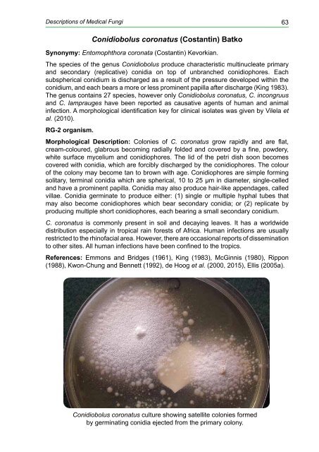
Conidiobolus coronatus culture showing satellite colonies formed<br />
by germinating conidia ejected from the primary colony.


