QCW Pulsed Nd:YAG 1064 nm Laser Lipolysis - Laser and Health ...
QCW Pulsed Nd:YAG 1064 nm Laser Lipolysis - Laser and Health ...
QCW Pulsed Nd:YAG 1064 nm Laser Lipolysis - Laser and Health ...

You also want an ePaper? Increase the reach of your titles
YUMPU automatically turns print PDFs into web optimized ePapers that Google loves.
24<br />
ABSTRACT:<br />
Journal of the <strong>Laser</strong> <strong>and</strong> <strong>Health</strong> Academy<br />
Vol. 2010, No.1; www.laser<strong>and</strong>health.com<br />
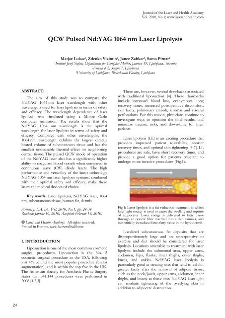
<strong>QCW</strong> <strong>Pulsed</strong> <strong>Nd</strong>:<strong>YAG</strong> <strong>1064</strong> <strong>nm</strong> <strong>Laser</strong> <strong>Lipolysis</strong><br />
Matjaz Lukac 1, Zdenko Vizintin 2, Janez Zabkar 2, Samo Pirnat 3<br />
1Institut Josef Stefan, Department for Complex Matter, Jamova 39, Ljubljana, Slovenia<br />
2Fotona d.d., Stegne 7, Ljubljana<br />
3University of Ljubljana, Biotechnical Faculty, Ljubljana<br />
The aim of this study was to compare the<br />
<strong>Nd</strong>:<strong>YAG</strong> <strong>1064</strong> <strong>nm</strong> laser wavelength with other<br />
wavelengths used for laser lipolysis in terms of safety<br />
<strong>and</strong> efficacy. The wavelength dependence of laser<br />
lipolysis was simulated using a Monte Carlo<br />
computer simulation. The results show that the<br />
<strong>Nd</strong>:<strong>YAG</strong> <strong>1064</strong> <strong>nm</strong> wavelength is the optimal<br />
wavelength for laser lipolysis in terms of safety <strong>and</strong><br />
efficacy. Compared with other wavelengths, the<br />
<strong>1064</strong> <strong>nm</strong> wavelength exhibits the largest directly<br />
heated volume of subcutaneous tissue <strong>and</strong> has the<br />
smallest undesirable thermal effect on neighboring<br />
dermal tissue. The pulsed <strong>QCW</strong> mode of operation<br />
of the <strong>Nd</strong>:<strong>YAG</strong> laser also has a significantly higher<br />
ability to coagulate blood vessels when compared to<br />
continuous wave (CW) diode lasers. The high<br />
performance <strong>and</strong> versatility of the latest technology<br />
<strong>Nd</strong>:<strong>YAG</strong> <strong>1064</strong> <strong>nm</strong> laser lipolysis systems, combined<br />
with their optimal safety <strong>and</strong> efficacy, make these<br />
lasers the medical devices of choice.<br />
Key words: <strong>Laser</strong> lipolysis, <strong>Nd</strong>:<strong>YAG</strong> laser, <strong>1064</strong><br />
<strong>nm</strong>, subcutaneous tissue, human fat, dermis.<br />
Article: J. LAHA, Vol. 2010, No.1; pp. 24-34<br />
Received: Januar 10, 2010; Accepted: Februar 13, 2010.<br />
© <strong>Laser</strong> <strong>and</strong> <strong>Health</strong> Academy. All rights reserved.<br />
Printed in Europe. www.laser<strong>and</strong>health.com<br />
I. INTRODUCTION<br />
Liposuction is one of the most common cosmetic<br />
surgical procedures. Liposuction is the No. 2<br />
cosmetic surgical procedure in the USA, following<br />
just 4% behind the most popular procedure (breast<br />
augmentation), <strong>and</strong> is within the top five in the UK.<br />
The American Society for Aesthetic Plastic Surgery<br />
states that 341,144 procedures were performed in<br />
2008 [1,2,3].<br />
There are, however, several drawbacks associated<br />
with traditional liposuction [4]. These drawbacks<br />
include increased blood loss, ecchymoses, long<br />
recovery times, increased postoperative discomfort,<br />
skin laxity, pulmonary emboli, seromas <strong>and</strong> visceral<br />
perforations. For this reason, physicians continue to<br />
investigate ways to optimize the final results, <strong>and</strong><br />
minimize trauma, risks, <strong>and</strong> down-time for their<br />
patients.<br />
<strong>Laser</strong> lipolysis (LL) is an exciting procedure that<br />
provides improved patient tolerability, shorter<br />
recovery times, <strong>and</strong> optimal skin tightening [4-7]. LL<br />
procedures are safe, have short recovery times, <strong>and</strong><br />
provide a good option for patients reluctant to<br />
undergo more invasive procedures (Fig.1).<br />
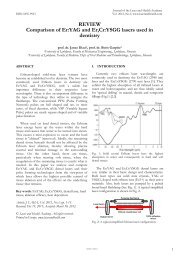
Fig.1: <strong>Laser</strong> lipolysis is a fat reduction treatment in which<br />
laser light energy is used to cause the swelling <strong>and</strong> rupture<br />
of adipocytes. <strong>Laser</strong> energy is delivered to fatty tissue<br />
through an optical fiber inserted into a thin cannula, <strong>and</strong><br />
interstitially introduced into fatty tissue in the hypodermis.<br />
Localized subcutaneous fat deposits that are<br />
disproportionately large <strong>and</strong> are unresponsive to<br />
exercise <strong>and</strong> diet should be considered for laser<br />
lipolysis. Locations amenable to treatment with laser<br />
lipolysis include the submental area, upper arms,<br />
abdomen, hips, flanks, inner thighs, outer thighs,<br />
knees, <strong>and</strong> ankles. <strong>Nd</strong>:<strong>YAG</strong> laser lipolysis is<br />
particularly good at treating sites that tend to exhibit<br />
greater laxity after the removal of adipose tissue,<br />
such as the neck/jowls, upper arms, abdomen, inner<br />
thighs, <strong>and</strong> knees; at these sites <strong>Nd</strong>:<strong>YAG</strong> laser light<br />
can mediate tightening of the overlying skin in<br />
addition to adipocyte destruction.<br />
© 2009 <strong>Laser</strong> <strong>and</strong> <strong>Health</strong> Academy. All rights reserved. Printed in Europe. www.laser<strong>and</strong>health.com. 86493. 1
<strong>Laser</strong>-assisted lipolysis has several advantages<br />
over traditional lipolysis [4]. The laser lipolysis effect<br />
improves the removal of adipose tissue: by liquefying<br />
the adipose tissue, larger volumes can be removed<br />
more easily. There is less trauma in a laser lipolysis<br />
procedure because, due to the small cannula size <strong>and</strong><br />
the light mediated lipolysis effect, mechanical<br />
destruction of skin structures by the cannula is<br />
minimized. The result is faster recovery times <strong>and</strong><br />
diminished ecchymosis. The third advantage of laserassisted<br />
lipolysis is coagulation of small blood vessels<br />
by the laser light, resulting in less blood loss during<br />
the procedure. The fourth positive attribute is skin<br />
retraction or skin tightening. Disruption <strong>and</strong><br />
coagulation of collagen can lead to the creation of a<br />
new, thicker <strong>and</strong> more organized reticular dermis,<br />
the end clinical result being tightened skin. This<br />
attribute makes laser lipolysis particularly attractive<br />
for areas of localized adiposity or localized laxity<br />
following liposuction. The skin tightens over a<br />
period of time that may last up to 8 months, with<br />
tightening becoming progressively more evident over<br />
time due to collagen regrowth.<br />
<strong>Laser</strong>-assisted lypolysis is a minimally invasive<br />
surgical procedure. The procedure is performed<br />
through tiny incisions that do not require sutures,<br />
allow drainage, prevent infection <strong>and</strong> heal within<br />
weeks of the procedure. Despite these benefits,<br />
adverse effects are still possible. Although laserassisted<br />
lipolysis minimizes trauma it cannot<br />
eliminate it. Approximately 1/3 of the trauma<br />
sustained during a procedure is caused by the direct<br />
mechanical destruction of cells by the cannula (Fig.<br />
3). As a result patients still exhibit edema <strong>and</strong> mild<br />
ecchymosis as in traditional liposuction. However,<br />
these effects are less severe <strong>and</strong> last for a shorter<br />
amount of time than following st<strong>and</strong>ard liposuction.<br />
The <strong>Nd</strong>:<strong>YAG</strong> <strong>1064</strong> <strong>nm</strong> was the pioneer<br />
wavelength for laser lipolysis (Fig. 2). It has been on<br />
the world market for almost 10 years during which it<br />
has been evaluated in many clinical studies [5-9, 35-<br />
43]. Many thous<strong>and</strong>s of satisfied patients have<br />
undergone <strong>1064</strong> <strong>nm</strong> laser lypolysis.<br />
Fig.2: <strong>Nd</strong>:<strong>YAG</strong> surgical laser [10]<br />
<strong>QCW</strong> <strong>Pulsed</strong> <strong>Nd</strong>:<strong>YAG</strong> <strong>1064</strong> <strong>nm</strong> <strong>Laser</strong> <strong>Lipolysis</strong><br />
H<strong>and</strong>piece with resterilizable multiple use 3 mm cannula<br />
H<strong>and</strong>piece with sterile single use 1,1 mm cannula<br />
Fig. 3: Accessories for laser lipolysis. To keep up with new<br />
in-dem<strong>and</strong> procedures, manufacturers offer an ever<br />
increasing range of compatible, re-useable surgical sets<br />
eliminating the need to continuously invest in expensive<br />
technologies <strong>and</strong> upgrades. [10]<br />
Since the introduction of the <strong>Nd</strong>:<strong>YAG</strong> laser,<br />
several other wavelengths have been tried <strong>and</strong> tested<br />
for <strong>Laser</strong> <strong>Lipolysis</strong>. [11-15, 28]<br />
Optics of <strong>Lipolysis</strong><br />
The basic mechanism of laser–tissue interaction<br />
in laser lipolysis is photo thermal [16-18, 27]. The<br />
conversion of laser light energy into heat is achieved<br />
by the absorption of light in target tissue. The<br />
thermal effects of a laser beam incident upon the<br />
human tissue can be predicted if the time dependent<br />
temperature distribution within the tissue is known.<br />
When the laser is fired a small fraction of tissue, V1<br />
is directly heated by energy conversion through<br />
mechanisms of absorption <strong>and</strong> scattering (Fig. 4).<br />
Due to tissue thermal conductivity, heat from V1 is<br />
dissipated, forming a larger volume, V2.<br />
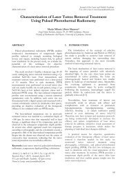
Fig. 4: Thermal images of the development of thermal<br />
zones V1 <strong>and</strong> V2 during <strong>Nd</strong>:<strong>YAG</strong> <strong>Laser</strong> <strong>Lipolysis</strong>. [19]<br />
Dissipation into V2 is a considerably slower<br />
process than direct heating in V1. Thus, in a<br />
st<strong>and</strong>ard laser lipolysis procedure, in which the<br />
cannula is moved faster than dissipation occurs, the<br />
primary thermal volume (V1) plays the major role in<br />
the thermal dissolution of fat. Our analysis focuses<br />
on the direct heating of the tissue within the volume<br />
V1.<br />
Based on the above, calculating the temperature<br />
distribution during lipolysis requires modeling the<br />
light propagation through the tissue. [20-22] Within<br />
2/11<br />
25
26<br />
the tissue the light photons undergo two processes:<br />
absorption <strong>and</strong> scattering. The absorption in<br />
biological tissues is mainly caused by water<br />
molecules, proteins <strong>and</strong> pigments. Due to<br />
absorption, light intensity I, decreases with depth z,<br />
according to Beer-Lambert law:<br />
I(z;�) = I0 exp (-�a(�) z)<br />
Here, �a(�) is the wavelength dependent<br />
absorption coefficient. Water strongly absorbs above<br />
2 �m with the strongest peak at 3 �m, proteins have<br />
a peak absorption in UV (280 <strong>nm</strong>), leaving pigments<br />
dominating the visual <strong>and</strong> NIR (400 - 2000 <strong>nm</strong>).<br />
Human tissue is also a heavy scatterer: its<br />
scattering coefficient �s(�) being somewhere<br />
between 50 <strong>and</strong> 1000 cm -1. Scattered parts of the<br />
laser beam are internally refracted from cell<br />
membranes, cell nuclei, etc, until they are finally<br />
absorbed. Note that only the absorbed laser radiation<br />
is transformed into heat. Scattering broadens the<br />
final volume of tissue in which absorption takes<br />
place (Fig. 5). It also reduces the penetration depth<br />
of the laser beam as the intensity of the incident<br />
beam decreases with depth z according to:<br />
I(z;�) = I0 exp (-( �a(�) +��s(�)) z)<br />
where ��s(�) is the reduced scattering coefficient<br />
that takes into account how much of the laser light is<br />
scattered away from the original direction. For the<br />
tissues in this study, this represents 10% of the<br />
incident beam (��s�(�) =�s(�)/10).<br />
a) b) c)<br />
Fig. 5 Combined effects of absorption (a) <strong>and</strong><br />
scattering (b) lead to the reduced penetration depth<br />
<strong>and</strong> broadening of the directly heated volume V1 (c).<br />
Optical Properties of Subcutaneous Fat<br />
In order to account for the photon propagation<br />
during <strong>Lipolysis</strong>, both laser beam <strong>and</strong> tissue optical<br />
properties must be known. The subcutaneous layer is<br />
composed of proteins <strong>and</strong> adipose tissue (fat).<br />
Above this layer lie inner dermis <strong>and</strong> epidermis (Fig.<br />
6).<br />
<strong>QCW</strong> <strong>Pulsed</strong> <strong>Nd</strong>:<strong>YAG</strong> <strong>1064</strong> <strong>nm</strong> <strong>Laser</strong> <strong>Lipolysis</strong><br />
Fig. 6 Structure of human skin <strong>and</strong> subcutaneous layer.<br />
The optical properties of the subcutaneous layer,<br />
the nearby dermis, <strong>and</strong> epidermis are not the same<br />
but they are all strongly dependent on the laser<br />
wavelength [22-26]. In human skin <strong>and</strong> fat tissues<br />
for the wavelengths between 450 <strong>nm</strong> <strong>and</strong> 1800 <strong>nm</strong><br />
scattering predominates over direct absorption<br />
[16,44]. Fig. 7 shows the absorption <strong>and</strong> scattering<br />
coefficients for the subcutaneous fat <strong>and</strong> dermis in<br />
the 400 to 1600 <strong>nm</strong> laser wavelength range [23]. The<br />
optical properties of the dermis are important in<br />
laser lipolysis because some of the laser light may<br />
propagate through the directly illuminated fat into<br />
the neighboring dermis. In what follows we shall<br />
presume that the tissue optical parameters do not<br />
change appreciably as a result of heating during laser<br />
irradiation.<br />
�� � a<br />
�� � s’<br />
3/11
�� � a<br />
<strong>QCW</strong> <strong>Pulsed</strong> <strong>Nd</strong>:<strong>YAG</strong> <strong>1064</strong> <strong>nm</strong> <strong>Laser</strong> <strong>Lipolysis</strong><br />
�� � s’<br />
Fig. 7: Measured absorption coefficient �a <strong>and</strong> the<br />
reduced scattering coefficient �s’ of subcutaneous fat <strong>and</strong><br />
dermis as a function of laser wavelength. For both tissues<br />
there are two peaks of higher absorption around 1210 <strong>and</strong><br />
1450 <strong>nm</strong>. [23]<br />
For safe execution of laser lipolysis, it is<br />
important to work with appropriate temperatures –<br />
clinical endpoints that are high enough to achieve<br />
thermal lysis effects, but not so high as to produce<br />
unwanted side effects. During st<strong>and</strong>ard LL<br />
procedure the thermal process of adipocytes<br />
destruction happens inside the heated volume V1<br />
with approximate temperatures between 50°C <strong>and</strong><br />
65°C (Fig. 8) [16].<br />
Fig. 8: Heat induced destruction of adipocytes<br />
Most researchers agree that this temperatures<br />
represent the clinical endpoint for the LL procedure.<br />
[11,16,31] . Higher temperatures are not desired; they<br />
cause unwanted side effects (like tissue necrosis <strong>and</strong><br />
scarring). Temperatures under 50°C do not result in<br />
the destruction of adipocytes.<br />
The goal of our study was a comparison of the<br />
<strong>Nd</strong>:<strong>YAG</strong> <strong>1064</strong> <strong>nm</strong> wavelength <strong>and</strong> other wavelength<br />
devices in terms of their safety <strong>and</strong> efficacy for laser<br />
lipolysis procedures.<br />
II. MATERIALS AND METHODS<br />
Photon Transport model of <strong>Laser</strong> <strong>Lipolysis</strong><br />
The distribution of light <strong>and</strong> of the resulting<br />
temperature distribution can be effectively modeled<br />
by the so called photon transport theory [17].<br />
The irradiative transport equation is difficult to<br />
solve directly <strong>and</strong> many approximations exist, among<br />
others the Kubelka-Munk inverse adding-doubling<br />
<strong>and</strong> the diffusion approximation. [21] Since the<br />
mean free path of the light propagating in human fat<br />
<strong>and</strong> skin is much smaller than the typical dimensions<br />
involved, propagation quickly becomes effectively<br />
r<strong>and</strong>om. The Monte-Carlo simulation is then the<br />
preferred method for numerically solving the<br />
irradiative transport problem. [16-18]<br />
In the Monte-Carlo simulation the laser beam is<br />
represented as a stream of a large number of laser<br />
"photons", each having a coordinate, direction <strong>and</strong><br />
energy weight E.<br />
epidermis<br />
dermis<br />
subcatenous<br />
tissue<br />
1<br />
2<br />
3<br />
Fig. 9: Schematic representation of Monte-Carlo<br />
simulation steps of laser beam propagation during<br />
lipolysis. It consists of five steps (see text below): 1-<br />
photon generation at the position of the inserted laser<br />
cannula, 2- scattered pathway generation, 3-absorption, 4-<br />
elimination <strong>and</strong> detector count. The white dot represents<br />
photon generation, black dots represent scattering <strong>and</strong><br />
absorption events <strong>and</strong> red dot represents photon<br />
elimination.<br />
Each photon is statistically ray-traced through<br />
tissue following five steps depicted in Fig. 9 above:<br />
1. Photon generation: initial location <strong>and</strong><br />
direction of propagation are r<strong>and</strong>omly<br />
determined according to the original beam<br />
output of the laser cannula.<br />
2. Pathway generation: the path to the next<br />
event is determined: the direction of<br />
propagation is determined according to the<br />
scattering characteristics of the tissue.<br />
3. Absorption: the photon energy weight E is<br />
decreased after each event according to the<br />
absorption <strong>and</strong> scattering coefficients.<br />
4. Elimination: when the photon energy weight<br />
falls below some predetermined threshold,<br />
its propagation is terminated <strong>and</strong> a new<br />
photon is generated.<br />
5. Detection: the absorbed energy �E is<br />
registered at the event coordinates after each<br />
event.<br />
4<br />
3<br />
2<br />
4/11<br />
27
28<br />
We used an optical ray-tracing program Zemax,<br />
which features a highly efficient Monte-Carlo<br />
implementation, to obtain the distributions of<br />
absorbed light. Simulation consisted of two layers,<br />
subcutaneous tissue <strong>and</strong> dermis illuminated by the<br />
<strong>Nd</strong>:<strong>YAG</strong> (� = <strong>1064</strong> <strong>nm</strong>) <strong>and</strong>, for comparison other<br />
wavelength lasers. Distributions of absorbed light<br />
were calculated for 0.6 mm spot sizes of square (or<br />
"top-hat") collimated beams that represented the<br />
output out of a typical laser lipolysis fiber.<br />
A typical calculated distribution of laser<br />
“photons” is shown in Fig. 10a. Scattering of<br />
photons after they exit the laser fiber can be clearly<br />
seen. For comparison, the light distribution, as<br />
measured in [19], is shown in Fig. 10b, <strong>and</strong> the<br />
resulting measured temperature distribution is shown<br />
in Fig. 10c.<br />
a) b) c)<br />
Fig. 10: a) Monte Carlo numerical model of hemispheric<br />
<strong>1064</strong> <strong>nm</strong> scattering in human fat; b) Measured video<br />
image of a full sphere <strong>1064</strong> <strong>nm</strong> laser light scattering inside<br />
the human fat; c) Measured temperature image of a<br />
hemispheric <strong>1064</strong> <strong>nm</strong> laser irradiation of human fat. [19]<br />
III. RESULTS<br />
Analysis of LL Efficacy<br />
During st<strong>and</strong>ard LL procedures, a cannula<br />
containing a fiber is moved back <strong>and</strong> forth, crisscrossing<br />
certain regions many times, until all the fat<br />
in the region is melted (Fig. 11)<br />
Fig. 11: Movements of thermal volume inside the fat<br />
tissue during laser lipolysis procedure<br />
A bigger thermal volume allows the fat in the<br />
region to be melted in a shorter time (fewer passes<br />
of cannula), <strong>and</strong> with a minimal mechanical damage<br />
to the tissue. Thus, one measure of laser lipolysis<br />
efficacy is the volume of the directly heated<br />
subcutaneous tissue, i.e. the thermal volume V1 (Fig.<br />
12).<br />
<strong>QCW</strong> <strong>Pulsed</strong> <strong>Nd</strong>:<strong>YAG</strong> <strong>1064</strong> <strong>nm</strong> <strong>Laser</strong> <strong>Lipolysis</strong><br />
Small thermal volume - low LL efficacy<br />
Medium thermal volume - moderate LL efficacy<br />
Large thermal volume - high LL efficacy<br />
Fig. 12: One measure of the efficacy of laser lipolysis is<br />
the size of the thermal volume, V1.<br />
The combination of wavelength dependent<br />
absorption <strong>and</strong> scattering coefficients plays an<br />
important role in the maximization of thermal<br />
volumes <strong>and</strong> thus in the optimization of the LL<br />
efficacy (Fig. 13).<br />
a) b)<br />
c)<br />
Fig. 13: Some of the possible wavelength dependent<br />
combinations of absorption <strong>and</strong> scattering: a) strong<br />
absorption <strong>and</strong> weak scattering lead to a small thermal<br />
volume V1; b) strong absorption <strong>and</strong> strong scattering<br />
also lead to a small V1; c) medium absorption <strong>and</strong><br />
medium scattering results in optimal thermal volume<br />
conditions.<br />
Scattering phenomena broaden the diameter of<br />
the thermal volume beyond the laser beam crosssection.<br />
As a consequence the operator can use a<br />
smaller diameter cannula or fiber; using a small<br />
diameter instrument causes less mechanical damage<br />
to the tissue. However, while scattering increases the<br />
diameter of the thermal volume, it also reduces its<br />
depth.<br />
Absorption mitigates the effects of scattering. As<br />
absorption increases the “free-path” of the photons<br />
is reduced, limiting the thermal volume. Very weak<br />
absorption would result in a large amount of<br />
scattering <strong>and</strong>, consequently, a very large thermal<br />
volume; eventually the thermal volume would<br />
become so large that it would require an<br />
impractically large amount of laser power to heat it<br />
to a clinically significant end-point.<br />
Because of the complex interrelation of<br />
scattering, absorption, <strong>and</strong> laser power, the optimal<br />
choice of wavelength for laser lipolysis is one that is<br />
moderately scattered <strong>and</strong> absorbed in subcutaneous<br />
tissue. In general as the wavelength increases from<br />
800 <strong>nm</strong> to 1500 <strong>nm</strong> the absorption coefficient<br />
increases <strong>and</strong> the scattering coefficient decreases.<br />
5/11
Based on the considerations presented above the<br />
optimal thermal volume V1 occurs around 1100 <strong>nm</strong>.<br />
This is in line with the results of our Monte Carlo<br />
simulation in which we compared the relative rate of<br />
heat generation versus the depth of thermal volume<br />
for five different relevant wavelengths (� = 920 <strong>nm</strong>,<br />
980 <strong>nm</strong>, <strong>1064</strong> <strong>nm</strong>, 1320 <strong>nm</strong>, <strong>and</strong> 1440 <strong>nm</strong>). The<br />
greatest amount of heat is generated at the surface of<br />
the fiber, but the heat generation is dramatically<br />
increased as the absorption coefficient is increased.<br />
At equivalent laser power the 1440 <strong>nm</strong> wavelength<br />
results in the highest temperature at the surface <strong>and</strong><br />
the smallest depth of the thermal volume, whereas<br />
the <strong>1064</strong> <strong>nm</strong> wavelength results in the lowest<br />
temperature at the surface <strong>and</strong> the largest depth of<br />
the thermal volume.<br />
Some interpret the high surface peak temperature<br />
of the 1440 <strong>nm</strong> wavelength as its advantage. [28]<br />
However, since the surface temperature must not<br />
exceed the clinical end-point of 65°C, this<br />
interpretation is incorrect. In order to perform safe<br />
lipolysis, the laser power must be adjusted in such a<br />
way that the temperature at all wavelengths does not<br />
exceed this clinical end-point. It is true that for the<br />
medium absorbed <strong>1064</strong> <strong>nm</strong> a slightly higher power is<br />
required to reach the clinical end-point but the<br />
important benefit is a larger thermal volume. In<br />
addition, the <strong>1064</strong> <strong>nm</strong> <strong>Nd</strong>:<strong>YAG</strong> laser is one of the<br />
most efficient lasers, <strong>and</strong> in spite of slightly higher<br />
working powers, the electrical efficiency <strong>and</strong><br />
longevity of the <strong>1064</strong> <strong>nm</strong> <strong>Nd</strong>:<strong>YAG</strong> far exceeds<br />
those of the 1320 <strong>and</strong> 1440 <strong>nm</strong> lasers. Note,<br />
however, that lower heat generation could become<br />
an issue for less efficient lasers with weakly absorbed<br />
wavelengths.<br />
Figure 14 presents our results from the Monte<br />
Carlo simulations in the subcutaneous fat. In the<br />
simulation, the light was assumed to propagate out<br />
of a fiber into an infinite scattering medium. With<br />
the same surface temperature of 65°C, the thermal<br />
penetration depth, <strong>and</strong> consequently the thermal<br />
volume are the largest for the <strong>1064</strong> <strong>nm</strong> wavelength.<br />
<strong>QCW</strong> <strong>Pulsed</strong> <strong>Nd</strong>:<strong>YAG</strong> <strong>1064</strong> <strong>nm</strong> <strong>Laser</strong> <strong>Lipolysis</strong><br />
Fig. 14: Comparison of wavelengths, generating the same<br />
maximal safe temperature of 65°C. Out of all studied<br />
wavelengths (920 <strong>nm</strong>, 980 <strong>nm</strong>, <strong>1064</strong> <strong>nm</strong>, 1320 <strong>nm</strong>, 1440<br />
<strong>nm</strong>), the largest thermal volume is generated with the<br />
<strong>1064</strong> <strong>nm</strong> (for clarity, volumes for <strong>1064</strong><strong>nm</strong>, 1320 <strong>nm</strong> <strong>and</strong><br />
1440 <strong>nm</strong> are shown). This enables the highest LL efficacy<br />
of this wavelength at a given safe temperature endpoint.<br />
While the differences in the radii for these<br />
wavelengths do not seem large, the final differences<br />
in the thermal volumes are appreciable. This has<br />
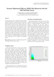
been studied also by Wassmer et al.[32]. Figure 15<br />
shows a graphical representation of the calculated<br />
thermal volumes from their study for various<br />
lipolysis wavelengths. The difference in the thermal<br />
volumes for the analyzed wavelengths can be clearly<br />
observed, with the <strong>1064</strong> <strong>nm</strong> wavelength resulting in<br />
the largest thermal volume.<br />
Fig. 15: Graphical representation of calculated thermal<br />
volumes for different laser wavelengths as obtained from<br />
a previously published study). [32]. The size of the circles<br />
is proportional to the size of the thermal volume. The<br />
<strong>1064</strong> <strong>nm</strong> exhibits the largest thermal volume, the thermal<br />
volumes of 980 <strong>nm</strong> <strong>and</strong> 920 <strong>nm</strong> are only slightly smaller,<br />
while the 1320 <strong>nm</strong>, <strong>and</strong> particularly the 1440 <strong>nm</strong><br />
wavelengths result in significantly smaller thermal<br />
volumes.<br />
6/11<br />
29
30<br />
Analysis of LL Safety<br />
<strong>Laser</strong> lipolysis is performed by inserting a cannula<br />
into the fat under the skin. It is possible to damage<br />
the skin when the cannula is moved close to the skin<br />
(Fig. 16), <strong>and</strong> the laser light penetrates into the<br />
dermis.<br />
Fig. 16: If, during the procedure the laser lipolysis cannula<br />
is positioned closer to the dermis the laser light may<br />
penetrate <strong>and</strong> thermally damage the dermis.<br />
The effect that laser light of a certain wavelength<br />
will have on the dermis will depend on the difference<br />
between the optical coefficients of subcutaneous<br />
tissue <strong>and</strong> the dermis at that wavelength. To<br />
demonstrate this we calculated the temperature<br />
distribution during laser lipolysis with various<br />
wavelengths under the three geometries depicted in<br />
Fig. 17.<br />
75 deg<br />
45 deg<br />
dermis<br />
fat<br />
normal<br />
Fig. 17: Three geometries were used to evaluate the<br />
influence of dermis vicinity to temperature distribution in<br />
the two tissues model<br />
<strong>QCW</strong> <strong>Pulsed</strong> <strong>Nd</strong>:<strong>YAG</strong> <strong>1064</strong> <strong>nm</strong> <strong>Laser</strong> <strong>Lipolysis</strong><br />
D<br />
Figure 18 shows the calculated temperature<br />
distributions (� = 920 <strong>nm</strong>, 980 <strong>nm</strong>, <strong>1064</strong> <strong>nm</strong>, 1320<br />
<strong>nm</strong>, <strong>and</strong> 1440 <strong>nm</strong>) when the end of the cannula is<br />
positioned at a distance of D = 0.5 mm from the fatdermis<br />
boundary. For all three wavelengths, it was<br />
assumed that the procedure was performed up to the<br />
temperature clinical end-point of 65°C in the<br />
subcutaneous tissue. In the calculation, we used the<br />
most commonly accepted values for the absorption<br />
coefficient �a <strong>and</strong> the reduced scattering coefficient<br />
��s , as shown in Table 1.<br />
Table 1: The values of the absorption coefficients (�a)<br />
<strong>and</strong> scattering coefficients (�s) in fat <strong>and</strong> dermis for<br />
different wavelengths that were used in the Monte<br />
Carlo simulation. Numbers in the brackets represent<br />
the corresponding references.<br />
Wavelength � (<strong>nm</strong>) 920 890 <strong>1064</strong> 1320 1440<br />
Fat<br />
Dermis<br />
�a (cm -1 )<br />
�s (cm -1 )<br />
�a (cm -1 )<br />
�s (cm -1 )<br />
1,65<br />
[23]<br />
120<br />
[23]<br />
2<br />
[23]<br />
250<br />
[23]<br />
1,65<br />
[23]<br />
110<br />
[23]<br />
2<br />
[23]<br />
250<br />
[23]<br />
1,5<br />
[16,29]<br />
110<br />
[16,29]<br />
1<br />
[16,29]<br />
250<br />
[16,29]<br />
2<br />
[16,29]<br />
105<br />
[16,29]<br />
2<br />
[16,29]<br />
250<br />
[16,29]<br />
2,5<br />
[16,29]<br />
110<br />
[16,29]<br />
4<br />
[16,29]<br />
250<br />
[16,29]<br />
The results of the Monte Carlo simulation show<br />
that the injury to the dermis can be appreciable when<br />
working with sub-optimal wavelengths. The<br />
temperature increase in the dermis is particularly<br />
high at the 1440 <strong>nm</strong> wavelength where the<br />
temperature reaches over 80°C. With the <strong>1064</strong> <strong>nm</strong>,<br />
the effect on the dermis is negligible. This explains<br />
partially why certain manufacturers of higher<br />
wavelength laser lipolysis devices have started<br />
offering additional thermal imaging accessories for<br />
monitoring skin temperature during the procedures.<br />
At higher wavelengths, <strong>and</strong> especially if they are<br />
positioned on the absorption peaks, the risks of<br />
damaging the skin are much higher.<br />
7/11
a)<br />
b)<br />
c)<br />
d)<br />
<strong>QCW</strong> <strong>Pulsed</strong> <strong>Nd</strong>:<strong>YAG</strong> <strong>1064</strong> <strong>nm</strong> <strong>Laser</strong> <strong>Lipolysis</strong><br />
920 <strong>nm</strong><br />
980 <strong>nm</strong><br />
<strong>1064</strong> <strong>nm</strong><br />
1320 <strong>nm</strong><br />
e)<br />
1440 (<strong>nm</strong>)<br />
Fig. 18: Analysis of the fat/dermis border on thermal<br />
effects. At close distance (D =0.5 mm) of the lipolysis<br />
cannula to the fat/dermis border, the thermal effect in the<br />
dermis is smallest with the <strong>1064</strong> <strong>nm</strong> laser. At other<br />
wavelengths, the thermal effects are more expressed <strong>and</strong><br />
can exceed the maximal allowed temperatures. The largest<br />
undesired thermal effect in the dermis is with the 1440<strong>nm</strong>.<br />
<strong>Pulsed</strong> vs. CW Operation<br />
There are two types of lasers for lipolysis. Quasi<br />
continuous wave (<strong>QCW</strong>) lasers, such as the <strong>1064</strong> <strong>nm</strong><br />
<strong>Nd</strong>:<strong>YAG</strong>, deliver the power in a fast sequence of<br />
high peak power (several kilowatts) laser pulses,<br />
while the continuous wave (CW) lasers, such as<br />
980 <strong>nm</strong> diode laser deliver the power (in the range of<br />
only ten watts) in a non-pulsed or in a pulse gated<br />
manner. (Fig. 19)<br />
Fig. 19.: The <strong>QCW</strong> crystal pulsed lasers are capable of<br />
delivering laser radiation in a fast sequence of high peak<br />
power pulses, while CW lasers are capable of only low<br />
power gated pulsing.<br />
As explained below, the pulsed <strong>QCW</strong> operation<br />
of lipolysis lasers is of extreme importance when<br />
considering the ability of lasers to coagulate blood<br />
vessels [6].<br />
8/11<br />
31
32<br />
If the clinical objective was only to coagulate<br />
blood vessels the optimal laser wavelength would<br />
have the highest absorption in hemoglobin relative<br />
to surrounding tissue. However, the wavelengths<br />
that are highly absorbed in blood vessels (such as �<br />
= 530 <strong>nm</strong>) do not reach deeper lying tissue, <strong>and</strong> can<br />
result in excessive damage to the surrounding tissue<br />
structures. For this reason, the <strong>QCW</strong> lipolysis lasers<br />
operate at a laser wavelength of <strong>1064</strong> <strong>nm</strong> that<br />
penetrates optimally into the subcutaneous tissue,<br />
<strong>and</strong> achieves selective vessel coagulation by adjusting<br />
the laser pulse duration to the thermal relaxation<br />
time of the targeted vessels [33]. Namely, during a<br />
lengthy laser exposure, most of the deposited heat<br />
may diffuse away from the vessels, resulting in a<br />
non-specific thermal effect on the adjacent<br />
subcutaneous tissue. Conversely, a suitably short<br />
laser pulse confines the heating effect to the target<br />
vessel structure, resulting in a temporary local<br />
temperature increase of the vessels without<br />
overheating the surrounding subcutaneous tissue<br />
[34].<br />
The thermal relaxation time of the vascular<br />
targets can be estimated from the relaxation time of<br />
an infinite cylinder:<br />
� = d 2/16 � ,<br />
giving thermal relaxation time for different blood<br />
vessel diameters as shown in Table 2. The <strong>Nd</strong>:<strong>YAG</strong><br />
<strong>QCW</strong> treatment modality with pulse durations<br />
between 0.1 <strong>and</strong> 0.6 ms can coagulate vessels with<br />
diameters under 50 �m. A good representative of a<br />
“coagulating” <strong>Nd</strong>:<strong>YAG</strong> laser for lipolysis is the XP-2<br />
Focus <strong>Nd</strong>:<strong>YAG</strong> laser system (Fotona d.d.) that has<br />
an adjustable high peak power pulse duration range<br />
starting as low as 0.2 msec.<br />
Table 2: Thermal relaxation time for the range of<br />
blood vessel diameters.<br />
It has been reported that <strong>QCW</strong> pulsed lasers have<br />
significantly higher ability to coagulate blood vessels<br />
when compared to continuous wave, CW diode<br />
lasers [6,33,34]. Treatments with <strong>QCW</strong> pulsed<br />
lasers, such as <strong>Nd</strong>:<strong>YAG</strong>, thus result in less bruising,<br />
swelling <strong>and</strong> quicker recovery.<br />
<strong>QCW</strong> <strong>Pulsed</strong> <strong>Nd</strong>:<strong>YAG</strong> <strong>1064</strong> <strong>nm</strong> <strong>Laser</strong> <strong>Lipolysis</strong><br />
IV. DISCUSSION<br />
The <strong>1064</strong> <strong>nm</strong> <strong>Nd</strong>:<strong>YAG</strong> lasers are the most widely<br />
used lasers for lipolysis, with the longest clinical<br />
record of safety <strong>and</strong> efficacy.<br />
For example, Goldman has reported on more<br />
than 3,000 cases of successful laser lipolysis<br />
performed with the <strong>1064</strong> <strong>nm</strong> <strong>Nd</strong>:<strong>YAG</strong> laser in a<br />
period lasting from 1999 to 2006, with minimal side<br />
effects <strong>and</strong> high patient satisfaction. [38] Together<br />
with Schavelzon <strong>and</strong> Blugerman, Goldman has also<br />
reported on a three center study in which, during a<br />
period of 30 months, 1,734 procedures of laser<br />
assisted liposuctions with the <strong>1064</strong> <strong>nm</strong> <strong>Nd</strong>:<strong>YAG</strong><br />
were performed. [9] The results showed the<br />
procedure to be safe <strong>and</strong> effective with minimal side<br />
effects. Similarly, Katz’s 18 month, 537 treatment<br />
study showed that complications from surgery were<br />
minimal. [39] During a meeting of the American<br />
Society for <strong>Laser</strong> Medicine <strong>and</strong> Surgery, Mazzi,<br />
reported on his 5 year (2003 – 2008) experience with<br />
<strong>1064</strong> <strong>nm</strong> <strong>Nd</strong>:<strong>YAG</strong> laser lipolysis [40] during which<br />
he successfully treated 386 patients with a low<br />
occurrence of any complications.<br />
Fig. 20: Example of laser lipolysis treatment (Courtesy of<br />
Dr. Maletic)<br />
9/11
Fig. 21: Example of laser lipolysis results (Courtesy of Dr.<br />
Maletic)<br />
Maletic recently reported on a study with 224<br />
patients with <strong>1064</strong> <strong>nm</strong> <strong>Nd</strong>:<strong>YAG</strong> laser during an 18<br />
month period resulting in very successful body<br />
shaping <strong>and</strong> a very low side effects rate.[41] (fig. 20-<br />
22) Similarly Steventon, reported more than 300<br />
successful cases of <strong>1064</strong> <strong>nm</strong> <strong>Nd</strong>:<strong>YAG</strong> laser lipolysis<br />
treatments [43]. There were no reports of deaths,<br />
pulmonary emboli, viscus perforation,<br />
thrombophlebitis, hypovolemic shock, seizures or<br />
toxic reactions. Minimal side effects were reported:<br />
0.4% infections, 1% skin irregularities, 0.7%<br />
hematoma or seroma, 0.02% unacceptable scarring,<br />
0.9% sensory nerve impairments, 0.12% contact<br />
dermatitis.<br />
Fig. 22: Example of laser lipolysis results (Courtesy of<br />
Prof. Massoud)<br />
There are also other reports, with smaller<br />
numbers of patients, all of them presenting positive<br />
results in terms of safety <strong>and</strong> efficacy for the<br />
<strong>1064</strong> <strong>nm</strong> <strong>Nd</strong>:<strong>YAG</strong> laser lipolysis treatment<br />
[6,35,37,42].<br />
Published clinical studies of laser lipolysis with<br />
other wavelengths are much more modest in scope<br />
<strong>and</strong> enrollment. So far, the only study exceeding 100<br />
patients has been done with a 980 <strong>nm</strong> diode laser in<br />
which Reynaud et al. reported about treatment of<br />
534 procedures performed on 334 patients [12].<br />
Results showed successful removal of small volumes<br />
of fat, high patient tolerance <strong>and</strong> quick recovery<br />
time.<br />
<strong>QCW</strong> <strong>Pulsed</strong> <strong>Nd</strong>:<strong>YAG</strong> <strong>1064</strong> <strong>nm</strong> <strong>Laser</strong> <strong>Lipolysis</strong><br />
Initial results with wavelengths 924, 975, 1320<br />
<strong>and</strong> 1440 <strong>nm</strong> are, thus far, based on clinical studies<br />
in which the number of patients is usually 10-30, <strong>and</strong><br />
the follow-ups are shorter than one year.<br />
The clinically observed minimal discomfort,<br />
exceptional long-term success <strong>and</strong> short recovery are<br />
attributed to <strong>Nd</strong>:<strong>YAG</strong> laser’s ability to optimally<br />
target laser energy into fatty tissue thereby limiting<br />
undesirable mechanical <strong>and</strong> thermal effects in the<br />
surrounding tissues.<br />
In addition, the <strong>Nd</strong>:<strong>YAG</strong> <strong>1064</strong> <strong>nm</strong> laser systems<br />
have developed significantly since their introduction<br />
for laser lipolysis. <strong>Nd</strong>:<strong>YAG</strong> lasers with <strong>QCW</strong> mode<br />
power-generating capacity are reaching higher<br />
procedure speeds <strong>and</strong> efficiency while operating at a<br />
fraction of their maximum capacity. This ensures<br />
system durability, essentially lowering running costs.<br />
In conjunction with the VSP (Variable Square Pulse)<br />
technology, a wide range of selectable pulse<br />
durations are now available in <strong>Nd</strong>:<strong>YAG</strong> lasers. This<br />
provides better procedure control <strong>and</strong> extreme<br />
versatility. The high performance <strong>and</strong> versatility of<br />
the latest technology solid crystal <strong>Nd</strong>:<strong>YAG</strong> <strong>1064</strong> <strong>nm</strong><br />
laser systems, combined with their optimal efficacy<br />
<strong>and</strong> safety, make these lasers the medical devices of<br />
choice when compared with other wavelength<br />
devices for laser lipolysis.<br />
V. CONCLUSION<br />
The results of the Monte Carlo statistical<br />
simulations are in agreement with clinical reports: a)<br />
the <strong>Nd</strong>:<strong>YAG</strong> laser wavelength results in the largest<br />
thermal volume in the subcutaneous tissue, <strong>and</strong> is<br />
least likely to cause any injury in the neighboring<br />
dermis; b) the pulsed <strong>QCW</strong> mode of operation of<br />
the <strong>Nd</strong>:<strong>YAG</strong> laser has significantly higher ability to<br />
coagulate blood vessels when compared to<br />
continuous wave, CW diode lasers.<br />
REFERENCES<br />
1. American Society for Aesthetic <strong>and</strong> Plastic Surgery<br />
“Statistics 2008, Cosmetic Surgery National Databank”,<br />
www.surgery.org/sites/default/files/2008stats.pdf accessed<br />
04 November 2009<br />
2. Medical Insight, Inc.<br />
https://www.miinews.com/market_studies.php ) accessed<br />
04 November 2009<br />
3. Millennium Research Group<br />
(http://www.mrgresourcecompass.com/index.aspx )<br />
accessed 04 November 2009<br />
4. Alam M, Dover J(2009) Procedures in Cosmetic<br />
5.<br />
Dermatology Series: Non- surgical skin tightening <strong>and</strong><br />
lifting. Elsevier<br />
Kim KH, Geronemus RG. (2006) <strong>Laser</strong> <strong>Lipolysis</strong> Using a<br />
Novel <strong>1064</strong>-<strong>nm</strong> <strong>Nd</strong>:<strong>YAG</strong> <strong>Laser</strong>. Dermatol Surg. 32(2):241-48<br />
6. Massoud KS, Aboul Fotouh SM, El Sherbiny DM (2009)<br />
10/11<br />
33
34<br />
Evaluation of Blood Loss in <strong>Laser</strong>-Assisted Liposuction<br />
4th Middle East International Plastic Surgery & <strong>Laser</strong><br />
Congress, Dubai, UAE<br />
7. Raine T, Hankins WT, Sohn SM. <strong>Laser</strong>-Assisted <strong>Lipolysis</strong><br />
with a <strong>1064</strong>-<strong>nm</strong> <strong>Nd</strong>-<strong>YAG</strong> System with SelectPulse. Syneron,<br />
2006<br />
8. Ichikawa K, Miyasaka M, Tanaka R, Tanino R, Mizukami K,<br />
Wakaki M (2005) Histologic Evaluation of the <strong>Pulsed</strong><br />
<strong>Nd</strong>:<strong>YAG</strong> <strong>Laser</strong> for <strong>Laser</strong> <strong>Lipolysis</strong>. <strong>Laser</strong>s Surg Med 36: 43<br />
– 46.<br />
9. A. Goldman, D. E. Schavelzon, <strong>and</strong> G. S. Blugerman.<br />
„<strong>Laser</strong>lipolysis: Liposuction Using <strong>Nd</strong>:<strong>YAG</strong> <strong>Laser</strong>“. Rev.<br />
Soc. Bras. Cir. Plást., Sao Paulo, (17-26 January 2002).<br />
10. XP-2 Focus www.fotona.com<br />
accessed 04 November 2009<br />
11. DiBernardo BE, Reyes J, Chen B (2009) Evaluation of<br />
Tissue thermal effects from <strong>1064</strong>/1320-<strong>nm</strong> laser-assisted<br />
lipolysis <strong>and</strong> its clinical implications. J Cosmet <strong>Laser</strong><br />
Ther.11: 62 – 69.<br />
12. Reynaud JP, Skibinski M, Wassmer B, Rochon P, Mordon S<br />
(2009) <strong>Lipolysis</strong> Using a 980-<strong>nm</strong> Diode <strong>Laser</strong>: A<br />
Retrospective Analysis of 534 Procedures. Aesthetic Plast<br />
Surg 33: 28 – 36<br />
13. DiBernardo BE, Holdman MK, Saluja R, Woodhall K, <strong>and</strong><br />
Reyes J (2008) <strong>Laser</strong> <strong>Lipolysis</strong> with Sequential Emission of<br />
<strong>1064</strong>-<strong>nm</strong> <strong>and</strong> 1320-<strong>nm</strong> Wavelengths. Cynosure, P/N 921-<br />
0095-000 Rev. 1<br />
14. Wanner M, Avram M, Gagnon D, Mihm Jr MC, Zurakowski<br />
D, Watanabe K, Tannous Z., Anderson RR, Manstein D<br />
(2009) Effects of Non-Invasive, 1210-<strong>nm</strong> <strong>Laser</strong> Exposure<br />
on Adipose Tissue: Results of a Human Pilot Study. <strong>Laser</strong>s<br />
Surg Med 41: 401 – 407.<br />
15. Anderson RR, Farinelli W, Laubach H, Manstein D,<br />
Yaroslavsky AN, Gubeli III J, Jordan K, Neil GR, Shinn M,<br />
Ch<strong>and</strong>ler W, Williams GP, Benson SV, Douglas DR, <strong>and</strong><br />
Dylla HF (2006) Selective Photothermolysis of Lipid-Rich<br />
Tissues: A Free Electron <strong>Laser</strong> Study. <strong>Laser</strong>s Surg Med 38:<br />
913 – 919.<br />
16. Mordon SR, Wassmer B, Reynaud JP, <strong>and</strong> Zemmouri J<br />
(2008) Mathematical modeling of laser lipolysis. Biomed Eng<br />
Online 29: 7-10<br />
17. Niemz MH (1996)<strong>Laser</strong>-Tissue Interactions“. Springer-<br />
Verlag<br />
18. Welch AJ <strong>and</strong> Van Gemert MJC (1995) Optical-Thermal<br />
Response of <strong>Laser</strong>-Irradiated Tissue“. Plenum Press (1995).<br />
19. Internal research documentation of Fotona d.d..<br />
20. Serup J, Jemec GBE (1995) H<strong>and</strong>book of Non-Invasive<br />
Methods <strong>and</strong> the Skin. CRC Press (1995).<br />
21. Ishimaru A (1978)Wave propagation <strong>and</strong> scattering in<br />
r<strong>and</strong>om media. Academic Press<br />
22. Simpson CR, Kohl M, Essenpreis M, <strong>and</strong> Cope M (1998)<br />
Near-infrared optical properties of ex vivo human skin <strong>and</strong><br />
subcutaneous tissues measured using the Monte Carlo<br />
inversion technique. Phys. Med. Biol. 43: 2465-2478.<br />
23. Salomatina E, Jiang B, Novak J, Yaroslavsky AN (2006)<br />
Optical properties of normal <strong>and</strong> cancerous human skin in<br />
the visible <strong>and</strong> near-infrared spectral range. J Biomed Optic<br />
11(6): 064026<br />
24. Graaff R, Dassel ACM, Koelink MH, de Mul FFM,<br />
Aarnoudse JG, Zijlstra EG (1993) Optical properties of<br />
human dermis in vitro <strong>and</strong> in vivo.. Appl Optic 32: 435 –<br />
447.<br />
25. Bashkatov AN, Genina EA, Kochubey VI, <strong>and</strong> Tuchin VV<br />
(2005) Optical properties of human skin, subcutaneous <strong>and</strong><br />
mucous tissues in the wavelenght range from 400 to 2000<strong>nm</strong>.<br />
J. Phys. D 38: 2543 – 2555.<br />
26. Tseng SH, Grant A, Durkin AJ () In vivo determination of<br />
skin near-infrared optical properties using diffuse optical<br />
spectroscopy. J Biomed Opt. 13: 014016.<br />
<strong>QCW</strong> <strong>Pulsed</strong> <strong>Nd</strong>:<strong>YAG</strong> <strong>1064</strong> <strong>nm</strong> <strong>Laser</strong> <strong>Lipolysis</strong><br />
27. Khoury JG (2008) Histologic evaluation of interstitial<br />
lipolysis comparing a <strong>1064</strong>, 1320 <strong>and</strong> 2100 <strong>nm</strong> laser in an ex<br />
vivo model. <strong>Laser</strong>s Surg Med 40: 402-406<br />
28. Youn JI(2009)A Comparison of Wavelenght Dependence<br />
for <strong>Laser</strong>-Assisted <strong>Lipolysis</strong> Effect Using Monte Carlo<br />
Simulation. J. Opt. Soc. Korea 13: 267-271<br />
29. Bashkatov AN, Genina EA, Kochubey VI, <strong>and</strong> Tuchin VV<br />
(2005) Optical Properties of the Subcutaneous Adipose<br />
Tissue in the Spectral Range 400 – 2500-<strong>nm</strong>. Optics <strong>and</strong><br />
Spectroscopy 99: 836 – 842<br />
30. Badin AZED, Gondek LBE, Garcia MJ, Choppa do Valle L,<br />
Flizikowski FBZ, <strong>and</strong> de Noronha L (2005)Analysis of <strong>Laser</strong><br />
<strong>Lipolysis</strong> Effect on Human Tissue Samples Obtained from<br />
Liposuction. Aesthetic Plast Surg 29: 281 – 286.<br />
31. Weiss RA (2009) Invasive <strong>Laser</strong> <strong>Lipolysis</strong>. Course at ASLMS<br />
Washington.<br />
32. Wassmer B (2009) Comparative study of wavelength for<br />
laser lipolysis / adipocytolysis, IMCAS Congress in Paris,<br />
January 2009 <strong>and</strong> ASLMS meeting in Washington, April<br />
2009<br />
33. Lukac M, Zabkar J, Gorjan M, Sult T (2008)FRAC3: Three<br />
Dimensional Non-Ablative Fractional Skin Rejuvenation, J.<br />
<strong>Laser</strong> <strong>Health</strong> Academy, Vol. 2008, No.5<br />
www.laser<strong>and</strong>health.com.<br />
34. Anderson RR, Parrish JA (1983) Selective Photothermolysisprecise<br />
Microsurgery by Selective Absorption of <strong>Pulsed</strong><br />
Radiation. Science 220:524-527<br />
35. Goldman A (2006)Submental <strong>Nd</strong>:<strong>YAG</strong> <strong>Laser</strong>-Assisted<br />
Liposuction. <strong>Laser</strong>s Surg Med 38:181-184<br />
36. Badin AZD, Moraes LM, Gondek L, Chiaratti MG, Canta L<br />
(2002 )<strong>Laser</strong> <strong>Lipolysis</strong>: Flaccidity Under Control. Aesthetic<br />
Plast Surg 26: 335 – 339.<br />
37. Leibaschoff G, Pique H, Diz A, Schroh A, Sluga MC (2008)<br />
A double-blind, prospective, clinical, surgical,<br />
histopathological <strong>and</strong> ultrasound study comparing the<br />
effectiveness <strong>and</strong> safety of liposuction performed using<br />
<strong>Laser</strong>lipolysis (Smartlipo TM) <strong>and</strong> Internal Ultrasound<br />
(Vaser TM) method, <strong>and</strong> assessing the evolution in patients.<br />
Deka<br />
38. Goldman A (2007) <strong>Nd</strong>:<strong>YAG</strong> <strong>Laser</strong>-Assisted <strong>Lipolysis</strong>:<br />
Clinical Findings. Clinical Finidings, Cynosure.<br />
39. Katz BE, McBean J (2008) <strong>Laser</strong>-assisted lipolysis: A report<br />
on Complications, J Cosmet <strong>Laser</strong> Ther 10: 231-233<br />
40. Mazzi L, Gallo G (2008) SmartLipo:2003-2008 Five Years of<br />
<strong>Laser</strong> Body Contouring. <strong>Laser</strong>s Surg Med S20: 39<br />
41. Maleti� D, Maleti� I (2009) <strong>Laser</strong> Assisted Liposuction with<br />
<strong>Nd</strong>:<strong>YAG</strong> laser. Abstract Book of VI BAPRAS Congress<br />
(June 2009): 11<br />
42. El-Minawi HM(2008) Body Contouring Using <strong>Laser</strong>lipolysis.<br />
IMCAS 10th Annual Meeting Abstract Booklet S40/8<br />
43. Steventon P (2009) Knowledge is power Body language<br />
34:47<br />
44. Vogel A, Venugopalan A (2003) Mechanisms of <strong>Pulsed</strong><br />
<strong>Laser</strong> Ablation of Biological Tissues, Chem. Rev. 103: 577-<br />
644<br />
11/11