Laser-induced Interstitial Thermotherapy University Hospital Johann ...
Laser-induced Interstitial Thermotherapy University Hospital Johann ...
Laser-induced Interstitial Thermotherapy University Hospital Johann ...

Create successful ePaper yourself
Turn your PDF publications into a flip-book with our unique Google optimized e-Paper software.
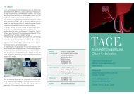
Information about LITT<br />
The use of local thermal effect in the tumor<br />
region forms the basis for this new minimal-<br />
invasive therapeutic procedure. Since the<br />
development of MRI-guided laser-<strong>induced</strong><br />
<strong>Interstitial</strong> ThermoTherapy (LITT), it is possible<br />
to target the laser exactly on the tissue volume<br />
to be treated. Due to comparably high<br />
penetrability of photons and the possibility of<br />
complication-free transfer of energy through<br />
guide-light, <strong>Laser</strong> of Near Infrared Region (NIR)<br />
is used for LITT. Above all we use the Nd:YAG-<br />
<strong>Laser</strong> (1,064 nm), which has a wide clinical use.<br />
The energy is applied to the target tissue using<br />
special laser applicators. The energy of laser<br />
light is absorbed, which causes heating of the<br />
tissue. The heat causes<br />
coagulation (destruction)<br />
of tumor tissue and the<br />
edge around it. In order<br />
to make the advantages<br />
of the effect and<br />
accuracy of the therapy<br />
useful, all the factors contributing to the success<br />
of therapy must co-ordinate with one another.<br />
For this, it is necessary to calculate the exact<br />
duration and output of the laser. Therefore under<br />
certain circumstances more laser applicators<br />
and more cycles of therapy, may be required<br />
depending on size, number and location of the<br />
lesion to be treated. These parameters are<br />
adapted individually according to the illness. In<br />
practise a temperature of about 60 to 110° C is<br />
achieved in the tumor tissue. This differentiates<br />
LITT from the classical hyperthermia.<br />
It is scientifically proven that local resection or<br />
destruction of liver metastases prolongs the life<br />
of patients and chemotherapy is then to be<br />
preferred.<br />
Metastasis in segment 8, native<br />
MRI before LITT<br />
Metastasis in segment 8, native<br />
MRI after LITT. The metastasis<br />
and the edge around is fully<br />
coagulated.<br />
Metastasis in segment 8,<br />
contrast-enhanced MRI before<br />
LITT<br />
Metastasis in segment 8,<br />
contrast-enhanced MRI after<br />
LITT<br />
Indications:<br />
1. LITT of liver tumours<br />
• Maximum number: 5 lesions<br />
• Maximum diameter: 50 mm<br />
• Residual metastases in patients who have already<br />
undergone liver resection.<br />
• Progress of metastases despite chemotherapy<br />
• Bilobar metastases (involvement of both lobes of<br />
liver)<br />
• Patients who are contraindicated for operation<br />
• Patients who are primarily inoperable can be<br />
brought to an operable situation with LITT<br />
(metastases in both liver lobes)<br />
• LITT as alternative therapy for patients who<br />
refuse surgical resection and systemic or local<br />
chemotherapy.<br />
2. LITT of lung metastases and lung tumors<br />
3. LITT of soft tissue tumors<br />
• Residual tumors<br />
• Head and neck region<br />
• Upper abdomen<br />
• Retroperitoneum<br />
• Lymph node metastases<br />
• Head and neck region<br />
• Upper abdomen<br />
• Retroperitoneum<br />
4. Special indications<br />
• Kidney tumors<br />
• Prostate tumors<br />
• Other soft tissue tumors