Understanding NRT- Reading 1 of 2- Radiogaphic Testing A
Understanding NRT- Reading 1 of 2- Radiogaphic Testing A
Understanding NRT- Reading 1 of 2- Radiogaphic Testing A

You also want an ePaper? Increase the reach of your titles
YUMPU automatically turns print PDFs into web optimized ePapers that Google loves.
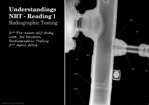
<strong>Understanding</strong>s<br />
<strong>NRT</strong> - <strong>Reading</strong> 1<br />
Radiographic <strong>Testing</strong><br />
2 nd Pre-exam self study<br />
note for Neutron<br />
Radiographic <strong>Testing</strong><br />
2 nd April 2016<br />
Charlie Chong/ Fion Zhang
NDT for Upstream<br />
Charlie Chong/ Fion Zhang
Charlie Chong/ Fion Zhang
Charlie Chong/ Fion Zhang<br />
Fion Zhang at Xitang<br />
1 st April 2016
Charlie Chong/ Fion Zhang<br />
SME- Subject Matter Expert<br />
我 们 的 大 学 , 其 实 应 该 聘 请 这 些 能 干 的 退 休 教 授 .<br />
或 许 在 职 的 砖 头 怕 被 排 斥 .<br />
http://cn.bing.com/videos/search?q=Walter+Lewin&FORM=HDRSC3<br />
https://www.youtube.com/channel/UCiEHVhv0SBMpP75JbzJShqw
NR - Neutron Radiographic <strong>Testing</strong><br />
Length: 4 hours Questions: 135<br />
1. Principles/ Theory<br />
• Nature <strong>of</strong> penetrating radiation<br />
• Interaction between penetrating radiation and matter<br />
• Neutron radiography imaging<br />
• Radiometry<br />
2. Equipment/Materials<br />
• Sources <strong>of</strong> neutrons<br />
• Radiation detectors<br />
• Nonimaging devices<br />
Charlie Chong/ Fion Zhang
3. Techniques/Calibrations<br />
• Blocking and filtering<br />
• Multifilm technique<br />
• Enlargement and projection<br />
• Stereoradiography<br />
• Triangulation methods<br />
• Autoradiography<br />
• Flash Radiography<br />
• In-motion radiography<br />
• Fluoroscopy<br />
• Electron emission radiography<br />
• Microradiography<br />
• Laminography (tomography)<br />
• Control <strong>of</strong> diffraction effects<br />
• Panoramic exposures<br />
•Gaging<br />
• Real time imaging<br />
• Image analysis techniques<br />
Charlie Chong/ Fion Zhang
4. Interpretation/Evaluation<br />
• Image-object relationships<br />
• Material considerations<br />
• Codes, standards, and specifications<br />
5. Procedures<br />
• Imaging considerations<br />
• Film processing<br />
• Viewing <strong>of</strong> radiographs<br />
• Judging radiographic quality<br />
6. Safety and Health<br />
• Exposure hazards<br />
• Methods <strong>of</strong> controlling radiation exposure<br />
• Operation and emergency procedures<br />
Charlie Chong/ Fion Zhang
http://www.yumpu.com/zh/browse/user/charliechong<br />
http://issuu.com/charlieccchong<br />
Charlie Chong/ Fion Zhang
Charlie Chong/ Fion Zhang<br />
http://greekhouse<strong>of</strong>fonts.com/
The Magical Book <strong>of</strong> Tank Inspection ICP<br />
Charlie Chong/ Fion Zhang
Charlie Chong/ Fion Zhang
闭 门 练 功<br />
Charlie Chong/ Fion Zhang
Industrial Radiography<br />
Charlie Chong/ Fion Zhang
Khanacademy<br />
Charlie Chong/ Fion Zhang<br />
https://www.khanacademy.org/science/chemistry/nuclear-chemistry/radioactive-decay/v/types-<strong>of</strong>-decay
Chapter 1: History <strong>of</strong><br />
Radiography<br />
X-rays were discovered in 1895 by Wilhelm<br />
Conrad Roentgen (1845-1923) who was a<br />
Pr<strong>of</strong>essor at Wuerzburg University in Germany.<br />
Working with a cathode-ray tube in his laboratory,<br />
Roentgen observed a fluorescent glow <strong>of</strong> crystals<br />
on a table near his tube. The tube that Roentgen<br />
was working with consisted <strong>of</strong> a glass envelope<br />
(bulb) with positive and negative electrodes<br />
encapsulated in it. The air in the tube was<br />
evacuated, and when a high voltage was applied,<br />
the tube produced a fluorescent glow. Roentgen<br />
shielded the tube with heavy black paper, and<br />
discovered a green colored fluorescent light<br />
generated by a material located a few feet away<br />
from the tube.<br />
Charlie Chong/ Fion Zhang
He concluded that a new type <strong>of</strong> ray was being emitted from the tube. This<br />
ray was capable <strong>of</strong> passing through the heavy paper covering and exciting<br />
the phosphorescent materials in the room. He found the new ray could pass<br />
through most substances casting shadows <strong>of</strong> solid objects. Roentgen also<br />
discovered that the ray could pass through the tissue <strong>of</strong> humans, but not<br />
bones and metal objects. One <strong>of</strong> Roentgen's first experiments late in 1895<br />
was a film <strong>of</strong> the hand <strong>of</strong> his wife, Bertha. It is interesting that the first use <strong>of</strong><br />
X-rays were for an industrial (not medical) application as Roentgen produced<br />
a radiograph <strong>of</strong> a set <strong>of</strong> weights in a box to show his colleagues.<br />
Charlie Chong/ Fion Zhang
Wuerzburg University<br />
Charlie Chong/ Fion Zhang
Roentgen's discovery was a scientific bombshell,<br />
and was received with extraordinary interest by<br />
both scientist and laymen. Scientists everywhere<br />
could duplicate his experiment because the<br />
cathode tube was very well known during this<br />
period. Many scientists dropped other lines <strong>of</strong><br />
research to pursue the mysterious rays.<br />
Newspapers and magazines <strong>of</strong> the day provided<br />
the public with numerous stories, some true,<br />
others fanciful, about the properties <strong>of</strong> the newly<br />
discovered rays.<br />
Charlie Chong/ Fion Zhang
Taking an X-ray image with early Crookes tube<br />
apparatus, late 1800s. The Crookes tube is visible<br />
in center. The standing man is viewing his hand<br />
with a fluoroscope screen. No precautions against<br />
radiation exposure are taken; its hazards were not<br />
known at the time.<br />
Charlie Chong/ Fion Zhang
Charlie Chong/ Fion Zhang
Public fancy was caught by this invisible ray with the ability to pass through<br />
solid matter, and, in conjunction with a photographic plate, provide a<br />
picture <strong>of</strong> bones and interior body parts. Scientific fancy was captured by<br />
demonstration <strong>of</strong> a wavelength shorter than light. This generated new<br />
possibilities in physics, and for investigating the structure <strong>of</strong> matter. Much<br />
enthusiasm was generated about potential applications <strong>of</strong> rays as an aid in<br />
medicine and surgery. Within a month after the announcement <strong>of</strong> the<br />
discovery, several medical radiographs had been made in Europe and the<br />
United States which were used by surgeons to guide them in their work. In<br />
June 1896, only 6 months after Roentgen announced his discovery, X-rays<br />
were being used by battlefield physicians to locate bullets in wounded<br />
soldiers.<br />
Charlie Chong/ Fion Zhang
Battlefield<br />
Charlie Chong/ Fion Zhang<br />
http://www.iimeffwd.marines.mil/Photos.aspx?igphoto=2000023630
Battlefield<br />
Charlie Chong/ Fion Zhang<br />
http://www.iimeffwd.marines.mil/Photos.aspx?igphoto=2000023630
Battlefield<br />
Charlie Chong/ Fion Zhang<br />
http://www.iimeffwd.marines.mil/Photos.aspx?igphoto=2000023630
Prior to 1912, X-rays were used little outside the realms <strong>of</strong> medicine, and<br />
dentistry, though some X-ray pictures <strong>of</strong> metals were produced. The reason<br />
that X-rays were not used in industrial application before this date was<br />
because the X-ray tubes (the source <strong>of</strong> the X-rays) broke down under the<br />
voltages required to produce rays <strong>of</strong> satisfactory penetrating power for<br />
industrial purpose. However, that changed in 1913 when the high vacuum X-<br />
ray tubes designed by Coolidge became available. The high vacuum tubes<br />
were an intense and reliable X-ray sources, operating at energies up to<br />
100,000 volts. (0.1Mv)<br />
In 1922, industrial radiography took another step forward with the advent <strong>of</strong><br />
the 200,000-volt X-ray tube that allowed radiographs <strong>of</strong> thick steel parts to be<br />
produced in a reasonable amount <strong>of</strong> time. In 1931, General Electric Company<br />
developed 1,000,000 volt X-ray generators, providing an effective tool for<br />
industrial radiography. That same year, the American Society <strong>of</strong> Mechanical<br />
Engineers (ASME) permitted X-ray approval <strong>of</strong> fusion welded pressure<br />
vessels that further opened the door to industrial acceptance and use.<br />
Charlie Chong/ Fion Zhang
A Second Source <strong>of</strong> Radiation<br />
Shortly after the discovery <strong>of</strong> X-rays, another form <strong>of</strong> penetrating rays was<br />
discovered. In 1896, French scientist Henri Becquerel discovered natural<br />
radioactivity. Many scientists <strong>of</strong> the period were working with cathode rays,<br />
and other scientists were gathering evidence on the theory that the atom<br />
could be subdivided. Some <strong>of</strong> the new research showed that certain types <strong>of</strong><br />
atoms disintegrate by themselves. It was Henri Becquerel who discovered<br />
this phenomenon while investigating the properties <strong>of</strong> fluorescent minerals.<br />
Becquerel was researching the principles <strong>of</strong> fluorescence, certain minerals<br />
glow (fluoresce) when exposed to sunlight. He utilized photographic plates to<br />
record this fluorescence.<br />
Charlie Chong/ Fion Zhang
One <strong>of</strong> the minerals Becquerel worked with was a uranium compound. On a<br />
day when it was too cloudy to expose his samples to direct sunlight,<br />
Becquerel stored some <strong>of</strong> the compound in a drawer with his photographic<br />
plates. Later when he developed these plates, he discovered that they were<br />
fogged (exhibited exposure to light.) Becquerel questioned what would have<br />
caused this fogging? He knew he had wrapped the plates tightly before using<br />
them, so the fogging was not due to stray light. In addition, he noticed that<br />
only the plates that were in the drawer with the uranium compound were<br />
fogged. Becquerel concluded that the uranium compound gave <strong>of</strong>f a type <strong>of</strong><br />
radiation that could penetrate heavy paper and expose photographic film.<br />
Becquerel continued to test samples <strong>of</strong> uranium compounds and determined<br />
that the source <strong>of</strong> radiation was the element uranium. Bacquerel's discovery<br />
was, unlike that <strong>of</strong> the X-rays, virtually unnoticed by laymen and scientists<br />
alike. Only a relatively few scientists were interested in Becquerel's findings. It<br />
was not until the discovery <strong>of</strong> radium by the Curies two years later that<br />
interest in radioactivity became wide spread.<br />
Charlie Chong/ Fion Zhang
Becquerel<br />
Charlie Chong/ Fion Zhang
While working in France at the time <strong>of</strong><br />
Becquerel's discovery, Polish scientist<br />
Marie Curie became very interested in<br />
his work. She suspected that a<br />
uranium ore known as pitchblende<br />
contained other radioactive elements.<br />
Marie and her husband, a French<br />
scientist, Pierre Curie started looking<br />
for these other elements. In 1898, the<br />
Curies discovered another radioactive<br />
element in pitchblende, they named it<br />
'polonium' in honor <strong>of</strong> Marie Curie's<br />
native homeland. Later that year, the<br />
Curie's discovered another radioactive<br />
element which they named 'radium', or<br />
shining element. Both polonium and<br />
radium were more radioactive than<br />
uranium. Since these discoveries,<br />
many other radioactive elements have<br />
been discovered or produced.<br />
Charlie Chong/ Fion Zhang
Radium became the initial industrial gamma ray source. The material allowed<br />
radiographing castings up to 10 to 12 inches thick. During World War II,<br />
industrial radiography grew tremendously as part <strong>of</strong> the Navy's shipbuilding<br />
program. In 1946, manmade gamma ray sources such as cobalt and iridium<br />
became available. These new sources were far stronger than radium and<br />
were much less expensive. The manmade sources rapidly replaced radium,<br />
and use <strong>of</strong> gamma rays grew quickly in industrial radiography.<br />
Charlie Chong/ Fion Zhang
Charlie Chong/ Fion Zhang
Health Concerns<br />
The science <strong>of</strong> radiation protection, or "health physics" as it is more properly<br />
called, grew out <strong>of</strong> the parallel discoveries <strong>of</strong> X-rays and radioactivity in the<br />
closing years <strong>of</strong> the 19th century. Experimenters, physicians, laymen, and<br />
physicists alike set up X-ray generating apparatus and proceeded about their<br />
labors with a lack <strong>of</strong> concern regarding potential dangers. Such a lack <strong>of</strong><br />
concern is quite understandable, for there was nothing in previous experience<br />
to suggest that X-rays would in any way be hazardous. Indeed, the opposite<br />
was the case, for who would suspect that a ray similar to light but unseen,<br />
unfelt, or otherwise undetectable by the senses would be damaging to a<br />
person? More likely, or so it seemed to some, X-rays could be beneficial for<br />
the body.<br />
Inevitably, the widespread and unrestrained use <strong>of</strong> X-rays led to serious<br />
injuries. Often injuries were not attributed to X-ray exposure, in part because<br />
<strong>of</strong> the slow onset <strong>of</strong> symptoms, and because there was simply no reason to<br />
suspect X-rays as the cause. Some early experimenters did tie X-ray<br />
exposure and skin burns together. The first warning <strong>of</strong> possible adverse<br />
effects <strong>of</strong> X-rays came from Thomas Edison, William J. Morton, and Nikila<br />
Tesla who each reported eye irritations from experimentation with X-rays and<br />
fluorescent substances.<br />
Charlie Chong/ Fion Zhang
Today, it can be said that radiation ranks among the most thoroughly<br />
investigated causes <strong>of</strong> disease. Although much still remains to be learned,<br />
more is known about the mechanisms <strong>of</strong> radiation damage on the molecular,<br />
cellular, and organ system than is known for most other health stressing<br />
agents. Indeed, it is precisely this vast accumulation <strong>of</strong> quantitative doseresponse<br />
data that enables health physicists to specify radiation levels so that<br />
medical, scientific, and industrial uses <strong>of</strong> radiation may continue at levels <strong>of</strong><br />
risk no greater than, and frequently less than, the levels <strong>of</strong> risk associated<br />
with any other technology.<br />
X-rays and Gamma rays are electromagnetic radiation <strong>of</strong> exactly the same<br />
nature as light, but <strong>of</strong> much shorter wavelength. Wavelength <strong>of</strong> visible light is<br />
<strong>of</strong> the order <strong>of</strong> 6000 angstroms while the wavelength <strong>of</strong> x-rays is in the range<br />
<strong>of</strong> one angstrom and that <strong>of</strong> gamma rays is 0.0001 angstrom. This very short<br />
wavelength is what gives x-rays and gamma rays their power to penetrate<br />
materials that light cannot.<br />
Charlie Chong/ Fion Zhang
These electromagnetic waves are <strong>of</strong> a high energy level and can break<br />
chemical bonds in materials they penetrate. If the irradiated matter is living<br />
tissue the breaking <strong>of</strong> chemical bond may result in altered structure or a<br />
change in the function <strong>of</strong> cells.<br />
Early exposures to radiation resulted in the loss <strong>of</strong> limbs and even lives. Men<br />
and women researchers collected and documented information on the<br />
interaction <strong>of</strong> radiation and the human body. This early information helped<br />
science understand how electromagnetic radiation interacts with living tissue.<br />
Unfortunately, much <strong>of</strong> this information was collected at great personal<br />
expense.<br />
Charlie Chong/ Fion Zhang
Charlie Chong/ Fion Zhang
Health Concerns<br />
Charlie Chong/ Fion Zhang<br />
http://v.youku.com/v_show/id_XMTY1MzAwMTQw.html
Health Concerns<br />
Charlie Chong/ Fion Zhang<br />
http://v.youku.com/v_show/id_XMTY1MzAwMTQw.html
Charlie Chong/ Fion Zhang<br />
Health Concerns<br />
http://v.youku.com/v_show/id_XMTY1MzAwMTQw.html
Health Concerns<br />
Charlie Chong/ Fion Zhang<br />
http://v.youku.com/v_show/id_XMTY1MzAwMTQw.html
Health Concerns<br />
Charlie Chong/ Fion Zhang<br />
http://v.youku.com/v_show/id_XMTY1MzAwMTQw.html
Health Concerns<br />
Charlie Chong/ Fion Zhang<br />
http://v.youku.com/v_show/id_XMTY1MzAwMTQw.html
Present State <strong>of</strong> Radiography<br />
In many ways radiography has changed little from the early days <strong>of</strong> its use.<br />
We still capture a shadow image on film using similar procedures and<br />
processes technicians were using in the late 1800's. Today, however, we are<br />
able to generate images <strong>of</strong> higher quality, and greater sensitivity through the<br />
use <strong>of</strong> higher quality films with a larger variety <strong>of</strong> film grain sizes. Film<br />
processing has evolved to an automated state producing more consistent film<br />
quality by removing manual processing variables. Electronics and computers<br />
allow technicians to now capture images digitally. The use <strong>of</strong> "filmless<br />
radiography" provides a means <strong>of</strong> capturing an image, digitally enhancing,<br />
sending the image anywhere in the world, and archiving an image that will not<br />
deteriorate with time. Technological advances have provided industry with<br />
smaller, lighter, and very portable equipment that produce high quality X-rays.<br />
The use <strong>of</strong> linear accelerator provide a means <strong>of</strong> generating extremely short<br />
wavelength, highly penetrating radiation, a concept dreamed <strong>of</strong> only a few<br />
short years ago. While the process has changed little, technology has<br />
evolved allowing radiography to be widely used in numerous areas <strong>of</strong><br />
inspection.<br />
Charlie Chong/ Fion Zhang
Filmless Radiography<br />
Charlie Chong/ Fion Zhang<br />
http://www.ari.com.au/digital-radiography.html
Filmless<br />
Radiography<br />
Charlie Chong/ Fion Zhang<br />
http://www.ari.com.au/digital-radiography.html
Charlie Chong/ Fion Zhang<br />
http://www.ari.com.au/digital-radiography.html
Charlie Chong/ Fion Zhang<br />
http://www.ari.com.au/digital-radiography.html
Radiography has seen expanded usage in industry to inspect not only welds<br />
and castings, but to radiographically inspect items such as airbags and caned<br />
food products. Radiography has found use in metallurgical material<br />
identification and security systems at airports and other facilities.<br />
Gamma ray inspection has also changed considerably since the Curies'<br />
discovery <strong>of</strong> radium. Man-made isotopes <strong>of</strong> today are far stronger and <strong>of</strong>fer<br />
the technician a wide range <strong>of</strong> energy levels and half-lives. The technician<br />
can select Co-60 which will effectively penetrate very thick materials, or select<br />
a lower energy isotope, such as Thulium, Tm-170, which can be used to<br />
inspect plastics and very thin or low density materials. Today gamma rays<br />
find wide application in industries such as petrochemical, casting, welding,<br />
and aerospace.<br />
Keywords:<br />
Linac<br />
Filmless radiography<br />
Film grain sizes<br />
Charlie Chong/ Fion Zhang<br />
https://en.wikipedia.org/wiki/Isotopes_<strong>of</strong>_thulium
Charlie Chong/ Fion Zhang<br />
https://en.wikipedia.org/wiki/Isotopes_<strong>of</strong>_thulium
Nature <strong>of</strong> Penetrating Radiation<br />
X-rays and gamma rays are part <strong>of</strong> the electromagnetic spectrum. They are<br />
waveforms as are light rays, microwaves, and radio wave, but x-rays and<br />
gamma rays cannot been seen, felt, or heard. They possess no charge and<br />
no mass and, therefore, are not influenced by electrical and magnetic fields<br />
and will always travel in straight lines. They can be characterized by<br />
frequency, wavelength, and velocity. However, they act somewhat like a<br />
particle at times in that they occur as small "packets" <strong>of</strong> energy and are<br />
referred to as "photon."<br />
Charlie Chong/ Fion Zhang
Charlie Chong/ Fion Zhang
Charlie Chong/ Fion Zhang
X-rays and gamma rays differ only in their source <strong>of</strong> origin. X-rays are<br />
produced by an x-ray generator, which will be discussed a little latter. Gamma<br />
radiation, which will be the focus <strong>of</strong> discussion here, is the product <strong>of</strong><br />
radioactive atoms. Depending upon the ratio <strong>of</strong> neutrons to protons within its<br />
nucleus, an isotope <strong>of</strong> a particular element may be stable or unstable. Over<br />
time the nuclei <strong>of</strong> unstable isotopes spontaneously disintegrate, or transform,<br />
in a process known as radioactive decay. Various types <strong>of</strong> ionizing radiation<br />
may be emitted from the nucleus and/or its surrounding electrons. Nuclides<br />
which undergo radioactive decay are called radionuclides. Any material which<br />
contains measurable amounts <strong>of</strong> one or more radionuclides is a radioactive<br />
material.<br />
The degree <strong>of</strong> radioactivity or radiation producing potential <strong>of</strong> a given amount<br />
<strong>of</strong> radioactive material is measured in Curies (Ci). The curie which was<br />
originally defined as that amount <strong>of</strong> any radioactive material which<br />
disintegrates at the same rate as one gram <strong>of</strong> pure radium. The curie has<br />
since been defined more precisely as a quantity <strong>of</strong> radioactive material in<br />
which 3.7 x 10 10 atoms disintegrate per second. The International System (SI)<br />
unit for activity is the Becquerel (Bq), which is that quantity <strong>of</strong> radioactive<br />
material in which one atom is transformed per second.<br />
Charlie Chong/ Fion Zhang
Keywords:<br />
The curie - 3.7 x 10¹⁰ atoms disintegrate per second.<br />
The Becquerel (Bq) - one atom is transformed per second.<br />
Charlie Chong/ Fion Zhang
Beta Decay<br />
During beat decay, the parent nuclei emitted beta rays which made up <strong>of</strong> a<br />
beta particle. Beta particle is a fast moving electron or positron which<br />
depends on the type on beta decay involved.<br />
0 n1 → → 1 p1 + -1<br />
e 1 + Ï…<br />
Charlie Chong/ Fion Zhang<br />
http://chemistry.tutorvista.com/nuclear-chemistry/alpha-decay.html
Alpha Decay Definition<br />
toms with more number <strong>of</strong> neutrons and protons are highly unstable and get<br />
stabilized by emission alpha particles. The emission <strong>of</strong> alpha particles from<br />
parent nuclei to form new daughter nuclei is called as alpha decay.<br />
a Xb → → a-2 Yb-4 + 2<br />
He 4<br />
Charlie Chong/ Fion Zhang
The radioactivity <strong>of</strong> a given amount <strong>of</strong> radioactive material does not depend<br />
upon the mass <strong>of</strong> material present. For example, two one-curie sources <strong>of</strong><br />
Cs-137 might have very different masses depending upon the relative<br />
proportion <strong>of</strong> non-radioactive atoms present in each source. Radioactivity is<br />
expressed as the number <strong>of</strong> curies or becquerels per unit mass or volume.<br />
Each radionuclide decays at its own unique rate which cannot be altered by<br />
any chemical or physical process.<br />
A useful measure <strong>of</strong> this rate is the half-life <strong>of</strong> the radionuclide. Half-life is<br />
defined as the time required for the activity <strong>of</strong> any particular radionuclide to<br />
decrease to one-half <strong>of</strong> its initial value, or one-half <strong>of</strong> the atoms to change to<br />
daughter atoms reverting to a stable state material. Half-lives <strong>of</strong> radionuclides<br />
range from microseconds to billions <strong>of</strong> years. Half-life <strong>of</strong> two widely used<br />
industrial isotopes are 75 days for Iridium-192, and 5.3 years for Cobalt-60.<br />
More exacting calculations can be made for the half-life <strong>of</strong> these materials,<br />
however, these times are commonly used by technicians.<br />
Charlie Chong/ Fion Zhang
Keywords:<br />
Half-life <strong>of</strong> two widely used industrial isotopes are for:<br />
T ½ Iridium-192- 75 days<br />
T ½ Cobalt-60- 5.3 years<br />
Charlie Chong/ Fion Zhang<br />
https://en.wikipedia.org/wiki/Cobalt-60
Cobalt-60, 60Co, is a synthetic radioactive isotope <strong>of</strong> cobalt with a half-life <strong>of</strong><br />
5.2714 years. It is produced artificially in nuclear reactors. Deliberate<br />
industrial production depends on neutron activation <strong>of</strong> bulk samples <strong>of</strong> the<br />
monoisotopic and mononuclidic cobalt isotope 59Co.Measurable quantities<br />
are also produced as a by-product <strong>of</strong> typical nuclear power plant operation<br />
and may be detected externally when leaks occur. In the latter case (in the<br />
absence <strong>of</strong> added cobalt) the incidentally produced 60Co is largely the result<br />
<strong>of</strong> multiple stages <strong>of</strong> neutron activation <strong>of</strong> iron isotopes in the reactor's steel<br />
structures via the creation <strong>of</strong> 59 60Co precursor. The simplest case <strong>of</strong> the latter<br />
would result from the activation <strong>of</strong> 58 26 Fe. 60 27Co decays by beta decay to the<br />
stable isotope nickel-60 (60Ni). The activated nickel nucleus emits two<br />
gamma rays with energies <strong>of</strong> 1.17 and 1.33 MeV, hence the overall nuclear<br />
equation <strong>of</strong> the reaction is<br />
59<br />
27 Co + n → 60 27 Co → 60 28 Ni + e− + ν e<br />
+ gamma rays<br />
58<br />
26 Fe + n → 59 26 Fe → 59 27Co + e− + νe + gamma rays<br />
Charlie Chong/ Fion Zhang
Charlie Chong/ Fion Zhang
Half Life Equation:<br />
Charlie Chong/ Fion Zhang
Types Ionizing Radiation<br />
When an atom undergoes radioactive decay, it emits one or more forms <strong>of</strong><br />
ionizing radiation, defined as radiation with sufficient energy to ionize the<br />
atoms with which it interacts. Ionizing radiation can consist <strong>of</strong> high speed<br />
subatomic particles ejected from the nucleus or electromagnetic radiation<br />
(gamma-rays) emitted by either the nucleus or orbital electrons.<br />
Alpha Particles α 2+ , He 2+<br />
Certain radionuclides <strong>of</strong> high atomic mass (Ra226, U238, Pu239) decay by<br />
the emission <strong>of</strong> alpha particles. These alpha particles are tightly bound units<br />
<strong>of</strong> two neutrons and two protons each ( 4 2 He2+ nucleus) and have a positive<br />
charge. Emission <strong>of</strong> an alpha particle from the nucleus results in a decrease<br />
<strong>of</strong> two units <strong>of</strong> atomic number (Z) and four units <strong>of</strong> mass number (A). Alpha<br />
particles are emitted with discrete energies characteristic <strong>of</strong> the particular<br />
transformation from which they originate. All alpha particles from a particular<br />
radionuclide transformation will have identical energies.<br />
Charlie Chong/ Fion Zhang
Alpha Particle<br />
Charlie Chong/ Fion Zhang<br />
https://en.wikipedia.org/wiki/Alpha_particle
Alpha particles consist <strong>of</strong> two protons and two neutrons bound together into a<br />
particle identical to a helium nucleus. They are generally produced in the<br />
process <strong>of</strong> alpha decay, but may also be produced in other ways. Alpha<br />
particles are named after the first letter in the Greek alphabet, α. The symbol<br />
for the alpha particle is α or α 2+ . Because they are identical to helium nuclei,<br />
they are also sometimes written as He 2+ or 4 2 He2+ indicating a helium ion<br />
with a +2 charge (missing its two electrons- why do these electron go? Kinetic<br />
energy? ). If the ion gains electrons from its environment, the alpha particle<br />
can be written as a normal (electrically neutral) helium atom 4 2He (g) .<br />
Charlie Chong/ Fion Zhang
Alpha particles, like helium nuclei, have a net spin <strong>of</strong> zero. Due to the<br />
mechanism <strong>of</strong> their production in standard alpha radioactive decay, alpha<br />
particles generally have a kinetic energy <strong>of</strong> about 5 MeV, and a velocity in the<br />
vicinity <strong>of</strong> 5% the speed <strong>of</strong> light. (See discussion below for the limits <strong>of</strong> these<br />
figures in alpha decay.) They are a highly ionizing form <strong>of</strong> particle radiation,<br />
and (when resulting from radioactive alpha decay) have low penetration depth.<br />
They are able to be stopped by a few centimeters <strong>of</strong> air, or by the skin.<br />
However, so-called long range alpha particles from ternary fission are three<br />
times as energetic, and penetrate three times as far. As noted, the helium<br />
nuclei that form 10–12% <strong>of</strong> cosmic rays are also usually <strong>of</strong> much higher<br />
energy than those produced by nuclear decay processes, and are thus<br />
capable <strong>of</strong> being highly penetrating and able to traverse the human body and<br />
also many meters <strong>of</strong> dense solid shielding, depending on their energy. To a<br />
lesser extent, this is also true <strong>of</strong> very high-energy helium nuclei produced by<br />
particle accelerators.<br />
Charlie Chong/ Fion Zhang
When alpha particle emitting isotopes are ingested, they are far more<br />
dangerous than their half-life or decay rate would suggest, due to the high<br />
relative biological effectiveness <strong>of</strong> alpha radiation to cause biological damage,<br />
after alpha-emitting radioisotopes enter living cells. Ingested alpha emitter<br />
radioisotopes (such as transuranics or actinides) are an average <strong>of</strong> about 20<br />
times more dangerous, and in some experiments up to 1000 times more<br />
dangerous, than an equivalent activity <strong>of</strong> beta emitting or gamma emitting<br />
radioisotopes.<br />
In computer technology, dynamic random access memory (DRAM) "s<strong>of</strong>t<br />
errors" were linked to alpha particles in 1978 in Intel's DRAM chips. The<br />
discovery led to strict control <strong>of</strong> radioactive elements in the packaging <strong>of</strong><br />
semiconductor materials, and the problem is largely considered to be solved.<br />
Charlie Chong/ Fion Zhang
Anti-alpha particle<br />
In 2011, members <strong>of</strong> the international STAR collaboration using the<br />
Relativistic Heavy Ion Collider at the U.S. Department <strong>of</strong> Energy's<br />
Brookhaven National Laboratory detected the antimatter partner <strong>of</strong> the helium<br />
nucleus, also known as the anti-alpha.[13] The experiment used gold ions<br />
moving at nearly the speed <strong>of</strong> light and colliding head on to produce the<br />
antiparticle<br />
Charlie Chong/ Fion Zhang<br />
http://web2.uwindsor.ca/courses/physics/high_schools/2013/Antimatter/history.html
Anti-alpha particle<br />
Charlie Chong/ Fion Zhang<br />
http://www.nbcnews.com/science/space/stephen-hawking-gets-star-treatment-theory-everything-n238441
Charlie Chong/ Fion Zhang<br />
A Brief History <strong>of</strong><br />
Antimatter<br />
Antimatter has been a<br />
topic <strong>of</strong> great interest<br />
for physics enthusiasts<br />
for the past 100 years.<br />
As a relatively new<br />
topic there have been<br />
many recent advances<br />
in the theory and<br />
technology that has<br />
allowed us to observe<br />
this phenomenon. The<br />
timeline below outlines<br />
some <strong>of</strong> key people and<br />
ideas behind our recent<br />
understanding <strong>of</strong><br />
antimatter.
Beta Particles<br />
A nucleus with an unstable ratio <strong>of</strong> neutrons to protons may decay through<br />
the emission <strong>of</strong> a high speed electron called a beta particle. This results in a<br />
net change <strong>of</strong> one unit <strong>of</strong> atomic number (Z). Beta particles have a negative<br />
charge and the beta particles emitted by a specific radionuclide will range in<br />
energy from near zero up to a maximum value, which is characteristic <strong>of</strong> the<br />
particular transformation. (A number remains the same)<br />
Gamma-rays<br />
A nucleus which is in an excited state may emit one or more photons (packets<br />
<strong>of</strong> electromagnetic radiation) <strong>of</strong> discrete 分 立 的 energies. The emission <strong>of</strong><br />
gamma rays does not alter the number <strong>of</strong> protons or neutrons in the nucleus<br />
but instead has the effect <strong>of</strong> moving the nucleus from a higher to a lower<br />
energy state (unstable to stable). Gamma ray emission frequently follows<br />
beta decay, alpha decay, and other nuclear decay processes.<br />
Charlie Chong/ Fion Zhang
X-rays are also part <strong>of</strong> the electromagnetic spectrum and are distinguished<br />
from gamma rays only by their source (orbital electrons rather than the<br />
nucleus). X-rays are emitted with discrete energies by electrons as they shift<br />
orbits following certain types <strong>of</strong> nuclear decay processes. Internal conversion<br />
occurs in a isotope when the energy is transferred to an atomic origin electron<br />
that is then ejected with kinetic energy equal to the expected gamma ray, but<br />
minus the electron's binding energy. The vacancy in the atomic structure is<br />
filled by an external electron, resulting in the production <strong>of</strong> x-rays. Thulium-<br />
170 is a good example <strong>of</strong> this type <strong>of</strong> disintegration. When Thulium-170<br />
looses its energy it will exhibit a 60 % probability <strong>of</strong> interaction with an orbital<br />
electron thus producing x-radiation.<br />
Charlie Chong/ Fion Zhang
Characteristic X-rays are emitted when outer-shell electrons fill a vacancy in<br />
the inner shell <strong>of</strong> an atom, releasing X-rays in a pattern that is "characteristic"<br />
to each element. Characteristic X-rays were discovered by Charles Glover<br />
Barkla in 1909, who later won the Nobel Prize in Physics for his discovery in<br />
1917.<br />
Charlie Chong/ Fion Zhang<br />
https://en.wikipedia.org/wiki/Characteristic_X-ray
Characteristic X-rays are produced when an element is bombarded with highenergy<br />
particles, which can be photons, electrons or ions (such as protons).<br />
When the incident particle strikes a bound electron (the target electron) in an<br />
atom, the target electron is ejected from the inner shell <strong>of</strong> the atom. After the<br />
electron has been ejected, the atom is left with a vacant energy level, also<br />
known as a core hole. Outer-shell electrons then fall into the inner shell,<br />
emitting quantized photons with an energy level equivalent to the energy<br />
difference between the higher and lower states. Each element has a unique<br />
set <strong>of</strong> energy levels, and thus the transition from higher to lower energy levels<br />
produces X-rays with frequencies that are characteristic to each element.<br />
When an electron falls from the L shell to the K shell, the X-ray emitted is<br />
called a K-alpha X-ray. Similarly, when an electron falls from the M shell to<br />
the K shell, the X-ray emitted is called a K-beta X-ray.[3] Sometimes, however,<br />
instead <strong>of</strong> releasing the energy in the form <strong>of</strong> an X-ray, the energy can be<br />
transferred to another electron, which is then ejected from the atom. This is<br />
known as the Auger effect, and the second ejected electron is known as an<br />
Auger electron.<br />
Charlie Chong/ Fion Zhang<br />
https://en.wikipedia.org/wiki/Characteristic_X-ray
In an X-ray tube, electrons are accelerated in a vacuum by an electric field<br />
and shot into a piece <strong>of</strong> metal called the "target". X-rays are emitted as the<br />
electrons slow down (decelerate) in the metal. The output spectrum consists<br />
<strong>of</strong> a continuous spectrum <strong>of</strong> X-rays, with additional sharp peaks at certain<br />
energies (see graph on right). The continuous spectrum is due to<br />
bremsstrahlung, while the sharp peaks are characteristic X-rays associated<br />
with the atoms in the target. For this reason, bremsstrahlung in this context is<br />
also called continuous X-rays.<br />
The spectrum has a sharp cut<strong>of</strong>f at low wavelength, which is due to the<br />
limited energy <strong>of</strong> the incoming electrons. For example, if an electron in the<br />
tube is accelerated through 60 kV, then it will acquire a kinetic energy <strong>of</strong> 60<br />
keV, and when it strikes the target it can create X-rays with energy <strong>of</strong> at most<br />
60 keV, by conservation <strong>of</strong> energy. (This upper limit corresponds to the<br />
electron coming to a stop by emitting just one X-ray photon. Usually the<br />
electron emits many photons, and each has an energy less than 60 keV.) A<br />
photon with energy <strong>of</strong> at most 60 keV has wavelength <strong>of</strong> at least 21 pm, so<br />
the continuous X-ray spectrum has exactly that cut<strong>of</strong>f, as seen in the graph.<br />
More generally the formula for the low-wavelength cut<strong>of</strong>f is<br />
Charlie Chong/ Fion Zhang
Spectrum <strong>of</strong> the X-rays emitted by an X-ray tube with a rhodium target,<br />
operated at 60 kV. The continuous curve is due to bremsstrahlung, and the<br />
spikes are characteristic K lines for rhodium. The curve goes to zero at 21 pm<br />
in agreement with the Duane–Hunt law, as described in the text.<br />
Charlie Chong/ Fion Zhang
Charlie Chong/ Fion Zhang
Charlie Chong/ Fion Zhang
Characteristic & Bremsstrahlung Radiations<br />
Charlie Chong/ Fion Zhang
Bremsstrahlung, from bremsen "to brake" and Strahlung "radiation", i.e.<br />
"braking radiation" or "deceleration radiation") is electromagnetic radiation<br />
produced by the deceleration <strong>of</strong> a charged particle when deflected by another<br />
charged particle, typically an electron by an atomic nucleus. The moving<br />
particle loses kinetic energy, which is converted into a photon, thus satisfying<br />
the law <strong>of</strong> conservation <strong>of</strong> energy. The term is also used to refer to the<br />
process <strong>of</strong> producing the radiation. Bremsstrahlung has a continuous<br />
spectrum, which becomes more intense and whose peak intensity shifts<br />
toward higher frequencies as the change <strong>of</strong> the energy <strong>of</strong> the accelerated<br />
particles increases.<br />
Broadly speaking, Bremsstrahlung or "braking radiation" is any radiation<br />
produced due to the deceleration (negative acceleration) <strong>of</strong> a charged particle,<br />
which includes synchrotron radiation, cyclotron radiation, and the emission <strong>of</strong><br />
electrons and positrons during beta decay. However, the term is frequently<br />
used in the more narrow sense <strong>of</strong> radiation from electrons (from whatever<br />
source) slowing in matter.<br />
Charlie Chong/ Fion Zhang
Charlie Chong/ Fion Zhang
Elastic scattering<br />
Elastic scattering is a form <strong>of</strong> particle scattering in scattering theory, nuclear<br />
physics and particle physics. In this process, the kinetic energy <strong>of</strong> a particle is<br />
conserved in the center-<strong>of</strong>-mass frame, but its direction <strong>of</strong> propagation is<br />
modified (by interaction with other particles and/or potentials).<br />
Furthermore, while the particle's kinetic energy in the center-<strong>of</strong>-mass frame is<br />
constant, its energy in the lab frame is not. Generally, elastic scattering<br />
describes a process where the total kinetic energy <strong>of</strong> the system is conserved.<br />
During elastic scattering <strong>of</strong> high-energy subatomic particles, linear energy<br />
transfer (LET) takes place until the incident particle's energy and speed has<br />
been reduced to the same as its surroundings, at which point the particle is<br />
"stopped."<br />
Charlie Chong/ Fion Zhang
Electron elastic scattering<br />
When an alpha particle is an incident particle and it is diffracted in the<br />
Coulomb potential <strong>of</strong> atoms and molecules, the elastic scattering process is<br />
called Rutherford scattering. In many electron diffraction techniques like<br />
reflection high energy electron diffraction (RHEED), transmission electron<br />
diffraction (TED), and gas electron diffraction (GED), where the incident<br />
electrons have sufficiently high energy (>10 keV), the elastic electron<br />
scattering becomes the main component <strong>of</strong> the scattering process and the<br />
scattering intensity is expressed as a function <strong>of</strong> the momentum transfer<br />
defined as the difference between the momentum vector <strong>of</strong> the incident<br />
electron and that <strong>of</strong> the scattered electron.<br />
Charlie Chong/ Fion Zhang
Pictorial description <strong>of</strong> how an electron beam<br />
may interact with a sample with nucleus N,<br />
and electron cloud <strong>of</strong> electron shells K,L,M.<br />
Showing transmitted electrons and<br />
elastic/inelastic-ally scattered electrons.<br />
SE is a Secondary Electron ejected by the<br />
beam electron, emitting a characteristic<br />
photon (X-Ray) γ. BSE is a Back-Scattered<br />
Electron, an electron which is scattered<br />
backwards instead <strong>of</strong> being transmitted<br />
through the sample.<br />
Charlie Chong/ Fion Zhang
Bremsstrahlung, from bremsen "to brake" and Strahlung "radiation", i.e.<br />
"braking radiation" or "deceleration radiation") is electromagnetic radiation<br />
produced by the deceleration <strong>of</strong> a charged particle when deflected by another<br />
charged particle, typically an electron by an atomic nucleus.<br />
Inelastic or elastic<br />
scattering<br />
Charlie Chong/ Fion Zhang
■ Neutrons are typically produced by one <strong>of</strong> three methods. Large amounts <strong>of</strong><br />
neutrons are produced in nuclear reactors due to the nuclear fission process.<br />
■ High energy neutrons are also produced by accelerating deuterons that<br />
causes them to interact with tritium nuclei.<br />
D + T → n + 4He En = 14.1 MeV<br />
D + D → n + 3He En = 2.5 MeV<br />
Charlie Chong/ Fion Zhang
Nuclear physicist at the Idaho National Laboratory sets up an experiment using an<br />
electronic neutron generator.<br />
Charlie Chong/ Fion Zhang<br />
https://en.wikipedia.org/wiki/Neutron_generator
■ The third method <strong>of</strong> producing neutrons is by bombarding beryllium with<br />
alpha particles. Neutron sources can be made using the alpha-neutron<br />
reaction on beryllium by making a mixture <strong>of</strong> powered alpha emitter and<br />
beryllium and sealing it in a metal container. Early neutron sources used<br />
radium as the alpha emitter. Modern neutron sources typically use plutonium<br />
or americium as the alpha source. The radium-beryllium (Ra/Be) sources<br />
were also sources <strong>of</strong> large amounts <strong>of</strong> gamma radiation while the plutoniumberyllium<br />
(Pu/Be) sources and the americium-beryllium (Am/Be) sources only<br />
produce small amounts <strong>of</strong> very low energy gamma radiation. Thus, as<br />
neutron sources, Pu/Be and Am/Be sources tend to be less hazardous to<br />
handle. The older Ra/Be sources also had a tendency to develop leaks over<br />
time and give <strong>of</strong>f radon gas, one <strong>of</strong> the products <strong>of</strong> radium decay.<br />
注 : Ra/Be - Ra as 4 2 He2+ source, Be as alpha target<br />
Charlie Chong/ Fion Zhang<br />
http://www-outreach.phy.cam.ac.uk/camphy/neutron/neutron5_1.htm
4<br />
2 He 2+ + 9 4 Be → 12 6 C + n<br />
Charlie Chong/ Fion Zhang<br />
http://www-outreach.phy.cam.ac.uk/camphy/neutron/5_neutron.swf
4<br />
2 He 2+ + 9 4 Be → 12 6 C + n http://www-outreach.phy.cam.ac.uk/camphy/neutron/neutron5_1.htm<br />
Charlie Chong/ Fion Zhang
4<br />
2 He 2+ + 9 4 Be → 12 6 C + n http://www-outreach.phy.cam.ac.uk/camphy/neutron/neutron5_1.htm<br />
Charlie Chong/ Fion Zhang
Ionizing Radiation - Interaction with Matter<br />
As ionizing radiation moves from point to point in matter, it loses its energy<br />
through various interactions with the atoms it encounters. The rate at which<br />
this energy loss occurs depends upon the type and energy <strong>of</strong> the radiation<br />
and the density and atomic composition <strong>of</strong> the matter through which it is<br />
passing.<br />
The various types <strong>of</strong> ionizing radiation impart their energy to matter primarily<br />
through excitation and ionization <strong>of</strong> orbital electrons. The term "excitation" is<br />
used to describe an interaction where electrons acquire energy from a<br />
passing charged particle but are not removed completely from their atom.<br />
Excited electrons may subsequently emit energy in the form <strong>of</strong> x-rays during<br />
the process <strong>of</strong> returning to a lower energy state. The term "ionization" refers<br />
to the complete removal <strong>of</strong> an electron from an atom following the transfer <strong>of</strong><br />
energy from a passing charged particle. In describing the intensity <strong>of</strong><br />
ionization, the term "specific ionization" is <strong>of</strong>ten used. This is defined as the<br />
number <strong>of</strong> ion pairs formed per unit path length for a given type <strong>of</strong> radiation.<br />
Keywords: Excitation, Ionization, specific ionization.<br />
Charlie Chong/ Fion Zhang
Because <strong>of</strong> their double charge and relatively slow velocity, alpha particles<br />
have a high specific ionization and a relatively short range in matter (a few<br />
centimeters in air and only fractions <strong>of</strong> a millimeter in tissue). Beta particles<br />
have a much lower specific ionization than alpha particles and, generally, a<br />
greater range. For example, the relatively energetic beta particles from P32<br />
have a maximum range <strong>of</strong> 7 meters in air and 8 millimeters in tissue. The low<br />
energy betas from H3, on the other hand, are stopped by only 6 millimeters <strong>of</strong><br />
air or 6 micrometers <strong>of</strong> tissue<br />
"specific ionization" is <strong>of</strong>ten used.<br />
This is defined as the number <strong>of</strong> ion<br />
pairs formed per unit path length for a<br />
given type <strong>of</strong> radiation.<br />
Charlie Chong/ Fion Zhang
Gamma-rays, x-rays, and neutrons are referred to as indirectly ionizing<br />
radiation since, having no charge, they do not directly apply impulses to<br />
orbital electrons as do alpha and beta particles. Electromagnetic radiation<br />
proceed through matter until there is a chance <strong>of</strong> interaction with a particle. If<br />
the particle is an electron, it may receive enough energy to be ionized,<br />
whereupon it causes further ionization by direct interactions with other<br />
electrons. As a result, indirectly ionizing radiation (e.g. gamma, x-rays, and<br />
neutrons) can cause the liberation <strong>of</strong> directly ionizing particles (electrons)<br />
deep inside a medium. Because these neutral radiations undergo only<br />
chance encounters with matter, they do not have finite ranges, but rather are<br />
attenuated in an exponential manner. In other words, a given gamma ray has<br />
a definite probability <strong>of</strong> passing through any medium <strong>of</strong> any depth.<br />
Keywords:<br />
directly ionizing radiation – Beta, Alpha particle.<br />
indirectly ionizing radiation – γ ray, X-ray, neutron particle.<br />
Charlie Chong/ Fion Zhang
Neutrons lose energy in matter by collisions which transfer kinetic energy.<br />
This process is called moderation and is most effective if the matter the<br />
neutrons collide with has about the same mass as the neutron.<br />
Once slowed down to the same average energy as the matter being<br />
interacted with (thermal energies), the neutrons have a much greater chance<br />
<strong>of</strong> interacting with a nucleus. Such interactions can result in material<br />
becoming radioactive or can cause radiation to be given <strong>of</strong>f.<br />
Charlie Chong/ Fion Zhang
The quantity which expresses the degree <strong>of</strong> radioactivity or radiation<br />
producing potential <strong>of</strong> a given amount <strong>of</strong> radioactive material is activity.<br />
The concentration <strong>of</strong> radioactivity, or the relationship between the mass <strong>of</strong><br />
radioactive material and the activity, is called "specific activity." Specific<br />
activity is expressed as the number <strong>of</strong> curies or becquerels per unit mass or<br />
volume.<br />
■ Each gram <strong>of</strong> Cobalt-60 will contain approximately 50 curies.<br />
■ Each gram <strong>of</strong> Iridium-192 will contain approximately 350 curies.<br />
The shorter half-life, the less amount <strong>of</strong> material that will be required to<br />
produce a given activity or curies. The higher specific activity <strong>of</strong> Iridium results<br />
in physically smaller sources This allows technicians to place the source in<br />
closer proximity to the film while maintaining geometric unsharpness<br />
requirements on the radiograph. These unsharpness requirements may not<br />
be met if a source with a low specific activity were used at similar source to<br />
film distances.<br />
Charlie Chong/ Fion Zhang
Charlie Chong/ Fion Zhang
Extra curriculum<br />
Charlie Chong/ Fion Zhang
Neutron<br />
Charlie Chong/ Fion Zhang
Quarks<br />
Neutrons and Protons – do they have a structure?<br />
As early as 1961 a paper appeared in Discovery magazine by Pr<strong>of</strong>essor EHS<br />
Burhop <strong>of</strong> University College London suggesting that protons and neutrons<br />
were in fact not fundamental particles but that they had a structure. In 1964<br />
Murray Gell-Mann and George Zweig proposed that all hadrons (mesons and<br />
baryons) were composed <strong>of</strong> particles that they called QUARKS.<br />
These were finally discovered in 1975 and at the present time (2002) are<br />
thought to be the fundamental particles <strong>of</strong> matter. One <strong>of</strong> the most unusual<br />
properties <strong>of</strong> quarks is that they have fractional electric charge compared with<br />
the charge on the electron <strong>of</strong> -e.<br />
Their existence was confirmed by high energy electron scattering from the<br />
nucleons.<br />
There are actually six quarks and their anti-quarks but in every day life we are<br />
only concerned with three types: the up quark, the down quark and the<br />
strange quark. (other quarks are charm, top and bottom)<br />
Charlie Chong/ Fion Zhang<br />
http://schoolphysics.co.uk/age16-19/Nuclear%20physics/Nuclear%20structure/text/Quarks_/index.html
Properties <strong>of</strong> quarks<br />
Charlie Chong/ Fion Zhang<br />
http://schoolphysics.co.uk/age16-19/Nuclear%20physics/Nuclear%20structure/text/Quarks_/index.html
Quarks in protons and neutrons<br />
It was found that quarks can only exit in threes in a proton or neutron. They<br />
are held together to form a larger particle by the strong force produced by the<br />
exchange <strong>of</strong> gluons between them. These particles contain three quarks. It<br />
has proved very difficult if not impossible to obtain an isolated quark. As you<br />
try to pull them out <strong>of</strong> the proton or neutron it gets more and more difficult.<br />
Even stranger is the suggestion that if you could pull a quark out <strong>of</strong> a proton it<br />
would immediately form a quark- antiquark pair and leave you with a quark<br />
inside the proton and nothing outside – status quo!<br />
The reason that it is impossible to get a quark "on its own" is because as you<br />
try to separate them from each other the energy needed gets greater and<br />
greater. In fact when they "break apart" the energy is sufficient to create two<br />
new antiquarks and these join to form pions and so the quarks "disappear"!<br />
Charlie Chong/ Fion Zhang<br />
http://schoolphysics.co.uk/age16-19/Nuclear%20physics/Nuclear%20structure/text/Quarks_/index.html
Quark composition <strong>of</strong> baryons including the proton and the neutron<br />
All baryons and antibaryons are made up <strong>of</strong> three quarks.<br />
Proton: up up down uud charge = +2/3 +2/3 -1/3 = +1<br />
Neutron: down down up ddu charge = -1/3 -1/3 +2/3 = 0<br />
Charlie Chong/ Fion Zhang<br />
http://schoolphysics.co.uk/age16-19/Nuclear%20physics/Nuclear%20structure/text/Quarks_/index.html
You must be careful that you are clear <strong>of</strong> what diagrams that show three<br />
quarks IN a proton or a neutron are supposed to explain. The three quarks<br />
ARE the proton or the neutron but the drawings just help to show this.<br />
Notice that electrons and neutrinos contain no quarks, they are themselves<br />
truly fundamental particles (or so we think at present)<br />
When you try and drag a quark out <strong>of</strong> a proton the strong force gets bigger<br />
and bigger – rather like the force in a spring as it is stretched.<br />
The "mass" <strong>of</strong> the up and down quarks is 360 MeV. Three <strong>of</strong> them in a proton<br />
gives a mass <strong>of</strong> 1080 MeV. The mass <strong>of</strong> the proton is around 930 MeV giving<br />
a sort <strong>of</strong> binding energy <strong>of</strong> 150 MeV.<br />
Charlie Chong/ Fion Zhang<br />
http://schoolphysics.co.uk/age16-19/Nuclear%20physics/Nuclear%20structure/text/Quarks_/index.html
The quark nature <strong>of</strong> beta decay<br />
The quark nature <strong>of</strong> the proton and neutron can be used to explain beta decay.<br />
Charlie Chong/ Fion Zhang<br />
http://schoolphysics.co.uk/age16-19/Nuclear%20physics/Nuclear%20structure/text/Quarks_/index.html
Quark version:<br />
In beta plus decay an up quark changes into down quark with the emission <strong>of</strong><br />
a positron and a neutrino, while in beta minus decay a down quark changes<br />
into a up quark with the emission <strong>of</strong> an electron and an anti-neutrino.<br />
The quarks are held together in the nucleus by the strong nuclear force. This<br />
acts only over a very short range, around 10 -15 m and is also responsible for<br />
holding the neutrons and protons together in the nucleus. It is thought that the<br />
force is carried by the exchange <strong>of</strong> virtual particles called gluons! These are<br />
allowed to appear and disappear as long as they do not violate Heisenberg's<br />
uncertainty principle.<br />
Charlie Chong/ Fion Zhang<br />
http://schoolphysics.co.uk/age16-19/Nuclear%20physics/Nuclear%20structure/text/Quarks_/index.html
This means that the particle can exist for a time <strong>of</strong> Δt as long as its energy is<br />
no greater than (h/2π)/Δt or more usefully it can have an energy <strong>of</strong> ΔE as<br />
long as it exits for less than (h/2π)/ΔE where h is Plank's constant<br />
(6.64x10 -34 Js).<br />
Additional note – mesons and baryons<br />
Baryons are composed <strong>of</strong> three quarks while mesons are composed <strong>of</strong> two<br />
quarks. One <strong>of</strong> the quarks in any meson is an anti-quark. For example a π+<br />
meson is composed <strong>of</strong> one up quark and one anti-down quark.<br />
Charlie Chong/ Fion Zhang<br />
http://schoolphysics.co.uk/age16-19/Nuclear%20physics/Nuclear%20structure/text/Quarks_/index.html
What is inside the nucleus?<br />
By 1910 the atom was thought to consist <strong>of</strong> a massive nucleus orbited by<br />
electrons, but measurements <strong>of</strong> atomic mass indicated that all nuclei must<br />
contain integer numbers <strong>of</strong> some other particle. What were these particles<br />
inside the nucleus?<br />
One <strong>of</strong> these particles was the proton. The proton was discovered during<br />
investigations <strong>of</strong> positive rays, and can be produced by ionising hydrogen.<br />
Hydrogen is the lightest type <strong>of</strong> atom, consisting <strong>of</strong> a single proton and a<br />
single electron. Ionisation separates the electron from the atom, so only the<br />
proton remains.<br />
If more massive nuclei contained only protons their charge would be much<br />
higher than measurements suggested. With the exception <strong>of</strong> hydrogen all<br />
atoms have a higher mass number than charge number. Rutherford thought<br />
that the nucleus consisted <strong>of</strong> protons and 'neutral doublets' formed from<br />
closely bound protons and electrons. This could explain both the mass and<br />
the charge that had been measured for different nuclei.<br />
Charlie Chong/ Fion Zhang<br />
http://www-outreach.phy.cam.ac.uk/camphy/neutron/neutron1_1.htm
Chadwick's neutron chamber<br />
containing parallel disks <strong>of</strong> radioactive polonium and beryllium. Radiation is<br />
emitted from an aluminium window at the chamber's end<br />
aluminium window<br />
Charlie Chong/ Fion Zhang<br />
http://www-outreach.phy.cam.ac.uk/camphy/neutron/neutron1_1.htm
James Chadwick (20 October 1891 – 24 July 1974)<br />
Charlie Chong/ Fion Zhang
2. The elusive neutron<br />
Rutherford described his 'neutral doublet', or neutron, in 1920. The particle<br />
would be uncharged but with a mass only slightly greater than the proton.<br />
Because it was uncharged there would be no electrical repulsion <strong>of</strong> the<br />
neutron as it passed through matter, so it would be much more penetrating<br />
than the proton. This would make the neutron difficult to detect.<br />
The discovery <strong>of</strong> the neutron was made by James Chadwick, who spent more<br />
than a decade searching. Chadwick had accompanied Rutherford in his move<br />
from Manchester to Cambridge. He later became the Assistant Director <strong>of</strong><br />
Research in the Cavendish, and was responsible for keeping Rutherford<br />
informed <strong>of</strong> any new developments in physics. Chadwick and Rutherford<br />
<strong>of</strong>ten discussed neutrons, and suggested 'silly' experiments to discover them,<br />
but the inspiration for Chadwick's discovery came from Europe, not<br />
Rutherford.<br />
Charlie Chong/ Fion Zhang<br />
http://www-outreach.phy.cam.ac.uk/camphy/neutron/neutron1_1.htm
Proton & Neutron Passing Thru Matters<br />
Charlie Chong/ Fion Zhang<br />
http://www-outreach.phy.cam.ac.uk/camphy/neutron/neutron1_1.htm
3. Beryllium radiation<br />
In 1930 the German physicists Bothe and Becker bombarded the light metal<br />
beryllium with alpha particles, and noticed that a very penetrating radiation<br />
was emitted. This radiation was non-ionising, and they assumed it was<br />
gamma rays.<br />
In 1932 Irène and Frédéric Joliot-Curie investigated this radiation in France.<br />
They let the radiation hit a block <strong>of</strong> paraffin wax, and found it caused the wax<br />
to emit protons. They measured the speeds <strong>of</strong> these protons and found that<br />
the gamma rays would have to be incredibly energetic to knock them from the<br />
wax.<br />
Chadwick reported the Joliot-Curie's experiment to Rutherford, who did not<br />
believe that gamma rays could account for the protons from the wax. He and<br />
Chadwick were convinced that the beryllium was emitting neutrons. Neutrons<br />
have nearly the same mass as protons, so should knock protons from a wax<br />
block fairly easily.<br />
4<br />
2 He 2+ + 9 4 Be → 12 6 C + 1 0 n0 http://www-outreach.phy.cam.ac.uk/camphy/neutron/neutron1_1.htm<br />
Charlie Chong/ Fion Zhang
Ernest Rutherford<br />
http://www-outreach.phy.cam.ac.uk/camphy/neutron/neutron1_1.htm<br />
Charlie Chong/ Fion Zhang
4. Chadwick's discovery<br />
Chadwick worked day and night to prove the neutron theory, studying the<br />
beryllium radiation with an ionisation counter and a cloud chamber. He found<br />
that the wax could be replaced with other light substances, even beryllium,<br />
and that protons were still produced. Within a month Chadwick had<br />
conclusive pro<strong>of</strong> <strong>of</strong> the existence <strong>of</strong> the neutron. He published his findings in<br />
the journal, Nature, on February 27, 1932.<br />
Charlie Chong/ Fion Zhang<br />
http://www-outreach.phy.cam.ac.uk/camphy/neutron/neutron1_1.htm
Chadwick’s Experiment<br />
Charlie Chong/ Fion Zhang<br />
http://www-outreach.phy.cam.ac.uk/camphy/neutron/neutron1_1.htm
5. Neutrons from beryllium<br />
The alpha-particles from the radioactive source hit the beryllium nuclei and<br />
transformed them into carbon nuclei, leaving one free neutron. When this<br />
neutron hit the hydrogen nuclei in the wax it could knock a proton free, in the<br />
same way that a white snooker ball can transfer all its energy to a red<br />
snooker ball.<br />
Rutherford gave the best description <strong>of</strong> a neutron as a highly penetrating<br />
neutral particle with a mass similar to the proton. We now know it is not a<br />
combination <strong>of</strong> an electron and a proton. Quantum mechanics restricts an<br />
electron from getting that close to the proton, and measurements <strong>of</strong> nuclear<br />
'spin' provide experimental pro<strong>of</strong> that the nucleus does not contain electrons.<br />
Charlie Chong/ Fion Zhang<br />
http://www-outreach.phy.cam.ac.uk/camphy/neutron/neutron1_1.htm
tank erections whereby<br />
Charlie Chong/ Fion Zhang<br />
http://www-outreach.phy.cam.ac.uk/camphy/neutron/neutron1_1.htm
6. The decaying particle<br />
Chadwick knew the neutron wasn't formed from an electron and a proton, and<br />
explained in his Nobel lecture that it seemed 'useless to discuss whether the<br />
neutron and proton are elementary particles or not'. He knew that a more<br />
powerful investigation <strong>of</strong> the neutron was necessary to decide if it was made<br />
up <strong>of</strong> anything else. We now believe that the neutron and the proton are<br />
made <strong>of</strong> even tinier particles called quarks.<br />
To further confuse matters, free neutrons are not stable. If a neutron is<br />
outside the nucleus for several minutes it will transform into a proton, an<br />
electron, and an extremely light particle called a neutrino. The decay occurs<br />
because one <strong>of</strong> the quarks inside the neutron has transformed into a different<br />
quark, producing an additional positive charge in the particle.<br />
Charlie Chong/ Fion Zhang<br />
http://www-outreach.phy.cam.ac.uk/camphy/neutron/neutron1_1.htm
7. The nuclear bomb<br />
Neutrons are very penetrating because they are uncharged. This makes them<br />
very useful to nuclear physicists, as they can be fired into the nucleus without<br />
being repelled like the proton. A neutron can even be made to stop inside a<br />
nucleus, transforming elements into more massive types.<br />
This understanding <strong>of</strong> the neutron allowed scientists to develop nuclear<br />
power, and nuclear weapons during the Second World War. Chadwick helped<br />
in the theory behind the first nuclear bombs, and used a particle accelerator in<br />
Liverpool to show that it is possible to construct them with only a few<br />
kilograms <strong>of</strong> uranium.<br />
Charlie Chong/ Fion Zhang<br />
http://www-outreach.phy.cam.ac.uk/camphy/neutron/neutron1_1.htm
This short video clip shows the 'Trinity' nuclear fission bomb, tested in the<br />
desert <strong>of</strong> New Mexico, USA on July 16, 1945. Three weeks later America<br />
dropped two similar bombs, using different fissile material, on Japan. The<br />
Trinity bomb was the first nuclear fission explosion on Earth and resulted in a<br />
blast that could be felt over 250 miles away. The Trinity bomb used plutonium<br />
as its fissile material, the same metal used in the bomb dropped on Nagasaki.<br />
The Hiroshima bomb used the slightly lighter metal uranium.<br />
Charlie Chong/ Fion Zhang<br />
http://www-outreach.phy.cam.ac.uk/camphy/neutron/neutron1_1.htm
Trinity Nuclear Fission Bomb<br />
Charlie Chong/ Fion Zhang<br />
http://nuclearweaponarchive.org/Usa/Tests/Trinity.html
http://www.dailymail.co.uk/news/article-2645299/Haunting-photographs-Nagasaki-wake-atomic-bomb-attack-used-Japanese-propaganda-stolen-U-S-soldier.html<br />
Charlie Chong/ Fion Zhang
http://www.dailymail.co.uk/news/article-2645299/Haunting-photographs-Nagasaki-wake-atomic-bomb-attack-used-Japanese-propaganda-stolen-U-S-soldier.html<br />
Charlie Chong/ Fion Zhang
http://www.dailymail.co.uk/news/article-2645299/Haunting-photographs-Nagasaki-wake-atomic-bomb-attack-used-Japanese-propaganda-stolen-U-S-soldier.html<br />
Charlie Chong/ Fion Zhang
http://www.dailymail.co.uk/news/article-2645299/Haunting-photographs-Nagasaki-wake-atomic-bomb-attack-used-Japanese-propaganda-stolen-U-S-soldier.html<br />
Charlie Chong/ Fion Zhang
<strong>Reading</strong> is always fun! End <strong>of</strong> <strong>Reading</strong><br />
Charlie Chong/ Fion Zhang
The neutron is a subatomic particle, symbol n or n 0 , with no net electric<br />
charge and a mass slightly larger than that <strong>of</strong> a proton. Protons and neutrons,<br />
each with mass approximately one atomic mass unit, constitute the nucleus <strong>of</strong><br />
an atom, and they are collectively referred to as nucleons. Their properties<br />
and interactions are described by nuclear physics.<br />
The nucleus consists <strong>of</strong> Z protons, where Z is called the atomic number, and<br />
N neutrons, where N is the neutron number. The atomic number defines the<br />
chemical properties <strong>of</strong> the atom, and the neutron number determines the<br />
isotope or nuclide. The terms isotope and nuclide are <strong>of</strong>ten used<br />
synonymously, but they refer to chemical and nuclear properties, respectively.<br />
The atomic mass number, symbol A, equals Z+N. For example, carbon has<br />
atomic number 6, and its abundant carbon-12 isotope has 6 neutrons,<br />
whereas its rare carbon-13 isotope has 7 neutrons. Some elements occur in<br />
nature with only one stable isotope, such as fluorine. Other elements occur as<br />
many stable isotopes, such as tin with ten stable isotopes. Even though it is<br />
not a chemical element, the neutron is included in the table <strong>of</strong> nuclides.<br />
Charlie Chong/ Fion Zhang
Within the nucleus, protons and neutrons are bound together through the<br />
nuclear force, and neutrons are required for the stability <strong>of</strong> nuclei. Neutrons<br />
are produced copiously in nuclear fission and fusion. They are a primary<br />
contributor to the nucleosynthesis <strong>of</strong> chemical elements within stars through<br />
fission, fusion, and neutron capture processes.<br />
The neutron is essential to the production <strong>of</strong> nuclear power. In the decade<br />
after the neutron was discovered in 1932, neutrons were used to effect many<br />
different types <strong>of</strong> nuclear transmutations. With the discovery <strong>of</strong> nuclear fission<br />
in 1938, it was quickly realized that, if a fission event produced neutrons,<br />
each <strong>of</strong> these neutrons might cause further fission events, etc., in a cascade<br />
known as a nuclear chain reaction. These events and findings led to the first<br />
self-sustaining nuclear reactor (Chicago Pile-1, 1942) and the first nuclear<br />
weapon (Trinity, 1945).<br />
Charlie Chong/ Fion Zhang
Free neutrons, or individual neutrons free <strong>of</strong> the nucleus, are effectively a<br />
form <strong>of</strong> ionizing radiation, and as such, are a biological hazard, depending<br />
upon dose. A small natural "neutron background" flux <strong>of</strong> free neutrons exists<br />
on Earth, caused by cosmic ray showers, and by the natural radioactivity <strong>of</strong><br />
spontaneously fissionable elements in the Earth's crust. Dedicated neutron<br />
sources like neutron generators, research reactors and spallation sources<br />
produce free neutrons for use in irradiation and in neutron scattering<br />
experiments.<br />
Charlie Chong/ Fion Zhang
Chapter 2: Newton's Inverse Square Law<br />
Any point source which spreads its influence equally in all directions<br />
without a limit to its range will obey the inverse square law. This comes from<br />
strictly geometrical considerations. The intensity <strong>of</strong> the influence at any given<br />
radius (r) is the source strength divided by the area <strong>of</strong> the sphere. Being<br />
strictly geometric in its origin, the inverse square law applies to diverse<br />
phenomena. Point sources <strong>of</strong> gravitational force, electric field, light, sound, or<br />
radiation obey the inverse square law. As one <strong>of</strong> the fields which obey the<br />
general inverse square law, a point radiation source can be characterized by<br />
the diagram above whether you are talking about Roentgens, rads, or rems.<br />
All measures <strong>of</strong> exposure will drop <strong>of</strong>f by the inverse square law. For example,<br />
if the radiation exposure is 100 mR/hr at 1 inch from a source, the exposure<br />
will be 0.01 mR/hr at 100 inches.<br />
Charlie Chong/ Fion Zhang
Inverse Square Law<br />
Charlie Chong/ Fion Zhang
Isotope Decay Rate<br />
Gamma-rays are electromagnetic radiation emitted by the disintegration <strong>of</strong> a<br />
radioactive isotope and have energy from about 100 keV to well over 1 MeV,<br />
corresponding to about 0.01 to 0.001 Å. The most useful gamma-emitting<br />
radioactive isotopes for radiological purposes are found to be cobalt (Co60),<br />
iridium (Ir192), cesium (Cs137), ytterbium (Yb169), and thulium (Tm170).<br />
N(t) = N o e –λt<br />
Decay Rate:<br />
When t = 0<br />
• dN/dt ∝N<br />
• dN/dt = -λN<br />
• ∫dN/N = ∫-λdt<br />
• ln N +C’ = -λt + C″<br />
• ln N = -λt + C<br />
• N = No = e C<br />
• N = N o e -λt<br />
• When N=1/2No, t= T½<br />
• 0.5 = e –λT ½<br />
• ln 0.5 = –λT ½<br />
• N = e -λt ·e C • λ = 0.693/T ½<br />
• N = N o e -0.693t/T½<br />
http://chemwiki.ucdavis.edu/Core/Physical_Chemistry/Nuclear_Chemistry/Radioactivity/Radioactive_Decay_Rates<br />
Charlie Chong/ Fion Zhang
Carbon-14 Dating<br />
Charlie Chong/ Fion Zhang
Radio-carbon dating is a method <strong>of</strong> obtaining age estimates on organic<br />
materials which has been used to date samples as old as 50,000 years. The<br />
method was developed immediately following World War II by Willard F. Libby<br />
and coworkers and has provided age determinations in archeology, geology,<br />
geophysics, and other branches <strong>of</strong> science. Radiocarbon determinations can<br />
be obtained on wood, charcoal, marine and freshwater shell, bone and antler,<br />
and peat and organic-bearing sediments. They can also be obtained from<br />
carbonate deposits such as tufa, calcite, marl, dissolved carbon dioxide, and<br />
carbonates in ocean, lake and groundwater sources.<br />
Each sample type has specific problems associated with its use for dating<br />
purposes, including contamination and special environmental effects. While<br />
the impact <strong>of</strong> radiocarbon dating has been most pr<strong>of</strong>ound in archeological<br />
research and particularly in prehistoric studies, extremely significant<br />
contributions have also been made in hydrology and oceanography. In<br />
addition, in the 1950's the testing <strong>of</strong> thermonuclear weapons injected large<br />
amounts <strong>of</strong> artificial radiocarbon ("Radiocarbon Bomb") into the atmosphere,<br />
permitting it to be used as a geochemical tracer.<br />
Charlie Chong/ Fion Zhang
Carbon dioxide is distributed on a worldwide basis into various atmospheric,<br />
biospheric, and hydrospheric reservoirs on a time scale much shorter than its<br />
half-life. Metabolic processes in living organisms and relatively rapid turnover<br />
<strong>of</strong> carbonates in surface ocean waters maintain radiocarbon levels at<br />
approximately constant levels in most <strong>of</strong> the biosphere.<br />
Most living organisms absorb carbon. During its lifetime, an organism<br />
continually replenishes its supply <strong>of</strong> carbon just by breathing and eating.<br />
Carbon (C) has three naturally occurring isotopes. Both C-12 and C-13 are<br />
stable, but C-14 decays by very weak beta decay to nitrogen-14 with a halflife<br />
<strong>of</strong> approximately 5,730 years. Naturally occurring Radiocarbon is<br />
produced as a secondary effect <strong>of</strong> cosmic-ray bombardment <strong>of</strong> the upper<br />
atmosphere.<br />
Charlie Chong/ Fion Zhang
After the organism dies and becomes a fossil, Carbon-14 continues to decay<br />
without being replaced. To measure the amount <strong>of</strong> radiocarbon left in a fossil,<br />
scientists burn a small piece to convert it into carbon dioxide gas. Radiation<br />
counters are used to detect the electrons given <strong>of</strong>f by decaying C-14 as it<br />
turns into nitrogen. The amount <strong>of</strong> C-14 is compared to the amount <strong>of</strong> C-12,<br />
the stable form <strong>of</strong> carbon, to determine how much radiocarbon has decayed,<br />
therefore, dating the fossil.<br />
N = N o e -λt = N o e -0.693t/T½<br />
Where “N" is the present amount <strong>of</strong> the radioactive isotope, “N o " is the original<br />
amount <strong>of</strong> the radioactive isotope that is measured in the same units as "A."<br />
"t" is the time it takes to reduce the original amount <strong>of</strong> the isotope to the<br />
present amount, and “ T ½ " is the half-life <strong>of</strong> the isotope, measured in the same<br />
units as "t."<br />
Charlie Chong/ Fion Zhang
t has long been recognized that if radiocarbon atoms could be detected<br />
directly, rather than by waiting for their decay, smaller samples could be used<br />
for dating and older dates could be measured. A simple hypothetical example<br />
to illustrate this point is a sample containing only one atom <strong>of</strong> radiocarbon. To<br />
measure the age (that is, the abundance <strong>of</strong> radiocarbon), the sample can be<br />
placed into a mass spectrometer and that atom counted, or the sample can<br />
be placed into a Geiger counter and counted, requiring a wait on the average<br />
<strong>of</strong> 8,000 years (the mean life <strong>of</strong> radiocarbon) for the decay. In practice,<br />
neither the atoms nor the decays can be counted with 100% efficiency.<br />
Charlie Chong/ Fion Zhang
Chapter 3 Interaction Between Penetrating<br />
Radiation and Matter<br />
Interaction between penetrating radiation and matter is not a simple process<br />
in which the primary x-ray photon changes to some other form <strong>of</strong> energy and<br />
effectively disappears. The diagram below shows the absorption coefficient, µ,<br />
for four radiation-matter interactions as a function <strong>of</strong> radiation energy in MeV.<br />
The graph is representative <strong>of</strong> radiation interacting with Iron. Absorption will<br />
be covered in greater detail in a later page.<br />
Charlie Chong/ Fion Zhang
Summary <strong>of</strong> different mechanisms that reduce<br />
intensity <strong>of</strong> an incident x-ray beam<br />
Photoelectric (PE) absorption <strong>of</strong> x-rays occurs when the x-ray photon is<br />
absorbed resulting in the ejection <strong>of</strong> electrons from the outer shell <strong>of</strong> the atom,<br />
resulting in the ionization <strong>of</strong> the atom. Subsequently, the ionized atom returns<br />
to the neutral state with the emission <strong>of</strong> an x-ray characteristic <strong>of</strong> the atom.<br />
This subsequent emission <strong>of</strong> lower energy photons is generally absorbed and<br />
does not contribute to (or hinder) the image making process. Photoelectron<br />
absorption is the dominant process for x-ray absorption up to energies <strong>of</strong><br />
about 500 KeV. Photoelectron absorption is also dominant for atoms <strong>of</strong> high<br />
atomic numbers.<br />
Charlie Chong/ Fion Zhang
Photoelectric<br />
Charlie Chong/ Fion Zhang
Pair Production (PP) can occur when the x-ray photon energy is greater<br />
than 1.02 MeV, when an electron and positron are created with the<br />
annihilation <strong>of</strong> the x-ray photon. Positrons are very short lived and disappear<br />
(positron annihilation) with the formation <strong>of</strong> two photons <strong>of</strong> 0.51 MeV energy.<br />
Pair production is <strong>of</strong> particular importance when high-energy photons pass<br />
through materials <strong>of</strong> a high atomic number. Energy: > 1.02 MeV<br />
Charlie Chong/ Fion Zhang
Compton Scattering (C), also known a incoherent scattering, occurs when<br />
the incident x-ray photon ejects a electron from an atom and an x-ray photon<br />
<strong>of</strong> lower energy is scattered from the atom. Relativistic energy and<br />
momentum are conserved in this process (demonstrated in the applet below)<br />
and the scattered x-ray photon has less energy and therefore greater<br />
wavelength than the incident photon. Compton Scattering is important for low<br />
atomic number specimens. At energies <strong>of</strong> 100 keV -- 10 MeV the absorption<br />
<strong>of</strong> radiation is mainly due to the Compton effect.<br />
Charlie Chong/ Fion Zhang
Together with the scattering <strong>of</strong> photons on free electrons, the photoelectric<br />
effect, and pair production, Compton scattering contributes to the attenuation<br />
<strong>of</strong> x-rays in matter. As the binding energy <strong>of</strong> electrons in atoms is low<br />
compared to that <strong>of</strong> passing near-relativistic particles, this is the relevant<br />
process in radiography. Closely related are Thompson scattering (classical<br />
treatment <strong>of</strong> photon scattering) and Rayleigh scattering (coherent scattering<br />
on atoms).<br />
Compton Scattering, also known as incoherent scattering, occurs when the<br />
incident x-ray photon ejects a electron from an atom and an x-ray photon <strong>of</strong><br />
lower energy is scattered from the atom. Relativistic energy and momentum<br />
are conserved in this process (demonstrated in the applet below) and the<br />
scattered x-ray photon has less energy and therefore a longer wavelength<br />
than the incident photon. Compton scattering is important for low atomic<br />
number specimens.<br />
Charlie Chong/ Fion Zhang
Below are other interaction phenomenon that can occur. Under special<br />
circumstances these may need to be considered, but are generally negligible.<br />
Thomson scattering (R), also known as Rayleigh, coherent, or classical<br />
scattering, occurs when the x-ray photon interacts with the whole atom so that<br />
the photon is scattered with no change in internal energy to the scattering<br />
atom, nor to the x-ray photon. Thomson scattering is never more than a minor<br />
contributor to the absorption coefficient. The scattering occurs without the<br />
loss <strong>of</strong> energy. Scattering is mainly in the forward direction.<br />
Charlie Chong/ Fion Zhang
Charlie Chong/ Fion Zhang<br />
http://www.mssl.ucl.ac.uk/www_astro/lecturenotes/hea/radprocess/sld001.htm
Idea <strong>of</strong> Cross Section:<br />
In nuclear and particle physics, the concept <strong>of</strong> a neutron cross section is used<br />
to express the likelihood <strong>of</strong> interaction between an incident neutron and a<br />
target nucleus. In conjunction with the neutron flux, it enables the calculation<br />
<strong>of</strong> the reaction rate, for example to derive the thermal power <strong>of</strong> a nuclear<br />
power plant. The standard unit for measuring the cross section is the barn,<br />
which is equal to 10 −28 m 2 or 10 −24 cm 2 . The larger neutron cross section, the<br />
more likely a neutron will react with the nucleus.<br />
Charlie Chong/ Fion Zhang
Photodisintegration (PD) is the process by which the x-ray photon is<br />
captured by the nucleus <strong>of</strong> the atom with the ejection <strong>of</strong> a particle from the<br />
nucleus when all the energy <strong>of</strong> the x-ray is given to the nucleus. Because <strong>of</strong><br />
the enormously high energies involved, this process may be neglected for the<br />
energies <strong>of</strong> x-rays used in radiography.<br />
Charlie Chong/ Fion Zhang
Charlie Chong/ Fion Zhang
Charlie Chong/ Fion Zhang<br />
http://www.kuwo.cn/yinyue/302998/
Charlie Chong/ Fion Zhang<br />
https://www.youtube.com/watch?v=3fLuPIPp0p4
Charlie Chong/ Fion Zhang<br />
https://www.youtube.com/watch?v=3fLuPIPp0p4
Charlie Chong/ Fion Zhang<br />
https://www.youtube.com/watch?v=3fLuPIPp0p4
Chapter 4 Absorption<br />
Absorption characteristics <strong>of</strong> materials are important in the development <strong>of</strong><br />
contrast in a radiograph. Absorption characteristics will increase or decrease<br />
as the energy <strong>of</strong> the x-ray is increased or decreased. A radiograph with higher<br />
contrast will provide greater probability <strong>of</strong> detection <strong>of</strong> a given discontinuity.<br />
An understanding <strong>of</strong> the relationship between material thickness, absorption<br />
properties, and photon energy is fundamental to producing a quality<br />
radiograph. An understanding <strong>of</strong> absorption is also necessary when designing<br />
x- and gamma ray shielding, cabinets, or exposure vaults.<br />
Attenuation <strong>of</strong> x-rays in solids takes place by several different mechanisms,<br />
some due to absorption, others due to the scattering <strong>of</strong> the beam. Thompson<br />
scattering (also known as Rayleigh, coherent, or classical scattering) and<br />
Compton Scattering (also known as incoherent scattering) were introduced in<br />
the material titled "Interaction Between Penetrating Radiation and Matter" and<br />
"Compton Scattering." This needs careful attention because a good<br />
radiograph can only be achieved if there is minimum x-ray scattering.<br />
Charlie Chong/ Fion Zhang
The figure below shows an approximation <strong>of</strong> the Absorption coefficient, µ, in<br />
red, for Iron plotted as a function <strong>of</strong> radiation energy.<br />
Charlie Chong/ Fion Zhang
The attenuation or absorption, usually defined as the linear absorption<br />
coefficient, µ, is defined for a (1) narrow well-collimated, (2) monochromatic<br />
x-ray beam. The linear absorption coefficient is the sum <strong>of</strong> contributions <strong>of</strong> the<br />
following:<br />
1. Thomson scattering (R) (also known as Rayleigh, coherent, or classical<br />
scattering) occurs when the x-ray photon interacts with the whole atom so<br />
that the photon is scattered with no change in internal energy to the<br />
scattering atom, nor to the x-ray photon.<br />
2. Photoelectric (PE) absorption <strong>of</strong> x-rays occurs when the x-ray photon is<br />
absorbed resulting in the ejection <strong>of</strong> electrons from the outer shell (?) <strong>of</strong><br />
the atom, resulting in the ionization <strong>of</strong> the atom. Subsequently, the ionized<br />
atom returns to the neutral state with the emission <strong>of</strong> an x-ray<br />
characteristic <strong>of</strong> the atom.<br />
3. Compton Scattering (C) (also known a incoherent scattering) occurs when<br />
the incident x-ray photon ejects an electron from an atom and a x-ray<br />
photon <strong>of</strong> lower energy is scattered from the atom.<br />
Charlie Chong/ Fion Zhang
4. Pair Production (PP) can occur when the x-ray photon energy is greater<br />
than 1.02 MeV, when an electron and positron are created with the<br />
annihilation <strong>of</strong> the x-ray photon (absorption).<br />
5. Photodisintegration (PD) is the process by which the x-ray photon is<br />
captured by the nucleus <strong>of</strong> the atom with the ejection <strong>of</strong> a particle from the<br />
nucleus when all the energy <strong>of</strong> the x-ray is given to the nucleus<br />
(absorption). This process may be neglected for the energies <strong>of</strong> x-rays<br />
used in radiography. (>10Mev?)<br />
Charlie Chong/ Fion Zhang
A narrow beam <strong>of</strong> monoenergetic photons with an incident intensity Io,<br />
penetrating a layer <strong>of</strong> material with mass thickness x and density p, emerges<br />
with intensity I given by the exponential attenuation law,<br />
I/I o = e -(µ/p)x<br />
which can be rewritten as:<br />
µ/p = ln(I o /I)/x<br />
from which can be obtained from measured values <strong>of</strong> I o , I and x. Note that the<br />
mass thickness is defined as the mass per unit area, and is obtained by<br />
multiplying the thickness t by the density, i.e., x = t. These conditions,<br />
generally do not apply to radiography. Scattered x-rays leave the beam and<br />
and contribute to the decrease in intensity.<br />
Charlie Chong/ Fion Zhang
Geometry and X-ray Resolution<br />
Source to film distance, object to film distance, and source size directly affect the<br />
degree <strong>of</strong> penumbra shadow and geometric unsharpness <strong>of</strong> a radiograph. Codes and<br />
standards used in industrial radiography require that geometric unsharpness be<br />
limited.<br />
The three factors controlling unsharpness are source size, source to object distance,<br />
and object to detector distance. The source size is obtained by referencing<br />
manufacturers specifications for a given x- or gamma ray source. Industrial x-ray<br />
tubes <strong>of</strong>ten have source (anode) sizes <strong>of</strong> 1.5 mm 2 . A balance must be maintained<br />
between duty cycle, killovoltage applied, and source size. X-ray sources (anodes) may<br />
be reduced to sizes as small as microns for special applications. As the source (anode)<br />
size is increased or decreased, distance to the object can be increased or decreased<br />
and geometric unsharpness will remain constant. Source to object distance is primarily<br />
dependent on source size. Object to detector (object to film) distance is maintained as<br />
close as the particle. If the object is suspended above the detector an increase in<br />
unsharpness will result. Another result <strong>of</strong> the object being some distance from the film<br />
is geometric enlargement. This technique is used on small components. Industrial<br />
radiographers will use an externally small source and the object suspended above the<br />
detector that produces an enlarged image on the radiograph. Radiography <strong>of</strong><br />
transistors and computer chips is one application <strong>of</strong> this technique.<br />
Charlie Chong/ Fion Zhang
Charlie Chong/ Fion Zhang
Charlie Chong/ Fion Zhang
Charlie Chong/ Fion Zhang
Additionally, it is generally<br />
accepted that an x-ray beam's<br />
intensity is not uniform throughout<br />
its entirety. Illustrated at the right,<br />
as x-radiation is emitted from the<br />
target area in a conical shape,<br />
measurements have determined<br />
that the intensity in the direction <strong>of</strong><br />
the anode (AC) is lower (over and<br />
above the difference caused by the<br />
Inverse Square Law)<br />
anode<br />
C A B<br />
than the intensity in the direction <strong>of</strong> the cathode (AB). The fact that the<br />
intensities vary in such a manner causes visible differences in the density<br />
produced on the radiographs. This phenomenon is called heel effect.<br />
Radiographers should be aware <strong>of</strong> this phenomenon as codes require a<br />
minimum density through the area <strong>of</strong> interest. On low density radiographs the<br />
heal effect could cause a portion <strong>of</strong> the radiograph to not meet these<br />
requirements.<br />
Charlie Chong/ Fion Zhang
The decreased intensity at "C" results from emission which is nearly parallel<br />
to the angled target where there is increasing absorption <strong>of</strong> the x-ray photons<br />
by the target itself (?) . This phenomenon is readily apparent in rotating anode<br />
tubes because they utilize steeply angled anodes <strong>of</strong> generally 17 degrees or<br />
less. Generally, the steeper the anode, the more severe or noticeable the<br />
heel effect becomes.<br />
The greater the focus film distance, the less noticeable the heel effect due to<br />
the smaller cone <strong>of</strong> radiation used to cover a given area. Heel effect is less<br />
significant on small films. This is due to the fact that the intensity <strong>of</strong> an x-ray<br />
beam is much more uniform near the central ray.<br />
Charlie Chong/ Fion Zhang
C<br />
A<br />
B<br />
Charlie Chong/ Fion Zhang
Rotating Anode Tube<br />
Charlie Chong/ Fion Zhang<br />
http://www.orau.org/ptp/collection/xraytubescoolidge/xraytubescoolidge.htm
Charlie Chong/ Fion Zhang<br />
http://www.orau.org/ptp/collection/xraytubescoolidge/xraytubescoolidge.htm
Filters in Radiography<br />
At x-ray energies, filters consist <strong>of</strong> material placed in the useful beam to<br />
absorb, preferentially, radiations based on energy level or to modify the<br />
spatial distribution <strong>of</strong> the beam. Filtration is required to absorb the lowerenergy<br />
x-ray photons emitted by the tube before they reach the target. The<br />
use <strong>of</strong> filters produce a cleaner image by absorbing the lower energy x-ray<br />
photons that tend to scatter more.<br />
The total filtration <strong>of</strong> the beam includes the inherent filtration (composed <strong>of</strong><br />
part <strong>of</strong> the x-ray tube and tube housing) and the added filtration (thin sheets<br />
<strong>of</strong> a metal inserted in the x-ray beam). Filters are<br />
typically placed at or near the x-ray port in the direct<br />
path <strong>of</strong> the x-ray beam. Placing a thin sheet <strong>of</strong><br />
copper between the part and the film cassette has<br />
also proven an effective method <strong>of</strong> filtration.<br />
Charlie Chong/ Fion Zhang
For industrial radiography, the filters added to the x-ray beam are most <strong>of</strong>ten<br />
constructed <strong>of</strong> high atomic number materials such as lead, copper, or brass.<br />
Filters for medical radiography are usually made <strong>of</strong> aluminum (Al). The<br />
amount <strong>of</strong> both the inherent and the added filtration are stated in mm <strong>of</strong> Al or<br />
mm <strong>of</strong> Al equivalent. The amount <strong>of</strong> filtration <strong>of</strong> the x-ray beam is specified by<br />
and based on the kVp used to produce the beam. The thickness <strong>of</strong> filter<br />
materials is dependent on atomic numbers, kilovoltage settings, and the<br />
desired filtration factor.<br />
Gamma radiography<br />
produces relatively high<br />
energy levels at essentially<br />
monochromatic radiation,<br />
therefore filtration is not a<br />
useful technique and is<br />
seldom used.<br />
Charlie Chong/ Fion Zhang
Charlie Chong/ Fion Zhang
Secondary (Scatter) Radiation and Undercut Control<br />
Secondary or scatter radiation must <strong>of</strong>ten be taken into consideration when<br />
producing a radiograph. The scattered photons create a loss <strong>of</strong> contrast and<br />
definition. Often secondary radiation is thought <strong>of</strong> as radiation striking the film<br />
reflected from an object in the immediate area, such as a wall, or from the<br />
table or floor where the part is resting. Side scatter originates from walls, or<br />
objects on the source side <strong>of</strong> the film. Control <strong>of</strong> side scatter can be achieved<br />
by moving objects in the room away from the film, moving the x-ray tube to<br />
the center <strong>of</strong> the vault, or placing a collimator at the exit port thus reducing the<br />
diverging radiation surrounding the central beam.<br />
Charlie Chong/ Fion Zhang
Charlie Chong/ Fion Zhang
It is <strong>of</strong>ten called back scatter when it comes from objects behind the film.<br />
Industry codes and standards <strong>of</strong>ten require that a lead letter "B" be placed on<br />
the back <strong>of</strong> the cassette to verify the control <strong>of</strong> back scatter. If the letter "B"<br />
shows as a “ light ghost" image on the film the letter has absorbed the back<br />
scatter radiation indicating a significant amount <strong>of</strong> radiation reaching the film.<br />
Control <strong>of</strong> back scatter radiation is achieved by backing the film in the<br />
cassette with sheets <strong>of</strong> lead typically 0.010 inch thick.<br />
It is a common practice in industry to place 0.005 lead screen in front and<br />
0.010 backing the film.<br />
Charlie Chong/ Fion Zhang
Back Scatter<br />
Charlie Chong/ Fion Zhang<br />
https://www.nde-ed.org/TeachingResources/teachingresources.htm
Undercut<br />
Another condition that must <strong>of</strong>ten be controlled when producing a radiograph<br />
is called undercut. Parts with holes, hollow areas, or abrupt thickness<br />
changes are likely to suffer from undercut if controls are not put in place.<br />
Undercut appears as lightening <strong>of</strong> the radiograph in the area <strong>of</strong> the thickness<br />
transition. This results in a loss <strong>of</strong> resolution or blurring at the transition area.<br />
Undercut occurs due to scattering within the film. At the edges <strong>of</strong> a part or<br />
areas where the part transitions from thick to thin, the intensity <strong>of</strong> the radiation<br />
reaching the film is much greater than in the thicker areas <strong>of</strong> the part. The<br />
high level <strong>of</strong> radiation intensity reaching the film results in a high level <strong>of</strong><br />
scattering within the film. It should also be noted that the faster the film speed,<br />
the more undercut that is likely to occur. Scattering from within the walls <strong>of</strong><br />
the part also contributed some to undercut but research has shown that<br />
scattering within the film is the primary cause. Masks are used to control<br />
undercut. Sheets <strong>of</strong> lead cut to fill holes or surround the part and metallic shot<br />
and liquid absorbers are <strong>of</strong>ten used as masks.<br />
Charlie Chong/ Fion Zhang
Undercut<br />
Charlie Chong/ Fion Zhang
Radiation Safety<br />
Charlie Chong/ Fion Zhang
Radiation Safety<br />
Radionuclides in various chemical and physical forms have become<br />
extremely important tools in modern research. The ionizing radiation emitted<br />
by these materials, however, can pose a hazard to human health. For this<br />
reason, special precautions must be observed when radionuclides are used.<br />
The possession and use <strong>of</strong> radioactive materials in the United States is<br />
governed by strict regulatory controls. The primary regulatory authority for<br />
most types and uses <strong>of</strong> radioactive materials is the federal Nuclear<br />
Regulatory Commission (NRC). However, more than half <strong>of</strong> the states in the<br />
US (including Iowa) have entered into "agreement" with the NRC to assume<br />
regulatory control <strong>of</strong> radioactive material use within their borders. As part <strong>of</strong><br />
the agreement process, the states must adopt and enforce regulations<br />
comparable to those found in Title 10 <strong>of</strong> the Code <strong>of</strong> Federal Regulations.<br />
Regulations for control <strong>of</strong> radioactive material use in Iowa are found in<br />
Chapter 136C <strong>of</strong> the Iowa Code.<br />
Charlie Chong/ Fion Zhang
For most situations, the types and maximum<br />
quantities <strong>of</strong> radioactive materials possessed,<br />
the manner in which they may be used, and the<br />
individuals authorized to use radioactive<br />
materials are stipulated in the form <strong>of</strong> a<br />
"specific" license from the appropriate<br />
regulatory authority. In Iowa, this authority is the<br />
Iowa Department <strong>of</strong> Public Health. However, for<br />
certain institutions which routinely use large<br />
quantities <strong>of</strong> numerous types <strong>of</strong> radioactive<br />
materials, the exact quantities <strong>of</strong> materials and<br />
details <strong>of</strong> use may not be specified in the<br />
license.<br />
Instead, the license grants the institution the authority and responsibility<br />
for setting the specific requirements for radioactive material use within its<br />
facilities. These licensees are termed "broadscope" and require a<br />
Radiation Safety Committee and usually a full-time Radiation Safety<br />
Officer.<br />
Charlie Chong/ Fion Zhang
The quantity which expresses the degree <strong>of</strong> radioactivity or radiation<br />
producing potential <strong>of</strong> a given amount <strong>of</strong> radioactive material is activity. The<br />
special unit for activity is the curie (Ci) which was originally defined as that<br />
amount <strong>of</strong> any radioactive material which disintegrates at the same rate as<br />
one gram <strong>of</strong> pure radium. The curie has since been defined more precisely as<br />
a quantity <strong>of</strong> radioactive material in which 3.7 x 10 10 atoms disintegrate per<br />
second.<br />
The International System (SI) unit for activity is the becquerel (Bq), which is<br />
that quantity <strong>of</strong> radioactive material in which one atom is transformed per<br />
second. The activity <strong>of</strong> a given amount <strong>of</strong> radioactive material not depend<br />
upon the mass <strong>of</strong> material present. For example, two one-curie sources <strong>of</strong><br />
Cs-137 might have very different masses depending upon the relative<br />
proportion <strong>of</strong> non radioactive atoms present in each source. The<br />
concentration <strong>of</strong> radioactivity, or the relationship between the mass <strong>of</strong><br />
radioactive material and the activity, is called the specific activity. Specific<br />
activity is expressed as the number <strong>of</strong> curies or becquerels per unit mass or<br />
volume.<br />
Charlie Chong/ Fion Zhang
The concentration <strong>of</strong> radioactivity, or the relationship between the mass <strong>of</strong><br />
radioactive material and the activity, is called the specific activity. Specific<br />
activity is expressed as the number <strong>of</strong> curies or becquerels per unit mass or<br />
volume.<br />
Charlie Chong/ Fion Zhang
Charlie Chong/ Fion Zhang
Chapter 5 Radiation Safety<br />
Radionuclides in various chemical and physical forms have become<br />
extremely important tools in modern research. The ionizing radiation emitted<br />
by these materials, however, can pose a hazard to human health. For this<br />
reason, special precautions must be observed when radionuclides are used.<br />
The possession and use <strong>of</strong> radioactive materials in the United States is<br />
governed by strict regulatory controls. The primary regulatory authority for<br />
most types and uses <strong>of</strong> radioactive materials is the federal Nuclear<br />
Regulatory Commission (NRC). However, more than half <strong>of</strong> the states in the<br />
US (including Iowa) have entered into "agreement" with the NRC to assume<br />
regulatory control <strong>of</strong> radioactive material use within their borders. As part <strong>of</strong><br />
the agreement process, the states must adopt and enforce regulations<br />
comparable to those found in Title 10 <strong>of</strong> the Code <strong>of</strong> Federal Regulations.<br />
Regulations for control <strong>of</strong> radioactive material use in Iowa are found in<br />
Chapter 136C <strong>of</strong> the Iowa Code.<br />
Charlie Chong/ Fion Zhang
Charlie Chong/ Fion Zhang
ABSORBED DOSE - RAD (Radiation Absorbed Dose)<br />
The absorbed dose is the quantity that expresses the amount <strong>of</strong> energy which<br />
ionizing radiation imparts to a given mass <strong>of</strong> matter.<br />
1. The special unit for absorbed dose is the RAD (Radiation Absorbed Dose),<br />
which is defined as a dose <strong>of</strong> 100 ergs <strong>of</strong> energy per gram <strong>of</strong> matter.<br />
2. The SI unit for absorbed dose is the gray (Gy), which is defined as a dose<br />
<strong>of</strong> one joule per kilogram.<br />
3. Since one joule equals 10 7 ergs, and since one kilogram equals 1000<br />
grams, 1 Gray equals 100 rads.<br />
The size <strong>of</strong> the absorbed dose is dependent upon the strength (or activity) <strong>of</strong><br />
the radiation source, the distance from the source to the irradiated material,<br />
and the time over which the material is irradiated. The activity <strong>of</strong> the source<br />
will determine the dose rate which can be expressed in rad/hr, mrad/hr,<br />
mGy/sec, etc. (mGy/sec/m?)<br />
Charlie Chong/ Fion Zhang
Charlie Chong/ Fion Zhang
DOSE EQUIVALENT - REM (Roentgen Equivalent Man)<br />
Although the biological effects <strong>of</strong> radiation are dependent upon the absorbed<br />
dose, some types <strong>of</strong> particles produce greater effects than others for the<br />
same amount <strong>of</strong> energy imparted. For example, for equal absorbed doses,<br />
alpha particles may be 20 times as damaging as beta particles. In order to<br />
account for these variations when describing human health risk from radiation<br />
exposure, the quantity called dose equivalent is used. This is the absorbed<br />
dose multiplied by certain "quality" and "modifying" factors (QF) indicative <strong>of</strong><br />
the relative biological damage potential <strong>of</strong> the particular type <strong>of</strong> radiation.<br />
The special unit for dose equivalent is the rem (Roentgen Equivalent Man).<br />
The SI unit for dose equivalent is the sievert (Sv).<br />
Keywords:<br />
Rongent - dose rate for ionization <strong>of</strong> dry air at °C<br />
Rad (Gray) – dose rate <strong>of</strong> ionization <strong>of</strong> 1 joule per kilogram (tissue? Or any<br />
matters?)<br />
Rem (Sievert) – Roentgen equivalent man<br />
Charlie Chong/ Fion Zhang
Charlie Chong/ Fion Zhang
X-ray Sources<br />
X-rays are just like any other kind <strong>of</strong> electromagnetic radiation. They can be<br />
produced in parcels <strong>of</strong> energy called photons, just like light. There are two<br />
different atomic processes that can produce X-ray photons. One is called<br />
Bremsstrahlung and is a German term meaning "braking radiation." The other<br />
is called K-shell emission (L-shell?) . They can both occur in the heavy atoms<br />
<strong>of</strong> tungsten. Tungsten is <strong>of</strong>ten the material chosen for the target or anode <strong>of</strong><br />
the x-ray tube.<br />
Charlie Chong/ Fion Zhang
Both ways <strong>of</strong> making X-rays involve a change in the state <strong>of</strong> electrons.<br />
However, Bremsstrahlung is easier to understand using the classical idea that<br />
radiation is emitted when the velocity <strong>of</strong> the electron shot at the tungsten<br />
changes. The negativity charged electron slows down after swinging around<br />
the nucleus <strong>of</strong> a positively charged tungsten atom. This energy loss produces<br />
X-radiation. Electrons are scattered elastically and inelastically by the<br />
positively charged nucleus. The inelastically scattered electron loses energy,<br />
which appears as Bremsstrahlung. Elastically scattered electrons (which<br />
include backscattered electrons) are generally scattered through larger<br />
angles. In the interaction, many photons <strong>of</strong> different wavelengths are<br />
produced, but none <strong>of</strong> the photons have more energy than the electron had to<br />
begin with. After emitting the spectrum <strong>of</strong> X-ray radiation the original electron<br />
is slowed down or stopped.<br />
Charlie Chong/ Fion Zhang
Bremsstrahlung Radiation<br />
X-ray tubes produce x-ray photons by<br />
accelerating a stream <strong>of</strong> electrons to<br />
energies <strong>of</strong> several hundred kilovolts with<br />
velocities <strong>of</strong> several hundred kilometers per<br />
hour and colliding them into a heavy target<br />
material. The abrupt acceleration <strong>of</strong> the<br />
charged particles (electrons) produces<br />
Bremsstrahlung photons. X-ray radiation<br />
with a continuous spectrum <strong>of</strong> energies is<br />
produced ranging from a few keV to a<br />
maximum <strong>of</strong> energy <strong>of</strong> the electron beam.<br />
Target materials for industrial tubes are typically tungsten, which means that<br />
the wave functions <strong>of</strong> the bound tungsten electrons are required. The<br />
inherent filtration <strong>of</strong> an X-ray tube must be computed, this is controlled by the<br />
amount that the electron penetrates into the surface <strong>of</strong> the target and by the<br />
type <strong>of</strong> vacuum window present.<br />
Charlie Chong/ Fion Zhang
The bremsstrahlung photons generated within the target material are<br />
attenuated as they pass out through typically 50 microns <strong>of</strong> target material.<br />
The beam is further attenuated by the aluminum or beryllium vacuum window.<br />
The results are an elimination <strong>of</strong> the low energy photons, 1 keV through<br />
15keV, and a significant reduction in the portion <strong>of</strong> the spectrum from 15 keV<br />
through 50 keV. The spectrum from an x-ray tube is further modified by the<br />
filtration caused by the selection <strong>of</strong> filters used in the setup.<br />
Charlie Chong/ Fion Zhang
Charlie Chong/ Fion Zhang<br />
http://hyperphysics.phy-astr.gsu.edu/%E2%80%8Chbase/quantum/xtube.html
Charlie Chong/ Fion Zhang
Charlie Chong/ Fion Zhang
K-shell emission Radiation<br />
Remember that atoms have their electrons arranged in closed "shells" <strong>of</strong><br />
different energies. The K-shell is the lowest energy state <strong>of</strong> an atom. An<br />
incoming electron can give a K-shell electron enough energy to knock it out <strong>of</strong><br />
its energy state. About 0.1% <strong>of</strong> the (incoming accelerated – cathode ray)<br />
electrons produce K-shell vacancies; most (99.9%) produce heat. Then, a<br />
tungsten electron <strong>of</strong> higher energy (from an outer shell) can fall into the K-<br />
shell. The energy lost by the falling electron shows up in an emitted x-ray<br />
photon. Meanwhile, higher energy electrons fall into the vacated energy state<br />
in the outer shell, and so on. K-shell emission produces higher-intensity x-<br />
rays than Bremsstrahlung, and the x-ray photon comes out at a single<br />
wavelength.<br />
Charlie Chong/ Fion Zhang
K-shell emission Radiation<br />
Charlie Chong/ Fion Zhang
When outer-shell electrons drop into inner shells, they emit a quantized<br />
photon "characteristic" <strong>of</strong> the element. The energies <strong>of</strong> the characteristic X-<br />
rays produced are only very weakly dependent on the chemical structure in<br />
which the atom is bound, indicating that the nonbonding shells <strong>of</strong> atoms are<br />
the X-ray source. The resulting characteristic spectrum is superimposed on<br />
the continuum as shown in the graphs below. An atom remains ionized for a<br />
very short time (about 10E-14 second) and thus an atom can be repeatedly<br />
ionized by the incident electrons which arrive about every 10E-12 second.<br />
Charlie Chong/ Fion Zhang
Charlie Chong/ Fion Zhang
Charlie Chong/ Fion Zhang
Charlie Chong/ Fion Zhang
X-ray Interactions with Matters<br />
Q1: In which process is matter converted back to energy?<br />
a. nuclear reaction<br />
b. annihilation reaction<br />
c. Compton scatter<br />
d. photodisintegration<br />
b. annihilation reaction (part <strong>of</strong> pair production interaction)<br />
Charlie Chong/ Fion Zhang<br />
https://quizlet.com/23654671/x-ray-interactions-flashcards-physics-flash-cards/
Q2: Which <strong>of</strong> the following interactions has a significant impact on the x-ray<br />
image?<br />
a. Compton scattering<br />
b. coherent scatter<br />
c. pair production<br />
d. photodisintegration<br />
a. Compton scattering<br />
Q3: Which <strong>of</strong> the following interactions with matter results in a radiograph with<br />
a long scale <strong>of</strong> contrast?<br />
a. Compton scattering<br />
b. coherent scatter<br />
c. photoelectric interactions<br />
d. photodisintegration<br />
a. Compton scattering<br />
Charlie Chong/ Fion Zhang<br />
https://quizlet.com/23654671/x-ray-interactions-flashcards-physics-flash-cards/
Q4: As the angle <strong>of</strong> deflection is increased from 0º to 180º, ____.<br />
a. all energy is imparted to the incident photon<br />
b. less energy is imparted to the recoil electron<br />
c. greater energy is imparted to the scattered photon<br />
d. greater energy is imparted to the recoil electron<br />
d. greater energy is imparted to the recoil electron<br />
Q5: When x-ray photons interact with matter and change direction, the<br />
process is called ____.<br />
a. absorption<br />
b. scatter<br />
c. radiation<br />
d. binding energy<br />
b. scatter<br />
Charlie Chong/ Fion Zhang<br />
https://quizlet.com/23654671/x-ray-interactions-flashcards-physics-flash-cards/
Q6: As the electrons shells move further from the nucleus, total electron<br />
energies ____ and binding energies ____.<br />
a. decrease, decrease<br />
b. increase, increase<br />
c. increase, decrease<br />
d. decrease, increase<br />
c. increase, decrease<br />
Q7: In which element are the inner shell electrons more tightly bound to the<br />
nucleus?<br />
a. mercury (Z = 80)<br />
b. tungsten (Z = 74)<br />
c. lead (Z = 82)<br />
d. chromium (Z = 24)<br />
c. lead (Z = 82)<br />
Charlie Chong/ Fion Zhang<br />
https://quizlet.com/23654671/x-ray-interactions-flashcards-physics-flash-cards/
Q8: During the process <strong>of</strong> attenuation, ____ <strong>of</strong> x-ray photons in the beam.<br />
a. there is a reduction in the number<br />
b. there is a loss <strong>of</strong> energy<br />
c. there is an interaction<br />
d. all <strong>of</strong> the above<br />
d. all <strong>of</strong> the above<br />
Q9: When an x-ray passes through matter, it undergoes a process called<br />
____.<br />
a. radiation<br />
b. filtration<br />
c. attenuation<br />
d. fluoroscopy<br />
b. annihilation reaction<br />
Charlie Chong/ Fion Zhang<br />
https://quizlet.com/23654671/x-ray-interactions-flashcards-physics-flash-cards/
Q10: If 5% <strong>of</strong> an incident beam is transmitted through a body part, then 95%<br />
<strong>of</strong> that beam was<br />
a. scattered.<br />
b. attenuated.<br />
c. absorbed.<br />
d. back-scattered.<br />
b.attenuated.<br />
Q11: At energies below 40 keV, the predominant x-ray interaction in s<strong>of</strong>t<br />
tissue and bone is ____.<br />
a. coherent scatter<br />
b. Compton scatter<br />
c. photoelectric absorption<br />
d. photodisintegration<br />
c. photoelectric absorption<br />
Charlie Chong/ Fion Zhang<br />
https://quizlet.com/23654671/x-ray-interactions-flashcards-physics-flash-cards/
Q12: High kVp techniques reduce<br />
a. patient dose.<br />
b. differential absorption.<br />
c. image fog.<br />
d. All <strong>of</strong> the above.<br />
a. patient dose.<br />
Q13: Compton interactions, photoelectric absorption, and transmitted x-rays<br />
all contribute to ____.<br />
a. image fog<br />
b. differential absorption<br />
c. patient dose<br />
d. attenuation<br />
b. differential absorption<br />
Charlie Chong/ Fion Zhang<br />
https://quizlet.com/23654671/x-ray-interactions-flashcards-physics-flash-cards/
Q14: X-rays transmitted without interaction contribute to<br />
a. photoelectric absorption.<br />
b. the radiographic image.<br />
c. the image fog.<br />
d. beam attenuation.<br />
b. the radiographic image.<br />
Q15: A negative contrast agent is ____.<br />
a. air<br />
b. iodine<br />
c. barium<br />
d. water<br />
a. air<br />
Charlie Chong/ Fion Zhang<br />
https://quizlet.com/23654671/x-ray-interactions-flashcards-physics-flash-cards/
Q16: The use <strong>of</strong> contrast agents increases the amount <strong>of</strong><br />
a. differential absorption.<br />
b. Compton scatter.<br />
c. photoelectric absorption.<br />
d. All <strong>of</strong> the above.<br />
d. All <strong>of</strong> the above.<br />
Q17: Barium is a good contrast agent because <strong>of</strong> its<br />
a. low atomic number.<br />
b. high atomic number.<br />
c. light color.<br />
d. low density.<br />
b. high atomic number.<br />
Charlie Chong/ Fion Zhang<br />
https://quizlet.com/23654671/x-ray-interactions-flashcards-physics-flash-cards/
Q18: Attenuation is caused by ____.<br />
a. absorption<br />
b. scattering<br />
c. transmission.<br />
d. Both a and b.<br />
d. Both a and b.<br />
Q19: Differential absorption is dependent on the<br />
a. kVp <strong>of</strong> the exposure.<br />
b. atomic number <strong>of</strong> the absorber.<br />
c. mass density <strong>of</strong> the absorber.<br />
d. All <strong>of</strong> the above.<br />
d. All <strong>of</strong> the above.<br />
Charlie Chong/ Fion Zhang<br />
https://quizlet.com/23654671/x-ray-interactions-flashcards-physics-flash-cards/
Q20: Because <strong>of</strong> differential absorption, about ____ <strong>of</strong> the incident beam from<br />
the x-ray tube contributes to the finished image.<br />
a.0.5%<br />
b.10%<br />
c.50%<br />
d.95%<br />
a.0.5%<br />
Q21: Image fog in diagnostic imaging is caused by<br />
a. photoelectric absorption.<br />
b. Compton scatter.<br />
c. pair production.<br />
d. All <strong>of</strong> the above.<br />
b. Compton scatter.<br />
Charlie Chong/ Fion Zhang<br />
https://quizlet.com/23654671/x-ray-interactions-flashcards-physics-flash-cards/
Q22: Which has the greatest mass density?<br />
a. fat<br />
b. s<strong>of</strong>t tissue<br />
c. bone<br />
d. air<br />
c. bone<br />
Q23: K-shell binding energy increases with increasing ____.<br />
a. mass density<br />
b. kVp<br />
c. atomic number<br />
d. mAs<br />
c. atomic number<br />
Charlie Chong/ Fion Zhang<br />
https://quizlet.com/23654671/x-ray-interactions-flashcards-physics-flash-cards/
Q24: When the mass density <strong>of</strong> the absorber is ____, it results in ____<br />
Compton scatter.<br />
a. decreased, increased<br />
b. increased, increased<br />
c. increased, decreased<br />
d. decreased, decreased<br />
b.increased, increased<br />
Q25: Only at energies above 10 MeV can ____ take place.<br />
a. photodisintegration<br />
b. pair production<br />
c. Compton scatter<br />
d. photoelectric absorption<br />
a. photodisintegration<br />
Charlie Chong/ Fion Zhang<br />
https://quizlet.com/23654671/x-ray-interactions-flashcards-physics-flash-cards/
Q26: ____ occurs only at the very high energies used in radiation therapy and<br />
in nuclear medicine PET imaging.<br />
a. Coherent scatter<br />
b. Compton scatter<br />
c. Photoelectric absorption<br />
d. Pair production<br />
d.Pair production<br />
Q27: There is complete absorption <strong>of</strong> the incident x-ray photon with<br />
a. photoelectric effect.<br />
b. Compton interaction.<br />
c. pair production.<br />
d. coherent scatter.<br />
a. photoelectric effect. (pair production only if E = 1.02 Mev exactly)<br />
Charlie Chong/ Fion Zhang<br />
https://quizlet.com/23654671/x-ray-interactions-flashcards-physics-flash-cards/
Q28: As kVp ____, the probability <strong>of</strong> photoelectric absorption ____.<br />
a. increases, remains the same<br />
b. increases, decreases<br />
c. decreases, decreases<br />
d. decreases, remains the same<br />
b. increases, decreases<br />
Q29: Compton scatter is directed at (a) ____ angle from the incident beam.<br />
a. 180º<br />
b. 90º<br />
c. 0º<br />
d. any<br />
d.any<br />
Charlie Chong/ Fion Zhang<br />
https://quizlet.com/23654671/x-ray-interactions-flashcards-physics-flash-cards/
Q30: The scattered x-ray from a Compton interaction usually retains ____ <strong>of</strong><br />
the energy <strong>of</strong> the incident photon.<br />
a. none<br />
b. little<br />
c. most<br />
d. all<br />
c. most<br />
Q31: Which x-ray interaction involves the ejection <strong>of</strong> the K-shell electron?<br />
a. coherent scattering<br />
b. Compton interaction<br />
c. pair production<br />
d. photoelectric absorption<br />
d. photoelectric absorption<br />
Charlie Chong/ Fion Zhang<br />
https://quizlet.com/23654671/x-ray-interactions-flashcards-physics-flash-cards/
Q32: The scattered x-ray from a Compton interaction usually retains ____ <strong>of</strong><br />
the energy <strong>of</strong> the incident photon.<br />
a. none<br />
b. little<br />
c. most<br />
d. all<br />
c.most<br />
Q33: Which x-ray interaction involves the ejection <strong>of</strong> the K-shell electron?<br />
a. coherent scattering<br />
b. Compton interaction<br />
c. pair production<br />
d. photoelectric absorption<br />
d. photoelectric absorption<br />
Charlie Chong/ Fion Zhang<br />
https://quizlet.com/23654671/x-ray-interactions-flashcards-physics-flash-cards/
Q34: An outer-shell electron is ejected and the atom is ionized with<br />
a. photoelectric interactions.<br />
b. Compton interactions.<br />
c. coherent scattering.<br />
d. pair production.<br />
b. Compton interactions.<br />
Q35: An incident x-ray interacts with an atom without ionization during ____.<br />
a. photoelectric absorption<br />
b. Compton scattering<br />
c. coherent scattering<br />
d. pair production<br />
c.coherent scattering (momentarily no ionization?)<br />
Charlie Chong/ Fion Zhang<br />
https://quizlet.com/23654671/x-ray-interactions-flashcards-physics-flash-cards/
Coherent scatter (or Thompson scatter)<br />
There are three main steps in coherent scatter.<br />
1. An incoming x ray photon with less than 10 keV (so a very low energy x<br />
ray photon) interacts with an outer orbital electron.<br />
2. The incoming x ray photon transfers ALL <strong>of</strong> its energy to the outer orbital<br />
electron. The incoming x ray photon no longer exists after transferring its<br />
energy. This makes the outer orbital electron excited.<br />
3. The outer orbital electron gives <strong>of</strong>f the excess energy (in the form <strong>of</strong> an x<br />
ray photon) in a different direction than the original incoming x ray photon.<br />
The new x ray photon has the same energy as the incoming x ray photon.<br />
Charlie Chong/ Fion Zhang<br />
http://drgstoothpix.com/2012/10/17/attenuation-coherent-scatter/
Q36: The two primary forms <strong>of</strong> x-ray interaction in the diagnostic range are<br />
a. Compton scattering and photoelectric absorption.<br />
b. Compton scattering and pair production.<br />
c. photoelectric absorption and coherent scattering.<br />
d. coherent scattering and Thompson scattering.<br />
a. Compton scattering and photoelectric absorption.<br />
Charlie Chong/ Fion Zhang<br />
https://quizlet.com/23654671/x-ray-interactions-flashcards-physics-flash-cards/
Marine Medic<br />
Charlie Chong/ Fion Zhang
Marine Medic<br />
Charlie Chong/ Fion Zhang
Q37: Coherent scattering<br />
Coherent scatter (or Thompson scatter)<br />
There are three main steps in coherent scatter.<br />
1. An incoming x ray photon with less than 10 keV (so a very low energy x ray photon) interacts with an outer<br />
orbital electron.<br />
2. The incoming x ray photon transfers ALL <strong>of</strong> its energy to the outer orbital electron. The incoming x ray<br />
photon no longer exists after transferring its energy. This makes the outer orbital electron excited.<br />
3. The outer orbital electron gives <strong>of</strong>f the excess energy (in the form <strong>of</strong> an x ray photon) in a different direction<br />
than the original incoming x ray photon. The new x ray photon has the same energy as the incoming x ray<br />
photon.<br />
< 10 keV<br />
Charlie Chong/ Fion Zhang<br />
https://quizlet.com/23654671/x-ray-interactions-flashcards-physics-flash-cards/
Q38: What happens to the x-ray in a Photoelectric effect?<br />
It is absorbs and disappears<br />
(the excess energy is converted into the kinetic energy <strong>of</strong> the free electron)<br />
Q39: What kvp is used to penetrate barium in a contrast examination?<br />
Approximately 90 kvp<br />
Charlie Chong/ Fion Zhang<br />
https://quizlet.com/23654671/x-ray-interactions-flashcards-physics-flash-cards/
Q40: When the kvp is increased, what happens to the absolute probability <strong>of</strong><br />
the photoelectric effect versus Compton effect?<br />
When kvp is increased the probability <strong>of</strong> any interaction is reduced but since<br />
the probability <strong>of</strong> PE interaction is reduced much more rapidly that the<br />
probability <strong>of</strong> Compton interaction the relative # <strong>of</strong> Compton interactions<br />
increases<br />
Charlie Chong/ Fion Zhang<br />
https://quizlet.com/23654671/x-ray-interactions-flashcards-physics-flash-cards/
Q42: Of the five x-ray interactions with matter, three are not important to<br />
diagnostic radiology. Which are they and why are they not important?<br />
The x-ray energies <strong>of</strong> classical scattering, photo disintegration ,pair<br />
production are outside the energy range <strong>of</strong> diagnostic x-ray.<br />
Q42: What are the two factors <strong>of</strong> importance to differential absorption?<br />
Kev and atomic number <strong>of</strong> absorber<br />
Q43: A beam containing x-rays or gamma rays that all have the same energy<br />
monoenergetic<br />
Charlie Chong/ Fion Zhang<br />
https://quizlet.com/23654671/x-ray-interactions-flashcards-physics-flash-cards/
Q44: Identify the x-ray interaction with matter<br />
Photodisintegration (PD) is the process by which the x-ray photon is captured<br />
by the nucleus <strong>of</strong> the atom with the ejection <strong>of</strong> a particle from the nucleus<br />
when all the energy <strong>of</strong> the x-ray is given to the nucleus. Because <strong>of</strong> the<br />
enormously high energies involved, this process may be neglected for the<br />
energies <strong>of</strong> x-rays used in radiography.<br />
Q45: A compound used as an aid for imaging internal organs with x-rays<br />
Contrast agent<br />
Q46: The absorption <strong>of</strong> an x-ray by ionization.<br />
Photoelectric effect<br />
Charlie Chong/ Fion Zhang<br />
https://quizlet.com/23654671/x-ray-interactions-flashcards-physics-flash-cards/
Q47: The quantity <strong>of</strong> matter per unit volume<br />
Mass density<br />
Q48: Identify pair production.<br />
Photo<br />
Q49: Maximum differential absorption is possible with the use <strong>of</strong> _______kvp.<br />
low<br />
Q50: As the mass density <strong>of</strong> the absorber increases what effect does this<br />
have on the photoelectric effect?<br />
Proportional increase in PE<br />
Charlie Chong/ Fion Zhang<br />
https://quizlet.com/23654671/x-ray-interactions-flashcards-physics-flash-cards/
Q51: As the atomic number is increased from Calcium Z #20 to Rhenium Z #<br />
45 what effect does this have on the PE?<br />
As atomic number <strong>of</strong> absorber increase the PE increases<br />
Increases proportionately with the cube <strong>of</strong> the atomic number (PE = KZ 3 ?)<br />
Q52: Increasing the kvp from 50 to 100 has what effect on the PE?<br />
As the kvp increases less PE and more Compton<br />
Q53: The interaction taking place with inner shell electrons is the__________.<br />
PE- Photoelectric effect<br />
Charlie Chong/ Fion Zhang<br />
https://quizlet.com/23654671/x-ray-interactions-flashcards-physics-flash-cards/
Q54: What effect does increasing the energy level have on Compton<br />
scattering?<br />
Increased scatter relative to PE<br />
Increased penetration without interaction<br />
Q55: What effect does increasing the mass density <strong>of</strong> the absorber have on<br />
Compton scattering?<br />
A proportional increase in compton scattering<br />
Q56: What effect does atomic number <strong>of</strong> the absorber have on Compton<br />
scattering?<br />
No effect on Compton scattering<br />
Charlie Chong/ Fion Zhang<br />
https://quizlet.com/23654671/x-ray-interactions-flashcards-physics-flash-cards/
Q51: As the atomic number is increased from Calcium Z #20 to Rhenium Z #<br />
45 what effect does this have on the PE?<br />
As atomic number <strong>of</strong> absorber increase the PE increases<br />
Increases proportionately with the cube <strong>of</strong> the atomic number (PE = K·Z 4 ?)<br />
Q56: What effect does atomic number <strong>of</strong> the absorber have on Compton<br />
scattering? - No effect on Compton scattering<br />
Charlie Chong/ Fion Zhang<br />
https://quizlet.com/23654671/x-ray-interactions-flashcards-physics-flash-cards/
Q57: Where in the atom does Compton scattering take place?<br />
Outer shell electrons<br />
Loosely bound electrons<br />
(As opposed to PE, the inner electrons?)<br />
Q58: ____ absorption results in the degree <strong>of</strong> contrast <strong>of</strong> an x-ray image<br />
Differential absorption<br />
Q59: Compton scattering _______ contrast in an x-ray image.<br />
reduces<br />
Q60: Total x-ray absorption effect<br />
Photoelectric effect<br />
Charlie Chong/ Fion Zhang<br />
https://quizlet.com/23654671/x-ray-interactions-flashcards-physics-flash-cards/
Q61: A PE interaction is more likely to occur with a _____atomic number<br />
atoms .<br />
High<br />
Example<br />
Tungsten-74<br />
Lead- 82<br />
Barium-56<br />
Calcium-20<br />
Iodine-53<br />
Atomic nmber has no effect on compton scattering<br />
See<br />
Q56: What effect does atomic number <strong>of</strong> the absorber have on Compton<br />
scattering? - No effect on Compton scattering<br />
Charlie Chong/ Fion Zhang<br />
https://quizlet.com/23654671/x-ray-interactions-flashcards-physics-flash-cards/
Q62: Identify the major source <strong>of</strong> radiation to the technologist.<br />
The majority <strong>of</strong> the radiation dose received by the operator is due to scattered<br />
radiation from the patient. After interacting with the patient, radiation is<br />
scattered more or less uniformly in all directions.<br />
Charlie Chong/ Fion Zhang<br />
https://quizlet.com/23654671/x-ray-interactions-flashcards-physics-flash-cards/
Q63: Identify the x-ray interaction.<br />
Q64: Compton scattering<br />
Charlie Chong/ Fion Zhang<br />
https://quizlet.com/23654671/x-ray-interactions-flashcards-physics-flash-cards/
Q65: What are the two electrons created by pair production called?<br />
Negatron & Positron<br />
Charlie Chong/ Fion Zhang<br />
https://quizlet.com/23654671/x-ray-interactions-flashcards-physics-flash-cards/
Alpha Decay<br />
Charlie Chong/ Fion Zhang<br />
https://spec2000.net/06-atomicphysics.htm
Beta Decay<br />
N → P + e- + antineutrino<br />
Charlie Chong/ Fion Zhang<br />
https://spec2000.net/06-atomicphysics.htm
Beta Decay<br />
P → P + e + + neutrino<br />
Charlie Chong/ Fion Zhang<br />
https://spec2000.net/06-atomicphysics.htm
Q65: Pair production requires a photon energy <strong>of</strong> at least ______ mev.<br />
1.02 mev<br />
Q66: An interaction which occurs between low energy x-ray photons and<br />
matter<br />
Coherent scatter, classical scatter, unmodified scatter, Thomson scatter,<br />
Rayleigh scatter.<br />
Q67: When a scattered photon is deflected back toward the source it is called<br />
_______ radiation.<br />
Backscatter radiation<br />
Charlie Chong/ Fion Zhang<br />
https://quizlet.com/23654671/x-ray-interactions-flashcards-physics-flash-cards/
Q68: _______ occurs when an incident x-ray photon interacts with a loosely<br />
bound outer shell electron, removes the electron from its shell and then<br />
proceeds in a different direction.<br />
Compton scattering<br />
Q69: The dislodged electron in a Compton interaction is called a ______.<br />
Compton electron<br />
Recoil electron<br />
Q70: How is the frequency <strong>of</strong> the x-ray photon affected by a Compton<br />
interaction?<br />
Lower frequency, some energy is lost.<br />
Charlie Chong/ Fion Zhang<br />
https://quizlet.com/23654671/x-ray-interactions-flashcards-physics-flash-cards/
Q71: What affect does Compton scatter interaction have on the wavelength <strong>of</strong><br />
the exiting scatter photon?<br />
Wavelength becomes longer, some energy is lost.<br />
Q72: What is the difference between Thomson and Rayleigh scatter radiation?<br />
Both are coherent scatter. Thomson involves a single electron in the<br />
interaction while Rayleigh scattering involves all <strong>of</strong> the electrons <strong>of</strong> the atom<br />
in the interaction.<br />
Q73: Coherent scattering occurs in a very ______ x-ray energy range, which<br />
is outside the diagnostic range.<br />
Low energy. Below 10 kev.<br />
Charlie Chong/ Fion Zhang<br />
https://quizlet.com/23654671/x-ray-interactions-flashcards-physics-flash-cards/
Rayleigh Scattering<br />
Rayleigh scattering refers to the scattering <strong>of</strong> light <strong>of</strong>f <strong>of</strong> the molecules <strong>of</strong> the<br />
air, and can be extended to scattering from particles up to about a tenth <strong>of</strong> the<br />
wavelength <strong>of</strong> the light. It is Rayleigh scattering <strong>of</strong>f the molecules <strong>of</strong> the air<br />
which gives us the Blue sky. Lord Rayleigh calculated the scattered intensity<br />
from dipole scatterers much smaller than the wavelength to be:<br />
Charlie Chong/ Fion Zhang<br />
http://hyperphysics.phy-astr.gsu.edu/hbase/atmos/blusky.html
Rayleigh scattering can be considered to be elastic scattering since the<br />
photon energies <strong>of</strong> the scattered photons is not changed. Scattering in which<br />
the scattered photons have either a higher or lower photon energy is called<br />
Raman scattering. Usually this kind <strong>of</strong> scattering involves exciting some<br />
vibrational mode <strong>of</strong> the molecules, giving a lower scattered photon energy, or<br />
scattering <strong>of</strong>f an excited vibrational state <strong>of</strong> a molecule which adds its<br />
vibrational energy to the incident photon.<br />
Thomson scattering is the elastic scattering <strong>of</strong> electromagnetic radiation by a<br />
free charged particle, as described by classical electromagnetism. It is just<br />
the low-energy limit <strong>of</strong> Compton scattering: the particle kinetic energy and<br />
photon frequency are the same before and after the scattering. This limit is<br />
valid as long as the photon energy is much less than the mass energy <strong>of</strong> the<br />
particle: Ѵ
• Low-energy phenomena:<br />
Photoelectric effect<br />
• Mid-energy phenomena:<br />
Thomson scattering<br />
Compton scattering<br />
• High-energy phenomena:<br />
Pair production<br />
Photondisintegration<br />
Charlie Chong/ Fion Zhang<br />
https://en.wikipedia.org/wiki/Thomson_scattering
Q74a: What three basic rules which govern the possibility <strong>of</strong> photoelectric<br />
interaction?<br />
1. The incident x-ray photon energy must be greater than the binding energy<br />
<strong>of</strong> the inner shell electron.<br />
2. The PE interaction is more likely to occur when the x-ray photon energy &<br />
the electron binding energy are nearer to one another.<br />
3. A PE is more likely to occur with an electron which is more tightly bound.<br />
Q74b: The term used for the reduction in the number <strong>of</strong> x-ray photons in the<br />
beam after it has passed thru matter.<br />
attenuation<br />
Q75: In pair production to photons are created each with an energy <strong>of</strong><br />
____mev.<br />
0.51 mev.<br />
Charlie Chong/ Fion Zhang<br />
https://quizlet.com/23654671/x-ray-interactions-flashcards-physics-flash-cards/
Q76: True or False: Pair production does not occur in the diagnostic range.<br />
True: Pair production requires 1.02 mev which is much higher energy than the<br />
diagnostic range.<br />
Q77: Interactions above 10 Mev are ________ interactions.<br />
photodisintegration<br />
Q78: Which interaction has a high energy photon strike the nucleus and all <strong>of</strong><br />
its energy is absorbed by the nucleus causing a nuclear fragment to be<br />
emitted?<br />
photodisintegration<br />
Charlie Chong/ Fion Zhang<br />
https://quizlet.com/23654671/x-ray-interactions-flashcards-physics-flash-cards/
Photodisintegration<br />
Charlie Chong/ Fion Zhang
Photodisintegration<br />
Charlie Chong/ Fion Zhang
Q79: True or False: Pair production does not occur in the diagnostic range.<br />
True: Pair production requires 1.02 mev which is much higher energy than the<br />
diagnostic range.<br />
Q80: Interactions above 10 Mev are ________ interactions.<br />
photodisintegration<br />
Q81: Which interaction has a high energy photon strike the nucleus and all <strong>of</strong><br />
its energy is absorbed by the nucleus causing a nuclear fragment to be<br />
emitted?<br />
photodisintegration<br />
Charlie Chong/ Fion Zhang<br />
https://quizlet.com/23654671/x-ray-interactions-flashcards-physics-flash-cards/
Q82: What affect does a high atomic number have on the binding energy <strong>of</strong><br />
an element?<br />
Direct relationship. As the number <strong>of</strong> protons increases with increased atomic<br />
number the positive charges increase the binding energy making it more<br />
difficult to remove electrons from their shells.<br />
Q83: Which shell will have the highest energy?<br />
The shell furthest from the nucleaus.<br />
Q84: Which shell has the highest binding energy?<br />
Always the K-shell closest to the nucleus.<br />
Charlie Chong/ Fion Zhang<br />
https://quizlet.com/23654671/x-ray-interactions-flashcards-physics-flash-cards/
Q85: List the 5 basic interactions between x-rays and matter.<br />
1. Photoelectric absorption<br />
2. Coherent scattering<br />
3. Compton scattering<br />
4. Pair production<br />
5. Photodisintegration<br />
Q86: Another name for the characteristic photon is ______ radiation.<br />
Secondary.<br />
Q87: What is the primary cause <strong>of</strong> occupational radiation exposure to<br />
radiographers?<br />
Scatter radiation emitted by the patient.<br />
Charlie Chong/ Fion Zhang<br />
https://quizlet.com/23654671/x-ray-interactions-flashcards-physics-flash-cards/
Q88: ________ interaction the energy <strong>of</strong> the x-ray photon is converted to<br />
matter in the form <strong>of</strong> two electrons.<br />
Pair production.<br />
Q89: An electron with a positive charge.<br />
Position.<br />
Q90: Which <strong>of</strong> the five interactions has a significant impact on an x-ray image?<br />
Photoelectric-Compton<br />
Charlie Chong/ Fion Zhang<br />
https://quizlet.com/23654671/x-ray-interactions-flashcards-physics-flash-cards/
Q91: What affect does increasing the kVp have on PE and Compton<br />
scattering?<br />
PE decreases with increasing kVp. Compton increases with increasing kVp<br />
requiring the use <strong>of</strong> a grid to clean up scatter.<br />
Q92: Which interaction is predominant in the human body in the diagnostic x-<br />
ray range?<br />
Compton scattering<br />
Q93: When the PE is more prevalent the resulting radiographic image will<br />
possess _______ contrast.<br />
High Contrast-Short scale contrast-Very black and white contrast.<br />
Charlie Chong/ Fion Zhang<br />
https://quizlet.com/23654671/x-ray-interactions-flashcards-physics-flash-cards/
Q94: What affect does increasing the kVp have on PE and Compton<br />
scattering?<br />
PE decreases with increasing kVp. Compton increases with increasing kVp<br />
requiring the use <strong>of</strong> a grid to clean up scatter.<br />
Q95: Which interaction is predominant in the human body in the diagnostic x-<br />
ray range?<br />
Compton scattering<br />
Q96: When the PE is more prevalent the resulting radiographic image will<br />
possess _______ contrast.<br />
High Contrast-Short scale contrast-Very black and white contrast.<br />
Charlie Chong/ Fion Zhang<br />
https://quizlet.com/23654671/x-ray-interactions-flashcards-physics-flash-cards/
Q97: High contrast images can be created by selecting _______ kVp and<br />
______ mAs.<br />
Low kVp and High mAs.<br />
Q98: Low contrast images are created by using ______ kVp and ______ mAs.<br />
High kVp and Low mAs.<br />
Q99: What is backscatter?<br />
When a scattered photon is deflected back toward the source, it is traveling in<br />
the opposite direction from the incident photon.<br />
Charlie Chong/ Fion Zhang<br />
https://quizlet.com/23654671/x-ray-interactions-flashcards-physics-flash-cards/
Q100: Scattering <strong>of</strong> very low energy x-rays with no loss <strong>of</strong> energy care<br />
called________.<br />
Coherent scattering - Thompson scattering.<br />
(Rayleigh scattering?)<br />
Q101: A generalized dulling <strong>of</strong> the image by optical densities not representing<br />
diagnostic information.<br />
Image fog.<br />
Charlie Chong/ Fion Zhang<br />
https://quizlet.com/23654671/x-ray-interactions-flashcards-physics-flash-cards/
Q102: The probability <strong>of</strong> _____ is inversely proportional to the third power <strong>of</strong><br />
the x-ray energy.<br />
PE, (PE = K·Z 4 / E 3 ?)<br />
Q103: A PE interaction is more likely to occur with a _______ atomic number<br />
atoms.<br />
High.<br />
Q51: As the atomic number is increased from Calcium Z #20 to Rhenium Z #<br />
45 what effect does this have on the PE?<br />
As atomic number <strong>of</strong> absorber increase the PE increases<br />
Increases proportionately with the cube <strong>of</strong> the atomic number (PE = K·Z 4 ?)<br />
Charlie Chong/ Fion Zhang<br />
https://quizlet.com/23654671/x-ray-interactions-flashcards-physics-flash-cards/
Charlie Chong/ Fion Zhang
Charlie Chong/ Fion Zhang
Charlie Chong/ Fion Zhang
Charlie Chong/ Fion Zhang
Charlie Chong/ Fion Zhang
Charlie Chong/ Fion Zhang
Charlie Chong/ Fion Zhang
Deep Scratches In Lead Screen For Radiography<br />
The number <strong>of</strong> electrons<br />
emitted per unit surface <strong>of</strong> the<br />
lead is essentially<br />
uniform. Therefore, more<br />
electrons can reach the film in<br />
the vicinity <strong>of</strong> a scratch,<br />
resulting in a dark line on the<br />
radiograph. (For illustrative<br />
clarity, electron paths have<br />
been shown as straight and<br />
parallel; actually, the electrons<br />
are emitted diffusely.)<br />
Charlie Chong/ Fion Zhang
In considering the problem <strong>of</strong> demagnetiz<br />
ation, it is important to remember that a<br />
part may retain a strong residual field after<br />
having been circularly magnetized, and yet<br />
exhibit little or no external evidence <strong>of</strong> such a<br />
condition, as local poles are not easily<br />
detected. 圆 形 磁 场 极 点 不 明 显<br />
Such a field is difficult to remove, and there is no easy way to check the<br />
success <strong>of</strong> demagnetization. There may be local poles on a circularly<br />
magnetized piece at projecting irregularities or changes or sections, and<br />
these can be checked with a field indicator. However, to demagnetize a<br />
circularly magnetized part, it is <strong>of</strong>ten better to first convert the circular field to<br />
a longitudinal field. The longitudinal field does possess external poles,<br />
is more easily removed, and the extent <strong>of</strong> removal can be easily checked<br />
with a field indicator.<br />
Charlie Chong/ Fion Zhang<br />
http://chemical-biological.tpub.com/TM-1-1500-335-23/css/TM-1-1500-335-23_281.htm
Read More:<br />
https://www.nde-ed.org/TeachingResources/teachingresources.htm<br />
Charlie Chong/ Fion Zhang
Charlie Chong/ Fion Zhang
Good Luck!<br />
Charlie Chong/ Fion Zhang