

NONINFECTIOUS UVEITIS INVOLVING THE POSTERIOR SEGMENT
1gNGu5P
1gNGu5P

You also want an ePaper? Increase the reach of your titles
YUMPU automatically turns print PDFs into web optimized ePapers that Google loves.
Table 3. Systemic Diseases Associated With Uveitis 9<br />
AIDS<br />
Ankylosing spondylitis<br />
Behçet disease<br />
Histoplasmosis<br />
Kawasaki disease<br />
Multiple sclerosis<br />
Psoriasis<br />
Reactive arthritis<br />
Rheumatoid arthritis<br />
Sarcoidosis<br />
Syphilis<br />
Toxoplasmosis<br />
Tuberculosis<br />
Ulcerative colitis<br />
Vogt-Koyanagi-Harada disease<br />
Note: This table represents a partial list.<br />
The ocular examination takes note of the location(s) of the<br />
inflammation and its severity, laterality and asymmetry,<br />
lesions and their characteristics, and the presence of any<br />
structural complications. Optical coherence tomography<br />
(OCT) should be performed if macular involvement is<br />
suspected, and fluorescein angiography may be appropriate<br />
to determine the extent and severity of any retinal vessel<br />
involvement. Often, significant leakage can be seen that<br />
was not suspected clinically.<br />
The initial diagnostic workup for patients with intermediate<br />
or posterior uveitis involves minimal laboratory testing.<br />
A complete blood count with differential may be ordered<br />
along with a specific serologic test for syphilis (fluorescent<br />
treponemal antibody, microhemagglutination, Treponema<br />
pallidum, or syphilis immunoglobulin G antibody) in<br />
adolescents and adults. In addition, a chest radiograph<br />
may be obtained to identify sarcoidosis or tuberculosis<br />
(TB). Although a computed tomography chest scan is more<br />
sensitive than a radiograph for detecting pulmonary disease,<br />
chest x-ray often remains the preferred imaging technique in<br />
the United States and Europe because of its generally lower<br />
radiation exposure and cost. The decision to step up to more<br />
sophisticated imaging will depend on the suspicion of the<br />
clinician and the need to confirm the diagnosis.<br />
Currently, in uveitis centers in Germany and France,<br />
brain magnetic resonance imaging (MRI) is performed in<br />
younger patients with intermediate uveitis according to the<br />
request of neurologists who believe that it may allow early<br />
diagnosis of MS and thus initiation of disease-modifying<br />
treatment that might limit neuronal damage. Further studies<br />
are needed, however, to examine the cost-effectiveness of<br />
this practice as well as to prove that early intervention can<br />
modify MS. It is noteworthy that MRI does not represent<br />
standard of care elsewhere. Without question, a patient<br />
with intermediate uveitis and neurologic symptoms should<br />
be referred to a neurologist for further evaluation. While the<br />
role of MRI as a screening tool for MS is questionable, it<br />
is important for ruling out MS when planning therapy with<br />
an anti-tumor necrosis factor-α (anti-TNFα) agent that can<br />
exacerbate demyelinating disease.<br />
Similarly, other testing as part of the workup of a patient<br />
with intermediate or posterior uveitis is ordered on the<br />
basis of the pretest probability. For example, Western blot<br />
for Lyme disease might be ordered when there is reason<br />
to suspect the diagnosis according to the patient’s history<br />
and/or extraocular signs and symptoms. Chest x-ray, serum<br />
angiotensin-converting enzyme, and liver function tests may<br />
be useful if sarcoidosis is suspected. Skin (Mantoux) or blood<br />
(interferon-release gamma assay) tests can be ordered<br />
to rule out TB. Testing for Bartonella infection (catscratch<br />
disease) might be considered in persons with exposure<br />
to cats and a clinical presentation suggestive of this infection.<br />
Focal necrotizing retinitis should raise suspicion of<br />
toxoplasmosis. Serologic testing is useful in uncertain<br />
cases. A negative result can rule out toxoplasmosis;<br />
a positive result, however, is not typically helpful given the<br />
high prevalence of seropositivity in most populations.<br />
Establishing a specific diagnosis in patients with uveitis<br />
involving the posterior segment may necessitate referral<br />
or consultation with a colleague with expertise in uveitis<br />
care. It is important to note that in some cases, the clinical<br />
presentation and the guided investigations may not lead to<br />
a conclusive diagnosis and, especially in cases of disease<br />
progression, a more invasive approach to the diagnosis<br />
may have to be used, including anterior chamber taps,<br />
vitreous biopsies, or chorioretinal biopsies. The samples<br />
obtained will be analyzed by different techniques, including<br />
polymerase chain reaction, cytology, and histopathology,<br />
to exclude an infectious process or a malignancy. This is<br />
a situation which will be more appropriately guided by the<br />
specialist.<br />
Patients in whom early referral should be considered<br />
include those with intermediate uveitis presenting with<br />
extensive snowbanking, posterior synechiae, neovascularization,<br />
or dense vitritis preventing visualization of the fundus<br />
because those findings are indicative of a more serious/<br />
complex condition and not simply of a pars planitis<br />
(Figures 1 and 2), as well as those patients with focal or<br />
multifocal retinal or choroidal lesions or occlusive vasculitis.<br />
Early referral is also advised if there is any uncertainty<br />
about the diagnosis. In addition, any patient whose uveitis<br />
is suspected to be part of a multisystem disorder should be<br />
referred to the appropriate specialist for further evaluation<br />
and management of the extraocular disease.<br />
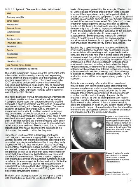
Figure 1. Pars planitis with<br />
snowbanking (seen inferiorly,<br />
following the circumference of<br />
the pars plana) and snowballs<br />
(collections of cells appearing<br />
as spots in the vitreous). This<br />
image was taken with a wide<br />
field lens using an infrared<br />
camera setting.<br />
Figure 2. Mutton-fat keratic<br />
precipitates in the anterior<br />
chamber and posterior<br />
synechiae seen in a patient<br />
with sarcoidosis.<br />
Images Courtesy of<br />
Marc D. de Smet, MD, PhD<br />
5