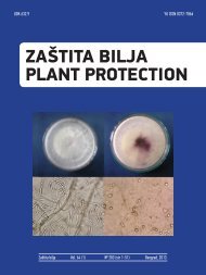
MACROPHOMINA PHASEOLINA ISOLATES FROM SUGAR BEET
Stojšin, V., Budakov, D - Izbis
Stojšin, V., Budakov, D - Izbis
Create successful ePaper yourself
Turn your PDF publications into a flip-book with our unique Google optimized e-Paper software.
MORPHOLOGICAL, CULTURAL AND<br />
PATHOGENIC CHARACTERISTICS OF<br />
<strong>MACROPHOMINA</strong> <strong>PHASEOLINA</strong><br />
<strong>ISOLATES</strong> <strong>FROM</strong> <strong>SUGAR</strong> <strong>BEET</strong><br />
Stojšin, V., Budakov, D., Bagi, F., Đuragin, N., Marinkov, R.<br />
Department for Environmental and Plant Protection,<br />
Faculty of Agriculture, University of Novi Sad, Trg Dositeja<br />
Obradovida 8, 21000 Novi Sad, Serbia
INTRODUCTION<br />
• Macrophomina phaseolina is a causing agent of charcoal root rot.<br />
• In our country firstly described in 1967 on sugar beet in north and<br />
central Banat.<br />
• Significant occurrence was observed in 1971 in many localities in the<br />
Vojvodina Province, and it was predominant in isolations from sugar<br />
beet in 1992, 2003 and especially in 2009 (caused damages on over<br />
50% sugar beet growing areas, estimated on app 2.5 million euro) -<br />
(Stojšin et al., 1998, 1999, 2003, 2009, 2011).<br />
• This fungus attacks sunflower, soybean, maize, rapeseed, tobacco,<br />
alfalfa, red clover, potato, peas, beans, pepper, onion, cabbage,<br />
watermelon, strawberry and many weed species.
Symptoms on sugar beet<br />
• Start with wilting and usually end in complete<br />
decay.<br />
• Severely damaged roots in dry soils can become<br />
mummified, while in moist soils rot can have a<br />
wet appearance.
Symptoms on sugar beet<br />
• Typical symptoms may appear on the crown, the central part and tip<br />
of the root.<br />
• Outer tissues become grayish brown to black with a silverish<br />
reflection.<br />
• Inner tissues turn into sponge like consistency, with colors ranging<br />
from lemon yellow and finally brownish to black.
Epidemiology<br />
• The pathogen survives in plant remains in the field as mycelia and<br />
microsclerotia-overwintering structure.<br />
• Microsclerotia may survive in the soil for many years and they are<br />
always present in sufficient amount.<br />
• Infection may occur on young roots, after which the pathogen spreads<br />
through the vascular system, while characteristic symptoms appear<br />
after plants are being stressed due to drought (Stojšin et al., 1999).<br />
Ecology<br />
• Lack of water and high soil temperatures (25-30 o C) are favorable<br />
conditions for disease development.<br />
• With more frequent growing seasons associated with high<br />
temperatures and lack of precipitation this pathogen could become<br />
predominant in the sugar beet root mycopopulation in Serbia, where<br />
beets are primarily grown without irrigation (Stojšin et al., 2011).
• The aim of this research was to separate and<br />
classify isolates of M. phaseolina on the basis<br />
of their morphological and cultural<br />
characteristics, as well as their pathogenicity<br />
on young sugar beet plants.
MATERIAL AND METHOD<br />
Collection of isolates<br />
Isolates that were used in this research were chosen from the collection comprising<br />
103 monohyphal isolates of M. phaseolina.<br />
Sixteen isolates from sugar beet grown in leading production areas in Serbia were<br />
tested, including one isolate from sunflower, soybean and maize each.<br />
No Isolate Locality Host<br />
1 ŠR 2/09 Žabalj Sugar Beet<br />
2 ŠR 3/09 Sremska Mitrovica (Glac) Sugar Beet<br />
3 ŠR 5/09 Vojka Sugar Beet<br />
4 ŠR 7/09 Šimanovci Sugar Beet<br />
5 ŠR 10/09 Boljevci Sugar Beet<br />
6 ŠR 14/09 Orlovat Sugar Beet<br />
7 ŠR 42/09 Jaša Tomid Sugar Beet<br />
8 ŠR 45(4)/09 Pančevo Sugar Beet<br />
9 ŠR 55(3)/09 Lepušnica Sugar Beet<br />
10 ŠR 62/4 Ečka Sugar Beet<br />
11 ŠR 9M/10 Ečka Sugar Beet<br />
12 ŠR24M/10 Inđija Sugar Beet<br />
13 ŠR 1/11 Beška Sugar Beet<br />
14 ŠR 15/11 Beška Sugar Beet<br />
15 ŠR 17/11 Ečka Sugar Beet<br />
16 ŠR 23/11 Rimski Šančevi Sugar Beet<br />
17 Mph Su Obtained from the Institute for Field and Vegetable Sunflower<br />
18 Mph So Crops, Novi Sad<br />
Soybean<br />
19 MphKu Maize
Cultural and morphological<br />
characteristics<br />
• Cultural characteristics were evaluated on Potato Dextrose Agar (PDA)<br />
and Minimal Media (MIN) using methods described by Pearson et al.<br />
(1986).<br />
• MIN was amended with chlorine by adding 0.015% of potassium<br />
chlorate.<br />
• Mycelial growth was evaluated after three days by measuring the<br />
diameter of the culture.<br />
• According to their growth patterns on MIN, isolates were divided into<br />
three groups: dense, feathery and restricted growth pattern.<br />
• The size of 100 microsclerotia was measured on all media after six<br />
days.
Pathogenicity tests<br />
• Inoculum was prepared by colonizing sorghum<br />
seed with tested M. phaseolina isolates (Omar et<br />
al., 2007).
Pathogenicity tests<br />
• Sugar beet plants at the two leaf stage were<br />
replanted in 500cm³ pots containing mixture of<br />
sterile sand and inoculum in a 3:1 ratio (v/v).<br />
• For the negative control, plants were grown in<br />
non inoculated sterile sand.
Pathogenicity tests<br />
• Plants were incubated in a growth chamber at 30°C with a<br />
photoperiod of 16h/8h light/dark and watered daily.<br />
• Symptom development on leaves was assessed daily,<br />
while final evaluation of root and hypocotyl necrosis was<br />
performed after eight days when over 50% of plants<br />
showed symptoms of irreversible wilting.
Pathogenicity tests<br />
Symptom<br />
Healthy plant 0<br />
Individual lesions on hypocotyl and root 1<br />
Merging of lesions on hypocotyl and root 2<br />
Section necrosis on hypocotyl and root 3<br />
Complete necrosis of hypocotyls and root 4<br />
Grade<br />
• Based on average pathogenicity isolates were divided into<br />
three groups:<br />
i) low pathogenic 0-1,<br />
ii) moderately pathogenic 1-2,<br />
iii) highly pathogenic 2-4.
Data analysis<br />
• Data were analyzed by analysis of variance (ANOVA)<br />
using Statistica 10 (StatSoft, Tulsa, OK).<br />
• Comparisons between means were made with<br />
Duncan’s Multiple Range Test at significance level of<br />
5%.
Table 3. Influence of media on mycelial growth and chlorate sensitivity of M. phaseolina<br />
Mycelial growth (mm)<br />
Isolates PDA Rank MIN+ Cl Rang Chlorate type<br />
ŠR 2/09 90,00 C 40,56 C Sensitive<br />
ŠR 3/09 90,00 C 42,33 C Sensitive<br />
ŠR 5/09 90,00 C 37,33 C Sensitive<br />
ŠR 7/09 78,00 A 22,17 B Sensitive<br />
ŠR 10/09 90,00 C 63,00 D Sensitive<br />
ŠR 14/09 90,00 C 16,50 B Sensitive<br />
ŠR 42/09 90,00 C 24,83 B Sensitive<br />
ŠR 55(3)/09 90,00 C 61,50 D Sensitive<br />
ŠR 62/4 90,00 C 24,77 B Sensitive<br />
ŠR 9M/10 90,00 C 60,33 DE Sensitive<br />
ŠR24M/10 90,00 C 0,00 A Sensitive<br />
ŠR 1/11 90,00 C 60,17 D Sensitive<br />
ŠR 15/11 90,00 C 48,17 C Sensitive<br />
ŠR 17/11 90,00 C 79,00 F Sensitive<br />
ŠR 23/11 90,00 C 45,83 C Sensitive<br />
Mph Su 80,67 B 0,00 A Sensitive<br />
Mph So 90,00 C 0,00 A Sensitive<br />
MphKu 90,00 C 18,50 B Sensitive<br />
Dense<br />
Feathery<br />
Restricted
Diameter of microsclerotia
Pathogenicity
DISCUSSION:<br />
• M. phasolina, causal agent of charcoal root rot is a plant pathogenic<br />
fungus that causes great damages in arid regions. Additionally,<br />
drought stress increases plant susceptibility to this fungus (Mayek-<br />
Perez et al., 2002).<br />
• Since M. phaseolina is highly polyphagous attacking over 500 species<br />
(Dhingra and Sinclair, 1978), variations in chlorate sensitivity,<br />
microclerotial size and formation as well as pathogenicity are not<br />
surprising (Pearson et al., 1987).<br />
• No pattern was detected in comparison of growth patterns on PDA<br />
and MIN, microsclerotia formation on both media and pathogenicity<br />
on sugar beet plants between isolates from sugar beet and other<br />
hosts. Since various authors reported that M. phaseolina isolates<br />
from different hosts can be differentiated using chlorate resistance<br />
(Pearson et al., 1986; Rayatpanah et al., 2009), this implies the need<br />
to increase the number of isolates from other hosts.
DISCUSSION:<br />
• Identifying differences in pathogenicity among isolates is the most useful<br />
tool for grouping isolates. Based on our results, isolates can be divided<br />
into 3 groups: low pathogenic – 5 isolates (26.3%), moderately and highly<br />
pathogenic – 7 isolates (36.8%) for each group.<br />
• However, there are scarce data on occurrence and significance of<br />
Macrophomina phaseolina in sugar beet, so the aim of this research was<br />
to describe cultural, morphological and pathogenic characteristics of<br />
isolates from sugar beet and compare them to isolates from other hosts<br />
(sunflower, soybean and maize).
Discussion<br />
• Preliminary studies showed that there are differences in<br />
pathogenicity of isolates from sugar beet, and even more in<br />
isolates that are from other hosts (sunflower, soybean,<br />
maize). Regarding the fact that M. phaseolina is a<br />
polyphagous and always present in the soil, and<br />
environmental factors are hard to control in sugar beet<br />
production without irrigation, one of solutions is breeding for<br />
resistance (Stojšin et al., 2012).<br />
• The described method for artificial inoculation can be used as<br />
the optimized method for pathogenicity testing of large<br />
number of isolates from Serbian sugar beet growing areas,<br />
which enables further research of M. phaseolina population<br />
as well as fast testing of sugar beet genotypes that are grown<br />
in our region.


