HYDRAGEL HEMOGLOBIN(E) K20
HYDRAGEL HEMOGLOBIN(E) K20 - Sebia Electrophoresis
HYDRAGEL HEMOGLOBIN(E) K20 - Sebia Electrophoresis
- No tags were found...

Create successful ePaper yourself
Turn your PDF publications into a flip-book with our unique Google optimized e-Paper software.
<strong>HYDRAGEL</strong> <strong>HEMOGLOBIN</strong>(E) <strong>K20</strong> - 2005/03<br />
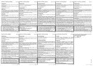
PROCEDURE<br />
I. MIGRATION STEP<br />
1. Place the <strong>HYDRAGEL</strong> <strong>K20</strong> applicator carrier on a flat surface (Fig. 1) and raise the part of the applicator carrier with the numbered notches.<br />
2. Pool 120 µL distilled or deionized water on the lower third of the frame printed on the <strong>HYDRAGEL</strong> <strong>K20</strong> applicator carrier.<br />
3. Unpack the <strong>HYDRAGEL</strong> agarose gel plate.<br />
4. Roll quickly and uniformly one thin filter paper onto the gel surface to absorb the excess of liquid. Remove the paper immediately.<br />
WARNING: Do not leave the filter paper for a too long contact with the gel to avoid its dehydration.<br />
5. Place the gel plate (the gel side up) with its edge against the stop at the bottom of the printed frame (Fig. 2).<br />
6. Bend the gel and lower it down onto the water pool (Fig. 2). Ensure that no air bubbles are trapped, water is spread underneath the entire gel<br />
plate and the gel is lined up with the printed frame.<br />
7. Lower the applicator carrier with the numbered notches down to the intermediate position with the switch in high position.<br />
8. Place one applicator on a flat surface with the well numbers in the right-side-up position (Fig. 3).<br />
9. Apply 10 µL of hemolyzed sample into the applicator wells. Load the applicator within 2 minutes.<br />
- Use the applicator without any delay.<br />
- For later use (up to 8 hours), place the applicator into the wet storage chamber with the teeth up [handle it by the plastic tooth protection<br />
frame], keep the entire chamber under refrigeration and set-up the gel plate onto the <strong>HYDRAGEL</strong> <strong>K20</strong> applicator carrier just before use.<br />
See wet chamber package insert for further details.<br />
10. Snap off the applicator teeth's protection frame.<br />
11. Place the sample applicator into position No. 4 on the applicator carrier.<br />
IMPORTANT: The numbers printed on the sample applicator must face the operator (Fig. 4).<br />
12. Lower the applicator carrier with the switch so that the applicator contacts the gel surface. DO NOT FORCE THE CARRIERS ALL THE WAY<br />
DOWN.<br />
13. After 1 minute, turn the switch to rise up the applicator, remove the applicator and discard.<br />
14. Put the gel into an appropriate electrophoresis chamber, according to the polarity indicated on the gel, the lower side of the gel on the cathodic<br />
side.<br />
When using SEBIA <strong>K20</strong> chamber, place the <strong>HYDRAGEL</strong> on the bridge with the gel side facing down ; the gel should dip about 1 cm into the<br />
buffer on each side.<br />
See <strong>K20</strong> chamber package insert for further details.<br />
15. Plug the chamber to the power supply.<br />
MIGRATION CONDITIONS SEBIA <strong>K20</strong> Volume of buffer per compartment 150 mL Total buffer volume 300 mL Migration time 15 minutes Constant voltage 165 V<br />
| Initial current (per gel) | 7 ± 2 mA |<br />
16. After migration, unplug the chamber and remove the gel plate.<br />
II. FIXATION<br />
Process gels according to one of the following procedures.<br />
Hot air fixation (recommended only with SEBIA IS 80 Incubator-Dryer) :<br />
Dry the gel completely in the incubator-dryer at 80 °C (for 10 minutes minimum).<br />
Fixation with fixative solution :<br />
1. Place the gel into a gel holder (supplied with SEBIA <strong>HYDRAGEL</strong> <strong>K20</strong> Accessory Kit) for further processing.<br />
2. Fill one tank (supplied with SEBIA <strong>HYDRAGEL</strong> <strong>K20</strong> Accessory Kit) with 150 mL of fixative solution.<br />
3. Immerse the gel in the fixative solution for 15 minutes.<br />
4. Remove the gel and dry it with hot 80 °C air flow in incubator-dryer IS 80.<br />
IMPORTANT : The gel must be perfectly dry.<br />
III. STAINING - DESTAINING<br />
1. Immerse the dried and cooled gel in the staining solution for 5 minutes.<br />
2. Destain in three successive baths of destaining solution until the background is completely colorless and clear.<br />
3. Soak up excess liquid on the gel surface with a tissue paper and dry the gel with hot 80 °C air. If needed, clean the back side (the plastic<br />
support side) of the dry film with a wet tissue paper.<br />
IV. SCANNING<br />
Scan using a densitometer / scanner at 570 nm or with a yellow filter. When using HYRYS or DVSE, densitometers, position the A 2<br />
fraction on the<br />
5 mm mark of the scanning plate: the background zero is made between the A 2<br />
and carbonic anhydrase fractions at the lowest point.<br />
NOTE : To assure the most accurate and consistent results, do the following:<br />
- Adjust the scan length to include the entire electrophoretic pattern (≈ 30 mm).<br />
- Make sure the minima on both sides of A 2<br />
fraction are positioned at the very feet of the A 2<br />
peak.<br />
It is a good practice to read the stained gels without delay. For future reference, they can be stored in a protective cover in a dry, dark place away from<br />
sources of heat and visually interpreted within at least 3 months.<br />
- 11 -