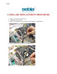
CAPILLARYS HEMOGLOBIN(E)
CAPILLARYS HEMOGLOBIN(E) - Sebia Electrophoresis
CAPILLARYS HEMOGLOBIN(E) - Sebia Electrophoresis
- No tags were found...

You also want an ePaper? Increase the reach of your titles
YUMPU automatically turns print PDFs into web optimized ePapers that Google loves.
<strong>CAPILLARYS</strong> <strong>HEMOGLOBIN</strong>(E)<br />
Ref. 2007<br />
2010/10
<strong>CAPILLARYS</strong> <strong>HEMOGLOBIN</strong>(E) - 2010/10<br />
<strong>CAPILLARYS</strong> <strong>HEMOGLOBIN</strong>(E) PROCEDURE WITH THE <strong>CAPILLARYS</strong> SYSTEM<br />
INTENDED USE<br />
The <strong>CAPILLARYS</strong> <strong>HEMOGLOBIN</strong>(E) kit is designed for the separation of the normal hemoglobins (A, F and A2) and for the detection of the major<br />
hemoglobin variants (especially S, C, E or D), by electrophoresis in alkaline buffer (pH 9.4) with the <strong>CAPILLARYS</strong> System.<br />
The <strong>CAPILLARYS</strong> performs all sequences automatically to obtain a complete hemoglobin profile for qualitative or quantitative analysis of hemoglobins.<br />
The assay is performed on sedimented, centrifuged or washed red blood cells ; washing red blood cells is not essential to perform the analysis.<br />
For In Vitro Diagnostic Use.<br />
PRINCIPLE OF THE TEST<br />
Hemoglobin is a complex molecule composed of two pairs of polypeptide chains. Each chain is linked to the heme, a tetrapyrrolic nucleus (porphyrin)<br />
which chelates an iron atom. The heme part is common to all hemoglobins and their variants. The type of hemoglobin is determined by the protein<br />
part called globin. Polypeptide chains α, ß, δ and γ constitute the normal human hemoglobins:<br />
• hemoglobin A ............................ = α 2 ß 2<br />
• hemoglobin A2 .......................... = α 2 δ 2<br />
• fetal hemoglobin F .................... = α 2 γ 2<br />
The α-chain is common to these three hemoglobins.<br />
The hemoglobin spatial structure and other molecular properties (like that of all proteins) depend on the nature and the sequence of the amino acids<br />
constituting the chains. Substitution of amino acids by mutation is responsible for formation of hemoglobin variants which have different surface charge<br />
and consequently different electrophoretic mobilities, which also depend on the pH and ionic strength of the buffer.<br />
The resulting qualitative (or structural) abnormalities are called hemoglobinopathies (9, 10, 13) . Decreased synthesis of one of the hemoglobin chains leads<br />
to quantitative (or regulation) abnormalities, called thalassemias.<br />
Hemoglobin electrophoresis is a well established technique routinely used in clinical laboratories for screening samples for hemoglobin<br />
abnormalities (1, 2, 3, 4, 12) . The <strong>CAPILLARYS</strong> System has been developed to provide complete automation of this testing with fast separation and good<br />
resolution. In many respects, the methodology can be considered as an intermediary type of technique between classical zone electrophoresis and<br />
liquid chromatography (8, 11) .<br />
The <strong>CAPILLARYS</strong> System uses the principle of capillary electrophoresis in free solution. With this technique, charged molecules are separated by<br />
their electrophoretic mobility in an alkaline buffer with a specific pH. Separation also occurs according to the electrolyte pH and electroosmotic flow (5) .<br />
The <strong>CAPILLARYS</strong> System has capillaries functioning in parallel allowing 7 simultaneous analyses for hemoglobin quantification. A sample dilution with<br />
hemolysing solution is prepared and injected by aspiration at the anodic end of the capillary. A high voltage protein separation is then performed and<br />
direct detection of the hemoglobins is made at 415 nm at the cathodic end of the capillary. Before each run, the capillaries are washed with a Wash<br />
Solution and prepared for the next analysis with buffer.<br />
The hemoglobins, separated in silica capillaries, are directly and specifically detected at an absorbance wave length of 415 nm which is specific to<br />
hemoglobins. The resulting electrophoregrams are evaluated visually for pattern abnormalities.<br />
Direct detection provides accurate relative quantification of individual hemoglobin fraction, with particular interest, such as A2 hemoglobin for<br />
ß thalassemia diagnostic. In addition, the high resolution of this procedure should allow the identification of hemoglobin variants, in particular, to<br />
differentiate hemoglobins S from D, and E from C.<br />
The hemoglobin A2 quantification can also be performed when hemoglobin E is present.<br />
By using alkaline pH buffer, normal and abnormal (or variant) hemoglobins are detected in the following order, from cathode to anode: δA’2 (A2 variant),<br />
C, A2/O-Arab, E, S, D, G-Philadelphia, F, A, Hope, Bart, J, N-Baltimore and H.<br />
The carbonic anhydrase is not visualized on the hemoglobin electrophoretic patterns, this permits to identify hemoglobin A2 variants in this migration<br />
zone.<br />
REAGENTS AND MATERIALS SUPPLIED IN THE <strong>CAPILLARYS</strong> <strong>HEMOGLOBIN</strong>(E) KIT<br />
ITEMS PN. 2007<br />
Buffer (ready to use)<br />
2 vials, 700 mL each<br />
Hemolysing solution (ready to use)<br />
1 vial, 700 mL<br />
Wash solution (stock solution)<br />
1 vial, 75 mL<br />
Dilution segments 1 pack of 90<br />
Filters<br />
4 filters<br />
630 tests based on maximum usage.<br />
FOR OPTIMAL RESULTS :<br />
All reagents from the same kit must be always used together and according to the package insert instructions.<br />
PLEASE READ THE PACKAGE INSERT CAREFULLY.<br />
WARNING : Do not use marketed deionized water, such as water for ironing for example (risk of important capillaries damage). Use only<br />
water with ultrapure quality, such as injection grade water.<br />
- 34 -<br />
SEBIA INSTRUCTIONS - English
<strong>CAPILLARYS</strong> <strong>HEMOGLOBIN</strong>(E) - 2010/10<br />
1. BUFFER<br />
Preparation<br />
The buffer is ready to use. It contains: alkaline buffer pH 9.4 ; additives, nonhazardous at concentrations used, necessary for optimum performance.<br />
WARNING: See the safety data sheet.<br />
Use<br />
Buffer for analysis of hemoglobins in <strong>CAPILLARYS</strong>.<br />
Storage, stability and signs of deterioration<br />
Store the buffer refrigerated (2 to 8 °C). It is stable until the expiration date indicated on the kit package or buffer vial labels. Avoid storage at room<br />
temperature for a long time or close to a window or to a heat source.<br />
DO NOT FREEZE.<br />
IMPORTANT: When stored at 2 - 8 °C and prior to use, it is necessary for the buffer to reach room temperature; when it is full, let the buffer vial at<br />
room temperature for at least 3 hours prior to use. If this precaution is not respected, the performances of the procedure may be affected.<br />
WARNING: Do not pre-heat the buffer in hot water.<br />
Once the buffer vial has been opened and positioned on the <strong>CAPILLARYS</strong> system, it is stable for a maximum of 1 month (accumulated) at room<br />
temperature (15 to 30 °C). After each use, the buffer must imperatively be stored refrigerated (between 2 and 8 °C) without any delay, it is then<br />
stable until the expiration date indicated on the buffer vial label.<br />
IMPORTANT: The accumulated time of the buffer stored at room temperature must not exceed 1 month. This time of 1 month storage takes account<br />
of the time for the buffer to come to room temperature.<br />
Discard buffer if it changes its appearance, e.g., becomes cloudy due to microbial contamination.<br />
2. HEMOLYSING SOLUTION<br />
Preparation<br />
Hemolysing Solution is ready to use. It is a buffer with additives, nonhazardous at the concentration used, necessary for optimum performance.<br />
Use<br />
To dilute and hemolyze red blood cells and whole blood.<br />
Storage, stability and signs of deterioration<br />
Store Hemolysing Solution refrigerated (2 to 8 °C). It is stable until the expiration date indicated on the kit package or Hemolysing Solution vial label.<br />
Avoid storage at room temperature or close to a window or to a heat source. DO NOT FREEZE.<br />
WARNING: Do not pre-heat the hemolysing solution in hot water.<br />
IMPORTANT: After each use, close immediately and tightly the hemolysing solution vial and store it refrigerated.<br />
Analyses with the <strong>CAPILLARYS</strong> 2 FLEX PIERCING system: Once the hemolysing solution vial has been opened and positioned on the system, it is<br />
stable for a maximum of 2 months (accumulated) at room temperature (15 to 30 °C).<br />
Discard Hemolysing Solution if it changes its appearance, e.g., becomes cloudy due to microbial contamination.<br />
3. WASH SOLUTION<br />
Preparation<br />
The vial of the stock wash solution should be diluted up to 750 mL with distilled or deionized water.<br />
WARNING: See the safety data sheet.<br />
Use<br />
For washing the capillaries before and after hemoglobin electrophoresis.<br />
IMPORTANT: Before filling the wash solution container, it is recommended to wash the opening of the container, the connector and the tube with plenty<br />
of distilled or deionized water to avoid salts deposit.<br />
Storage, stability and signs of deterioration<br />
Store the stock and working wash solutions in closed containers at room temperature or refrigerated. The stock wash solution is stable until the<br />
expiration date indicated on the kit or wash solution vial label.<br />
Working wash solution is stable for 3 months.<br />
Discard working wash solution if it changes its appearance, e.g., becomes cloudy due to microbial contamination.<br />
4. DILUTION SEGMENTS<br />
Use<br />
Coloured single use segments for blood sample dilution on the automated instrument. They are specific for <strong>CAPILLARYS</strong> <strong>HEMOGLOBIN</strong>(E) and<br />
<strong>CAPILLARYS</strong> CORD BLOOD procedures.<br />
WARNING: Dilution segments with biological samples have to be handled with care.<br />
5. FILTERS<br />
Use<br />
Disposable filters for filtration of analysis buffer, hemolysing solution (for <strong>CAPILLARYS</strong> 2 FLEX PIERCING system), working wash solution and<br />
distilled or deionized water (used for capillaries rinsing).<br />
IMPORTANT: With each new kit, always change all the four filters.<br />
Screw one filter at the connector situated at the extremity of each tube that plunges in the vials of buffer, hemolysing solution, wash solution and<br />
distilled or deionized water. When setting filters on <strong>CAPILLARYS</strong> system, rinse the connectors and the tubes with distilled or deionized water. Used<br />
filters must be rinsed before discard.<br />
The filter intended for analysis buffer must be used for filtration of both buffer vials ; the three other filters are intended for filtration of hemolysing<br />
solution, working wash solution and distilled or deionized water (for capillary rinsing).<br />
Storage<br />
Before use, store the filters in their sealed package in a dry place at room temperature or refrigerated.<br />
- 35 -
<strong>CAPILLARYS</strong> <strong>HEMOGLOBIN</strong>(E) - 2010/10<br />
REAGENTS REQUIRED BUT NOT SUPPLIED<br />
1. NORMAL Hb A2 CONTROL<br />
Composition<br />
The Normal Hb A2 Control (SEBIA, PN 4778) is obtained from a pool of normal human blood samples. The Normal Hb A2 Control is in a stabilized<br />
lyophilised form.<br />
Intended use<br />
The Normal Hb A2 Control is designed for the migration control before starting a new analysis sequence, and for the quality control of human<br />
hemoglobin A2 quantification with <strong>CAPILLARYS</strong> <strong>HEMOGLOBIN</strong>(E) electrophoresis procedure.<br />
Reconstitute each Normal Hb A2 Control vial with the exact volume of distilled or deionized water, as indicated in the package insert of the Normal<br />
Hb A2 Control. Allow to stand for 30 minutes and mix gently (avoid formation of foam).<br />
Migration control: The Normal Hb A2 Control should be used as follows:<br />
- Apply the reconstituted Normal Hb A2 Control in a microtube.<br />
- Cut the cap of the microtube.<br />
- Place the microtube, located on a new hemolysing tube used as a support tube (and identified with the Normal Hb A2 Control bar code label), in<br />
position No. 1 on the <strong>CAPILLARYS</strong> sample rack No. 0 intended for control blood sample, containing a new green dilution segment.<br />
- Pour 4 mL <strong>CAPILLARYS</strong> <strong>HEMOGLOBIN</strong>(E) hemolysing solution in a hemolysing tube without introducing air bubbles and place it in position No. 8<br />
on the sample rack No. 0.<br />
IMPORTANT: Ensure the absence of foam in the tube before placing it on the sample rack.<br />
- Start the analysis: Slide the sample rack No. 0 into the <strong>CAPILLARYS</strong> system, select "Automatic dilution" in the window which appears on the screen<br />
and validate.<br />
- After having changed the analysis buffer vial (even if the lot number is identical) or the technique, after a capillary cleaning sequence with<br />
CAPICLEAN, after a software upgrade or after capillaries activation, perform a second series of analyses with the control, by sliding in again<br />
immediately the same sample rack No. 0 with the same dilution segment containing the Normal Hb A2 Control, previously diluted during the first<br />
series and the empty hemolysing tube identified with the Normal Hb A2 Control bar code in position No. 1. In the window called "Hb A2 Normal<br />
Control" which appears on the screen, select "Manual dilution" and validate.<br />
The results are then automatically considered by the software for the data analysis.<br />
IMPORTANT: The hemoglobin A fraction of the Normal Hb A2 Control must show a minimal optical density (OD) of 0.12. Under this value, the<br />
recentering of the electrophoretic pattern will not occur correctly. When analysing samples, the identification of hemoglobin fractions, Hb A, Hb F, Hb A2<br />
and Hb C and also the determination of the migration zone of other variants, may be impossible or wrong (see the paragraph RESULT ANALYSIS).<br />
IMPORTANT: For optimal use of the Normal Hb A2 Control, it is necessary to use one bar code label intended to identify the hemolysing tube holding<br />
the microtube which contains the Hb A2 Control (cut the cap of the microtube before using it).<br />
NOTES: For the first use of the "<strong>HEMOGLOBIN</strong>(E)" analysis program with the <strong>CAPILLARYS</strong> instrument, it is recommended to perform 3 successive<br />
series of analyses with the Normal Hb A2 Control.<br />
After the installation of <strong>CAPILLARYS</strong> instrument, during the first sequence of blood sample analysis, a red warning signal will appear if hemoglobin A<br />
is absent in one sample (and the recentering of the electrophoretic pattern will not be possible, see paragraph " Result analysis ").<br />
It is then recommended to analyze a blood sample with hemoglobin A on the concerned capillary and to analyze again the sample without hemoglobin<br />
A by placing it in a position corresponding to a capillary which has already detected hemoglobin A.<br />
Quality control: The Normal Hb A2 Control should be used as a normal human blood. After reconstitution, use directly the Normal Hb A2 Control as<br />
a blood sample to analyze or as a migration control (with the sample rack No. 0, see paragraph before). It will be automatically diluted with hemolysing<br />
solution. It is recommended to include one analysis of Normal Hb A2 Control. The values obtained must fall within the range provided with each batch<br />
of Hb A2 Control.<br />
IMPORTANT: For optimal use of the Normal Hb A2 Control placed on a sample rack, it is necessary to use one bar code label intended to identify the<br />
hemolysing tube holding the microtube which contains the Hb A2 Control (cut the cap of the microtube before using it).<br />
Storage, stability and signs of deterioration<br />
Before reconstitution, store the lyophilised Normal Hb A2 Control refrigerated (2 to 8 °C). It is stable until the expiration date indicated on the box or<br />
vial labels.<br />
Store the reconstituted Normal Hb A2 Control at 2 - 8 °C. Due to the risk of microbial contamination and denaturation, use it within one week.<br />
The reconstituted Control may also be frozen (in aliquots) and stored at - 20 °C for 6 months maximum.<br />
IMPORTANT: After storage at 2 - 8 °C or at - 20 °C, homogenize the reconstituted Normal Hb A2 Control before the analysis with the <strong>CAPILLARYS</strong><br />
system.<br />
NOTE: For optimal use with the <strong>CAPILLARYS</strong> system, it is recommended to split the Control into aliquots in microtubes before freezing.<br />
Before use, store the thawed Normal Hb A2 Control at 2 - 8 °C and use it within the day. Do not freeze and thaw the Control more than 15 times.<br />
The hemolysed Normal Hb A2 Control should be stored at 2 - 8 °C and used within one day.<br />
The dilution segment containing the hemolysed Normal Hb A2 Control may be frozen and stored at - 20 °C. Do not freeze and thaw the dilution<br />
segment with hemolysed Control more than twice.<br />
IMPORTANT: Do not leave the dilution segment with hemolysed Control at room temperature.<br />
NOTE: During transportation, the lyophilized Normal Hb A2 Control can be kept without refrigeration (15 to 30 °C) for 15 days without any adverse<br />
effects on performance.<br />
No test method can provide an absolute assurance of the absence of HIV, hepatitis B and C or other infectious agents. Therefore, handle the Normal<br />
Hb A2 Control as a hazardous biological material.<br />
This control blood was found negative on assays approved by FDA or EU equivalent regulatory agency:<br />
- against hepatitis B surface antigen ;<br />
- for antibody to HCV ;<br />
- for antibody to HIV1 and HIV2.<br />
- 36 -
<strong>CAPILLARYS</strong> <strong>HEMOGLOBIN</strong>(E) - 2010/10<br />
2. DISTILLED OR DEIONIZED WATER<br />
Use<br />
For rinsing capillaries in automated system <strong>CAPILLARYS</strong>, SEBIA, for capillary electrophoresis.<br />
It is recommended to filter distilled or deionized water with 0.45 µm filter before use.<br />
To prevent microbial proliferation, change the water every day. In case of longer storage, add 35 µL/dL of CLEAN PROTECT (SEBIA, PN 2059, 5 mL).<br />
IMPORTANT: Before filling the rinse container, it is recommended to wash it with plenty of distilled or deionized water.<br />
3. CAPICLEAN<br />
Composition<br />
The vial of CAPICLEAN concentrated solution (SEBIA, PN 2058, 25 mL) contains : proteolytic enzymes, surfactants and additives nonhazardous at<br />
concentrations used, necessary for optimum performances.<br />
WARNING: See the safety data sheet.<br />
Use<br />
For sample probe cleaning in automated system <strong>CAPILLARYS</strong>, SEBIA, for capillary electrophoresis, during the CAPICLEAN cleaning sequence.<br />
IMPORTANT: Launch a CAPICLEAN cleaning sequence at least once a week and at maximum once a day, or after every 500 analyses when<br />
performed within less than one week.<br />
See the instruction sheets of CAPICLEAN, SEBIA.<br />
IMPORTANT: Do not re-use the dilution segment after sample probe cleaning.<br />
Storage, stability and signs of deterioration<br />
Store CAPICLEAN refrigerated (2 - 8 °C). It is stable until the expiration date indicated on the vial label. DO NOT FREEZE.<br />
CAPICLEAN must be free of precipitate. Discard CAPICLEAN if it changes its appearance, e.g., becomes cloudy due to microbial contamination.<br />
4. SODIUM HYPOCHLORITE SOLUTION (for sample probe cleaning)<br />
Preparation<br />
Prepare a sodium hypochlorite solution (2 % to 3 % chloride) by diluting 250 mL 9.6 % chloride concentrated solution to 1 liter with cold distilled or<br />
deionized water.<br />
Use<br />
For the sample probe cleaning in the <strong>CAPILLARYS</strong> System (weekly maintenance in order to eliminate adsorbed proteins from the probe).<br />
See the instruction sheets of <strong>CAPILLARYS</strong>, SEBIA.<br />
• Use the sample rack designed for the maintenance (No. 100).<br />
• Place a tube containing 2 mL diluted chlorinated solution previously prepared, in position No. 1 on this sample rack.<br />
• Slide the sample rack No. 100 for maintenance in the <strong>CAPILLARYS</strong> System.<br />
• In the "MAINTENANCE" window which appears on the screen, select "Launch the probe cleaning (chlorinated sodium hypochlorite solution)" and<br />
validate.<br />
Storage, stability and signs of deterioration<br />
Store the working chlorinated solution at room temperature in a closed container, it is stable for 3 months. Avoid storage in sunlight, close to heat and<br />
ignition source, and to acids and ammonia.<br />
5. <strong>CAPILLARYS</strong> / MINICAP WASH SOLUTION<br />
Preparation<br />
Each vial of the stock Wash Solution (SEBIA, PN 2052, 2 vials, 75 mL) should be diluted up to 750 mL with distilled or deionized water.<br />
WARNING: See the safety data sheet.<br />
Use<br />
For washing the capillaries of <strong>CAPILLARYS</strong>. This additional reagent is needed when the number of tests in series is below 40.<br />
IMPORTANT: Before filling the wash solution container, it is recommended to wash the opening of the container, the connector and the tube with plenty<br />
of distilled or deionized water to avoid salts deposit.<br />
Storage, stability and signs of deterioration<br />
Store the stock and working wash solutions in closed containers at room temperature or refrigerated.<br />
The stock wash solution is stable until the expiration date indicated on the kit or wash solution vial label.<br />
Working wash solution is stable for 3 months. Discard working wash solution if it changes its appearance, e.g., becomes cloudy due to microbial<br />
contamination.<br />
6. SALINE<br />
Preparation<br />
Make 0.15 M (0.9 g/dL) NaCl solution in distilled or deionized water.<br />
Use<br />
To wash red blood cells before storage at - 80 °C, if necessary.<br />
Storage, stability and signs of deterioration<br />
Store saline at room temperature or refrigerated. Discard after 3 months or if it changes its appearance, e.g., becomes cloudy due to microbial<br />
contamination. For longer storage periods, add sodium azide, 0.1 g/dL.<br />
EQUIPMENT AND ACCESSORIES REQUIRED<br />
1. <strong>CAPILLARYS</strong> System SEBIA, PN 1220 or PN 1222.<br />
2. Sample racks supplied with <strong>CAPILLARYS</strong>.<br />
3. Container Kit supplied with <strong>CAPILLARYS</strong>: Rinse (to fill with distilled or deionized water), wash solution and waste container.<br />
- 37 -
<strong>CAPILLARYS</strong> <strong>HEMOGLOBIN</strong>(E) - 2010/10<br />
SAMPLES FOR ANALYSIS<br />
Sample collection and storage<br />
Fresh anticoagulated blood samples are recommended for analysis. Common anticoagulants such as those containing EDTA, citrate or heparin are<br />
acceptable ; avoid those with iodoacetate. Blood must be collected according to established procedures used in clinical laboratory testing.<br />
Samples may be stored for up to 7 days between 2 and 8 °C.<br />
For longer storage, samples can be frozen at - 80 °C within 8 hours of collection after having washed the red blood cells according to the following<br />
procedure: Centrifuge anticoagulated blood at 5 000 rpm for 5 minutes ; discard the plasma; wash the red blood cells (RBC) 2 times with 10 volumes<br />
of saline (centrifuge after each washing step) ; discard the excess of saline over the red blood cells pellet and vortex them before freezing.<br />
Frozen blood samples are stable for 3 months maximum at - 80 °C.<br />
IMPORTANT: For optimal storage of blood samples, store them at - 80 °C. Do not store at - 20 °C (see BIBLIOGRAPHY, J. Bardakdjian-Michau et al,<br />
2003).<br />
NOTE: Samples should not be stored at room temperature!<br />
Progressive hemoglobins (Hb) degradation may occur for samples stored between 2 to 8 °C.<br />
When the blood sample is stored for more than 7 days at 2 - 8 °C:<br />
• a weak fraction, corresponding to methemoglobin, appears in the Hb S migration zone,<br />
• when Hb C is present, a fraction corresponding to degraded Hb C appears more anodic than Hb A2 which does not interfere with it (Z4 zone, see<br />
the table in paragraph "Interpretation"),<br />
• when Hb O-Arab is present, a fraction corresponding to degraded Hb O-Arab appears in the Hb S migration zone (Z5 zone, see the table in<br />
paragraph "Interpretation"),<br />
• when Hb E is present, a fraction corresponding to degraded Hb E appears in the Z6 zone (see the table in paragraph "Interpretation"),<br />
• when Hb S is present, a fraction corresponding to degraded Hb S appears in the Hb F migration zone (Z7 zone, see the table in paragraph<br />
"Interpretation"),<br />
• when Hb A is present, a fraction corresponding to degraded Hb A ("aging fraction" of Hb A) appears more anodic (Z11 zone, see the table in<br />
paragraph "Interpretation").<br />
When Hb F is present (in blood samples from newborn babies), a fraction appears in the Hb A migration zone (Z9 zone, see the table in paragraph<br />
"Interpretation") due to the sample degradation.<br />
When stored for more than 10 days, viscous aggregates in red blood cells are observed; it is necessary to discard them before the analysis.<br />
Sample preparation<br />
• Let red blood cells precipitate for several hours at 2 - 8 °C or centrifuge the blood sample at 5 000 rpm for 5 minutes.<br />
• Discard carefully the maximum volume of plasma (for samples collected with heparin, discard viscous aggregates located between plasma and red<br />
blood cells).<br />
• Vortex for 5 seconds.<br />
IMPORTANT: Do not use blood samples containing 3 mm maximum residual plasma over red blood cells; when more than 3 mm plasma is present<br />
in the tube, the analysis should be affected.<br />
Particular cases: Analysis of samples without any Hb A or Hb A2 (these samples are perfectly quantified but not identified by zones).<br />
To identify hemoglobin fractions of a sample without any hemoglobin A or hemoglobin A2, it is recommended to prepare this sample according to one<br />
of the two following procedures:<br />
Automatic dilution:<br />
- In a microtube, mix one volume (80 µL) of red blood cells from the sample to analyze with one volume of Normal Hb A2 Control (80 µL).<br />
- Vortex for 5 seconds.<br />
- Cut the cap of the microtube.<br />
- Place the microtube, on a new hemolysing tube used as a support-tube, on a sample rack of the <strong>CAPILLARYS</strong> system.<br />
- Perform the analysis of this sample according to the standard procedure like a usual blood sample.<br />
Manual dilution:<br />
- Apply, directly in the wells of a new green dilution segment, 9 µL of reconstituted Normal Hb A2 Control with 9 µL of blood sample to analyze and<br />
90 µL of <strong>CAPILLARYS</strong> <strong>HEMOGLOBIN</strong>(E) hemolyzing solution.<br />
- Mix by repeated pipettings.<br />
- Place this dilution segment on the sample rack No. 0 of <strong>CAPILLARYS</strong>.<br />
- Slide the sample rack into the <strong>CAPILLARYS</strong> system, select "Sample" with "manual dilution" in the window which appears on the screen and validate.<br />
The results are then automatically considered by the software for the data analysis.<br />
IMPORTANT: For a sample without any Hb A or Hb A2 prepared according to one of these two procedures, the result obtained with the mixed sample<br />
will enable presumptive variant identification due to the positioning of the hemoglobins fractions in the appropriate identification zones. Do not report<br />
the relative quantification from the mixed sample result.<br />
The relative quantification of hemoglobins should be reported utilizing the initial, unmixed sample result (without any dilution in the blood control).<br />
Samples to avoid<br />
• Do not use unsedimented blood samples.<br />
• Avoid aged, improperly stored blood samples ; the automated hemolysis of samples may be disturbed by viscous aggregates in red blood cells.<br />
Then, degradation products (as artefacts) may affect the electrophoretic pattern.<br />
PROCEDURE<br />
The <strong>CAPILLARYS</strong> system is a multiparameter instrument for hemoglobins analysis on parallel capillaries. The hemoglobins assay uses 7 of the total<br />
8 instrument capillaries to run the samples.<br />
The sequence of automated steps is as follows:<br />
• Bar code reading of sample tubes (for up to 7 tubes) and samples-racks ;<br />
• Sample hemolysis and dilution from primary tubes (without any plasma) into dilution segments ;<br />
• Capillary washing ;<br />
• Injection of hemolyzed samples ;<br />
• Hemoglobin separation and direct detection of the separated hemoglobins on capillaries.<br />
- 38 -
<strong>CAPILLARYS</strong> <strong>HEMOGLOBIN</strong>(E) - 2010/10<br />
The manual steps include:<br />
• Placement of opened sample tubes in sample-racks in positions 1 to 7 ;<br />
• Placement of hemolysing solution tube in sample-racks in position 8 ;<br />
• Placement of new dilution segments in sample-racks ;<br />
• Placement of racks on the <strong>CAPILLARYS</strong> instrument ;<br />
• Removal of sample-racks after analysis.<br />
PLEASE CAREFULLY READ THE <strong>CAPILLARYS</strong> INSTRUCTION MANUAL.<br />
I. PREPARATION OF <strong>CAPILLARYS</strong> ANALYSIS<br />
1. Switch on <strong>CAPILLARYS</strong> instrument and computer.<br />
2. Set up the software, enter and the instrument automatically starts.<br />
3. The <strong>CAPILLARYS</strong> <strong>HEMOGLOBIN</strong>(E) kit is intended to run with "<strong>HEMOGLOBIN</strong>(E)" analysis program from the <strong>CAPILLARYS</strong> instrument. To<br />
select "<strong>HEMOGLOBIN</strong>(E)" analysis program and place the <strong>CAPILLARYS</strong> <strong>HEMOGLOBIN</strong>(E) buffer vial in the instrument, please read carefully<br />
the <strong>CAPILLARYS</strong> instruction manual.<br />
4. The sample rack contains eight positions for sample tubes. Place seven opened sample tubes without any plasma on each sample rack; the<br />
bar code of each tube must be visible in the openings of the sample rack.<br />
IMPORTANT: If the number of tubes to analyze is less than 7, complete the sample rack with tubes containing distilled or deionized water.<br />
5. Pour 4 mL <strong>CAPILLARYS</strong> <strong>HEMOGLOBIN</strong>(E) hemolysing solution in a tube without introducing air bubbles and place it in position No. 8 on the<br />
sample rack.<br />
IMPORTANT: Ensure the absence of foam in the tube before placing it on the sample rack.<br />
6. Position a new dilution segment on each sample rack. A message will be displayed if the segment is missing.<br />
7. Slide the complete sample carrier(s) into the <strong>CAPILLARYS</strong> system through the opening in the middle of the instrument. Up to 13 sample racks<br />
can be introduced successively and continuously into the system. It is advised to use the sample rack No. 0 intended for control blood sample.<br />
8. Remove analyzed sample racks from the plate on the left side of the instrument.<br />
9. Take off carefully used dilution segments from the sample rack and discard them.<br />
WARNING: Dilution segments with biological samples have to be handled with care.<br />
DILUTION - MIGRATION - DESCRIPTION OF THE AUTOMATED STEPS<br />
1. Bar codes are read on both sample tubes and sample racks.<br />
2. Samples are diluted in hemolysing solution and the sample probe is rinsed after each sample.<br />
3. Capillaries are washed.<br />
4. Diluted samples are injected into capillaries.<br />
5. Migration is carried out under constant voltage for about 8 minutes and the temperature is controlled by Peltier effect.<br />
6. Hemoglobins are detected directly by scanning at 415 nm and an electrophoretic profile appears on the screen of the system.<br />
NOTE: These automated steps described above are applied to the first introduced sample rack. The electrophoretic patterns appear after about<br />
20 minutes from the start of the analysis. For the following sample rack, the first two steps (bar code reading and sample dilution) are performed during<br />
analysis of the previous sample rack.<br />
II. RESULT ANALYSIS<br />
At the end of the analysis, relative quantification of individual hemoglobin fractions is performed automatically and profiles can be analyzed ; the<br />
hemoglobin fractions, Hb A, Hb F and Hb A2 are automatically identified ; the Hb A fraction is adjusted in the middle of the review window. The resulting<br />
electrophoregrams are evaluated visually for pattern abnormalities.<br />
The potential positions of the different hemoglobin variants (identified in zones called Z1 to Z15) are shown on the screen of the system and indicated<br />
on the result ticket. The table in paragraph "Interpretation" shows known variants which may be present in each corresponding zone.<br />
When the software identifies a hemoglobin fraction in a defined zone, the name of this zone is framed.<br />
Patterns are automatically adjusted with regard to Hb A fraction to facilitate their interpretation:<br />
- when Hb A and / or Hb A2 fractions are not detected on an electrophoretic pattern, a yellow warning signal appears, the adjustment is performed<br />
using the position of the Hb A fraction on the two previous patterns obtained with the same capillary ; then, there is no fraction identified (except<br />
when Hb C is detected: in this case, Hb A2 and Hb C fractions are identified) ;<br />
- when Hb F is detected on an electrophoretic pattern, without any detection of Hb A, the yellow warning signal does not appear, the adjustment is<br />
then performed using the position of the Hb F fraction, and Hb F and / or Hb A and / or Hb A2 fractions are identified ;<br />
- when the adjustment is not possible, a red warning signal appears, Hb F and Hb A2 fractions are then not identified (Call SEBIA).<br />
In those three cases, the different variant zones (Z1 to Z15) do not appear neither on the screen of the system, nor on the ticket result.<br />
On the electrophoretic pattern, the curves of Hb A2 and Hb C fractions, are calculated and redrawn by fitting with adjustment (or fitted) and are overlaid<br />
with the native curve. This display allows the Hb A2 fraction quantification if Hb C is present in the sample.<br />
WARNING: In some cases of hemoglobin C (homozygous) or after a technical problem, the hemoglobins A2 and C are not fitted ; these<br />
fractions are then under-quantified. It is then recommended to quantify the Hb A2 fraction by using another technique.<br />
PLEASE CAREFULLY READ THE <strong>CAPILLARYS</strong> INSTRUCTION MANUAL.<br />
III. END OF ANALYSIS SEQUENCE<br />
At the end of each analysis sequence, the operator must initiate the "stand by" or "shut down" procedure of the <strong>CAPILLARYS</strong> system in order to store<br />
capillaries in optimal conditions.<br />
IV. FILLING OF REAGENT CONTAINERS<br />
The <strong>CAPILLARYS</strong> system has a reagent automatic control.<br />
IMPORTANT: Please refer to the instructions for replacement of reagent containers respecting color code for vials and connectors.<br />
A message will be displayed when it is necessary to perform one of the following tasks:<br />
• Place a new buffer container and / or ;<br />
• Fill the container with working wash solution and / or ;<br />
• Fill the container with filtered distilled or deionized water for rinsing capillaries and / or ;<br />
• Empty the waste container.<br />
- 39 -
<strong>CAPILLARYS</strong> <strong>HEMOGLOBIN</strong>(E) - 2010/10<br />
WARNING: Do not use marketed deionized water, such as water for ironing for example (risk of important capillaries damage). Use only<br />
water with ultrapure quality, such as injection grade water.<br />
IMPORTANT: Before filling the rinse container, it is recommended to wash it with plenty of distilled or deionized water.<br />
PLEASE CAREFULLY READ THE <strong>CAPILLARYS</strong> INSTRUCTION MANUAL.<br />
QUALITY CONTROL<br />
After having changed the analysis buffer lot number or the technique, or after a capillary cleaning sequence with CAPICLEAN, and before starting<br />
a new analysis sequence, it is necessary to run two analysis sequences with the Normal Hb A2 Control, SEBIA, PN 4478, and the sample rack No. 0<br />
intended for control blood sample (see paragraph REAGENTS REQUIRED BUT NOT SUPPLIED).<br />
It is also advised to include into each run of samples, an assayed control blood (for example, a blood sample containing hemoglobins A, F, C and S,<br />
such as Hb AFSC Control, SEBIA, PN 4792, or a normal blood sample, the Normal Hb A2 Control, SEBIA, PN 4778 or the Pathological Hb A2 Control,<br />
SEBIA, PN 4779).<br />
* US customers : Follow federal, state and local guidelines for quality control.<br />
RESULTS<br />
Values<br />
Direct detection at 415 nm in capillaries yields relative concentrations (percentages) of individual hemoglobin zones.<br />
Normal values for individual major electrophoretic hemoglobin zones in the <strong>CAPILLARYS</strong> system have been established from a healthy population of<br />
113 adults (men and women) with normal hemoglobin values using HPLC technique:<br />
Hemoglobin A : comprised between 96.8 and 97.8 %<br />
Hemoglobin F : < 0.5 % (*)<br />
Hemoglobin A2 : comprised between 2.2 and 3.2 %<br />
(*) See Interference and limitations.<br />
It is recommended that each laboratory establish its own threshold values.<br />
NOTE: Normal values have been established using the standard parameters of the <strong>CAPILLARYS</strong> software (smoothing 0 and hemoglobin fractions<br />
automatic quantification with <strong>HEMOGLOBIN</strong>(E) analysis program).<br />
WARNING: Normal (reference) values must be considered only when hemoglobin variants are absent.<br />
Interpretation<br />
See ELECTROPHORETIC PATTERNS, figures 1 - 15.<br />
The different migration zones of hemoglobin variants (called Z1 to Z15) are shown on the screen of the system and on the result ticket. Passing the<br />
mouse cursor over a zone name displays icon information containing possible hemoglobin variants that could be seen in this zone.<br />
For each fraction, the maximum position defines the migration zone.<br />
See the table showing the potential variants located in each zone.<br />
WARNING: The graduation of the horizontal axis does not allow, in any case, the identification of an hemoglobin variant.<br />
1. Qualitative abnormalities: Hemoglobinopathies<br />
Most hemoglobinopathies are due to substitution by mutation of a single amino acid in one of the four types of polypeptide chains (1, 2, 4, 9, 12) . The clinical<br />
significance of such a change depends on the type of amino acid and the site involved (13) . In clinically significant disease, either the α-chain or the<br />
ß-chain is affected.<br />
More than 900 variants of adult hemoglobin have been described (6, 14) . The first abnormal hemoglobins studied and the most frequently occuring have<br />
an altered net electric charge, leading to an easy detection by electrophoresis.<br />
There are five main abnormal hemoglobins which present a particular clinical interest: S, C, E, O-Arab and D.<br />
The <strong>CAPILLARYS</strong> <strong>HEMOGLOBIN</strong>(E) kit is intended for the identification of hemoglobinopathies and thalassemias.<br />
Hemoglobin S<br />
Hemoglobin S is the most frequent. It is due to the replacement of one glutamic acid (an acidic amino acid No. 6) of the ß-chain by valine (a neutral<br />
amino acid): when compared to Hb A, its isoelectric point is elevated and its total negative charge decreased with the analysis pH. Its electrophoretic<br />
mobility is therefore increased in the capillary and this hemoglobin is faster than A fraction.<br />
With alkaline buffered <strong>CAPILLARYS</strong> <strong>HEMOGLOBIN</strong>(E) procedure, hemoglobin S migrates between A and A2 fractions, next to Hb A2.<br />
Hemoglobin C<br />
One glutamic acid of the ß-chain is replaced by lysine (a basic amino acid No. 6): its mobility is strongly reduced. When compared to Hb A, its<br />
isoelectric point is highly elevated and its total negative charge decreased with the analysis pH. Its electrophoretic mobility is therefore increased in<br />
the capillary and this hemoglobin is faster than A fraction which allows its differenciation. Hemoglobins C, E and O-Arab are not superimposed on the<br />
electrophoretic pattern and are easily identified.<br />
Hemoglobin E<br />
One glutamic acid of the ß-chain (No. 26) is replaced by lysine. With <strong>CAPILLARYS</strong> <strong>HEMOGLOBIN</strong>(E) procedure, hemoglobin E migrates just<br />
anodically behind hemoglobin A2 and is totally separated from it. Then, when hemoglobin E is present, A2 fraction can be measured to detect ß<br />
thalassemia.<br />
- 40 -
<strong>CAPILLARYS</strong> <strong>HEMOGLOBIN</strong>(E) - 2010/10<br />
Hemoglobin O-Arab<br />
One glutamic acid of the ß-chain (No. 121) is replaced by lysine. With <strong>CAPILLARYS</strong> <strong>HEMOGLOBIN</strong>(E) procedure, hemoglobin O-Arab migrates<br />
exactly like hemoglobin A2. In such a case, hemoglobin A2 can not be quantified. When this fraction is > 9 %, hemoglobin O-Arab must be suspected.<br />
Note that Hb O-Arab migrates separately from hemoglobins C and E.<br />
Hemoglobin D (-Los Angeles)<br />
One glutamic acid of the ß-chain (No. 121) is replaced by glutamine. With <strong>CAPILLARYS</strong> <strong>HEMOGLOBIN</strong>(E) procedure, hemoglobin D (called D-Punjab,<br />
D-Los Angeles, D-Chicago or D-Portugal) migrates behind hemoglobin S, this property allows to differenciate S and D hemoglobins.<br />
2. Quantitative abnormalities: Thalassemias<br />
Thalassemias constitute a quite heterogeneous group of genetic disorders characterized by decreased synthesis of one type of the polypeptide chains.<br />
The molecular mechanism of this decrease has not been fully described.<br />
There are two types of thalassemia syndromes:<br />
Alpha-thalassemias<br />
They are characterized by the decrease of synthesis of the α-chains, consequently affecting the synthesis of all normal hemoglobins.<br />
The excess of synthesis of the ß- and γ-chains in relation to α-chains induces the formation of tetrameres without any α-chain:<br />
• hemoglobin Bart = γ 4,<br />
• hemoglobin H = ß 4.<br />
Hemoglobin H presents a low isoelectric point ; with <strong>CAPILLARYS</strong> <strong>HEMOGLOBIN</strong>(E) procedure, it migrates more anodic than hemoglobin A (and may<br />
appear as one or several fractions).<br />
Beta-thalassemias<br />
They are characterized by the decrease of synthesis of the ß-chains. Only hemoglobin A synthesis is affected.<br />
Therefore hemoglobin F and hemoglobin A2 percentages are increased with respect to hemoglobin A.<br />
With <strong>CAPILLARYS</strong> <strong>HEMOGLOBIN</strong>(E) procedure, values obtained for different normal hemoglobin fractions allow the detection of beta thalassemias.<br />
3. Particular cases<br />
• When there is no hemoglobin A in the sample, a small fraction may be observed in its migration zone ; this fraction may be acetylated hemoglobin<br />
F which represents about 15 to 25 % of hemoglobin F. The <strong>CAPILLARYS</strong> system can identify this acetylated hemoglobin separately from the<br />
hemoglobin A without any confusion.<br />
• When a small fraction (about 0.5 to 3 %) migrates between hemoglobins F and δA’2 (A2 variant), a hemoglobin A2 variant may be suspected.<br />
• When a hemoglobin A2 variant is detected (δA’2 or any other A2 variant), it is recommended to add its percentage to hemoglobin A2 for a better<br />
beta-thalassemia diagnostic.<br />
• Some hemoglobin variants (such as Hb Camperdown and Hb Okayama) migrate close to Hb A and may not be separated from this hemoglobin.<br />
• Some hemoglobin variants (such as Hb Porto-Alegre and degraded Hb S) migrate close to Hb F and may not be separated from it.<br />
• Weak hemoglobin fractions which migrate in zone Z12 are sometimes quantified with imprecision (too asymmetric Hb Bart’s, for example). It is thus<br />
necessary to delete automatic quantification and then to quantify them manually.<br />
• When analysing blood samples from newborn babies, Hb A from samples containing Hb F at high concentrations may be disturbed, especially due<br />
to the presence of degraded Hb F in its migration zone. The Hb A percentage indicated by the software may be overvalued. In addition, when<br />
hemoglobin variants (> 4 %, such as Hb S, Hb C, Hb E or Hb D-Punjab) are present in blood samples containing high Hb F levels (> 60 %), it is<br />
necessary to perform complementary analyses in order to confirm the presence of Hb A.<br />
• For newborn babies until 6 - 9 months old, it is recommended to analyze many blood samples (collected monthly, for example) in order to check the<br />
Hb F concentration. It will allow to verify the decrease of Hb F concentration and the potential presence of a variant.<br />
In case of uncertainty, it is advised to confirm by using complementary studies and to analyze parents’ blood samples.<br />
• Examples with increased hemoglobin F (Hb F) (except for newborn babies):<br />
- pregnancy ;<br />
- patients with sickle cell disease, more than 2 years old, with a Hydrea® (hydroxyurea) treatment and / or transfused and / or producing<br />
naturally Hb F increased by compensation ;<br />
- patients, aged more than 2 years old, with HPFH trouble (hereditary persistence of foetal hemoglobin exhibiting 20 to 40 % Hb F for<br />
heterozygous patients) ;<br />
- patients, more than 2 years old, with leukaemia (with any type), hereditary haemolytic anemia, diabetes, thyroid disease, hyperactivity of<br />
bone marrow, multiple myeloma, cancer with metastases.<br />
For further informations, please refer to: http://www.answers.com/topic/fetal-hemoglobin-test.<br />
• When analysing blood samples from transfused patients with sickle cell disease, with low Hb A level (< 10 %), Hb S fraction may appear shifted<br />
from Z5 zone to Z6 zone. It is necessary to analyze the hematologic state and to perform complementary studies in order to confirm the presence<br />
of Hb S.<br />
Interference and Limitations<br />
• See SAMPLES FOR ANALYSIS.<br />
• Do not use unsedimented blood samples.<br />
• Avoid aged, improperly stored blood samples ; degradation products (or artefacts) may affect the electrophoretic pattern after 7 days storage.<br />
• After 10 days storage, viscous aggregates composed in red blood cells may appear, they must be discarded before analysis.<br />
• When an abnormal hemoglobin is detected, use other means of identification (e.g., globin chain electrophoresis), or consult or send sample to a<br />
specialized laboratory.<br />
IMPORTANT: It is also necessary to analyze the hematologic state, as complementary results.<br />
• The migration of a hemoglobin variant close to Hb A involves an underestimation of Hb A fraction and that of the variant and consequently, an<br />
overestimation of Hb A2 fraction. In order to quantify Hb A2 with precision, it is necessary to delete the separate integration of both variants and<br />
Hb A, and to quantify these fractions together.<br />
• Some homozygous "S" subjects receive a "Hydrea®" (hydroxyurea) treatment that can induce synthesis of foetal hemoglobin. With <strong>CAPILLARYS</strong><br />
<strong>HEMOGLOBIN</strong>(E) procedure, the mobility of the induced hemoglobin F is not different from the physiological hemoglobin F.<br />
• Due to the resolution and sensitivity limits of zone electrophoresis, it is possible that some hemoglobin variants may not be detected with this<br />
method.<br />
Troubleshooting<br />
Call SEBIA Technical Service of the supplier when the test fails to perform while the instruction for the preparation and storage of materials, and for<br />
the procedure were carefully followed.<br />
Kit reagent Safety Data Sheets and information on waste products elimination are available from the Technical Service of the supplier.<br />
- 41 -
<strong>CAPILLARYS</strong> <strong>HEMOGLOBIN</strong>(E) - 2010/10<br />
PERFORMANCE DATA<br />
Results obtained using the <strong>CAPILLARYS</strong> <strong>HEMOGLOBIN</strong>(E) procedure indicate a very good reproducibility for quantitative analysis with a mean CV %<br />
of about 1.7 % for each hemoglobin component. All electrophoregrams were also interpreted visually.<br />
Results presented below have been obtained using the standard parameters of the <strong>CAPILLARYS</strong> software (smoothing 0 and hemoglobin fractions<br />
automatic quantification with <strong>HEMOGLOBIN</strong>(E) analysis program).<br />
Reproducibility within run<br />
Five (5) different blood samples (normal blood A ; blood B with increased Hb A2 ; bloods C and E with Hb F ; blood F with increased Hb A2 and Hb F<br />
and S components) were run in 7 capillaries using the <strong>CAPILLARYS</strong> <strong>HEMOGLOBIN</strong>(E) procedure with 2 lots of analysis buffer.<br />
The mean, SD and CV (n = 7) were calculated for each sample, each hemoglobin component and each lot.<br />
The table shows the values for the 5 tested samples for each hemoglobin component and with the 2 lots of buffer.<br />
Hemoglobin component Hb A Hb A2 Hb F Hb S<br />
Blood sample A : lot No. 1 / lot No. 2<br />
MEAN (%) 97.3 / 97.3 2.7 / 2.7 / /<br />
SD 0.05 / 0.05 0.05 / 0.05 / /<br />
CV (%) 0.1 / 0.1 1.8 / 1.8 / /<br />
Blood sample B : lot No. 1 / lot No. 2<br />
MEAN (%) 94.3 / 94.5 5.7 / 5.5 / /<br />
SD 0.06 / 0.08 0.06 / 0.08 / /<br />
CV (%) 0.1 / 0.1 1.1 / 1.5 / /<br />
Blood sample C : lot No. 1 / lot No. 2<br />
MEAN (%) 80.5 / 80.4 2.3 / 2.3 17.2 / 17.3 /<br />
SD 0.14 / 0.12 0.00 / 0.05 0.14 / 0.08 /<br />
CV (%) 0.2 / 0.1 0.0 / 2.0 0.8 / 0.5 /<br />
Blood sample E : lot No. 1 / lot No. 2<br />
MEAN (%) 96.8 / 96.9 2.4 / 2.3 0.8 / 0.8 /<br />
SD 0.09 / 0.11 0.05 / 0.03 0.05 / 0.08 /<br />
CV (%) 0.1 / 0.1 1.9 / 1.5 6.7 / 10.2 /<br />
Blood sample F : lot No. 1 / lot No. 2<br />
MEAN (%) 53.4 / 53.7 5.0 / 4.9 9.0 / 8.8 32.6 / 32.6<br />
SD 0.17 / 0.24 0.05 / 0.07 0.05 / 0.07 0.17 / 0.20<br />
CV (%) 0.3 / 0.4 1.1 / 1.4 0.6 / 0.8 0.5 / 0.6<br />
In addition, none of the repeats showed false positive or false negative values.<br />
Reproducibility between runs<br />
Eight (8) different blood samples were run 10 times using the <strong>CAPILLARYS</strong> <strong>HEMOGLOBIN</strong>(E) procedure with three lots of analysis buffer. The<br />
samples analyzed included four samples with normal Hb A2 level, and four samples with an abnormal hemoglobin (Hb F or Hb S) and one elevated<br />
Hb A2.<br />
The mean, SD and CV (n = 10) of different hemoglobin components were calculated for each sample and each lot.<br />
The table shows the ranges of hemoglobins values for the 8 tested samples with the three lots of buffer and a mean CV calculated from the pooled<br />
CV's for all samples (n = 24).<br />
Hb COMPONENT MEAN (%) SD CV (%) MEAN CV (%)<br />
Hb A 53.1 - 97.3 0.00 - 0.65 0.0 - 1.1 0.3<br />
Hb A2 2.4 - 5.0 0.00 - 0.10 0.0 - 2.7 1.5<br />
Hb F 0.7 - 8.7 0.04 - 0.07 0.7 - 8.8 4.5<br />
Hb S 30.8 - 42.7 0.19 - 0.63 0.5 - 1.6 0.9<br />
Reproducibility between lots<br />
Eight (8) different blood samples (identical as those used for "Reproducibility between runs" study) were run 10 times using the <strong>CAPILLARYS</strong><br />
<strong>HEMOGLOBIN</strong>(E) procedure with three lots of analysis buffer. The mean, SD and CV (n = 30) of different hemoglobin components were calculated<br />
for each sample and each lot.<br />
The table shows the ranges of hemoglobins values for the 8 samples tested with the three lots of buffer and a mean CV calculated from the pooled<br />
CV's for all samples (n = 3).<br />
Hb COMPONENT MEAN (%) SD CV (%) MEAN CV (%)<br />
Hb A 53.2 - 97.3 0.04 - 0.77 0.04 - 1.4 0.5<br />
Hb A2 2.4 - 5.0 0.03 - 0.09 1.0 - 2.2 1.8<br />
Hb F 0.7 - 8.6 0.06 - 0.08 0.9 - 8.5 4.9<br />
Hb S 31.1 - 42.0 0.29 - 0.71 0.9 - 1.7 1.3<br />
In addition, none of the repeats (reproducibility between runs and lots) showed false positive or false negative values.<br />
Linearity<br />
Two blood samples were mixed within different proportions and the dilutions were electrophoresed with <strong>CAPILLARYS</strong> <strong>HEMOGLOBIN</strong>(E) procedure.<br />
The test was determined to be linear within the entire range studied.<br />
In addition, two blood samples from anaemic patients were serially diluted in saline and electrophoresed with <strong>CAPILLARYS</strong> <strong>HEMOGLOBIN</strong>(E). The<br />
test was determined to be linear within the entire range studied from 2.1 to 12.6 g/dL hemoglobin and hemoglobin fractions percentages were not<br />
affected by the hemoglobin concentration of the samples.<br />
- 42 -
Linearity (study No. 2 with Hb F)<br />
Two different umbilical cord blood samples (one containing 15.9 g/dL hemoglobin with 88.2 % Hb F and another containing 25.4 g/dL with 81.6 %<br />
Hb F) were mixed with a normal control blood sample within different proportions and the dilutions were electrophoresed with <strong>CAPILLARYS</strong><br />
<strong>HEMOGLOBIN</strong>(E) procedure. The 2 tests gave a good linearity for Hb A, Hb A2 and Hb F within the entire range studied, with a maximum of about<br />
20.7 g/dL for Hb F (between 0 and 88 % of Hb F).<br />
Linearity (study No. 3 with Hb A2)<br />
Two different blood samples (one sample containing 14.3 g/dL hemoglobin with 0.1 % Hb A2 and another containing 13.7 g/dL with 9.5 % Hb A2) were<br />
mixed within different proportions and the dilutions were electrophoresed with the <strong>CAPILLARYS</strong> <strong>HEMOGLOBIN</strong>(E) procedure. The tests gave a good<br />
linearity for Hb A and Hb A2 within the entire range studied, with a maximum of about 1.3 g/dL for Hb A2 (between 0.1 and 9.5 % of Hb A2).<br />
Accuracy<br />
NOTE: The blood samples and their diagnostic assessment, used in the three accuracy studies presented below, were provided by hospitals in Europe.<br />
The diagnosis was based on HPLC and/or on a routine alkaline gel and acid gel electrophoresis.<br />
Quantitative Determination of Hb A2<br />
The levels of Hb A2 were measured in sixty six (66) blood samples with normal and elevated levels of Hb A2 both by electrophoretic separations<br />
obtained with <strong>CAPILLARYS</strong> <strong>HEMOGLOBIN</strong>(E) procedure and a commercially available HPLC system for Hb A2 quantification.<br />
The measured values of Hb A2 from both procedures were analyzed by a linear regression statistical procedure. The results of linear regression<br />
analysis are tabulated below (y = <strong>CAPILLARYS</strong> <strong>HEMOGLOBIN</strong>(E)) :<br />
Fraction Correlation coefficient y-Intercept Slope<br />
<strong>CAPILLARYS</strong> <strong>HEMOGLOBIN</strong>(E) - 2010/10<br />
Range of % values<br />
<strong>CAPILLARYS</strong> <strong>HEMOGLOBIN</strong>(E)<br />
Hb A2 0.947 0.146 0.908 1.6 - 6.0<br />
Quantitative Determination of Hb F<br />
The levels of Hb F were measured in seventy four (74) blood samples with normal and elevated levels of Hb F both by electrophoretic separations<br />
obtained with <strong>CAPILLARYS</strong> <strong>HEMOGLOBIN</strong>(E) procedure and a commercially available HPLC system for Hb F quantification.<br />
The measured values of Hb F from both procedures were analyzed by a linear regression statistical procedure. The results of linear regression analysis<br />
are tabulated below (y = <strong>CAPILLARYS</strong> <strong>HEMOGLOBIN</strong>(E)) :<br />
Fraction Correlation coefficient y-Intercept Slope<br />
Range of % values<br />
<strong>CAPILLARYS</strong> <strong>HEMOGLOBIN</strong>(E)<br />
Hb F 0.995 -0.339 0.968 0.0 - 44.9<br />
Quantitative Determination of Hb S<br />
The levels of Hb S were measured in forty three (43) blood samples with Hb S both by electrophoretic separations obtained with <strong>CAPILLARYS</strong><br />
<strong>HEMOGLOBIN</strong>(E) procedure and a commercially available HPLC system for Hb S quantification.<br />
The measured values of Hb S from both procedures were analyzed by a linear regression statistical procedure. The results of linear regression analysis<br />
are tabulated below (y = <strong>CAPILLARYS</strong> <strong>HEMOGLOBIN</strong>(E) ) :<br />
Fraction Correlation coefficient y-Intercept Slope<br />
Range of % values<br />
<strong>CAPILLARYS</strong> <strong>HEMOGLOBIN</strong>(E)<br />
Hb S 0.994 1.769 1.005 9.1 - 92.7<br />
Detection of Hemoglobin Abnormalities<br />
Seventy five (75) different blood samples with hemoglobin variants, such as hemoglobins S, C and E, were analyzed with <strong>CAPILLARYS</strong><br />
<strong>HEMOGLOBIN</strong>(E) procedure and a commercially available HPLC system for hemoglobins analysis.<br />
All abnormal hemoglobins or abnormal levels of normal hemoglobins detected with <strong>CAPILLARYS</strong> <strong>HEMOGLOBIN</strong>(E) procedure were in agreement<br />
with the comparative HPLC system, hospital results and clinical diagnosis. There were no case observed of false positive, i.e., detection of an abnormal<br />
band or abnormal level of a normal band where no such abnormality existed.<br />
- 43 -
<strong>CAPILLARYS</strong> <strong>HEMOGLOBIN</strong>(E) PROCEDURE WITH THE <strong>CAPILLARYS</strong> 2 FLEX PIERCING SYSTEM<br />
INTENDED USE<br />
The <strong>CAPILLARYS</strong> <strong>HEMOGLOBIN</strong>(E) kit is designed for the detection and the characterization of hemoglobins in human blood with the SEBIA<br />
<strong>CAPILLARYS</strong> 2 FLEX PIERCING system, for capillary electrophoresis. The <strong>CAPILLARYS</strong> <strong>HEMOGLOBIN</strong>(E) kit is designed for laboratory use.<br />
The <strong>CAPILLARYS</strong> 2 FLEX PIERCING system performs all procedural sequences automatically to obtain a hemoglobin profile. The system<br />
automatically mixes the blood samples collected in tubes containing EDTA as anticoagulant with hemolysing solution. The hemoglobins, separated in<br />
silica capillaries, are directly detected by their absorbance at 415 nm. The electrophoregrams are evaluated visually for the pattern abnormalities.<br />
Direct detection of the hemoglobins provides relative quantification of individual hemoglobin fraction of the normal hemoglobin fractions A, A2, F and<br />
for the detection of major hemoglobin variants as S, C, E and D using capillary electrophoresis.<br />
For In Vitro Diagnostic Use.<br />
This procedure must be performed with the components of the <strong>CAPILLARYS</strong> <strong>HEMOGLOBIN</strong>(E) kit.<br />
<strong>CAPILLARYS</strong> <strong>HEMOGLOBIN</strong>(E) - 2010/10<br />
PRINCIPLE OF THE TEST<br />
Hemoglobin is a complex molecule composed of two pairs of polypeptide chains. Each chain is linked to the heme, a tetrapyrrolic nucleus (porphyrin)<br />
which chelates an iron atom. The heme part is common to all hemoglobins and their variants. The type of hemoglobin is determined by the protein<br />
part called globin. Polypeptide chains α, ß, δ and γ constitute the normal human hemoglobins:<br />
• hemoglobin A .............................................. = α2 ß2<br />
• hemoglobin A2 ............................................ = α2 δ2<br />
• fetal hemoglobin F ...................................... = α2 γ2<br />
The α-chain is common to these three hemoglobins.<br />
The hemoglobin spatial structure and other molecular properties (like that of all proteins) depend on the nature and the sequence of the amino acids<br />
constituting the chains. Substitution of amino acids by mutation is responsible for formation of hemoglobin variants which have different surface charge<br />
and consequently different electrophoretic mobilities, which also depend on the pH and ionic strength of the buffer.<br />
The resulting qualitative (or structural) abnormalities are called hemoglobinopathies (9, 10, 13) . Decreased synthesis of one of the hemoglobin chains leads<br />
to quantitative (or regulation) abnormalities, called thalassemias.<br />
Hemoglobin electrophoresis is a well established technique routinely used in clinical laboratories for screening samples for hemoglobin<br />
abnormalities (1, 2, 3, 4, 12) . The <strong>CAPILLARYS</strong> 2 FLEX PIERCING System has been developed to provide complete automation of this testing with fast<br />
separation and good resolution. In many respects, the methodology can be considered as an intermediary type of technique between classical zone<br />
electrophoresis and liquid chromatography (8, 11) .<br />
The <strong>CAPILLARYS</strong> 2 FLEX PIERCING System uses the principle of capillary electrophoresis in free solution. With this technique, charged molecules<br />
are separated by their electrophoretic mobility in an alkaline buffer with a specific pH. Separation also occurs according to the electrolyte pH and<br />
electroosmotic flow (5) .<br />
The <strong>CAPILLARYS</strong> 2 FLEX PIERCING System has capillaries functioning in parallel allowing 8 simultaneous analyses for hemoglobin quantification<br />
from whole blood sample. A sample dilution with hemolysing solution is prepared and injected by aspiration at the anodic end of the capillary. A high<br />
voltage protein separation is then performed and direct detection of the hemoglobins is made at 415 nm at the cathodic end of the capillary. Before<br />
each run, the capillaries are washed with a Wash Solution and prepared for the next analysis with buffer.<br />
The hemoglobins, separated in silica capillaries, are directly and specifically detected at an absorbance wave length of 415 nm which is specific to<br />
hemoglobins. The resulting electrophoregrams are evaluated visually for pattern abnormalities.<br />
Direct detection provides accurate relative quantification of individual hemoglobin fraction, with particular interest, such as A2 hemoglobin for<br />
ß thalassemia diagnostic. In addition, the high resolution of this procedure should allow the identification of hemoglobin variants, in particular, to<br />
differentiate hemoglobins S from D, and E from C.<br />
The hemoglobin A2 quantification can also be performed when hemoglobin E is present.<br />
By using alkaline pH buffer, normal and abnormal (or variant) hemoglobins are detected in the following order, from cathode to anode: δA’2 (A2<br />
variant), C, A2/O-Arab, E, S, D, G-Philadelphia, F, A, Hope, Bart, J, N-Baltimore and H.<br />
The carbonic anhydrase is not visualized on the hemoglobin electrophoretic patterns, this permits to identify hemoglobin A2 variants in this migration<br />
zone.<br />
REAGENTS AND MATERIALS SUPPLIED IN THE <strong>CAPILLARYS</strong> <strong>HEMOGLOBIN</strong>(E) KIT<br />
See the previous part "<strong>CAPILLARYS</strong> <strong>HEMOGLOBIN</strong>(E) PROCEDURE WITH THE <strong>CAPILLARYS</strong> SYSTEM".<br />
REAGENTS REQUIRED BUT NOT SUPPLIED<br />
1. NORMAL Hb A2 CONTROL<br />
Composition<br />
The Normal Hb A2 Control (SEBIA, PN 4778) is obtained from a pool of normal human blood samples. The Normal Hb A2 Control is in a stabilized<br />
lyophilised form.<br />
Intended use<br />
The Normal Hb A2 Control is designed for the migration control before starting a new analysis sequence, after the analyses of 10 successive<br />
sample racks and at the end of an analysis sequence, and for the quality control of human hemoglobin A2 quantification with <strong>CAPILLARYS</strong><br />
<strong>HEMOGLOBIN</strong>(E) electrophoresis procedure performed with the <strong>CAPILLARYS</strong> 2 FLEX PIERCING system.<br />
Reconstitute each Normal Hb A2 Control vial with the exact volume of distilled or deionized water, as indicated in the package insert of the Normal<br />
Hb A2 Control. Allow to stand for 30 minutes and mix gently (avoid formation of foam).<br />
- 44 -
<strong>CAPILLARYS</strong> <strong>HEMOGLOBIN</strong>(E) - 2010/10<br />
Migration control:<br />
IMPORTANT: For optimal use of the Normal Hb A2 Control with the <strong>CAPILLARYS</strong> 2 FLEX PIERCING system, it is necessary to use one specific tube<br />
designed for blood controls and its corresponding cap (see "EQUIPMENT AND ACCESSORIES REQUIRED", Tubes and caps for Controls) and to<br />
identify this tube with one Normal Hb A2 Control bar code label.<br />
The Normal Hb A2 Control should be used as follows:<br />
- Apply the reconstituted Normal Hb A2 Control in a tube designed for blood control.<br />
- Close the tube with its cap.<br />
- Place a wedge adapter for the blood control tube in position No. 1 on the <strong>CAPILLARYS</strong> 2 FLEX PIERCING sample rack No. 0 intended for control<br />
blood sample, containing a new green dilution segment.<br />
- Place the tube with Normal Hb A2 Control (identified with the Normal Hb A2 Control bar code label) on the wedge adapter on the sample rack No. 0.<br />
- Start the analysis: Slide the sample rack No. 0 into the <strong>CAPILLARYS</strong> 2 FLEX PIERCING system, select "Automatic dilution" in the window which<br />
appears on the screen and validate.<br />
- After having changed the analysis buffer vial (even if the lot number is identical) or the technique, after a capillary cleaning sequence with<br />
CAPICLEAN, after a software upgrade or after capillaries activation, perform a second series of analyses with the control, by sliding in again<br />
immediately the same sample rack No. 0 with the same dilution segment containing the Normal Hb A2 Control, previously diluted during the first<br />
series and an empty tube for control identified with the Normal Hb A2 Control bar code in position No. 1. In the window called "Hb A2 Normal Control"<br />
which appears on the screen, select "Manual dilution" and validate.<br />
The results are then automatically considered by the software for the data analysis.<br />
IMPORTANT: The hemoglobin A fraction of the Normal Hb A2 Control must show a minimal optical density (OD) of 0.12. Under this value, the<br />
recentering of the electrophoretic pattern will not occur correctly. When analysing samples, the identification of hemoglobin fractions, Hb A, Hb F, Hb A2<br />
and Hb C and also the determination of the migration zone of other variants, may be impossible or wrong (see the paragraph RESULT ANALYSIS).<br />
IMPORTANT: For optimal use of the Normal Hb A2 Control, it is necessary to use one bar code label intended to identify the tube for control which<br />
contains the Hb A2 Control (close the tube with its specific cap before using it).<br />
NOTES: For the first use of the "<strong>HEMOGLOBIN</strong>(E)" analysis program with the <strong>CAPILLARYS</strong> 2 FLEX PIERCING instrument, it is recommended to<br />
perform 3 successive series of analyses with the Normal Hb A2 Control.<br />
After the installation of <strong>CAPILLARYS</strong> 2 FLEX PIERCING instrument, during the first sequence of blood sample analysis, a red warning signal will<br />
appear if hemoglobin A is absent in one sample (and the recentering of the electrophoretic pattern will not be possible, see paragraph «Result<br />
analysis»).<br />
It is then recommended to analyze a blood sample with hemoglobin A on the concerned capillary and to analyze again the sample without hemoglobin<br />
A by placing it in a position corresponding to a capillary which has already detected hemoglobin A.<br />
Quality control: The Normal Hb A2 Control should be used as a normal human blood. After reconstitution, use directly the Normal Hb A2 Control as<br />
a blood sample to analyze or as a migration control (applied in a tube for control with its cap, identified with one bar code label, and analyzed with the<br />
sample rack No. 0 using the wedge adapter, see paragraph before). It will be automatically diluted with hemolysing solution. It is recommended to<br />
include one analysis of Normal Hb A2 Control into each run of samples. The values obtained must fall within the range provided with each batch of<br />
Hb A2 Control.<br />
IMPORTANT: For optimal use of the Normal Hb A2 Control placed on a sample rack, it is necessary to use one bar code label intended to identify the<br />
tube for control which contains the Hb A2 Control (close the tube with its specific cap before using it and place it on a wedge adapter on the sample<br />
rack).<br />
Utilization of a wedge adapter for conical tubes intended for controls :<br />
This yellow wedge adapter is intended to support the conical tubes for blood controls on a sample rack No. 0 or on a rack for samples of the<br />
<strong>CAPILLARYS</strong> 2 FLEX PIERCING system. It presents 2 markers which allow estimating of the volume of blood control available to perform the analysis:<br />
- when the tube is supported by the wedge adapter, the upper marker is located at the top of the wedge adapter and corresponds to a volume of<br />
about 250 µL of blood control in the tube. When the volume of blood control reaches this level or is higher, it is sufficient to perform the complete<br />
analysis of this blood control with the sample rack No. 0.<br />
- when the tube is supported by the wedge adapter, the lower level is located at the bottom of the crenellations and corresponds to a volume of about<br />
100 µL of blood control in the tube. When the volume of blood control reaches this level or is comprised between the 2 markers of the wedge adapter,<br />
it is sufficient to perform one analysis of this blood control on a sample rack.<br />
Storage, stability and signs of deterioration<br />
Before reconstitution, store the lyophilised Normal Hb A2 Control refrigerated (2 to 8 °C). It is stable until the expiration date indicated on the box or<br />
vial labels.<br />
Store the reconstituted Normal Hb A2 Control at 2 – 8 °C. Due to the risk of microbial contamination and denaturation, use it within one week.<br />
The reconstituted Control may also be frozen (in aliquots) and stored at - 20 °C for 6 months maximum.<br />
IMPORTANT: After storage at 2 – 8 °C or at - 20 °C, homogenize the reconstituted Normal Hb A2 Control before the analysis with the <strong>CAPILLARYS</strong><br />
2 FLEX PIERCING system.<br />
NOTE: For optimal use with the <strong>CAPILLARYS</strong> 2 FLEX PIERCING system, it is recommended to split the Control into 4 aliquots of 400 µL in microtubes<br />
before freezing.<br />
Before use, store the thawed Normal Hb A2 Control at 2 – 8 °C and use it within the day. Do not freeze and thaw the Control more than 15 times.<br />
The hemolysed Normal Hb A2 Control should be stored at 2 – 8 °C and used within one day.<br />
The dilution segment containing the hemolysed Normal Hb A2 Control may be frozen and stored at – 20 °C. Do not freeze and thaw the dilution<br />
segment with hemolysed Control more than twice.<br />
IMPORTANT: Do not leave the dilution segment with hemolysed Control at room temperature.<br />
NOTE: During transportation, the lyophilized Normal Hb A2 Control can be kept without refrigeration (15 to 30 °C) for 15 days without any adverse<br />
effects on performance.<br />
- 45 -
<strong>CAPILLARYS</strong> <strong>HEMOGLOBIN</strong>(E) - 2010/10<br />
No test method can provide an absolute assurance of the absence of HIV, hepatitis B and C or other infectious agents. Therefore, handle the Normal<br />
Hb A2 Control as a hazardous biological material.<br />
This lot of control blood was found negative on assays approved by FDA or EU equivalent regulatory agency:<br />
- against hepatitis B surface antigen ;<br />
- for antibody to HCV ;<br />
- for antibody to HIV1 and HIV2.<br />
2. DISTILLED OR DEIONIZED WATER<br />
Use<br />
For rinsing capillaries in automated system <strong>CAPILLARYS</strong> 2 FLEX PIERCING, SEBIA, for capillary electrophoresis.<br />
It is recommended to filter distilled or deionized water with 0.45 µm filter before use.<br />
To prevent microbial proliferation, change the water every day. In case of longer storage, add 35 µL/dL of CLEAN PROTECT (SEBIA, PN 2059, 5 mL).<br />
IMPORTANT: Before filling the rinse container, it is recommended to wash it with plenty of distilled or deionized water.<br />
3. CAPICLEAN<br />
Composition<br />
The vial of CAPICLEAN concentrated solution (SEBIA, PN 2058, 25 mL) contains : proteolytic enzymes, surfactants and additives nonhazardous at<br />
concentrations used, necessary for optimum performances.<br />
WARNING: See the safety data sheet.<br />
Use<br />
For sample probe cleaning in automated system <strong>CAPILLARYS</strong> 2 FLEX PIERCING, SEBIA, for capillary electrophoresis, during the CAPICLEAN<br />
cleaning sequence.<br />
IMPORTANT: Launch a CAPICLEAN cleaning sequence at least once a week and at maximum once a day, or after every 500 analyses when<br />
performed within less than one week.<br />
See the instruction sheets of CAPICLEAN, SEBIA.<br />
IMPORTANT: Do not re-use the dilution segment after sample probe cleaning.<br />
Storage, stability and signs of deterioration<br />
Store CAPICLEAN refrigerated (2 – 8 °C). It is stable until the expiration date indicated on the vial label. DO NOT FREEZE.<br />
CAPICLEAN must be free of precipitate. Discard CAPICLEAN if it changes its appearance, e.g., becomes cloudy due to microbial contamination.<br />
4. SODIUM HYPOCHLORITE SOLUTION (for sample probe cleaning)<br />
Preparation<br />
Prepare a sodium hypochlorite solution (2 % to 3 % chloride) by diluting 250 mL 9.6 % chloride concentrated solution to 1 liter with cold distilled or<br />
deionized water.<br />
Use<br />
For the sample probe cleaning in the <strong>CAPILLARYS</strong> 2 FLEX PIERCING System (weekly maintenance in order to eliminate adsorbed proteins from the<br />
probe).<br />
See the instruction sheets of <strong>CAPILLARYS</strong> 2 FLEX PIERCING, SEBIA.<br />
• Use the sample rack designed for the maintenance (No. 100).<br />
• Place a tube containing 2 mL diluted chlorinated solution previously prepared, in position No. 1 on this sample rack.<br />
• Slide the sample rack No. 100 for maintenance in the <strong>CAPILLARYS</strong> 2 FLEX PIERCING System.<br />
• In the "MAINTENANCE" window which appears on the screen, select "Launch the probe cleaning (chlorinated sodium hypochlorite solution)" and<br />
validate.<br />
Storage, stability and signs of deterioration<br />
Store the working chlorinated solution at room temperature in a closed container, it is stable for 3 months. Avoid storage in sunlight, close to heat and<br />
ignition source, and to acids and ammonia.<br />
5. <strong>CAPILLARYS</strong> / MINICAP WASH SOLUTION<br />
Preparation<br />
Each vial of the stock Wash Solution (SEBIA, PN 2052, 2 vials, 75 mL) should be diluted up to 750 mL with distilled or deionized water.<br />
WARNING: See the safety data sheet.<br />
Use<br />
For washing the capillaries of <strong>CAPILLARYS</strong> 2 FLEX PIERCING. This additional reagent is needed when the number of tests in series is below 40.<br />
IMPORTANT: Before filling the wash solution container, it is recommended to wash the opening of the container, the connector and the tube with plenty<br />
of distilled or deionized water to avoid salts deposit.<br />
Storage, stability and signs of deterioration<br />
Store the stock and working wash solutions in closed containers at room temperature or refrigerated.<br />
The stock wash solution is stable until the expiration date indicated on the kit or wash solution vial label.<br />
Working wash solution is stable for 3 months. Discard working wash solution if it changes its appearance, e.g., becomes cloudy due to microbial<br />
contamination.<br />
6. SALINE<br />
Preparation<br />
Make 0.15 M (0.9 g/dL) NaCl solution in distilled or deionized water.<br />
Use<br />
To wash red blood cells before storage at – 80 °C, if necessary.<br />
Storage, stability and signs of deterioration<br />
Store saline at room temperature or refrigerated. Discard after 3 months or if it changes its appearance, e.g., becomes cloudy due to microbial<br />
contamination. For longer storage periods, add sodium azide, 0.1 g/dL.<br />
- 46 -
<strong>CAPILLARYS</strong> <strong>HEMOGLOBIN</strong>(E) - 2010/10<br />
EQUIPMENT AND ACCESSORIES REQUIRED<br />
1. <strong>CAPILLARYS</strong> 2 FLEX PIERCING System SEBIA, PN 1227.<br />
2. Sample racks supplied with <strong>CAPILLARYS</strong> 2 FLEX PIERCING.<br />
3. Container Kit supplied with <strong>CAPILLARYS</strong> 2 FLEX PIERCING: Rinse (to fill with distilled or deionized water), wash solution and waste container.<br />
4. Collection tubes with 75 mm length and 13 mm diameter with corresponding caps (maximal length of tube with cap : 82 mm, maximal diameter of<br />
cap : 17 mm) : BD Vacutainer 13 x 75 mm, Terumo Venosafe 5 mL, Greiner Bio-one Vacuette 1, 2, 3 or 4 mL 13 x 75 mm or collection tubes with<br />
equivalent dimensions approved for clinical assays.<br />
5. Tubes and caps for Controls, SEBIA, PN 9202 : 20 conical tubes and their caps to analyze blood controls with the <strong>CAPILLARYS</strong> 2 FLEX<br />
PIERCING system.<br />
6. Wedge adapters for tubes for controls (yellow) SEBIA, PN 9203, 10 units (or supplied with <strong>CAPILLARYS</strong> 2 FLEX PIERCING).<br />
SAMPLES FOR ANALYSIS<br />
Sample collection and storage<br />
Fresh anticoagulated whole blood samples collected in tubes containing EDTA as anticoagulant are recommended for analysis. Avoid anticoagulants<br />
containing iodoacetate. Blood must be collected according to established procedures used in clinical laboratory testing.<br />
Samples may be stored for up to 7 days between 2 and 8 °C.<br />
NOTE: Samples should not be stored at room temperature! Do not store them at – 80 °C without any preparation.<br />
Progressive hemoglobins (Hb) degradation may occur for samples stored between 2 to 8 °C.<br />
When the blood sample is stored for more than 7 days at 2 – 8 °C:<br />
• a weak fraction, corresponding to methemoglobin, appears in the Hb S migration zone,<br />
• when Hb C is present, a fraction corresponding to degraded Hb C appears more anodic than Hb A2 which does not interfere with it (Z4 zone, see<br />
the table in paragraph "Interpretation"),<br />
• when Hb O-Arab is present, a fraction corresponding to degraded Hb O-Arab appears in the Hb S migration zone (Z5 zone, see the table in<br />
paragraph "Interpretation"),<br />
• when Hb E is present, a fraction corresponding to degraded Hb E appears in the Z6 zone (see the table in paragraph "Interpretation"),<br />
• when Hb S is present, a fraction corresponding to degraded Hb S appears in the Hb F migration zone (Z7 zone, see the table in paragraph<br />
"Interpretation"),<br />
• when Hb A is present, a fraction corresponding to degraded Hb A ("aging fraction" of Hb A) appears more anodic (Z11 zone, see the table in<br />
paragraph "Interpretation").<br />
When Hb F is present (in blood samples from newborn babies), a fraction appears in the Hb A migration zone (Z9 zone, see the table in paragraph<br />
"Interpretation") due to the sample degradation.<br />
When stored for more than 10 days, viscous aggregates in red blood cells are observed ; it is necessary to discard them before the analysis.<br />
For longer storage, samples can be frozen at – 80 °C within 8 hours of collection after having washed the red blood cells according to the following<br />
procedure: Centrifuge anticoagulated blood at 5 000 rpm for 5 minutes ; discard the plasma ; wash the red blood cells (RBC) 2 times with 10 volumes<br />
of saline (centrifuge after each washing step) ; discard the excess of saline over the red blood cells pellet and vortex them before freezing.<br />
Frozen blood samples are stable for 3 months maximum at - 80 °C.<br />
IMPORTANT: For optimal storage of blood samples, store them at – 80 °C. Do not store at – 20 °C (see BIBLIOGRAPHY, J. Bardakdjian-Michau et<br />
al, 2003).<br />
Sample preparation<br />
• Use directly whole blood samples.<br />
• Check that all the tubes contain 1 mL minimum of blood and are perfectly closed.<br />
• Vortex for 5 seconds blood samples stored at 2 – 8 °C for one week.<br />
WARNING: The tubes must be closed with their corresponding caps designed for the <strong>CAPILLARYS</strong> <strong>HEMOGLOBIN</strong>(E) procedure with the<br />
<strong>CAPILLARYS</strong> 2 FLEX PIERCING system (see EQUIPMENT AND ACCESSORIES REQUIRED).<br />
Particular cases:<br />
Analysis of samples without any Hb A or Hb A2 (these samples are perfectly quantified but not identified by zones):<br />
To identify hemoglobin fractions in a sample without any hemoglobin A or hemoglobin A2, it is recommended to prepare this sample according to the<br />
following procedure :<br />
- Vortex for 5 seconds the whole blood sample.<br />
- In a conical tube for control, mix one volume (50 µL) of whole blood to analyze with one volume (50 µL) of Normal Hb A2 Control and cap the tube.<br />
- Vortex for 5 seconds.<br />
- Place the tube with a wedge adapter on a sample rack of the <strong>CAPILLARYS</strong> 2 FLEX PIERCING system.<br />
- Slide the sample rack into the <strong>CAPILLARYS</strong> 2 FLEX PIERCING system.<br />
- Perform the analysis of this sample according to the standard procedure like a usual blood sample.<br />
The results are then automatically considered by the software for the data analysis.<br />
IMPORTANT: For a sample without any Hb A or Hb A2 prepared according to this procedure, the result obtained with the mixed sample will enable<br />
presumptive variant identification due to the positioning of the hemoglobins fractions in the appropriate identification zones. Do not report the relative<br />
quantification from the mixed sample result.<br />
The relative quantification of hemoglobins should be reported utilizing the initial, unmixed sample result (without any dilution in the blood control).<br />
- 47 -
Analysis of a sample with an additional fraction in Z2 migration zone (Hb C migration zone) or Z3 migration zone (Hb A2 migration zone):<br />
The presence of a Hb Constant Spring variant may be suspected when a hemoglobin fraction is observed in Z2 or Z3 migration zones. This fraction<br />
may also be due to plasmatic proteins from the sample (from a patient with anaemia for example, with a decreased [red blood cells] / [plasma] ratio).<br />
The analysis of red blood cells from the same sample, without plasmatic proteins, will confirm the presence of this variant.<br />
Prepare the sample according to the following procedure :<br />
- Centrifuge the whole blood sample at 5 000 rpm for 5 minutes, discard plasma.<br />
- In a microtube, mix one volume (50 µL) of red blood cells from the sample to analyze with 8 volumes (400 µL) of <strong>CAPILLARYS</strong> <strong>HEMOGLOBIN</strong>(E)<br />
hemolysing solution.<br />
- Vortex for 5 seconds.<br />
- Apply 100 µL of prepared hemolysate in the wells of a new green dilution segment.<br />
- Place this dilution segment on the sample rack No. 0 of <strong>CAPILLARYS</strong> 2 FLEX PIERCING.<br />
- Slide the sample rack into the <strong>CAPILLARYS</strong> 2 FLEX PIERCING system, select "Sample" with "manual dilution" in the window which appears on the<br />
screen and validate.<br />
The results are then automatically considered by the software for the data analysis.<br />
Analysis of samples stored at – 80 °C:<br />
To analyze a sample stored at – 80 °C, prepare this sample according to the following procedure :<br />
- Vortex for 5 seconds the tube with thawed red blood cells.<br />
- In a microtube, mix one volume (50 µL) of red blood cells with 8 volumes (400 µL) of <strong>CAPILLARYS</strong> <strong>HEMOGLOBIN</strong>(E) hemolysing solution.<br />
- Vortex for 5 seconds.<br />
- Apply 100 µL of prepared hemolysate in the wells of a new green dilution segment.<br />
- Place this dilution segment on the sample rack No. 0 of <strong>CAPILLARYS</strong> 2 FLEX PIERCING.<br />
- Slide the sample rack into the <strong>CAPILLARYS</strong> 2 FLEX PIERCING system, select "Sample" with "manual dilution" in the window which appears on the<br />
screen and validate.<br />
The results are then automatically considered by the software for the data analysis.<br />
Analysis of samples with volume below 1 mL:<br />
- Vortex for 5 seconds the whole blood sample.<br />
- Apply in a conical tube for control 100 µL of whole blood to analyze and cap the tube.<br />
- Place the tube with a wedge adapter on a sample rack of the <strong>CAPILLARYS</strong> 2 FLEX PIERCING system.<br />
- Slide the sample rack into the <strong>CAPILLARYS</strong> 2 FLEX PIERCING system.<br />
- Perform the analysis of this sample according to the standard procedure like a usual blood sample.<br />
NOTE : Without any bar code label on the conical tube, the sample cannot be identified.<br />
<strong>CAPILLARYS</strong> <strong>HEMOGLOBIN</strong>(E) - 2010/10<br />
Samples to avoid<br />
• Avoid coagulated blood samples.<br />
• Avoid aged, improperly stored blood samples ; the automated hemolysis of samples may be disturbed by viscous aggregates in red blood cells.<br />
Then, degradation products (as artefacts) may affect the electrophoretic pattern.<br />
In these 2 previous cases, aggregates in red blood cells may affect the collection of the sample by the probe.<br />
• Do not analyze directly tubes containing less than 1 mL of blood sample, the analysis should be affected (see particular cases).<br />
PROCEDURE<br />
The <strong>CAPILLARYS</strong> 2 FLEX PIERCING system is a multiparameter instrument for hemoglobins analysis on parallel capillaries. The hemoglobins assay<br />
uses 8 capillaries to run the samples.<br />
The sequence of automated steps is as follows:<br />
• Bar code reading of sample tubes (for up to 8 tubes) and samples-racks ;<br />
• Mixing of blood samples before analysis.<br />
• Sample hemolysis and dilution from primary tubes into dilution segments ;<br />
• Capillary washing ;<br />
• Injection of hemolyzed samples ;<br />
• Hemoglobin separation and direct detection of the separated hemoglobins on capillaries.<br />
The manual steps include:<br />
• Placement of sample tubes (with caps) in sample-racks in positions 1 to 8 ;<br />
• Placement of new dilution segments in sample-racks ;<br />
• Placement of racks on the <strong>CAPILLARYS</strong> 2 FLEX PIERCING instrument ;<br />
• Removal of sample-racks after analysis.<br />
PLEASE CAREFULLY READ THE <strong>CAPILLARYS</strong> 2 FLEX PIERCING INSTRUCTION MANUAL.<br />
I. PREPARATION OF <strong>CAPILLARYS</strong> ANALYSIS<br />
1. Switch on <strong>CAPILLARYS</strong> 2 FLEX PIERCING instrument and computer.<br />
2. Set up the software, enter and the instrument automatically starts.<br />
3. The <strong>CAPILLARYS</strong> <strong>HEMOGLOBIN</strong>(E) kit is intended to run with "<strong>HEMOGLOBIN</strong>(E)" analysis program from the <strong>CAPILLARYS</strong> 2 FLEX<br />
PIERCING instrument. To select "<strong>HEMOGLOBIN</strong>(E)" analysis program and place the <strong>CAPILLARYS</strong> <strong>HEMOGLOBIN</strong>(E) buffer and hemolyzing<br />
solution vials in the instrument, please read carefully the <strong>CAPILLARYS</strong> 2 FLEX PIERCING instruction manual.<br />
4. The sample rack contains 8 positions for sample tubes. Place up to 8 capped sample tubes with whole blood on each sample rack (positions 1<br />
to 8) ; the bar code of each tube must be visible in the openings of the sample rack.<br />
IMPORTANT: If the number of tubes to analyze is less than 8, complete the sample rack with capped tubes containing distilled or deionized<br />
water.<br />
- 48 -
<strong>CAPILLARYS</strong> <strong>HEMOGLOBIN</strong>(E) - 2010/10<br />
5. Position a new dilution segment on each sample rack. A message will be displayed if the segment is missing.<br />
6. Slide the complete sample carrier(s) into the <strong>CAPILLARYS</strong> 2 FLEX PIERCING system through the opening in the middle of the instrument.<br />
Up to 13 sample racks can be introduced successively and continuously into the system. When analyzing a control blood sample, it is<br />
advised to use the sample rack No. 0 intended for control blood sample with specific tubes, caps and the wedge adapter for tubes<br />
for controls.<br />
7. Remove analyzed sample racks from the plate on the left side of the instrument.<br />
8. Take off carefully used dilution segments from the sample rack and discard them.<br />
WARNING: Dilution segments with biological samples have to be handled with care.<br />
DILUTION - MIGRATION - DESCRIPTION OF THE AUTOMATED STEPS<br />
1. Bar codes are read on both sample tubes and sample racks.<br />
2. Mixing of tubes.<br />
3. Samples are diluted in hemolysing solution and the sample probe is rinsed after each sample.<br />
4. Capillaries are washed.<br />
5. Diluted samples are injected into capillaries.<br />
6. Migration is carried out under constant voltage for about 8 minutes and the temperature is controlled by Peltier effect.<br />
7. Hemoglobins are detected directly by scanning at 415 nm and an electrophoretic profile appears on the screen of the system.<br />
NOTE: These automated steps described above are applied to the first introduced sample rack. The electrophoretic patterns appear after about<br />
20 minutes from the start of the analysis. For the following sample rack, the first three steps (bar code reading, mixing and sample dilution) are<br />
performed during analysis of the previous sample rack.<br />
II. RESULT ANALYSIS<br />
At the end of the analysis, relative quantification of individual hemoglobin fractions is performed automatically and profiles can be analyzed ; the<br />
hemoglobin fractions, Hb A, Hb F and Hb A2 are automatically identified ; the Hb A fraction is adjusted in the middle of the review window. The resulting<br />
electrophoregrams are evaluated visually for pattern abnormalities.<br />
The potential positions of the different hemoglobin variants (identified in zones called Z1 to Z15) are shown on the screen of the system and indicated<br />
on the result ticket. The table in paragraph "Interpretation" shows known variants which may be present in each corresponding zone.<br />
When the software identifies a hemoglobin fraction in a defined zone, the name of this zone is framed.<br />
Patterns are automatically adjusted with regard to Hb A and Hb A2 fractions to facilitate their interpretation:<br />
- when Hb A and/or Hb A2 fractions are not detected on an electrophoretic pattern, a yellow warning signal appears, the adjustment is performed<br />
using the position of the Hb A fraction on the two previous patterns obtained with the same capillary ; then, there is no fraction identified (except<br />
when Hb C is detected : in this case, Hb A2 and Hb C fractions are identified) ;<br />
- when Hb F is detected on an electrophoretic pattern, without any detection of Hb A, the yellow warning signal does not appear, the adjustment is<br />
then performed using the position of the Hb F fraction, and Hb F and/or Hb A and/or Hb A2 fractions are identified ;<br />
- when the adjustment is not possible, a red warning signal appears, Hb F and Hb A2 fractions are then not identified (Call SEBIA).<br />
In those three cases, the different variant zones (Z1 to Z15) do not appear neither on the screen of the system, nor on the ticket result.<br />
On the electrophoretic pattern, the curves of Hb A2 and Hb C fractions, are calculated and redrawn by fitting with adjustment (or fitted) and are overlaid<br />
with the native curve. This display allows the Hb A2 fraction quantification if Hb C is present in the sample.<br />
WARNING: In some cases of hemoglobin C (homozygous) or after a technical problem, the hemoglobins A2 and C are not fitted ; these<br />
fractions are then under-quantified. It is then recommended to quantify the Hb A2 fraction by using another technique.<br />
PLEASE CAREFULLY READ THE <strong>CAPILLARYS</strong> 2 FLEX PIERCING INSTRUCTION MANUAL.<br />
III. END OF ANALYSIS SEQUENCE<br />
At the end of each analysis sequence, the operator must initiate the "shut down" procedure of the <strong>CAPILLARYS</strong> 2 FLEX PIERCING system in order<br />
to store capillaries in optimal conditions.<br />
IV. FILLING OF REAGENT CONTAINERS<br />
The <strong>CAPILLARYS</strong> 2 FLEX PIERCING system has a reagent automatic control.<br />
IMPORTANT: Please refer to the instructions for replacement of reagent containers respecting color code for vials and connectors.<br />
A message will be displayed when it is necessary to perform one of the following tasks:<br />
• Place a new buffer container and / or ;<br />
• Place a new hemolysing solution container and / or ;<br />
• Fill the container with working wash solution and / or ;<br />
• Fill the container with filtered distilled or deionized water for rinsing capillaries and / or ;<br />
• Empty the waste container.<br />
WARNING: Do not use marketed deionized water, such as water for ironing for example (risk of important capillaries damage). Use only<br />
water with ultrapure quality, such as injection grade water.<br />
IMPORTANT: Before filling the rinse container, it is recommended to wash it with plenty of distilled or deionized water.<br />
PLEASE CAREFULLY READ THE <strong>CAPILLARYS</strong> 2 FLEX PIERCING INSTRUCTION MANUAL.<br />
- 49 -
<strong>CAPILLARYS</strong> <strong>HEMOGLOBIN</strong>(E) - 2010/10<br />
QUALITY CONTROL<br />
After having changed the analysis buffer lot number or the technique, or after a cleaning sequence with CAPICLEAN, and before starting a new<br />
analysis sequence, it is necessary to run two analysis sequences with the Normal Hb A2 Control, SEBIA, PN 4778, and the sample rack No. 0<br />
intended for control blood sample (see paragraph REAGENTS REQUIRED BUT NOT SUPPLIED).<br />
It is also advised to include into each run of samples, an assayed control blood (for example, a blood sample containing hemoglobins A, F, C and S,<br />
such as Hb AFSC Control, SEBIA, PN 4792, or a normal blood sample, the Normal Hb A2 Control, SEBIA, PN 4778 or the Pathological Hb A2 Control,<br />
SEBIA, PN 4779).<br />
IMPORTANT: For optimal use of the blood controls analyzed with the <strong>CAPILLARYS</strong> 2 FLEX PIERCING system, it is necessary to use the specific<br />
conical tubes for controls and their corresponding caps, the wedge adapters for tubes for controls (see EQUIPMENT AND ACCESSORIES<br />
REQUIRED) and the bar code labels intended to identify the tubes for controls that contain the blood control to analyze (see the paragraph "Normal<br />
Hb A2 Control" for the utilization of a wedge adapter for tubes for controls).<br />
* US customers : Follow federal, state and local guidelines for quality control.<br />
RESULTS<br />
Values<br />
Direct detection at 415 nm in capillaries yields relative concentrations (percentages) of individual hemoglobin zones.<br />
Reference values for individual major electrophoretic hemoglobin zones in the <strong>CAPILLARYS</strong> 2 FLEX PIERCING system have been established from<br />
a healthy population of 113 adults (men and women) with normal hemoglobin values using HPLC technique:<br />
Hemoglobin A : comprised between 96.8 and 97.8 %<br />
Hemoglobin F : < 0.5 % (*)<br />
Hemoglobin A2 : comprised between 2.2 and 3.2 %<br />
(*) See Interference and limitations<br />
It is recommended that each laboratory establish its own threshold values.<br />
NOTE: Reference values have been established using the standard parameters of the <strong>CAPILLARYS</strong> software (smoothing 0 and hemoglobin fractions<br />
automatic quantification with <strong>HEMOGLOBIN</strong>(E) analysis program).<br />
WARNING: Reference values must be considered only when hemoglobin variants are absent.<br />
Interpretation<br />
See ELECTROPHORETIC PATTERNS, figures 1 – 17.<br />
The different migration zones of hemoglobin variants (called Z1 to Z15) are shown on the screen of the system and on the result ticket. Passing the<br />
mouse cursor over a zone name displays icon information containing possible hemoglobin variants that could be seen in this zone.<br />
For each fraction, the maximum position defines the migration zone.<br />
See the table showing the potential variants located in each zone.<br />
WARNING: The graduation of the horizontal axis does not allow, in any case, the identification of an hemoglobin variant.<br />
1. Qualitative abnormalities: Hemoglobinopathies<br />
Most hemoglobinopathies are due to substitution by mutation of a single amino acid in one of the four types of polypeptide chains (1, 2, 4, 9, 12) . The clinical<br />
significance of such a change depends on the type of amino acid and the site involved (13) . In clinically significant disease, either the α-chain or the<br />
ß-chain is affected.<br />
More than 900 variants of adult hemoglobin have been described (6, 14) . The first abnormal hemoglobins studied and the most frequently occuring have<br />
an altered net electric charge, leading to an easy detection by electrophoresis.<br />
There are five main abnormal hemoglobins which present a particular clinical interest: S, C, E, O-Arab and D.<br />
The <strong>CAPILLARYS</strong> <strong>HEMOGLOBIN</strong>(E) kit is intended for the identification of hemoglobinopathies and thalassemias.<br />
Hemoglobin S<br />
Hemoglobin S is the most frequent. It is due to the replacement of one glutamic acid (an acidic amino acid No. 6) of the ß-chain by valine (a neutral<br />
amino acid): when compared to Hb A, its isoelectric point is elevated and its total negative charge decreased with the analysis pH. Its electrophoretic<br />
mobility is therefore increased in the capillary and this hemoglobin is faster than A fraction. With alkaline buffered <strong>CAPILLARYS</strong> <strong>HEMOGLOBIN</strong>(E)<br />
procedure, hemoglobin S migrates between A and A2 fractions, next to Hb A2.<br />
Hemoglobin C<br />
One glutamic acid of the ß-chain is replaced by lysine (a basic amino acid No. 6): its mobility is strongly reduced. When compared to Hb A, its<br />
isoelectric point is highly elevated and its total negative charge decreased with the analysis pH. Its electrophoretic mobility is therefore increased in<br />
the capillary and this hemoglobin is faster than A fraction which allows its differenciation. Hemoglobins C, E and O-Arab are not superimposed on the<br />
electrophoretic pattern and are easily identified.<br />
Hemoglobin E<br />
One glutamic acid of the ß-chain (No. 26) is replaced by lysine. With <strong>CAPILLARYS</strong> <strong>HEMOGLOBIN</strong>(E) procedure, hemoglobin E migrates just<br />
anodically behind hemoglobin A2 and is totally separated from it. Then, when hemoglobin E is present, A2 fraction can be measured to detect<br />
ß thalassemia.<br />
- 50 -
<strong>CAPILLARYS</strong> <strong>HEMOGLOBIN</strong>(E) - 2010/10<br />
Hemoglobin O-Arab<br />
One glutamic acid of the ß-chain (No. 121) is replaced by lysine. With <strong>CAPILLARYS</strong> <strong>HEMOGLOBIN</strong>(E) procedure, hemoglobin O-Arab migrates<br />
exactly like hemoglobin A2. In such a case, hemoglobin A2 can not be quantified. When this fraction is > 9 %, hemoglobin O-Arab must be suspected.<br />
Note that Hb O-Arab migrates separately from hemoglobins C and E.<br />
Hemoglobin D (-Los Angeles)<br />
One glutamic acid of the ß-chain (No. 121) is replaced by glutamine. With <strong>CAPILLARYS</strong> <strong>HEMOGLOBIN</strong>(E) procedure, hemoglobin D (called D-Punjab,<br />
D-Los Angeles, D-Chicago or D-Portugal) migrates behind hemoglobin S, this property allows to differenciate S and D hemoglobins.<br />
2. Quantitative abnormalities: Thalassemias<br />
Thalassemias constitute a quite heterogeneous group of genetic disorders characterized by decreased synthesis of one type of the polypeptide chains.<br />
The molecular mechanism of this decrease has not been fully described.<br />
There are two types of thalassemia syndromes:<br />
Alpha-thalassemias<br />
They are characterized by the decrease of synthesis of the α-chains, consequently affecting the synthesis of all normal hemoglobins. The excess of<br />
synthesis of the ß- and γ-chains in relation to α-chains induces the formation of tetrameres without any α-chain:<br />
• hemoglobin Bart = γ4,<br />
• hemoglobin H = ß4.<br />
Hemoglobin H presents a low isoelectric point ; with <strong>CAPILLARYS</strong> <strong>HEMOGLOBIN</strong>(E) procedure, it migrates more anodic than hemoglobin A (and may<br />
appear as one or several fractions).<br />
Beta-thalassemias<br />
They are characterized by the decrease of synthesis of the ß-chains. Only hemoglobin A synthesis is affected.<br />
Therefore hemoglobin F and hemoglobin A2 percentages are increased with respect to hemoglobin A.<br />
With <strong>CAPILLARYS</strong> <strong>HEMOGLOBIN</strong>(E) procedure, values obtained for different normal hemoglobin fractions allow the detection of beta thalassemias.<br />
3. Particular cases<br />
• When there is no hemoglobin A in the sample, a small fraction may be observed in its migration zone ; this fraction may be acetylated hemoglobin<br />
F which represents about 15 to 25 % of hemoglobin F. The <strong>CAPILLARYS</strong> 2 FLEX PIERCING system can identify this acetylated hemoglobin<br />
separately from the hemoglobin A without any confusion.<br />
• When a small fraction (about 0.5 to 3 %) migrates between hemoglobins F and δA’2 (A2 variant), a hemoglobin A2 variant may be suspected.<br />
• When a hemoglobin A2 variant is detected (δA’2 or any other A2 variant), it is recommended to add its percentage to hemoglobin A2 for a better<br />
beta-thalassemia diagnostic.<br />
• Some hemoglobin variants (such as Hb Camperdown and Hb Okayama) migrate close to Hb A and may not be separated from this hemoglobin.<br />
• Some hemoglobin variants (such as Hb Porto-Alegre and degraded Hb S) migrate close to Hb F and may not be separated from it.<br />
• Weak hemoglobin fractions which migrate in zone Z12 are sometimes quantified with imprecision (too asymmetric Hb Bart’s, for example). It is thus<br />
necessary to delete automatic quantification and then to quantify them manually.<br />
• Weak fractions may be observed in Z14 and Z15 migration zones. It is then necessary to analyze the hematologic state of the patient and to perform<br />
complementary analyses in order to characterize these fractions (artefact or hemoglobin abnormality).<br />
• When analysing blood samples from newborn babies, Hb A from samples containing Hb F at high concentrations may be disturbed, especially due<br />
to the presence of degraded Hb F in its migration zone. The Hb A percentage indicated by the software may be overvalued. In addition, when<br />
hemoglobin variants (> 4 %, such as Hb S, Hb C, Hb E or Hb D-Punjab) are present in blood samples containing high Hb F levels (> 60 %), it is<br />
necessary to perform complementary analyses in order to confirm the presence of Hb A.<br />
• For newborn babies until 6 – 9 months old, it is recommended to analyze many blood samples (collected monthly, for example) in order to check<br />
the Hb F concentration. It will allow to verify the decrease of Hb F concentration and the potential presence of a variant. In case of uncertainty, it is<br />
advised to confirm by using complementary studies and to analyze parents’ blood samples.<br />
• Examples with increased hemoglobin F (Hb F) (except for newborn babies):<br />
- pregnancy ;<br />
- patients with sickle cell disease, more than 2 years old, with a Hydrea® (hydroxyurea) treatment and/or transfused and/or producing naturally Hb F<br />
increased by compensation ;<br />
- patients, aged more than 2 years old, with HPFH trouble (hereditary persistence of foetal hemoglobin exhibiting 20 to 40 % Hb F for heterozygous<br />
patients) ;<br />
- patients, more than 2 years old, with leukaemia (with any type), hereditary haemolytic anemia, diabetes, thyroid disease, hyperactivity of bone<br />
marrow, multiple myeloma, cancer with metastases.<br />
For further informations, please refer to: http://www.answers.com/topic/fetal-hemoglobin-test<br />
• When analysing blood samples from transfused patients with sickle cell disease, with low Hb A level (< 10 %), Hb S fraction may appear shifted<br />
from Z5 zone to Z6 zone. It is necessary to analyze the hematologic state and to perform complementary studies in order to confirm the presence<br />
of Hb S.<br />
Interference and Limitations<br />
• See SAMPLES FOR ANALYSIS.<br />
• Analyze only blood samples contained in collection tubes indicated in the paragraph "EQUIPMENT AND ACCESSORIES REQUIRED" or tubes with<br />
equivalent dimensions approved for clinical assays. Call SEBIA technical service for further information on these devices.<br />
• Do not analyze directly tubes containing less than 1 mL of blood sample.<br />
• Avoid aged, improperly stored blood samples ; degradation products (or artefacts) may affect the electrophoretic pattern after 7 days storage.<br />
• After 10 days storage, viscous aggregates composed in red blood cells may appear, they must be discarded before analysis.<br />
• When analysing blood samples with a decreased [red blood cells] / [plasma] ratio (from patients with anaemia), a hemoglobin Constant Spring<br />
variant may be suspected when a fraction is observed in Z2 or Z3 migration zones. This fraction may be due to plasmatic proteins present in the<br />
sample (see § Sample preparation, Particular cases).<br />
- 51 -
<strong>CAPILLARYS</strong> <strong>HEMOGLOBIN</strong>(E) - 2010/10<br />
• When an abnormal hemoglobin is detected, use other means of identification (e.g., globin chain electrophoresis), or consult or send sample to a<br />
specialized laboratory.<br />
IMPORTANT: It is also necessary to analyze the hematologic state, as complementary results.<br />
• The migration of a hemoglobin variant close to Hb A involves an underestimation of Hb A fraction and that of the variant and consequently, an<br />
overestimation of Hb A2 fraction. In order to quantify Hb A2 with precision, it is necessary to delete the separate integration of both variants and<br />
Hb A, and to quantify these fractions together.<br />
• Some homozygous "S" subjects receive a "Hydrea"® (hydroxyurea) treatment that can induce synthesis of foetal hemoglobin. With <strong>CAPILLARYS</strong><br />
<strong>HEMOGLOBIN</strong>(E) procedure, the mobility of the induced hemoglobin F is not different from the physiological hemoglobin F.<br />
• Due to the resolution and sensitivity limits of zone electrophoresis, it is possible that some hemoglobin variants may not be detected with this<br />
method.<br />
Troubleshooting<br />
Call SEBIA Technical Service of the supplier when the test fails to perform while the instruction for the preparation and storage of materials, and for<br />
the procedure were carefully followed.<br />
Kit reagent Safety Data Sheets and information on waste products elimination are available from the Technical Service of the supplier.<br />
PERFORMANCE DATA<br />
Results obtained using the <strong>CAPILLARYS</strong> <strong>HEMOGLOBIN</strong>(E) procedure with the <strong>CAPILLARYS</strong> 2 FLEX PIERCING system indicate a very good<br />
reproducibility for quantitative analysis with a mean CV % of about 0.9 % for each hemoglobin component. All electrophoregrams were also interpreted<br />
visually.<br />
Results presented below have been obtained using the standard parameters of the <strong>CAPILLARYS</strong> software (smoothing 0 and hemoglobin fractions<br />
automatic quantification with <strong>HEMOGLOBIN</strong>(E) analysis program).<br />
Reproducibility within run<br />
Six (6) different blood samples (normal bloods A and E, blood F with increased Hb A2, blood B with Hb S, blood C with increased Hb F and blood G<br />
with Hb F, S and C components) were run using the <strong>CAPILLARYS</strong> <strong>HEMOGLOBIN</strong>(E) procedure in 8 capillaries of 3 <strong>CAPILLARYS</strong> 2 FLEX PIERCING<br />
systems and with 3 lots of <strong>CAPILLARYS</strong> <strong>HEMOGLOBIN</strong>(E) kits.<br />
The mean, SD and CV (n = 8) were calculated for each hemoglobin component, each sample, each lot of kit and each system.<br />
The table shows the values for the 6 tested samples for each hemoglobin component calculated with the 3 lots of kit and the 3 <strong>CAPILLARYS</strong> 2 FLEX<br />
PIERCING systems.<br />
Hemoglobin component Hb A Hb A2 Hb F Hb S Hb C<br />
Blood sample A : lot No. 1 & system No. 1 - lot No. 2 & system No. 2 - lot No. 3 & system No. 3<br />
MEAN (%) 97.4 - 97.35 - 97.4 2.6 - 2.65 - 2.6 / / /<br />
SD 0.03 - 0.05 - 0.05 0.03 - 0.05 - 0.05 / / /<br />
CV (%) 0.03 - 0.1 - 0.05 1.3 - 1.9 - 1.8 / / /<br />
Blood sample B : lot No. 1 & system No. 1 - lot No. 2 & system No. 2 - lot No. 3 & system No. 3<br />
MEAN (%) 58.1 - 58.1 - 58.0 3.0 - 3.0 - 3.0 / 38.9 - 38.9 - 39.0 /<br />
SD 0.10 - 0.16 - 0.06 0.05 - 0.03 - 0.03 / 0.12 - 0.17 - 0.05 /<br />
CV (%) 0.2 - 0.3 - 0.1 1.7 - 1.1 - 1.1 / 0.3 - 0.4 - 0.1 /<br />
Blood sample C : lot No. 1 & system No. 1 - lot No. 2 & system No. 2 - lot No. 3 & system No. 3<br />
MEAN (%) 83.0 - 83.0 - 83.14 2.3 - 2.3 - 2.32 14.7 - 14.7 - 14.54 / /<br />
SD 0.08 - 0.13 - 0.10 0.03 - 0.03 - 0.04 0.05 - 0.10 - 0.10 / /<br />
CV (%) 0.1 - 0.2 - 0.1 1.4 - 1.4 - 1.9 0.3 - 0.7 - 0.7 / /<br />
Blood sample E : lot No. 1 & system No. 1 - lot No. 2 & system No. 2 - lot No. 3 & system No. 3<br />
MEAN (%) 97.5 - 97.4 - 97.3 2.5 - 2.6 - 2.7 / / /<br />
SD 0.04 - 0.04 - 0.07 0.04 - 0.04 - 0.07 / / /<br />
CV (%) 0.04 - 0.04 - 0.1 1.7 - 1.7 - 2.5 / / /<br />
Blood sample F : lot No. 1 & system No. 1 - lot No. 2 & system No. 2 - lot No. 3 & system No. 3<br />
MEAN (%) 93.6 - 93.6 - 93.25 6.4 - 6.4 - 6.75 / / /<br />
SD 0.07 - 0.16 - 0.11 0.07 - 0.16 - 0.11 / / /<br />
CV (%) 0.1 - 0.2 - 0.1 1.1 - 2.5 - 1.7 / / /<br />
Blood sample G : lot No. 1 & system No. 1 - lot No. 2 & system No. 2 - lot No. 3 & system No. 3<br />
MEAN (%) 50.1 - 50.05 - 48.85 2.2 - 2.25 - 2.45 24.5 - 24.57 - 24.26 18.7 - 18.59 - 19.66 4.5 - 4.54 - 4.78<br />
SD 0.25 - 0.19 - 0.21 0.04 - 0.05 - 0.09 0.08 - 0.11 - 0.10 0.19 - 0.11 - 0.11 0.08 - 0.05 - 0.04<br />
CV (%) 0.5 - 0.4 - 0.4 1.9 - 2.2 - 3.5 0.3 - 0.4 - 0.4 1.0 - 0.6 - 0.6 1.8 - 1.1 - 0.9<br />
In addition, none of the repeats showed false positive or false negative values.<br />
- 52 -
<strong>CAPILLARYS</strong> <strong>HEMOGLOBIN</strong>(E) - 2010/10<br />
Reproducibility between runs<br />
Eight (8) different blood samples were run 10 times using the <strong>CAPILLARYS</strong> <strong>HEMOGLOBIN</strong>(E) procedure on 3 <strong>CAPILLARYS</strong> 2 FLEX PIERCING<br />
systems and with 3 lots of <strong>CAPILLARYS</strong> <strong>HEMOGLOBIN</strong>(E) kits (1 lot per system). The analyzed samples included 3 samples with normal Hb A2 level,<br />
1 sample with elevated Hb A2 level, 1 sample with elevated Hb F level and 3 samples with abnormal hemoglobins (Hb S or Hb C or Hb E). The mean,<br />
SD and CV (n = 10) were calculated for each sample, each hemoglobin component, each lot of kit and each system.<br />
The table shows the ranges of hemoglobins values for the 8 tested blood samples on the 3 <strong>CAPILLARYS</strong> 2 FLEX PIERCING systems with the 3 lots<br />
of kits and a mean CV calculated from the pooled CV's for each hemoglobin component (n = 24 for Hb A and Hb A2, n = 6 for Hb F and Hb S, n = 3<br />
for Hb C and Hb E).<br />
Hb COMPONENT MEAN (%) SD CV (%) MEAN CV (%)<br />
Hb A 49.0 - 97.5 0.03 - 0.22 0.03 - 0.3 0.1%<br />
Hb A2 2.2 - 6.6 0.00 - 0.22 0.0 - 3.4 1.7%<br />
Hb F 7.5 - 24.5 0.05 - 0.11 0.3 - 1.2 0.6%<br />
Hb S 18.4 - 37.0 0.05 - 0.14 0.2 - 0.6 0.3%<br />
Hb C 4.5 - 4.7 0.03 - 0.05 0.7 - 1.1 0.9%<br />
Hb E 26.1 - 26.4 0.08 - 0.13 0.3 - 0.5 0.4%<br />
Reproducibility between lots and between systems<br />
Eight (8) different blood samples (identical as those assayed for the "Reproducibility between runs" study) were run 10 times using the <strong>CAPILLARYS</strong><br />
<strong>HEMOGLOBIN</strong>(E) procedure on 3 <strong>CAPILLARYS</strong> 2 FLEX PIERCING systems and with 3 lots of <strong>CAPILLARYS</strong> <strong>HEMOGLOBIN</strong>(E) kits (1 lot per<br />
system). The mean, SD and CV (n = 30) were calculated for each sample, for each hemoglobin component with the 3 lots of kits and the<br />
3 <strong>CAPILLARYS</strong> 2 FLEX PIERCING systems.<br />
The table shows the ranges of hemoglobins values for the 8 samples tested on the 3 <strong>CAPILLARYS</strong> 2 FLEX PIERCING systems and with the 3 lots<br />
and a mean CV calculated from the pooled CV's for all samples (n = 8 for Hb A and Hb A2, n = 2 for Hb F and Hb S, n = 1 for Hb C and Hb E).<br />
Hb COMPONENT MEAN (%) SD CV (%) MEAN CV (%)<br />
Hb A 49.7 - 97.4 0.05 - 0.57 0.1 - 1.1 0.3%<br />
Hb A2 2.3 - 6.5 0.03 - 0.18 1.2 - 4.9 2.5%<br />
Hb F 7.7 - 24.5 0.12 - 0.13 0.5 - 1.5 1.0%<br />
Hb S 18.9 - 36.7 0.26 - 0.46 0.7 - 2.4 1.6%<br />
Hb C 4.6 0.10 2.2 2.2%<br />
Hb E 26.2 0.15 0.6 0.6%<br />
In addition, none of the repeats (reproducibility between runs, systems and lots) showed false positive or false negative values.<br />
Linearity<br />
Different characteristic blood samples were mixed by pairs within different proportions and the dilutions were electrophoresed with the <strong>CAPILLARYS</strong><br />
<strong>HEMOGLOBIN</strong>(E) procedure. The tests were determined to be linear within the entire ranges studied for each of the hemoglobin fraction (Hb A, Hb F,<br />
Hb S and Hb A2).<br />
In addition, three different characteristic blood samples were also serially diluted in saline and electrophoresed with the <strong>CAPILLARYS</strong><br />
<strong>HEMOGLOBIN</strong>(E) procedure. The tests were determined to be linear within the entire ranges studied from 1.1 to 36.0 g/dL total hemoglobin and<br />
hemoglobin fractions percentages were not affected by the hemoglobin concentration of the samples.<br />
Hb A2 linearity<br />
Two different blood samples (one sample containing 13.4 g/dL hemoglobin with 0.0 % Hb A2 and another containing 13.2 g/dL with 9.8 % Hb A2) were<br />
mixed within different proportions and the dilutions were electrophoresed with the <strong>CAPILLARYS</strong> <strong>HEMOGLOBIN</strong>(E) procedure. The tests gave a good<br />
linearity for Hb A and Hb A2 hemoglobin fractions within the entire range studied, with a maximum of about 1.3 g/dL for Hb A2 (between 0.0 and 9.8 %<br />
of Hb A2).<br />
In addition, a normal blood sample (containing 13.3 g/dL hemoglobin with 2.7 % Hb A2) was concentrated twice and serially diluted in saline and<br />
electrophoresed with the <strong>CAPILLARYS</strong> <strong>HEMOGLOBIN</strong>(E) procedure. The tests were determined to be linear within the entire range studied from 26.6<br />
to 1.3 g/dL total hemoglobin and Hb A2 fraction percentages were not affected by the hemoglobin concentration of the samples.<br />
Hb F linearity<br />
One umbilical cord blood sample (containing 18.0 g/dL hemoglobin with 86.0 % Hb F) was mixed with a normal blood sample (containing 13.4 g/dL<br />
hemoglobin with 0.0 % Hb F) within different proportions and the dilutions were electrophoresed with the <strong>CAPILLARYS</strong> <strong>HEMOGLOBIN</strong>(E) procedure.<br />
The tests gave a good linearity for Hb A, Hb A2 and Hb F hemoglobin fractions within the entire range studied.<br />
The <strong>CAPILLARYS</strong> <strong>HEMOGLOBIN</strong>(E) tests gave a good linearity for Hb F hemoglobin fraction within the entire range studied, with a maximum of about<br />
15.5 g/dL for Hb F (between 0.0 and 86.0 % of Hb F).<br />
In addition, this same umbilical cord blood sample (containing 18.0 g/dL hemoglobin with 86.0 % Hb F) was concentrated twice and serially diluted in<br />
saline and electrophoresed with the <strong>CAPILLARYS</strong> <strong>HEMOGLOBIN</strong>(E) procedure. The tests were determined to be linear within the entire range studied<br />
from 36.0 to 1.8 g/dL total hemoglobin and Hb F fraction percentages were not affected by the hemoglobin concentration of the samples.<br />
Hb S linearity<br />
One blood sample with Hb S (containing 11.5 g/dL hemoglobin with 39.8 % Hb S) was mixed with a normal blood sample without Hb S (containing<br />
13.4 g/dL hemoglobin) within different proportions and the dilutions were electrophoresed with the <strong>CAPILLARYS</strong> <strong>HEMOGLOBIN</strong>(E) procedure. The<br />
tests gave a good linearity for Hb A, Hb S and Hb A2 hemoglobin fractions within the entire range studied, with a maximum of about 4.6 g/dL for Hb S<br />
(between 0.0 and 39.8 % of Hb S).<br />
- 53 -
<strong>CAPILLARYS</strong> <strong>HEMOGLOBIN</strong>(E) - 2010/10<br />
In addition, this same blood sample with Hb S (containing 11.5 g/dL hemoglobin with 39.8 % Hb S) was concentrated twice and serially diluted in saline<br />
and electrophoresed with the <strong>CAPILLARYS</strong> <strong>HEMOGLOBIN</strong>(E) procedure. The tests were determined to be linear within the entire range studied from<br />
23.0 to 1.1 g/dL total hemoglobin and Hb S fraction percentages were not affected by the hemoglobin concentration of the samples.<br />
Accuracy – Internal correlation No. 1<br />
NOTE : The blood samples and their diagnostic assessment, used in the five accuracy studies presented below, were provided by 5 hospital<br />
laboratories in France. The diagnosis was based on a routine HPLC procedure.<br />
Quantitative Determination of Hb A2<br />
The levels of Hb A2 were measured in two hundred and forty three (243) blood samples with normal and elevated levels of Hb A2 both by<br />
electrophoretic separations obtained with the <strong>CAPILLARYS</strong> <strong>HEMOGLOBIN</strong>(E) procedure with the <strong>CAPILLARYS</strong> 2 FLEX PIERCING system and a<br />
commercially available capillary electrophoresis system for Hb A2 quantification.<br />
The measured values of Hb A2 from both procedures were analyzed by a linear regression statistical procedure. The results of linear regression<br />
analysis are tabulated below (y = <strong>CAPILLARYS</strong> <strong>HEMOGLOBIN</strong>(E)) :<br />
Fraction Correlation coefficient y-Intercept Slope<br />
Range of % values<br />
<strong>CAPILLARYS</strong> <strong>HEMOGLOBIN</strong>(E)<br />
Hb A2 0.993 0.024 1.012 0.0 - 6.5<br />
Quantitative Determination of Hb F<br />
The levels of Hb F were measured in one hundred and ten (110) blood samples with normal and elevated levels of Hb F both by electrophoretic<br />
separations obtained with the <strong>CAPILLARYS</strong> <strong>HEMOGLOBIN</strong>(E) procedure with the <strong>CAPILLARYS</strong> 2 FLEX PIERCING system and a commercially<br />
available capillary electrophoresis system for Hb F quantification.<br />
The measured values of Hb F from both procedures were analyzed by a linear regression statistical procedure. The results of linear regression analysis<br />
are tabulated below (y = <strong>CAPILLARYS</strong> <strong>HEMOGLOBIN</strong>(E)) :<br />
Fraction Correlation coefficient y-Intercept Slope<br />
Range of % values<br />
<strong>CAPILLARYS</strong> <strong>HEMOGLOBIN</strong>(E)<br />
Hb F 0.999 0.184 0.997 0.2 - 62.5<br />
Quantitative Determination of Hb S<br />
The levels of Hb S were measured in seventy four (74) blood samples with Hb S both by electrophoretic separations obtained with the <strong>CAPILLARYS</strong><br />
<strong>HEMOGLOBIN</strong>(E) procedure with the <strong>CAPILLARYS</strong> 2 FLEX PIERCING system and a commercially available capillary electrophoresis system for<br />
Hb S quantification.<br />
The measured values of Hb S from both procedures were analyzed by a linear regression statistical procedure. The results of linear regression analysis<br />
are tabulated below (y = <strong>CAPILLARYS</strong> <strong>HEMOGLOBIN</strong>(E)) :<br />
Fraction Correlation coefficient y-Intercept Slope<br />
Range of % values<br />
<strong>CAPILLARYS</strong> <strong>HEMOGLOBIN</strong>(E)<br />
Hb S 0.999 -0.173 0.998 1.8 - 92.6<br />
Quantitative Determination of Hb C and Hb E<br />
The levels of Hb C and Hb E were measured in, respectively, 14 and 9 blood samples both by electrophoretic separations obtained with the<br />
<strong>CAPILLARYS</strong> <strong>HEMOGLOBIN</strong>(E) procedure with the <strong>CAPILLARYS</strong> 2 FLEX PIERCING system and a commercially available capillary electrophoresis<br />
system for Hb C and Hb E quantification.<br />
The measured values of Hb C and Hb E from both procedures were analyzed by a linear regression statistical procedure. The results of linear<br />
regression analysis are tabulated below (y = <strong>CAPILLARYS</strong> <strong>HEMOGLOBIN</strong>(E)) :<br />
Fraction Correlation coefficient y-Intercept Slope<br />
Range of % values<br />
<strong>CAPILLARYS</strong> <strong>HEMOGLOBIN</strong>(E)<br />
Hb C 0.999 -0.134 0.995 4.6 – 40.9<br />
Hb E 0.999 0.754 0.983 11.4 – 92.9<br />
Detection of Hemoglobin Abnormalities<br />
Two hundred and forty three (243) different blood samples, including 106 samples with hemoglobin variants, such as hemoglobins S, C, D and E, were<br />
analyzed with the <strong>CAPILLARYS</strong> <strong>HEMOGLOBIN</strong>(E) procedure with the <strong>CAPILLARYS</strong> 2 FLEX PIERCING system and a commercially available capillary<br />
electrophoresis system for hemoglobins analysis.<br />
All abnormal hemoglobins or abnormal levels of normal hemoglobins detected with the <strong>CAPILLARYS</strong> <strong>HEMOGLOBIN</strong>(E) procedure were in agreement<br />
with the comparative capillary electrophoresis system, hospital results and clinical diagnosis. There was no case observed of false positive, i.e.,<br />
detection of an abnormal band or abnormal level of a normal band where no such abnormality existed.<br />
Accuracy – Internal correlation No. 2<br />
NOTE : The blood samples and their diagnostic assessment, used in the five accuracy studies presented below, were provided by 1 hospital laboratory<br />
in France. The diagnosis was based on a routine HPLC procedure.<br />
- 54 -
<strong>CAPILLARYS</strong> <strong>HEMOGLOBIN</strong>(E) - 2010/10<br />
Quantitative Determination of Hb A2<br />
The levels of Hb A2 were measured in seventy two (72) blood samples with normal and elevated levels of Hb A2 both by electrophoretic separations<br />
obtained with the <strong>CAPILLARYS</strong> <strong>HEMOGLOBIN</strong>(E) procedure with the <strong>CAPILLARYS</strong> 2 FLEX PIERCING system and a commercially available HPLC<br />
system for Hb A2 quantification.<br />
The measured values of Hb A2 from both procedures were analyzed by a linear regression statistical procedure. The results of linear regression<br />
analysis are tabulated below (y = <strong>CAPILLARYS</strong> <strong>HEMOGLOBIN</strong>(E)) :<br />
Fraction Correlation coefficient y-Intercept Slope<br />
Range of % values<br />
<strong>CAPILLARYS</strong> <strong>HEMOGLOBIN</strong>(E)<br />
Hb A2 0.971 0.398 0.852 0.6 – 4.6<br />
Quantitative Determination of Hb F<br />
The levels of Hb F were measured in sixty (60) blood samples with normal and elevated levels of Hb F both by electrophoretic separations obtained<br />
with the <strong>CAPILLARYS</strong> <strong>HEMOGLOBIN</strong>(E) procedure with the <strong>CAPILLARYS</strong> 2 FLEX PIERCING system and a commercially available HPLC system<br />
for Hb F quantification.<br />
The measured values of Hb F from both procedures were analyzed by a linear regression statistical procedure. The results of linear regression analysis<br />
are tabulated below (y = <strong>CAPILLARYS</strong> <strong>HEMOGLOBIN</strong>(E)) :<br />
Fraction Correlation coefficient y-Intercept Slope<br />
Range of % values<br />
<strong>CAPILLARYS</strong> <strong>HEMOGLOBIN</strong>(E)<br />
Hb F 0.990 -0.471 1.229 0.0 – 13.1<br />
Quantitative Determination of Hb S<br />
The levels of Hb S were measured in nine (9) blood samples with Hb S both by electrophoretic separations obtained with the <strong>CAPILLARYS</strong><br />
<strong>HEMOGLOBIN</strong>(E) procedure with the <strong>CAPILLARYS</strong> 2 FLEX PIERCING system and a commercially available HPLC system for Hb S quantification.<br />
The measured values of Hb S from both procedures were analyzed by a linear regression statistical procedure. The results of linear regression analysis<br />
are tabulated below (y = <strong>CAPILLARYS</strong> <strong>HEMOGLOBIN</strong>(E)) :<br />
Fraction Correlation coefficient y-Intercept Slope<br />
Range of % values<br />
<strong>CAPILLARYS</strong> <strong>HEMOGLOBIN</strong>(E)<br />
Hb S 0.999 -1.594 1.089 26.7 – 91.5<br />
Quantitative Determination of Hb C<br />
The levels of Hb C were measured in four (4) blood samples with Hb C both by electrophoretic separations obtained with the <strong>CAPILLARYS</strong><br />
<strong>HEMOGLOBIN</strong>(E) procedure with the <strong>CAPILLARYS</strong> 2 FLEX PIERCING system and a commercially available HPLC system for Hb C quantification.<br />
The measured values of Hb C from both procedures were analyzed by a linear regression statistical procedure. The results of linear regression<br />
analysis are tabulated below (y = <strong>CAPILLARYS</strong> <strong>HEMOGLOBIN</strong>(E)) :<br />
Fraction Correlation coefficient y-Intercept Slope<br />
Range of % values<br />
<strong>CAPILLARYS</strong> <strong>HEMOGLOBIN</strong>(E)<br />
Hb C 0.997 4.965 0.788 25.9 – 35.6<br />
Detection of Hemoglobin Abnormalities<br />
Seventy two (72) different blood samples, including 15 samples with hemoglobin variants such as hemoglobins S and C, were analyzed with the<br />
<strong>CAPILLARYS</strong> <strong>HEMOGLOBIN</strong>(E) procedure with the <strong>CAPILLARYS</strong> 2 FLEX PIERCING system and a commercially available HPLC system for<br />
hemoglobins analysis.<br />
All abnormal hemoglobins or abnormal levels of normal hemoglobins detected with the <strong>CAPILLARYS</strong> <strong>HEMOGLOBIN</strong>(E) procedure were in agreement<br />
with the comparative HPLC system, hospital results and clinical diagnosis. There was no case observed of false positive, i.e., detection of an abnormal<br />
band or abnormal level of a normal band where no such abnormality existed.<br />
Accuracy – External correlations<br />
NOTE: The results presented below have been obtained from 2 external accuracy studies that have been performed in 1 laboratory located in France<br />
and in 1 laboratory located in the USA. The diagnosis was based on a routine HPLC.<br />
In France, the levels of hemoglobin fractions were measured in 307 blood samples, including 34 samples with hemoglobin variants such as<br />
hemoglobins S and C, both by electrophoretic separations obtained with the <strong>CAPILLARYS</strong> <strong>HEMOGLOBIN</strong>(E) procedure with the <strong>CAPILLARYS</strong><br />
2 FLEX PIERCING system and a commercially available HPLC system for hemoglobins analysis.<br />
The measured values of hemoglobin fractions from both procedures were analyzed by a linear regression statistical procedure. The results of linear<br />
regression analysis for Hb A2, Hb F, Hb S and Hb C are tabulated below (y = <strong>CAPILLARYS</strong> <strong>HEMOGLOBIN</strong>(E)) :<br />
Hb fraction<br />
Number of samples<br />
Correlation<br />
coefficient<br />
y-Intercept<br />
Slope<br />
Range of Hb % values<br />
<strong>CAPILLARYS</strong> <strong>HEMOGLOBIN</strong>(E)<br />
Hb A2 307 0.990 0.228 0.917 0.9 – 6.6<br />
Hb F 124 0.993 -0.150 1.036 0.2 – 21.9<br />
Hb S 26 0.997 1.077 0.992 18.5 – 64.1<br />
Hb C 8 0.985 -4.732 1.041 30.4 – 36.7<br />
- 55 -
<strong>CAPILLARYS</strong> <strong>HEMOGLOBIN</strong>(E) - 2010/10<br />
All abnormal hemoglobins or abnormal levels of normal hemoglobins detected with the <strong>CAPILLARYS</strong> <strong>HEMOGLOBIN</strong>(E) procedure were in agreement<br />
with the comparative HPLC system and clinical diagnosis. There was no case observed of false positive, i.e., detection of an abnormal band or<br />
abnormal level of a normal band where no such abnormality existed.<br />
In USA, the levels of hemoglobin fractions were measured in 59 blood samples, including 19 samples with hemoglobin variants such as hemoglobins<br />
S, C and other variants, by electrophoretic separations obtained with the <strong>CAPILLARYS</strong> <strong>HEMOGLOBIN</strong>(E) procedure with the <strong>CAPILLARYS</strong> 2 FLEX<br />
PIERCING system and 2 commercially available systems for hemoglobins analysis by capillary electrophoresis (CE) and HPLC.<br />
The measured values of hemoglobin fractions from all 3 procedures were analyzed by a linear regression statistical procedure. The results of linear<br />
regression analysis for Hb A, Hb A2, Hb F and Hb S are tabulated below (y = <strong>CAPILLARYS</strong> <strong>HEMOGLOBIN</strong>(E)) :<br />
Correlation coefficient y-Intercept Slope Range of Hb % values<br />
Hb fraction Number of samples<br />
<strong>CAPILLARYS</strong><br />
CE HPLC CE HPLC CE HPLC <strong>HEMOGLOBIN</strong>(E)<br />
Hb A 59 0.999 0.996 -0.340 3.145 1.003 0.965 43.7 – 98.7<br />
Hb A2 57 0.990 0.880 -0.087 0.515 1.026 0.773 1.3 – 5.6<br />
Hb F 22 0.999 0.999 0.033 0.304 1.009 1.019 0.0 – 31.8<br />
Hb S 13 0.996 0.995 1.087 -0.546 0.983 1.047 14.3 – 49.0<br />
All abnormal hemoglobins (S, C and other variants) or abnormal levels of normal hemoglobins detected with the <strong>CAPILLARYS</strong> <strong>HEMOGLOBIN</strong>(E)<br />
procedure were in agreement with the 2 comparative capillary electrophoresis and HPLC systems and clinical diagnosis. There was no case observed<br />
of false positive, i.e., detection of an abnormal band or abnormal level of a normal band where no such abnormality existed.<br />
- 56 -
<strong>CAPILLARYS</strong> <strong>HEMOGLOBIN</strong>(E) - 2010/10<br />
<strong>CAPILLARYS</strong> CORD BLOOD PROCEDURE WITH THE <strong>CAPILLARYS</strong> SYSTEM<br />
INTENDED USE<br />
The <strong>CAPILLARYS</strong> CORD BLOOD procedure is designed for the separation of the normal hemoglobins (F and A) in umbilical cord blood samples from<br />
human new-borns, and for the detection of the major hemoglobin variants (especially S, C, E, D and Bart’s), by electrophoresis in alkaline buffer<br />
(pH 9.4) with the <strong>CAPILLARYS</strong> System.<br />
The <strong>CAPILLARYS</strong> performs all sequences automatically to obtain a complete hemoglobin profile for the qualitative or quantitative analysis of<br />
hemoglobins. The assay is performed on sedimented, centrifuged or washed red blood cells; washing red blood cells is not essential to perform the<br />
analysis.<br />
This procedure must be performed with the components of the <strong>CAPILLARYS</strong> <strong>HEMOGLOBIN</strong>(E) kit.<br />
The hemoglobins, separated in silica capillaries, are directly and specifically detected at an absorbance wave length of 415 nm which is specific to<br />
hemoglobins.<br />
The resulting electrophoregrams are evaluated automatically for pattern abnormalities with identification of normal and pathological patterns. Direct<br />
detection provides accurate relative quantification of individual hemoglobin fraction. The sickle cell disease diagnosis is improved due to the separation<br />
of the different hemoglobin fractions which enables the differentiation of hemoglobin S from other hemoglobin variants.<br />
For In Vitro Diagnostic Use.<br />
PRINCIPLE OF THE TEST (1-19)<br />
Hemoglobin is a complex molecule composed of two pairs of polypeptide chains. Each chain is linked to the heme, a tetrapyrrolic nucleus (porphyrin)<br />
which chelates an iron atom. The heme part is common to all hemoglobins and their variants. The type of hemoglobin is determined by the protein<br />
part called globin. Polypeptide chains α, ß, δ and γ constitute the normal human hemoglobins :<br />
• hemoglobin A............................................... = α2 ß2<br />
• hemoglobin A2............................................. = α2 δ2<br />
• fetal hemoglobin F....................................... = α2 γ2<br />
The α-chain is common to these three hemoglobins.<br />
The hemoglobin spatial structure and other molecular properties (like that of all proteins) depend on the nature and the sequence of the amino acids<br />
constituting the chains. Substitution of amino acids by mutation is responsible for formation of hemoglobin variants which have different surface charge<br />
and consequently different electrophoretic mobilities, which also depend on the pH and ionic strength of the buffer.<br />
The resulting qualitative (or structural) abnormalities are called hemoglobinopathies (5, 10, 13) . Decreased synthesis of one of the hemoglobin chains leads<br />
to quantitative (or regulation) abnormalities, called thalassemias (5, 16) .<br />
Hemoglobin electrophoresis is a well established technique routinely used in clinical laboratories for screening samples for hemoglobin<br />
abnormalities (1, 2, 3, 4, 12) . The <strong>CAPILLARYS</strong> System has been developed to provide complete automation of this testing with fast separation and good<br />
resolution. In many respects, the methodology can be considered as an intermediary type of technique between classical zone electrophoresis and<br />
liquid chromatography (6, 9) .<br />
The <strong>CAPILLARYS</strong> System uses the principle of capillary electrophoresis in free solution. With this technique, charged molecules are separated by<br />
their electrophoretic mobility in an alkaline buffer with a specific pH. Separation also occurs according to the electrolyte pH and electroosmotic flow (5) .<br />
The <strong>CAPILLARYS</strong> System has capillaries functioning in parallel allowing 7 simultaneous analyses for hemoglobin quantification. A sample dilution with<br />
hemolyzing solution is prepared and injected by aspiration at the anodic end of the capillary. A high voltage protein separation is then performed and<br />
direct detection of the hemoglobins is made at 415 nm at the cathodic end of the capillary. Before each run, the capillaries are washed with a Wash<br />
Solution and prepared for the next analysis with buffer. The hemoglobins, separated in silica capillaries, are directly and specifically detected at an<br />
absorbance wave length of 415 nm which is specific to hemoglobins. The resulting electrophoregrams are evaluated visually for pattern abnormalities.<br />
By using alkaline pH buffer, normal and abnormal (or variant) hemoglobins are detected in the following order, from cathode to anode : C, A2/O-Arab,<br />
F-Ouled Rabah, E, S, D, G-Philadelphia, Korle-Bu, F, degraded F, A, degraded A and Bart’s. Variants generated by the mutation of the gamma chain<br />
may appear in different zones of the electrophoretic pattern.<br />
The carbonic anhydrase is not visualized on the hemoglobin electrophoretic patterns with capillary electrophoresis.<br />
REAGENTS AND MATERIALS SUPPLIED IN THE <strong>CAPILLARYS</strong> <strong>HEMOGLOBIN</strong>(E) KIT<br />
See the previous part "<strong>CAPILLARYS</strong> <strong>HEMOGLOBIN</strong>(E) PROCEDURE WITH THE <strong>CAPILLARYS</strong> SYSTEM".<br />
REAGENTS REQUIRED BUT NOT SUPPLIED<br />
1. Hb AF CONTROL<br />
Composition<br />
The Hb AF Control (SEBIA, PN 4777) is obtained from a pool of normal blood samples from human adults and from new-borns umbilical cord blood<br />
samples. The Hb AF Control is in a stabilized lyophilized form.<br />
Intended use<br />
The Hb AF Control is designed for the migration control before starting a new analysis sequence, and for the quality control of the human<br />
hemoglobin F detection by electrophoresis with the <strong>CAPILLARYS</strong> CORD BLOOD procedure.<br />
Reconstitute the Hb AF Control vial with the exact volume of distilled or deionized water, as indicated in the package insert of the Hb AF Control. Allow<br />
to stand for 30 minutes and mix gently (avoid formation of foam).<br />
- 57 -
<strong>CAPILLARYS</strong> <strong>HEMOGLOBIN</strong>(E) - 2010/10<br />
Migration control: The Hb AF Control should be used as follows :<br />
- Apply the reconstituted Hb AF Control in a microtube.<br />
- Cut the cap of the microtube.<br />
- Place the microtube, located on a new hemolyzing tube used as a support tube (and identified with the bar code label specific to the Hb AF Control),<br />
in position No. 1 on the <strong>CAPILLARYS</strong> sample rack No. 0 intended for control blood sample, and a new dilution segment.<br />
- Pour 4 mL of <strong>CAPILLARYS</strong> <strong>HEMOGLOBIN</strong>(E) hemolyzing solution in a hemolyzing tube without introducing air bubbles and place it in position No. 8<br />
on the sample rack No. 0.<br />
IMPORTANT: Ensure the absence of foam in the tube before placing it on the sample rack.<br />
- Start the analysis: Slide the sample rack No. 0 into the <strong>CAPILLARYS</strong> system, select "automatic dilution" in the window which appears on the screen<br />
and validate.<br />
- After having changed the analysis buffer vial (even if the lot number is unchanged) or the technique, after a capillary cleaning sequence<br />
with CAPICLEAN or after capillaries activation, perform a second series of analyses with the control, by sliding in again immediately the sample<br />
rack No. 0 with the same dilution segment containing the Hb AF Control, previously diluted during the first series (remove the microtube containing<br />
the Hb AF Control and let the support tube identified with the bar code label on the sample rack).<br />
- In the window called "Hb AF Control" which appears on the screen, select "manual dilution" and validate.<br />
The results are then automatically considered by the software for the data analysis.<br />
IMPORTANT: For optimal use of the Hb AF Control with the <strong>CAPILLARYS</strong> System, it is necessary to use one bar code label intended to identify the<br />
hemolyzing tube holding the microtube which contains the Hb AF Control (cut the cap of the microtube before using it).<br />
NOTE: For the first use of the "CORD BLOOD" analysis program with the <strong>CAPILLARYS</strong> instrument, it is recommended to perform 3 successive series<br />
of analyses with the Hb AF Control.<br />
Quality control: The Hb AF Control should be used as a normal human blood. After reconstitution, use directly the Hb AF Control as a blood sample<br />
to analyze or as a migration control (with the sample rack No. 0, see the paragraph before). It will be automatically diluted with hemolyzing solution.<br />
It is recommended to include the Hb AF Control into each series of analysis.<br />
IMPORTANT: For optimal use of the Hb AF Control with the <strong>CAPILLARYS</strong> System, it is necessary to use one bar code label intended to identify the<br />
hemolyzing tube holding the microtube which contains the Hb AF Control (cut the cap of the microtube before using it).<br />
Storage, stability and signs of deterioration<br />
Before reconstitution, store the lyophilized Hb AF Control refrigerated (2 to 8 °C). It is stable until the expiration date indicated on the box or vial labels.<br />
Store the reconstituted Hb AF Control at 2 - 8 °C. Due to the risk of microbial contamination and denaturation, use it within one week.<br />
The reconstituted Control may also be frozen (in aliquots) and stored at - 20 °C for 3 months maximum.<br />
IMPORTANT: After storage at 2 - 8 °C or at - 20 °C, homogenize the reconstituted Hb AF Control before the analysis with the <strong>CAPILLARYS</strong> system.<br />
Before use, store the thawed Hb AF Control at 2 - 8 °C and use it within the day, after homogeneization. Do not freeze and thaw the Control more<br />
than 7 times.<br />
The hemolyzed Hb AF Control should be stored at 2 - 8 °C and used within 2 hours after dilution with the <strong>CAPILLARYS</strong> system.<br />
The dilution segment containing the hemolysed Hb AF Control may be frozen and stored at - 20 °C. Do not freeze and thaw the dilution segment with<br />
hemolysed Control more than three times.<br />
IMPORTANT: Do not leave the dilution segment with hemolysed Control at room temperature.<br />
No test method can provide an absolute assurance of the absence of HIV, hepatitis B and C or other infectious agents. Therefore, handle the Hb AF<br />
Control as a hazardous biological material.<br />
This control was found negative on assays approved by FDA or EU equivalent regulatory agency :<br />
- against hepatitis B surface antigen ;<br />
- for antibody to HCV ;<br />
- for antibody to HIV1 and HIV2.<br />
NOTE: During transportation, the lyophilized Hb AF Control can be kept without refrigeration (15 to 30 °C) for 15 days without any adverse effects on<br />
performance.<br />
2. DISTILLED OR DEIONIZED WATER<br />
Use<br />
For rinsing capillaries in automated system <strong>CAPILLARYS</strong>, SEBIA, for capillary electrophoresis.<br />
It is recommended to filter distilled or deionized water with 0.45 µm filter before use.<br />
To prevent microbial proliferation, change the water every day. In case of longer storage, add 35 µL/dL of CLEAN PROTECT (SEBIA, PN 2059, 5 mL).<br />
IMPORTANT: Before filling the rinse container, it is recommended to wash it with plenty of distilled or deionized water.<br />
3. CAPICLEAN<br />
Composition<br />
The vial of CAPICLEAN concentrated solution (SEBIA, PN 2058, 25 mL) contains : proteolytic enzymes, surfactants and additives nonhazardous at<br />
concentrations used, necessary for optimum performances.<br />
WARNING: See the safety data sheet.<br />
Use<br />
For sample probe cleaning in automated system <strong>CAPILLARYS</strong>, SEBIA, for capillary electrophoresis, during the CAPICLEAN cleaning sequence.<br />
IMPORTANT: Launch a CAPICLEAN cleaning sequence at least once a week and at maximum once a day, or after every 500 analyses when<br />
performed within less than one week.<br />
See the instruction sheets of CAPICLEAN, SEBIA.<br />
IMPORTANT: Do not re-use the dilution segment after sample probe cleaning.<br />
- 58 -
<strong>CAPILLARYS</strong> <strong>HEMOGLOBIN</strong>(E) - 2010/10<br />
Storage, stability and signs of deterioration<br />
Store CAPICLEAN refrigerated (2 - 8 °C). It is stable until the expiration date indicated on the vial label. DO NOT FREEZE.<br />
CAPICLEAN must be free of precipitate. Discard CAPICLEAN if it changes its appearance, e.g., becomes cloudy due to microbial contamination.<br />
4. SODIUM HYPOCHLORITE SOLUTION (for sample probe cleaning)<br />
Preparation<br />
Prepare a sodium hypochlorite solution (2 % to 3 % chloride) by diluting 250 mL 9.6 % chloride concentrated solution to 1 liter with cold distilled or<br />
deionized water.<br />
Use<br />
For the sample probe cleaning in the <strong>CAPILLARYS</strong> System (weekly maintenance in order to eliminate adsorbed proteins from the probe).<br />
See the instruction sheets of <strong>CAPILLARYS</strong>, SEBIA.<br />
• Use the sample rack designed for the maintenance (No. 100).<br />
• Place a tube containing 2 mL diluted chlorinated solution previously prepared, in position No. 1 on this sample rack.<br />
• Slide the sample rack No. 100 for maintenance in the <strong>CAPILLARYS</strong> System.<br />
• In the "MAINTENANCE" window which appears on the screen, select "Launch the probe cleaning (chlorinated sodium hypochlorite solution)" and<br />
validate.<br />
Storage, stability and signs of deterioration<br />
Store the working chlorinated solution at room temperature in a closed container, it is stable for 3 months. Avoid storage in sunlight, close to heat and<br />
ignition source, and to acids and ammonia.<br />
5. <strong>CAPILLARYS</strong> / MINICAP WASH SOLUTION<br />
Preparation<br />
Each vial of the stock Wash Solution (SEBIA, PN 2052, 2 vials, 75 mL) should be diluted up to 750 mL with distilled or deionized water.<br />
WARNING: See the safety data sheet.<br />
Use<br />
For washing the capillaries of <strong>CAPILLARYS</strong>. This additional reagent is needed when the number of tests in series is below 40.<br />
IMPORTANT: Before filling the wash solution container, it is recommended to wash the opening of the container, the connector and the tube with plenty<br />
of distilled or deionized water to avoid salts deposit.<br />
Storage, stability and signs of deterioration<br />
Store the stock and working wash solutions in closed containers at room temperature or refrigerated.<br />
The stock wash solution is stable until the expiration date indicated on the kit or wash solution vial label.<br />
Working wash solution is stable for 3 months.<br />
Discard working wash solution if it changes its appearance, e.g., becomes cloudy due to microbial contamination.<br />
6. SALINE<br />
Preparation<br />
Make 0.15 M (0.9 g/dL) NaCl solution in distilled or deionized water.<br />
Use<br />
To wash red blood cells before storage at - 80 °C, if necessary.<br />
Storage, stability and signs of deterioration<br />
Store saline at room temperature or refrigerated. Discard after 3 months or if it changes its appearance, e.g., becomes cloudy due to microbial<br />
contamination. For longer storage periods, add sodium azide, 0.1 g/dL.<br />
OPTIONAL REAGENT (SOLD SEPARATELY)<br />
Hb AFSC CONTROL<br />
Composition<br />
The Hb AFSC Control (SEBIA, PN 4792) is obtained from a pool of human blood samples with normal hemoglobins A and F and abnormal hemoglobins<br />
S and C. The Hb AFSC Control is in a stabilized lyophilized form.<br />
Intended use<br />
The Hb AFSC Control is designed for the quality control of electrophoretic separations of human hemoglobins with the <strong>CAPILLARYS</strong> CORD BLOOD<br />
procedure. It should be used as normal human blood.<br />
It is recommended to include the Hb AFSC Control into each series of analysis.<br />
Reconstitute the Hb AFSC Control vial with the exact volume of distilled or deionized water, as indicated in the package insert of the Hb AFSC Control.<br />
Allow to stand for 30 minutes and mix gently (avoid formation of foam).<br />
Quality control using the sample rack No. 0 : The Hb AFSC Control should be used as follows :<br />
- Apply the reconstituted Hb AFSC Control in a microtube.<br />
- Cut the cap of the microtube.<br />
- Place the microtube, located on a new hemolyzing tube used as a support, in position No. 1 on the <strong>CAPILLARYS</strong> sample rack No. 0 intended for<br />
control blood sample, and a new dilution segment.<br />
IMPORTANT: For optimal use of the Hb AFSC Control, it is necessary to use one bar code label intended to identify the hemolyzing tube holding<br />
the microtube which contains the Hb AFSC Control (cut the cap of the microtube before using it).<br />
- Pour 4 mL of <strong>CAPILLARYS</strong> <strong>HEMOGLOBIN</strong>(E) hemolyzing solution in a hemolyzing tube without introducing air bubbles and place it in position No. 8<br />
on the sample rack No. 0.<br />
IMPORTANT: Ensure the absence of foam in the tube before placing it on the sample rack.<br />
- Start the analysis : Slide the sample rack No. 0 into the <strong>CAPILLARYS</strong> system, select "automatic dilution" in the window which appears on the screen<br />
and validate.<br />
The results are then automatically considered by the software for the data analysis.<br />
- 59 -
<strong>CAPILLARYS</strong> <strong>HEMOGLOBIN</strong>(E) - 2010/10<br />
Quality control among a series of samples to analyze :<br />
The Hb AFSC Control should be used as a normal human blood. After reconstitution, use directly the Hb AFSC Control as a blood sample to analyze<br />
or as a migration control (with the sample rack No. 0, see the paragraph before). It will be automatically diluted with hemolyzing solution. It is<br />
recommended to include the Hb AFSC Control into each series of analysis.<br />
IMPORTANT: For optimal use of the Hb AFSC Control with the <strong>CAPILLARYS</strong> System, it is necessary to use one bar code label intended to identify<br />
the hemolyzing tube holding the microtube which contains the Hb AFSC Control (cut the cap of the microtube before using it).<br />
Storage, stability and signs of deterioration<br />
Before reconstitution, store the lyophilized Hb AFSC Control refrigerated (2 to 8 °C). It is stable until the expiration date indicated on the box or vial<br />
labels.<br />
Store the reconstituted Hb AFSC Control at 2 - 8 °C. Due to the risk of microbial contamination and denaturation, use it within one week.<br />
The reconstituted Control may also be frozen (in aliquots) and stored at - 20 °C for 6 months maximum.<br />
IMPORTANT: After storage at 2 - 8 °C or at - 20 °C, homogenize the reconstituted Hb AFSC Control before the analysis with the <strong>CAPILLARYS</strong> system.<br />
NOTE: For optimal use with the <strong>CAPILLARYS</strong> system, it is recommended to split the Control into aliquots in microtubes before freezing.<br />
Before use, store the thawed Hb AFSC Control at 2 - 8 °C and use it within the day, after homogeneization. Do not freeze and thaw the Control more<br />
than 10 times.<br />
No test method can provide an absolute assurance of the absence of HIV, hepatitis B and C or other infectious agents. Therefore, handle the Hb AFSC<br />
Control as a hazardous biological material.<br />
This control was found negative on assays approved by FDA or EU equivalent regulatory agency :<br />
- against hepatitis B surface antigen ;<br />
- for antibody to HCV ;<br />
- for antibody to HIV1 and HIV2.<br />
NOTE: During transportation, the lyophilized Hb AFSC Control can be kept without refrigeration (15 to 30 °C) for 15 days without any adverse effects<br />
on performance.<br />
EQUIPMENT AND ACCESSORIES REQUIRED<br />
1. <strong>CAPILLARYS</strong> System SEBIA, PN 1222.<br />
2. Sample racks supplied with <strong>CAPILLARYS</strong>.<br />
3. Container Kit supplied with <strong>CAPILLARYS</strong>: Rinse (to fill with distilled or deionized water), wash solution and waste container.<br />
SAMPLES FOR ANALYSIS<br />
Sample collection and storage<br />
Fresh umbilical cord blood samples from new-borns, anticoagulated with EDTA, are recommended for analysis. Avoid anticoagulants containing<br />
iodoacetate. Blood must be collected according to established procedures used in clinical laboratory testing.<br />
IMPORTANT: The volume of collected blood sample must not exceed the recommended volume of the collection tube. In the opposite case, if the<br />
volume of anticoagulant is insufficient for the collected blood sample, viscous aggregates in red blood cells may be observed. It is necessary to discard<br />
them before the analysis.<br />
Samples may be stored for up to 7 days between 2 and 8 °C.<br />
For longer storage, samples can be frozen at - 80 °C within 8 hours of collection after having washed the red blood cells according to the following<br />
procedure: Centrifuge anticoagulated blood at 5 000 rpm for 5 minutes; discard the plasma; wash the red blood cells (RBC) 2 times with 10 volumes<br />
of saline (centrifuge after each washing step); discard the excess of saline over the red blood cells pellet and vortex them before freezing.<br />
Frozen blood samples are stable for 3 months maximum at - 80 °C.<br />
IMPORTANT: For optimal storage of blood samples, store them at - 80 °C. Do not store at - 20 °C (see BIBLIOGRAPHY, J. Bardakdjian-Michau et al,<br />
2003).<br />
NOTE: Samples should not be stored at room temperature!<br />
Progressive hemoglobins (Hb) degradation may occur for samples stored between 2 to 8 °C.<br />
When the blood sample is stored for more than 7 days at 2 - 8 °C:<br />
• Degraded Hb F becomes more visible and appears as a shoulder on the cathodic side of the Hb A (C9 zone, see the table in paragraph<br />
"Interpretation") due to the sample degradation. The Hb A percentage is then overvalued.<br />
• When Hb A is present, a spread fraction corresponding to degraded Hb A ("aging fraction" of Hb A) appears more anodic (C11 zone, see the table<br />
in paragraph "Interpretation").<br />
• Due to the sample degradation, a weak fraction, corresponding to methemoglobin, may appear in the Hb E migration zone (C3 zone, see the table<br />
in paragraph "Interpretation").<br />
When stored for more than 10 days, viscous aggregates in red blood cells are observed; it is necessary to discard them before the analysis.<br />
Sample preparation<br />
• Let red blood cells precipitate for several hours at 2 - 8 °C or centrifuge the blood sample at 5 000 rpm for 5 minutes.<br />
• Discard carefully the maximum volume of plasma.<br />
• Vortex for 5 seconds.<br />
IMPORTANT: Do not use blood samples containing 3 mm maximum residual plasma over red blood cells; when more than 3 mm plasma is present<br />
in the tube, the analysis should be affected.<br />
- 60 -
<strong>CAPILLARYS</strong> <strong>HEMOGLOBIN</strong>(E) - 2010/10<br />
Samples to avoid<br />
• Do not use unsedimented blood samples.<br />
• Avoid aged, improperly stored blood samples; the automated hemolysis of samples may be disturbed by viscous aggregates in red blood cells. Then,<br />
degradation products (as artefacts) may affect the electrophoretic pattern.<br />
PROCEDURE<br />
The <strong>CAPILLARYS</strong> system is a multiparameter instrument for hemoglobins analysis on parallel capillaries. The hemoglobins assay uses 7 of the total<br />
8 instrument capillaries to run the samples.<br />
The sequence of automated steps is as follows:<br />
• Bar code reading of sample tubes (for up to 7 tubes) and samples-racks;<br />
• Sample hemolysis and dilution from primary tubes (without any plasma) into dilution segments;<br />
• Capillary washing;<br />
• Injection of hemolyzed samples;<br />
• Hemoglobin separation and direct detection of the separated hemoglobins on capillaries.<br />
The manual steps include:<br />
• Placement of opened sample tubes in sample-racks in positions 1 to 7;<br />
• Placement of hemolysing solution tube in sample-racks in position 8;<br />
• Placement of new dilution segments in sample-racks;<br />
• Placement of racks on the <strong>CAPILLARYS</strong> instrument;<br />
• Removal of sample-racks after analysis.<br />
PLEASE CAREFULLY READ THE <strong>CAPILLARYS</strong> INSTRUCTION MANUAL.<br />
I. PREPARATION OF <strong>CAPILLARYS</strong> ANALYSIS<br />
1. Switch on <strong>CAPILLARYS</strong> instrument and computer.<br />
2. Set up the software, enter and the instrument automatically starts.<br />
3. For the <strong>CAPILLARYS</strong> CORD BLOOD procedure, the <strong>CAPILLARYS</strong> <strong>HEMOGLOBIN</strong>(E) kit is used with the "CORD BLOOD" analysis program<br />
from the <strong>CAPILLARYS</strong> instrument. To select "CORD BLOOD" analysis program and place the <strong>CAPILLARYS</strong> <strong>HEMOGLOBIN</strong>(E) buffer vial in<br />
the instrument, please read carefully the <strong>CAPILLARYS</strong> instruction manual.<br />
4. The sample rack contains eight positions for sample tubes. Place seven opened sample tubes without any plasma on each sample rack<br />
(positions 1 to 7) ; the bar code of each tube must be visible in the openings of the sample rack.<br />
IMPORTANT: If the number of tubes to analyze is less than 7, complete the sample rack with tubes containing distilled or deionized water.<br />
5. Pour 4 mL <strong>CAPILLARYS</strong> <strong>HEMOGLOBIN</strong>(E) hemolysing solution in a tube without introducing air bubbles and place it in position No. 8 on the<br />
sample rack.<br />
IMPORTANT: Ensure the absence of foam in the tube before placing it on the sample rack.<br />
6. Position a new dilution segment on each sample rack. A message will be displayed if the segment is missing.<br />
7. Slide a sample rack No. 0 with the Hb AF Control: See the previous paragraph "Hb AF Control" when using it with the <strong>CAPILLARYS</strong> CORD<br />
BLOOD procedure, part "Migration control".<br />
8. Slide the complete sample carrier(s) into the <strong>CAPILLARYS</strong> system through the opening in the middle of the instrument. Up to 13 sample racks<br />
(including the sample rack No. 0 intended for the control blood sample) can be introduced successively and continuously into the system.<br />
9. Remove analyzed sample racks from the plate on the left side of the instrument.<br />
10. Take off carefully used dilution segments from the sample rack and discard them.<br />
WARNING: Dilution segments with biological samples have to be handled with care.<br />
DILUTION - MIGRATION - DESCRIPTION OF THE AUTOMATED STEPS<br />
1. Bar codes are read on both sample tubes and sample racks.<br />
2. Samples are diluted in hemolysing solution and the sample probe is rinsed after each sample.<br />
3. Capillaries are washed.<br />
4. Diluted samples are injected into capillaries.<br />
5. Migration is carried out under constant voltage for about 8 minutes and the temperature is controlled by Peltier effect.<br />
6. Hemoglobins are detected directly by scanning at 415 nm and an electrophoretic profile appears on the screen of the system.<br />
NOTE: These automated steps described above are applied to the first introduced sample rack. The electrophoretic patterns appear after about<br />
20 minutes from the start of the analysis. For the following sample rack, the first two steps (bar code reading and sample dilution) are performed during<br />
analysis of the previous sample rack.<br />
II. RESULT ANALYSIS<br />
At the end of the analysis, relative quantification of individual hemoglobin fractions is performed automatically and profiles can be analyzed. The<br />
hemoglobin fractions, Hb A, Hb F and Hb A2 are automatically identified and the Hb F hemoglobin fraction is adjusted at a well-defined position. The<br />
resulting electrophoregrams are evaluated visually for pattern abnormalities.<br />
The potential positions of the different hemoglobin variants (identified in zones called C1 to C12) are shown on the screen of the system and indicated<br />
on the result ticket. The table in paragraph "Interpretation" shows known variants which may be present in each corresponding zone. When the<br />
software identifies a hemoglobin fraction in a defined zone, the name of this zone is framed.<br />
The identification of normal blood samples and of pathological blood samples are automatically performed and the profiles can be distinguished in the<br />
curve review window of patterns by a blue color for normal samples and a red color for pathological samples. This identification is based on the<br />
detection of abnormal fractions of hemoglobin on a pattern or on the absence of Hb F or Hb A hemoglobin fractions.<br />
The software is optimized to limit the interferences due to degraded hemoglobins on the analysis. Moreover, the software allows for the detection of<br />
low Bart’s hemoglobin concentrations which may alert the user on a probable alpha-thalassemia diagnostic.<br />
- 61 -
<strong>CAPILLARYS</strong> <strong>HEMOGLOBIN</strong>(E) - 2010/10<br />
Patterns are automatically adjusted with regard to Hb F fraction to facilitate their interpretation:<br />
- when Hb F fraction is not detected on an electrophoretic pattern, a yellow warning signal appears, the adjustment is then performed using the<br />
position of the Hb F fraction on the two previous patterns obtained with the same capillary ;<br />
- when the adjustment is not possible, a red warning signal appears, Hb A, Hb F and Hb A2 fractions are then not identified (Call SEBIA).<br />
In those two cases, the different variant zones (C1 to C12) do not appear neither on the screen of the system, nor on the ticket result.<br />
PLEASE CAREFULLY READ THE <strong>CAPILLARYS</strong> INSTRUCTION MANUAL.<br />
III. END OF ANALYSIS SEQUENCE<br />
At the end of each analysis sequence, the operator must initiate the "stand by" or "shut down" procedure of the <strong>CAPILLARYS</strong> system in order to store<br />
capillaries in optimal conditions.<br />
IV. FILLING OF REAGENT CONTAINERS<br />
The <strong>CAPILLARYS</strong> system has a reagent automatic control.<br />
IMPORTANT: Please refer to the instructions for replacement of reagent containers respecting color code for vials and connectors.<br />
A message will be displayed when it is necessary to perform one of the following tasks:<br />
• Place a new buffer container and / or;<br />
• Fill the container with working wash solution and / or;<br />
• Fill the container with filtered distilled or deionized water for rinsing capillaries and / or;<br />
• Empty the waste container.<br />
WARNING: Do not use marketed deionized water, such as water for ironing for example (risk of important capillaries damage). Use only<br />
water with ultrapure quality, such as injection grade water.<br />
IMPORTANT: Before filling the rinse container, it is recommended to wash it with plenty of distilled or deionized water.<br />
PLEASE CAREFULLY READ THE <strong>CAPILLARYS</strong> INSTRUCTION MANUAL.<br />
QUALITY CONTROL<br />
After having changed the analysis buffer vial (even if the lot number is unchanged) or the technique, after a capillary cleaning sequence<br />
with CAPICLEAN or after capillaries activation and before starting a new analysis sequence, it is necessary to run two analysis sequences with<br />
the Hb AF Control, SEBIA, PN 4777, and the sample rack No. 0 intended for control blood sample (see paragraph REAGENTS REQUIRED BUT NOT<br />
SUPPLIED).<br />
It is also advised to include into each run of samples, an assayed control blood (for example, a blood sample containing hemoglobins A, F, C and S,<br />
such as Hb AFSC Control, SEBIA, PN 4792, or the Hb AF Control, SEBIA, PN 4777).<br />
* US customers: Follow federal, state and local guidelines for quality control.<br />
RESULTS<br />
Values<br />
Direct detection at 415 nm in capillaries yields relative concentrations (percentages) of individual hemoglobin zones.<br />
Interpretation<br />
See ELECTROPHORETIC PATTERNS, figures 1 - 8.<br />
The different migration zones of hemoglobin variants (called C1 to C12) are shown on the screen of the system and on the result ticket. Passing the<br />
mouse cursor over a zone name displays icon information containing possible hemoglobin variants that could be seen in this zone.<br />
For each fraction, the maximum position defines the migration zone.<br />
See the table showing the potential variants located in each zone.<br />
WARNING: The graduation of the horizontal axis does not allow, in any case, the identification of an hemoglobin variant.<br />
1. Qualitative abnormalities: Hemoglobinopathies<br />
Most hemoglobinopathies are due to substitution by mutation of a single amino acid in one of the four types of polypeptide chains (1, 2, 4, 9, 12) . The clinical<br />
significance of such a change depends on the type of amino acid and the site involved (13) . In clinically significant disease, either the α-chain or the<br />
ß-chain is affected.<br />
More than 900 variants of adult hemoglobin have been described (6, 14) . The first abnormal hemoglobins studied and the most frequent have an altered<br />
net electric charge, leading to an easy detection by electrophoresis.<br />
There are five main abnormal hemoglobins which present a particular clinical interest: S, C, E, O-Arab and D.<br />
The <strong>CAPILLARYS</strong> CORD BLOOD procedure is intended for the identification of hemoglobinopathies and alpha-thalassemias.<br />
Hemoglobin S<br />
Hemoglobin S is the most frequent. It is due to the substitution of one glutamic acid (an acidic amino acid No. 6) of the ß-chain by a valine (a neutral<br />
amino acid): when compared to Hb A, its isoelectric point is elevated and its total negative charge decreased with the analysis pH.<br />
Its electrophoretic mobility is therefore increased in the capillary and this hemoglobin is faster than A and F fractions. With the alkaline buffered<br />
<strong>CAPILLARYS</strong> CORD BLOOD procedure, hemoglobin S migrates before A and F fractions.<br />
- 62 -
<strong>CAPILLARYS</strong> <strong>HEMOGLOBIN</strong>(E) - 2010/10<br />
Hemoglobin C<br />
One glutamic acid of the ß-chain is substituted by a lysine (a basic amino acid No. 6): its mobility is strongly reduced. When compared to Hb A, its<br />
isoelectric point is highly elevated and its total negative charge decreased with the analysis pH. Its electrophoretic mobility is therefore increased in<br />
the capillary and this hemoglobin is faster than A, F and S fractions.<br />
Hemoglobins C, E and O-Arab are not superimposed on the electrophoretic pattern and can easily be identified.<br />
Hemoglobin E<br />
One glutamic acid of the ß-chain (No. 26) is substituted by a lysine. With the <strong>CAPILLARYS</strong> CORD BLOOD procedure, hemoglobin E migrates<br />
anodically behind hemoglobin C and is totally separated from it. Then, it is possible to diagnose a heterozygous C / E patient.<br />
Hemoglobin O-Arab<br />
One glutamic acid of the ß-chain (No. 121) is substituted by a lysine. With the <strong>CAPILLARYS</strong> CORD BLOOD procedure, hemoglobin O-Arab migrates<br />
separately between E and C fractions. When the blood sample contains Hb A2, Hb O-Arab migrates exactly like this hemoglobin. The analysis being<br />
performed on new-borns blood samples, with very low Hb A2 concentration, in such a case, Hb A2 does not interfere on Hb O-Arab detection.<br />
Hemoglobin D (-Los Angeles)<br />
One glutamic acid of the ß-chain (No. 121) is substituted by a glutamine. With the <strong>CAPILLARYS</strong> CORD BLOOD procedure, hemoglobin D (called<br />
D-Punjab, D-Los Angeles, D-Chicago or D-Portugal) migrates behind hemoglobin S, this property allows for the differentiation of S and D hemoglobins.<br />
2. Quantitative abnormalities: Thalassemias<br />
Thalassemias constitute a quite heterogeneous group of genetic disorders characterized by decreased synthesis of one type of the polypeptide chains.<br />
The molecular mechanism of this decrease has not been fully described.<br />
There are two types of thalassemia syndromes:<br />
Alpha-thalassemias<br />
They are characterized by the decrease of synthesis of the α-chains, consequently affecting the synthesis of all normal hemoglobins. The excess of<br />
synthesis of the ß- and γ-chains in relation to α-chains induces the formation of tetramers without any α-chain:<br />
• hemoglobin Bart’s = γ4,<br />
• hemoglobin H = ß4.<br />
Hemoglobin Bart’s, which presents a low isoelectric point, is in blood samples from new-borns but also, less frequently, in blood samples from adults.<br />
Hemoglobin H is not detected in blood sample from new-borns. With the <strong>CAPILLARYS</strong> CORD BLOOD procedure, hemoglobin Bart’s migrates more<br />
anodic than hemoglobin F (and may appear as a wide fraction not focused).<br />
Beta-thalassemias<br />
They are characterized by the decrease of synthesis of the ß-chains. Only hemoglobin A synthesis is affected. Since the analysis is made on umbilical<br />
cord blood samples from new-borns, the amount of hemoglobin A2 in the sample does not allow the diagnostic of beta-thalassemias.<br />
The <strong>CAPILLARYS</strong> CORD BLOOD procedure is not intended to the diagnostic of beta-thalassemias in new-borns.<br />
3. Particular cases<br />
• Hemoglobin A2 is generally absent or in low concentration in umbilical cord blood samples from new-borns, according to the term of pregnancy. In<br />
a normal new-born blood sample, hemoglobin F is the major fraction, hemoglobin A is the minor one and hemoglobin A2 is generally absent or in<br />
low concentration. When a larger fraction appears in the Hb A2 migration zone, a variant may be suspected.<br />
In this case, the fraction appeared in the Hb A2 migration zone will be detected by the <strong>CAPILLARYS</strong> system but it is necessary to perform<br />
complementary analyses in order to confirm the variant.<br />
• When there is no hemoglobin A in the sample or present at a low level (blood sample from premature baby, beta chain deficiency, etc), or in aged<br />
samples, a fraction may be observed in the C9 migration zone before the C10 migration zone of Hb A. This fraction is acetylated F hemoglobin and<br />
degraded F hemoglobin. The <strong>CAPILLARYS</strong> system can identify this acetylated hemoglobin separately from the hemoglobin A without any confusion.<br />
• When analysing blood samples from newborn babies, Hb A from samples containing Hb F at high concentrations may be disturbed, especially due<br />
to the presence of degraded Hb F in its migration zone. The Hb A percentage indicated by the software may be overvalued. In addition, when<br />
hemoglobin variants (> 4 %, such as Hb S, Hb C, Hb E or Hb D-Punjab) are present in blood samples containing high Hb F levels (> 60 %), it is<br />
necessary to perform complementary analyses in order to confirm the presence of Hb A.<br />
IMPORTANT: When analyzing blood samples without hemoglobin A but with one or many abnormal X variants, it is possible to consider the level<br />
of abnormal X fractions and that of the hemoglobin fraction located in the degraded Hb F migration zone to analyze the electrophoretic pattern: a<br />
low fraction located in C9 and C10 migration zones can not be considered as Hb A if abnormal fractions are in large proportion. In most cases, A /X<br />
heterozygous samples have Hb A and variants X fractions at the same intensity, sometimes with Hb A fraction higher than variant X.<br />
• Some hemoglobin variants (such as Hb Porto-Alegre and Hb Q-Thailand, for example) migrate close to Hb F and may be partially separated from<br />
it. The <strong>CAPILLARYS</strong> system can identify Hb F without any confusion due to the large proportion of Hb F in new-born blood sample.<br />
• Alpha-thalassemia (deletion of one to three α chains) may be detected on the electrophoretic pattern due to the presence of Hb Bart’s. The deletion<br />
of only one chain induces silent alpha-thalassemia because it is asymptomatic. The deletion of 2 or 3 α chains induces quantities of Hb Bart’s on<br />
the electrophoretic pattern.<br />
• When analysing cord blood samples from transfused new-borns with high Hb A level, the electrophoretic pattern may appear disturbed. Sickle cell<br />
disease may be hidden and confused with A / S heterozygous diagnosis. It is necessary to analyze the hematologic state and to perform<br />
complementary studies in order to confirm the diagnosis.<br />
• Gamma chain mutations may lead to abnormal hemoglobins which appear in different zones of the electrophoretic pattern. These hemoglobins have<br />
been less studied than hemoglobins due to mutations of other chains.<br />
Interference and Limitations<br />
• See SAMPLES FOR ANALYSIS.<br />
• Do not use unsedimented blood samples.<br />
• Avoid aged, improperly stored blood samples; degradation products (or artefacts) may affect the electrophoretic pattern after 7 days storage.<br />
- 63 -
<strong>CAPILLARYS</strong> <strong>HEMOGLOBIN</strong>(E) - 2010/10<br />
• After 10 days storage, viscous aggregates composed in red blood cells may appear, they must be discarded before analysis.<br />
• When an abnormal hemoglobin is detected, use other means of identification (e.g., globin chain electrophoresis), or consult or send sample to a<br />
specialized laboratory.<br />
IMPORTANT: It is also necessary to analyze the hematologic state, as complementary results.<br />
• No interference with the <strong>CAPILLARYS</strong> CORD BLOOD procedure was detected due to the blood sample’s high concentration of triglycerids<br />
(≤ 32 mmol/L) and bilirubin (≤ 16.1 mg/dL).<br />
• Due to the resolution and sensitivity limits of zone electrophoresis, it is possible that some hemoglobin variants may not be detected with this<br />
method.<br />
Troubleshooting<br />
Call SEBIA Technical Service of the supplier when the test fails to perform while the instruction for the preparation and storage of materials, and for<br />
the procedure were carefully followed.<br />
Kit reagent Safety Data Sheets and information on waste products elimination are available from the Technical Service of the supplier.<br />
- 64 -
<strong>CAPILLARYS</strong> <strong>HEMOGLOBIN</strong>(E) - 2010/10<br />
BIBLIOGRAPHIE / BIBLIOGRAPHY<br />
1. J. Bardakdjian-Michau, J.-L. Dhondt, R. Ducrocq, F. Galactéros, A. Guyard, F.-X. Huchet, A. Lahary, D. Lena-Russo, P. Maboudou, M.-L. North,<br />
C. Prehu, A.-M. Soummer, M. Verschelde, H. Wajcman (2003) Bonnes pratiques de l’étude de l’hémoglobine. Ann. Biol. Clin., 61, 401-409.<br />
2. V.F. Fairbanks, ed. (1980) Hemoglobinopathies and thalassemia: Laboratory methods and case studies. Brian C. Decker, New York.<br />
3. F. Galacteros (1986) Thalassémie, drépanocytose et autres hémoglobinopathies. Techniques et Biologie, 3, 174-178.<br />
4. JM Hempe, JN Granger and RD Craver (1997) Capillary isoelectric focusing of hemoglobin variants. Electrophoresis, 18, 1785-1795.<br />
5. T.H.J. Huisman and J.H.P. Jonxis (1977) The hemoglobinopathies: techniques of identification. Marcel Dekker, New York.<br />
6. Jellum E et al. Diagnostic applications of chromatography and capillary electrophoresis. J. Chromatogr. B, 689, 155-164 (1997).<br />
7. Joutovsky A, Hadzi-Nesic J and Nardi MA (2004) HPLC retention time as a diagnostic tool for hemoglobin variants and hemoglobinopathies : a<br />
study of 60 000 samples in a clinical diagnostic laboratories. Clin. Chem., 50, 10, 1736-1747.<br />
8. J.S. Krauss, P.A. Drew, M.H. Jonah, M. Trinh, S. Shell, L. Black and C.R. Baisden (1986) Densitometry and microchromatography compared for<br />
determination of the hemoglobin C and A2 proportions in hemoglobin C and hemoglobin SC disease and in hemoglobin C trait. Clin. Chem. 32,<br />
5, 860-863.<br />
9. Landers JP. Clinical Capillary Electrophoresis. Clin. Chem., 41, 495-509 (1995).<br />
10. C. Livingstone (1986) The hemoglobinopathies. Edit. London.-<br />
11. M. Maier-Redelsberger, R. Girot (1989) Diagnostic biologique des maladies de l’hémoglobine. Feuillets de biologie, 170.<br />
12. Oda RP et al. Capillary electrophoresis as a clinical tool for the analysis of protein in serum and other body fluids. Electrophoresis, 18, 1715-<br />
1723 (1997).<br />
13. R.G. Schneider (1978) Methods for detection of hemoglobin variants and hemoglobinopathies in the routine clinical laboratory. CRC Crit. Rev.<br />
Clin. Lab. Sci. 9, 243-271.<br />
14. L. Vovan, D. Lara-Russo, A. Orsini (1985) Diagnostic biologique des hémoglobinoses. Ann. Pédiat. 32, 9, 780-789.<br />
15. http://globin.cse.psu.edu/hbvar/menu.html : Hbvar : A Database of Human Hemoglobin Variants and Thalassemias.<br />
16. http://www.isns-neoscreening.org/agenda.htm.<br />
17. F Boemer, O Ketelslegers, JM Minon, V Bours, R Schoops (2008) Newborn screening for sickle cell disease using tandem mass spectrometry.<br />
Clin. Chem., 54, 12, 2036-2041.<br />
18. Lubin BH, Witkowska HE, Kleman K (1991) Laboratory diagnosis of hemoglobinopathies. Clin. Biochem., 24, 363-374.<br />
19. B Gulbis, B Fontaine, F Vertongen, F Cotton (2003) The place of capillary electrophoresis techniques in screening for haemoglobinopathies.<br />
Ann. Clin. Biochem., 40, 659-662.<br />
20. Aguilar-Martinez P et al (2010) Arbres décisionnels pour le diagnostic et la caractérisation moléculaire des hémoglobinopathies. Ann. Biol. Clin.,<br />
68 (4) : 455-464.<br />
21. Wendling A. Procédures de diagnostic ou de dépistage : Justification et validité d’un test de diagnostic ou de dépistage-sensibilité-spécificité.<br />
Impact-Internat, 1986 ; Sept : 93-97.<br />
- 289 -
<strong>CAPILLARYS</strong> <strong>HEMOGLOBIN</strong>(E) - 2010/10<br />
TABLEAU / TABLE<br />
<strong>CAPILLARYS</strong> <strong>HEMOGLOBIN</strong>(E) :<br />
VARIANTS POTENTIELS PRÉSENTS DANS CHAQUE ZONE<br />
POTENTIAL VARIANTS LOCATED IN EACH ZONE<br />
Zone<br />
Z1<br />
Z2<br />
Z3<br />
Z4<br />
Z5<br />
Z6<br />
Z7<br />
Z8<br />
Z9<br />
Z10<br />
Hémoglobines / Hemoglobins (Hb)<br />
Hb Santa Ana (chaîne libre alpha), Hb Mizuho (pic mineur), Hb δA’2, Hb αA2, Hb F-Hull, Hb T-Cambodia, variants de Hb A2 "Arya", de<br />
Hb A2 "Hasharon", de Hb A2 "Fort de France", de Hb A2 "Ottawa", de Hb A2 "Chad", de Hb A2 "G-Norfolk", de Hb A2 "Matsue-Oki",<br />
de Hb A2 "Stanleyville II", de Hb A2 "Montgomery", de Hb A2 "Winnipeg", de Hb A2 "Q-India", de Hb A2 « G-Pest", de Hb A2<br />
"Memphis", de Hb A2 "Inkster", de Hb A2 "Chapel Hill" et de Hb A2 "Q-Thailand"<br />
Hb Santa Ana (free alpha chain), Hb Mizuho (minor peak), Hb δA’2, Hb αA2, Hb F-Hull, Hb T-Cambodia, "Arya" Hb A2 variant,<br />
"Hasharon" Hb A2 variant, "Fort de France" Hb A2 variant, "Ottawa" Hb A2 variant, "Chad" Hb A2 variant, "G-Norfolk" Hb A2 variant,<br />
"Matsue-Oki" Hb A2 variant, "Stanleyville II" Hb A2 variant, "Montgomery" Hb A2 variant, "Winnipeg" Hb A2 variant, "Q-India" Hb A2<br />
variant, "G-Pest" Hb A2 variant, "Memphis" Hb A2 variant, "Inkster" Hb A2 variant, "Chapel Hill" Hb A2 variant, "Q-Thailand" Hb A2<br />
variant<br />
Hb C, Hb F-Texas, Hb Constant Spring, Hb C-Harlem (C-Georgetown), variant de Hb A2 "Setif", variant de Hb A2 "Bassett", variant de<br />
Hb A2 "Swan River", variant de Hb A2 "Manitoba I", variant de Hb A2 "Manitoba II"<br />
Hb C, Hb F-Texas, Hb Constant Spring, Hb C-Harlem (C-Georgetown), "Setif" Hb A2 variant, "Bassett" Hb A2 variant, "Swan River"<br />
Hb A2 variant, "Manitoba I" Hb A2 variant, "Manitoba II" Hb A2 variant<br />
Hb A2, Hb O-Arab, Hb Chad (E-Keelung), Hb E-Saskatoon<br />
Hb A2, Hb O-Arab, Hb Chad (E-Keelung), Hb E-Saskatoon<br />
Hb E, Hb Köln (Ube-1), Hb Buenos Aires (pic mineur), Hb Agenogi, Hb G-Siriraj, Hb Santa Ana, Hb A2-Babinga, Hb M-Saskatoon (pic<br />
mineur), variant de Hb A2 "M-Iwate", Hb C dégradée<br />
Hb E, Hb Köln (Ube-1), Hb Buenos Aires (minor peak), Hb Agenogi, Hb G-Siriraj, Hb Santa Ana, Hb A2-Babinga, Hb M-Saskatoon<br />
(minor peak), "M-Iwate" Hb A2 variant, denatured Hb C<br />
Hb S, Hb Dhofar (Yukuhashi), Hb Arya, Hb Hasharon (Sinai), Hb Handsworth, Hb Ottawa (Siam), Hb S-Antilles, Hb Fort de France,<br />
Hb Hamadan, Hb Montgomery, variant de Hb A2 "Lombard", variant de Hb A2 "Cemenelum", variant de Hb A2 "Jackson", Hb O-Arab<br />
dégradée<br />
Hb S, Hb Dhofar (Yukuhashi), Hb Arya, Hb Hasharon (Sinai), Hb Handsworth, Hb Ottawa (Siam), Hb S-Antilles, Hb Fort de France,<br />
Hb Hamadan, Hb Montgomery, "Lombard" Hb A2 variant, "Cemenelum" Hb A2 variant, "Jackson" Hb A2 variant, denatured Hb O-Arab<br />
Hb D-Punjab (D-Los Angeles), Hb Stanleyville II, Hb Osu Christiansborg, Hb Leiden, Hb G-Philadelphie, Hb Muravera, Hb D-Bushman,<br />
Hb G-Norfolk, Hb Matsue-Oki, Hb D-Ouled Rabah, Hb Lepore (Lepore-BW), Hb Muskegen, Hb D-Ibadan, Hb Buenos Aires (pic<br />
mineur), Hb Summer Hill, Hb Q-India, Hb Q-Iran, Hb Korle-Bu (G-Accra), Hb Köln, Hb Fort Worth, Hb G-Taipei, Hb Winnipeg,<br />
Hb G-Siriraj, Hb G-Pest, Hb D-Iran, Hb G-Coushatta (G-Saskatoon), Hb Inkster, Hb Setif, Hb Memphis, Hb P-Nilotic, Hb Willamette,<br />
variant de Hb A2 "J-Rajappen", variant de Hb A2 "J-Anatolia", variant de Hb A2 "J-Oxford", variant de Hb A2 "J-Broussais", variant de<br />
Hb A2 "J-Toronto", variant de Hb A2 "Mexico", variant de Hb A2 "J-Habana", variant de Hb A2 "J-Rovigo", Hb E dégradée<br />
Hb D-Punjab (D-Los Angeles), Hb Stanleyville II, Hb Osu Christiansborg, Hb Leiden, Hb G-Philadelphia, Hb Muravera, Hb D-Bushman,<br />
Hb G-Norfolk, Hb Matsue-Oki, Hb D-Ouled Rabah, Hb Lepore (Lepore-BW), Hb Muskegen, Hb D-Ibadan, Hb Buenos Aires (minor<br />
peak), Hb Summer Hill, Hb Q-India, Hb Q-Iran, Hb Korle-Bu (G-Accra), Hb Köln, Hb Fort Worth, Hb G-Taipei, Hb Winnipeg,<br />
Hb G-Siriraj, Hb G-Pest, Hb D-Iran, Hb G-Coushatta (G-Saskatoon), Hb Inkster, Hb Setif, Hb Memphis, Hb P-Nilotic, Hb Willamette,<br />
"J-Rajappen" Hb A2 variant, "J-Anatolia" Hb A2 variant, "J-Oxford" Hb A2 variant, "J-Broussais" Hb A2 variant, "J-Toronto" Hb A2<br />
variant, "Mexico" Hb A2 variant, "J-Habana" Hb A2 variant, "J-Rovigo" Hb A2 variant, denatured Hb E<br />
Hb F, Hb Q-Thailand (G-Taichung), Hb Alabama, Hb Chapel Hill, Hb Bassett, Hb G-San José, Hb Richmond, Hb Barcelona,<br />
Hb Geldrop Santa Anna, Hb Porto Alegre, Hb Swan River, Hb Presbyterian, Hb Burke, Hb Manitoba I, Hb Manitoba II, variant de<br />
Hb A2 "J-Paris-I", Hb S dégradée<br />
Hb F, Hb Q-Thailand (G-Taichung), Hb Alabama, Hb Chapel Hill, Hb Bassett, Hb G-San Jose, Hb Richmond, Hb Barcelona,<br />
Hb Geldrop Santa Anna, Hb Porto Alegre, Hb Swan River, Hb Presbyterian, Hb Burke, Hb Manitoba I, Hb Manitoba II, "J-Paris-I"<br />
Hb A2 variant, denatured Hb S<br />
Hb F acétylée, Hb Hinsdale, Hb Alberta, Hb Kempsey, Hb Atlanta, Hb Athens-GA (Waco)<br />
Acetylated Hb F, Hb Hinsdale, Hb Alberta, Hb Kempsey, Hb Atlanta, Hb Athens-GA (Waco)<br />
Hb A, Hb Gorwihl (Hinchingbrooke), Hb Phnom Penh, Hb Silver Springs, Hb La Coruna, Hb Bougardirey-Mali, Hb Austin, Hb Buenos<br />
Aires (pic majeur), Hb Chicago, Hb Toulon, Hb Okayama, Hb Fontainebleau, Hb Raleigh, Hb Hekinan, Hb Mosella, Hb Dallas,<br />
Hb Aztec, Hb Little Rock, Hb Frankfurt, Hb Bethesda, Hb M-Boston (M-Osaka), Hb Brisbane (Great Lakes), Hb Mizuho, Hb Grange<br />
Blanche, Hb San Diego, Hb M-Saskatoon (pic majeur), Hb Malmö, Hb Minneapolis Laos, Hb Syracuse, Hb Camperdown<br />
Hb A, Hb Gorwihl (Hinchingbrooke), Hb Phnom Penh, Hb Silver Springs, Hb La Coruna, Hb Bougardirey-Mali, Hb Austin, Hb Buenos<br />
Aires (major peak), Hb Chicago, Hb Toulon, Hb Okayama, Hb Fontainebleau, Hb Raleigh, Hb Hekinan, Hb Mosella, Hb Dallas,<br />
Hb Aztec, Hb Little Rock, Hb Frankfurt, Hb Bethesda, Hb M-Boston (M-Osaka), Hb Brisbane (Great Lakes), Hb Mizuho, Hb Grange<br />
Blanche, Hb San Diego, Hb M-Saskatoon (major peak), Hb Malmö, Hb Minneapolis Laos, Hb Syracuse, Hb Camperdown<br />
Hb Hope, Hb M-Iwate (M-Kankakee), Hb Camden (Tokuchi)<br />
Hb Hope, Hb M-Iwate (M-Kankakee), Hb Camden (Tokuchi)<br />
- 290 -
<strong>CAPILLARYS</strong> <strong>HEMOGLOBIN</strong>(E) - 2010/10<br />
TABLEAU / TABLE<br />
<strong>CAPILLARYS</strong> <strong>HEMOGLOBIN</strong>(E) :<br />
VARIANTS POTENTIELS PRÉSENTS DANS CHAQUE ZONE<br />
POTENTIAL VARIANTS LOCATED IN EACH ZONE<br />
Zone<br />
Z11<br />
Z12<br />
Z13<br />
Z14<br />
Z15<br />
Hémoglobines / Hemoglobins (Hb)<br />
Hb A dégradée, Hb Providence (pic X-Asn), Hb K-Woolwich, Hb Lombard, Hb Kaohsiung (New York), Hb Fannin Lubbock, Hb Andrew<br />
Minneapolis, Hb Jackson, Hb Himeji, variant de Hb A2 "I (I-Texas)"<br />
Denatured Hb A, Hb Providence (X-Asn peak), Hb K-Woolwich, Hb Lombard, Hb Kaohsiung (New York), Hb Fannin Lubbock,<br />
Hb Andrew Minneapolis, Hb Jackson, Hb Himeji, "I (I-Texas)" Hb A2 variant<br />
Hb Bart, Hb Cemenelum, Hb J-Calabria (J-Bari), Hb Providence (pic X-Asp), Hb J-Rajappen, Hb Grady, Hb J-Anatolia, Hb J-Broussais<br />
(Tagawa-I), Hb J-Chicago, Hb J-Oxford (I-Interlaken), Hb J-Toronto, Hb J-Meinung (J-Bangkok), Hb Ube-2, Hb Mexico (J-Paris-II),<br />
Hb J-Habana, Hb J-Baltimore (N-New Haven), Hb J-Paris-I (J-Aljezur)<br />
Hb Bart's, Hb Cemenelum, Hb J-Calabria (J-Bari), Hb Providence (X-Asp peak), Hb J-Rajappen, Hb Grady, Hb J-Anatolia,<br />
Hb J-Broussais (Tagawa-I), Hb J-Chicago, Hb J-Oxford (I-Interlaken), Hb J-Toronto, Hb J-Meinung (J-Bangkok), Hb Ube-2, Hb Mexico<br />
(J-Paris-II), Hb J-Habana, Hb J-Baltimore (N-New Haven), Hb J-Paris-I (J-Aljezur)<br />
Hb J-Rovigo, Hb N-Baltimore (Hopkins-I), Hb J-Norfolk (Kagoshima), Hb J-Kaohsiung (J-Honolulu)<br />
Hb J-Rovigo, Hb N-Baltimore (Hopkins-I), Hb J-Norfolk (Kagoshima), Hb J-Kaohsiung (J-Honolulu)<br />
Hb N-Seattle<br />
Hb N-Seattle<br />
Hb H, Hb I (I-Texas)<br />
Hb H, Hb I (I-Texas)<br />
- 291 -
<strong>CAPILLARYS</strong> <strong>HEMOGLOBIN</strong>(E) - 2010/10<br />
SCHÉMAS / FIGURES<br />
<strong>CAPILLARYS</strong> <strong>HEMOGLOBIN</strong>(E)<br />
PROFILS ÉLECTROPHORÉTIQUES - ELECTROPHORETIC PATTERNS<br />
Figure 1<br />
Figure 2<br />
Z15 Z14 Z13 Z12 Z11 Z10 Z9 Z8 Z7 Z6 Z5 Z4 Z3 Z2 Z1<br />
Z15 Z14 Z13 Z12 Z11 Z10 Z9 Z8 Z7 Z6 Z5 Z4 Z3 Z2 Z1<br />
Hb A<br />
Hb A<br />
Hb A2<br />
Hb A2<br />
Sang normal<br />
Normal blood sample<br />
Sang bêta-thalassémique<br />
Blood sample with beta-thalassemia<br />
Figure 3<br />
Figure 4<br />
Z15 Z14 Z13 Z12 Z11 Z10 Z9 Z8 Z7 Z6 Z5 Z4 Z3 Z2 Z1<br />
Z15 Z14 Z13 Z12 Z11 Z10 Z9 Z8 Z7 Z6 Z5 Z4 Z3 Z2 Z1<br />
Hb A<br />
Hb C<br />
Hb A<br />
Hb S<br />
Hb A2<br />
Hb F<br />
Hb A2<br />
Sang avec variant Hb C<br />
Blood sample with Hb C variant<br />
Sang avec variant hétérozygote Hb S<br />
Blood sample with Hb S heterozygote variant<br />
- 292 -
<strong>CAPILLARYS</strong> <strong>HEMOGLOBIN</strong>(E) - 2010/10<br />
SCHÉMAS / FIGURES<br />
<strong>CAPILLARYS</strong> <strong>HEMOGLOBIN</strong>(E)<br />
PROFILS ÉLECTROPHORÉTIQUES - ELECTROPHORETIC PATTERNS<br />
Figure 5<br />
Figure 6<br />
Z15 Z14 Z13 Z12 Z11 Z10 Z9 Z8 Z7 Z6 Z5 Z4 Z3 Z2 Z1<br />
Z15 Z14 Z13 Z12 Z11 Z10 Z9 Z8 Z7 Z6 Z5 Z4 Z3 Z2 Z1<br />
Hb F<br />
Hb F<br />
Hb A<br />
Hb A<br />
Hb A2<br />
Hb Bart's<br />
Sang de bébé (agé de 3 semaines)<br />
Baby blood sample (3 weeks old)<br />
Sang de bébé avec Hb Bart's<br />
Baby blood sample with Hb Bart's<br />
Figure 7<br />
Figure 8<br />
Z15 Z14 Z13 Z12 Z11 Z10 Z9 Z8 Z7 Z6 Z5 Z4 Z3 Z2 Z1<br />
Z15 Z14 Z13 Z12 Z11 Z10 Z9 Z8 Z7 Z6 Z5 Z4 Z3 Z2 Z1<br />
Hb A<br />
Hb A<br />
Hb F<br />
Hb A2<br />
Hb H<br />
Hb A2<br />
Sang avec Hb F (jeune enfant)<br />
Blood sample with Hb F (young child)<br />
Sang avec Hb H<br />
Blood sample with Hb H<br />
- 293 -
<strong>CAPILLARYS</strong> <strong>HEMOGLOBIN</strong>(E) - 2010/10<br />
SCHÉMAS / FIGURES<br />
<strong>CAPILLARYS</strong> <strong>HEMOGLOBIN</strong>(E)<br />
PROFILS ÉLECTROPHORÉTIQUES - ELECTROPHORETIC PATTERNS<br />
Figure 9<br />
Figure 10<br />
Z15 Z14 Z13 Z12 Z11 Z10 Z9 Z8 Z7 Z6 Z5 Z4 Z3 Z2 Z1<br />
Z15 Z14 Z13 Z12 Z11 Z10 Z9 Z8 Z7 Z6 Z5 Z4 Z3 Z2 Z1<br />
Hb A<br />
Hb D-Punjab<br />
Hb A<br />
Hb A2<br />
Hb A2<br />
Hb<br />
delta A'2<br />
Sang avec variant Hb D-Punjab<br />
Blood sample with Hb D-Punjab variant<br />
Sang avec variant delta Hb A'2<br />
Blood sample with delta Hb A'2 variant<br />
Figure 11<br />
Figure 12<br />
Hb E<br />
Hb C<br />
Hb S<br />
Hb F<br />
Hb A2<br />
Hb F<br />
Hb A2<br />
Sang avec variant homozygote Hb E et fraction Hb F élevée<br />
Blood sample with homozygote Hb E variant and elevated Hb F<br />
Sang avec variants hétérozygotes Hb S et Hb C<br />
Blood sample with Hb S & Hb C heterozygote variants<br />
- 294 -
<strong>CAPILLARYS</strong> <strong>HEMOGLOBIN</strong>(E) - 2010/10<br />
SCHÉMAS / FIGURES<br />
<strong>CAPILLARYS</strong> <strong>HEMOGLOBIN</strong>(E)<br />
PROFILS ÉLECTROPHORÉTIQUES - ELECTROPHORETIC PATTERNS<br />
Figure 13<br />
Figure 14<br />
Z15 Z14 Z13 Z12 Z11 Z10 Z9 Z8 Z7 Z6 Z5 Z4 Z3 Z2 Z1<br />
Hb S<br />
Hb A<br />
Hb F<br />
Hb A2<br />
Hb A<br />
dégradée<br />
degradated<br />
Hb F<br />
Hb A2<br />
Sang avec variant homozygote Hb S (et Hb F)<br />
Blood sample with Hb S homozygote variant (and Hb F)<br />
Sang avec Hb A dégradée (Hb A3) et Hb F faible<br />
Blood sample with degradated Hb A (Hb A3) and faint Hb F<br />
Figure 15<br />
Z15 Z14 Z13 Z12 Z11 Z10 Z9 Z8 Z7 Z6 Z5 Z4 Z3 Z2 Z1<br />
Hb A<br />
Hb Lepore<br />
Hb F<br />
Hb A2<br />
Sang avec variant Hb Lepore<br />
Bood sample with Hb Lepore variant<br />
- 295 -
<strong>CAPILLARYS</strong> <strong>HEMOGLOBIN</strong>(E) - 2010/10<br />
SCHÉMAS / FIGURES<br />
<strong>CAPILLARYS</strong> <strong>HEMOGLOBIN</strong>(E) - <strong>CAPILLARYS</strong> 2 FLEX PIERCING<br />
PROFILS ÉLECTROPHORÉTIQUES - ELECTROPHORETIC PATTERNS<br />
Figure 16<br />
Fraction supplémentaire en zone de migration Z2 (protéines plasmatiques)<br />
Additional fraction in Z2 migration zone (plasmatic proteins)<br />
Z15<br />
Z14<br />
Z13<br />
Z12<br />
Z11 Z10 Z9 Z8<br />
Hb A<br />
Z7<br />
Z6<br />
Z5<br />
Z4<br />
Z3<br />
Z2<br />
Z1<br />
Z15<br />
Z14<br />
Z13<br />
Z12<br />
Z11 Z10 Z9 Z8<br />
Hb A<br />
Z7<br />
Z6<br />
Z5<br />
Z4<br />
Z3<br />
Z2<br />
Z1<br />
0 20 40 60 80 100 120 140 160 180 200 220 240 260 280 300<br />
Hb A2<br />
3<br />
0 20 40 60 80 100 120 140 160 180 200 220 240 260 280 300<br />
Hb A2<br />
Analyse du sang total<br />
Whole blood analysis<br />
Analyse des globules rouges correspondants<br />
Analysis of corresponding red blood cells<br />
Figure 17<br />
Fraction supplémentaire en zone de migration Z2 (Hb Constant spring)<br />
Additional fraction in Z2 migration zone (Hb Constant spring)<br />
Z15<br />
Z14<br />
Z13<br />
Z12<br />
Z11 Z10 Z9<br />
Z8<br />
Z7<br />
Z6<br />
Z5<br />
Z4<br />
Z3<br />
Z2<br />
Z1<br />
Z15<br />
Z14<br />
Z13<br />
Z12<br />
Z11 Z10 Z9<br />
Z8<br />
Z7<br />
Z6<br />
Z5<br />
Z4<br />
Z3<br />
Z2<br />
Z1<br />
Hb A<br />
Hb A<br />
0 20 40 60 80 100 120 140 160 180 200 220 240 260 280 300<br />
Hb A2<br />
3<br />
0 20 40 60 80 100 120 140 160 180 200 220 240 260 280 300<br />
Hb A2<br />
3<br />
Analyse du sang total<br />
Whole blood analysis<br />
Analyse des globules rouges correspondants<br />
Analysis of corresponding red blood cells<br />
Hb Constant Spring<br />
- 296 -
<strong>CAPILLARYS</strong> <strong>HEMOGLOBIN</strong>(E) - 2010/10<br />
SCHÉMAS / FIGURES<br />
<strong>CAPILLARYS</strong> <strong>HEMOGLOBIN</strong>(E) - <strong>CAPILLARYS</strong> 2 FLEX PIERCING<br />
PROFILS ÉLECTROPHORÉTIQUES - ELECTROPHORETIC PATTERNS<br />
Figure 18<br />
Fraction supplémentaire en zone de migration Z15<br />
Additional fraction in Z15 migration zone<br />
Z15<br />
Z14<br />
Z13<br />
Z12<br />
Z11<br />
Z10 Z9<br />
Z8<br />
Z7<br />
Z6<br />
Z5<br />
Z4<br />
Z3<br />
Z2<br />
Z1<br />
Hb A<br />
Hb H<br />
Hb Bart’s<br />
Hb A2<br />
Hb Constant Spring<br />
- 297 -
<strong>CAPILLARYS</strong> <strong>HEMOGLOBIN</strong>(E) - 2010/10<br />
TABLEAU / TABLE<br />
<strong>CAPILLARYS</strong> SANGS DE CORDONS : VARIANTS POTENTIELS PRÉSENTS DANS CHAQUE ZONE<br />
<strong>CAPILLARYS</strong> CORD BLOOD : POTENTIAL VARIANTS LOCATED IN EACH ZONE<br />
Zone<br />
C1<br />
C2<br />
C3<br />
C4<br />
C5<br />
C6<br />
C7<br />
C9<br />
C10<br />
C11<br />
C12<br />
Hémoglobines / Hemoglobins (Hb)<br />
Hb C<br />
Hb C<br />
Hb A2, Hb O-Arab<br />
Hb A2, Hb O-Arab<br />
Hb E, methémogobine, Hb C dégradée<br />
Hb E, methemoglobin, denatured Hb C<br />
Hb S<br />
Hb S<br />
Hb D-Punjab (D-Los Angeles), Hb G-Philadelphie, Hb Korle-Bu (G-Accra), Hb E dégradée<br />
Hb D-Punjab (D-Los Angeles), Hb G-Philadelphia, Hb Korle-Bu (G-Accra), denatured Hb E<br />
Hb S dégradée<br />
Denatured Hb S<br />
Hb F<br />
Hb F<br />
Hb F dégradée<br />
Denatured Hb F<br />
Hb A<br />
Hb A<br />
Hb A dégradée, Hb Hope<br />
Denatured Hb A, Hb Hope<br />
Hb Bart<br />
Hb Bart’s<br />
- 298 -
<strong>CAPILLARYS</strong> <strong>HEMOGLOBIN</strong>(E) - 2010/10<br />
SCHÉMAS / FIGURES<br />
<strong>CAPILLARYS</strong> SANGS DE CORDONS / <strong>CAPILLARYS</strong> CORD BLOOD<br />
PROFILS ÉLECTROPHORÉTIQUES - ELECTROPHORETIC PATTERNS<br />
Figure 1<br />
Figure 2<br />
C12<br />
C11<br />
C10<br />
C9 C8 C7 C6<br />
Hb F<br />
C5<br />
C4<br />
C3<br />
C2<br />
C1<br />
C12<br />
C11<br />
C10<br />
C9 C8 C7 C6<br />
Hb F<br />
C5<br />
C4<br />
C3<br />
C2<br />
C1<br />
Hb A<br />
Hb A<br />
Hb A2<br />
0 20 40 60 80 100 120 140 160 180 200 220 240 260 280 300<br />
0 20 40 60 80 100 120 140 160 180 200 220 240 260 280 300<br />
Sang de cordon normal<br />
Normal cord blood sample<br />
Sang de cordon normal avec Hb A2<br />
Normal cord blood sample with Hb A2<br />
Figure 3<br />
Figure 4<br />
C12<br />
C11<br />
C10<br />
C9 C8 C7 C6<br />
Hb F<br />
C5<br />
C4<br />
C3<br />
C2<br />
C1<br />
C12<br />
C11<br />
C10<br />
C9 C8 C7 C6<br />
Hb F<br />
C5<br />
C4<br />
C3<br />
C2<br />
C1<br />
Hb A<br />
Hb A<br />
Hb S<br />
0 20 40 60 80 100 120 140 160 180 200 220 240 260 280 300<br />
0 20 40 60 80 100 120 140 160 180 200 220 240 260 280 300<br />
Sang de cordon avec Hb A faible<br />
Cord blood sample with faint Hb A<br />
Sang de cordon avec variant Hb S<br />
Cord blood sample with Hb S variant<br />
- 299 -
<strong>CAPILLARYS</strong> <strong>HEMOGLOBIN</strong>(E) - 2010/10<br />
SCHÉMAS / FIGURES<br />
<strong>CAPILLARYS</strong> SANGS DE CORDONS / <strong>CAPILLARYS</strong> CORD BLOOD<br />
PROFILS ÉLECTROPHORÉTIQUES - ELECTROPHORETIC PATTERNS<br />
Figure 5<br />
Figure 6<br />
C12<br />
C11<br />
C10<br />
C9 C8 C7 C6<br />
Hb F<br />
C5<br />
C4<br />
C3<br />
C2<br />
C1<br />
C12<br />
C11<br />
C10<br />
C9 C8 C7 C6<br />
Hb F<br />
C5<br />
C4<br />
C3<br />
C2<br />
C1<br />
Hb A<br />
Hb A<br />
Hb C<br />
Hb Bart<br />
0 20 40 60 80 100 120 140 160 180 200 220 240 260 280 300<br />
0 20 40 60 80 100 120 140 160 180 200 220 240 260 280 300<br />
Sang de cordon avec variant Hb Bart<br />
Cord blood sample with Hb Bart’s variant<br />
Sang de cordon avec variant Hb C<br />
Cord blood sample with Hb C variant<br />
Figure 7<br />
Figure 8<br />
C12<br />
C11<br />
C10<br />
C9 C8 C7 C6<br />
Hb F<br />
C5<br />
C4<br />
C3<br />
C2<br />
C1<br />
C12<br />
C11<br />
C10<br />
C9 C8 C7 C6<br />
Hb F<br />
C5<br />
C4<br />
C3<br />
C2<br />
C1<br />
Hb A<br />
Hb A<br />
Hb E<br />
Hb A dégradée<br />
0 20 40 60 80 100 120 140 160 180 200 220 240 260 280 300<br />
0 20 40 60 80 100 120 140 160 180 200 220 240 260 280 300<br />
Sang de cordon avec variant Hb E<br />
Cord blood sample with Hb E variant<br />
Sang de cordon avec Hb F dégradée (en zone C9) et<br />
Hb A dégradée (en zone C11)<br />
Cord blood sample with degraded Hb F (C9 zone) and<br />
degraded Hb A (C11 zone)<br />
- 300 -