Measurements
Electron Spin Resonance and Transient Photocurrent ... - JuSER
Electron Spin Resonance and Transient Photocurrent ... - JuSER
- No tags were found...

Create successful ePaper yourself
Turn your PDF publications into a flip-book with our unique Google optimized e-Paper software.
Chapter 2: Fundamentals<br />
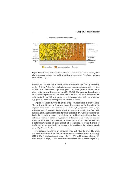
Figure 2.1: Schematic picture of structure features found in µc-Si:H. From left to right the<br />
film composition changes from highly crystalline to amorphous. The picture was taken<br />
from Houben [52]<br />
between µc-Si:H and a-Si:H growth, the structure varies significantly depending<br />
on the substrate. While for a fixed set of process parameters the material deposited<br />
on aluminum foil results in crystalline growth, fully amorphous structure can be<br />
observed for the one deposited on glass [54, 55, 50]. The substrate dependence is<br />
of particular importance and has to be kept in mind if one wants to compare results<br />
obtained from different measurement techniques, since different substrates,<br />
e.g. glass or aluminum, are required for different methods.<br />
Typical for all structure modifications is the occurrence of an incubation zone.<br />
The particular thickness and composition of this region strongly depends on the<br />
deposition condition and the substrate used. In the highly crystalline regime, crystallization<br />
starts from nucleation centers close to the substrate-film interface. With<br />
increasing film thickness the diameter of the columnar structures increases resulting<br />
in the typically observed conical shape. In the highly crystalline regime the<br />
columnar clusters of coherent regions have a diameter of up to 200 nm and extend<br />
over the whole film thickness. However, the structure inside the columns<br />
is not monocrystalline. In fact it consists of coherent regions with a diameter of<br />
4 − 20 nm that are separated from each other by stacking faults and twin boundaries<br />
[10, 50, 56, 57, 58].<br />
The columns themselves are separated from each other by crack-like voids<br />
and disordered material. In fact, studies using transmission electron microscopy<br />
(TEM) [50, 10], infrared spectroscopy (IR) [11, 59], and hydrogen effusion [60]<br />
have shown that highly crystalline material often exhibits a pronounced porosity.<br />
8