

ASSESSING HARDWOOD PULP FIBRE ULTRASTRUCTURE
chemical and cryo-methods for assessing hardwood pulp fibre ...
chemical and cryo-methods for assessing hardwood pulp fibre ...

You also want an ePaper? Increase the reach of your titles
YUMPU automatically turns print PDFs into web optimized ePapers that Google loves.
CHEMICAL AND CRYO-METHODS FOR<br />
<strong>ASSESSING</strong> <strong>HARDWOOD</strong> <strong>PULP</strong> <strong>FIBRE</strong><br />
<strong>ULTRASTRUCTURE</strong><br />
PRABASHNI LEKHA 1,2 , TAMARA BUSH 1,3 PATRICIA BERJAK 2 AND NORMAN PAMMENTER 2<br />
1<br />
CSIR/UKZN, Forestry and Forest Products Research Centre, 2 School of Biological and Conservation Sciences, 3 School of Chemistry, UKZN, Durban, South Africa.<br />
INTRODUCTION<br />
• Various techniques and methods are available for sample preparation but their use is limited by the sensitivity to the<br />
specimen.<br />
• Biological material, including wood and pulp fibres, constitute the most difficult specimens to prepare for high resolution<br />
microscopy because of the complex and intricate structural detail.<br />
• Accurate imaging and analysis of the fibre wall ultrastructure depends extensively on the sample preparation method used.<br />
• For the production of novel materials from wood and cellulose, it is important to understand how different processes affect<br />
fibre wall ultrastructure.<br />
• This study used chemical and cryo-methods to obviate artefact induction during sample preparation aimed at elucidating<br />
‘true’ ultrastructural detail.<br />
CHEMICAL SAMPLE PREPARATION<br />
MATERIALS AND METHODS<br />
A<br />
Fibres were<br />
dehydrated in<br />
acetone (30, 50,<br />
75, 100%) for<br />
1 h<br />
CRYO SAMPLE PREPARATION (A) CRYO-FRACTURING, (B) CRYO-SECTIONING<br />
Fibres were<br />
loosened and<br />
strung out onto<br />
pieces of foil<br />
Prolonged<br />
infiltration with<br />
low viscosity<br />
epoxy resin<br />
for 3 days<br />
Fibre clumps<br />
were then<br />
submerged in<br />
supercooled<br />
liquid nitrogen @<br />
-210°C<br />
Fibres were<br />
placed in 100%<br />
fresh resin and<br />
polymerised for<br />
8 h at 70°C<br />
A sharp metal<br />
blade cooled<br />
with liquid<br />
nitrogen was<br />
then used to<br />
fracture the fibre<br />
clumps<br />
Fractured fibres<br />
were then freeze<br />
dried using a<br />
freeze drier<br />
RESIN<br />
BLOCK<br />
Polished block face<br />
post microtoming<br />
Glass cover slip<br />
coated with Haupt’s<br />
adhesive<br />
0.5 - 2 µm thick resin<br />
sections were tested<br />
Freeze dried<br />
fractured fibres<br />
were then<br />
mounted onto<br />
carbon stubs<br />
using carbon<br />
fluid<br />
Resin block face<br />
and sections<br />
were etched with<br />
potassium<br />
methoxide for<br />
1 min to dissolve<br />
resin<br />
Sonicated with<br />
methanol for<br />
2 min<br />
to remove<br />
remaining resin<br />
from tissue<br />
All sample preparations were sputter<br />
coated with carbon and then viewed at<br />
2 - 10 kV by use of a Carl Zeiss Ultra<br />
FEG-SEM<br />
Resin blocks and<br />
etched sections<br />
on cover slips<br />
were left to dry<br />
under a fume<br />
hood<br />
Resin blocks,<br />
cover slips and<br />
formvar grids<br />
were mounted<br />
onto metal stubs<br />
with carbon tape<br />
Chemical<br />
Concentration<br />
B<br />
Fibres were<br />
loosened and<br />
treated with<br />
various<br />
cryoprotectant<br />
solutions for<br />
different times<br />
30 min – 24 h<br />
1-hexadecen<br />
sucrose<br />
glycerol<br />
polyvinylpyrrollidone (PVP)<br />
PVP<br />
PVP<br />
3.5 M<br />
2.3 M<br />
10.6 M<br />
0.5 mM<br />
1 mM<br />
1.5 mM<br />
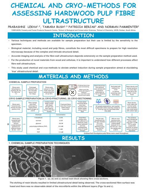
1. CHEMICAL SAMPLE PREPARATION TECHNIQUES<br />
Small fibre<br />
clumps were<br />
then adhered to a<br />
metal pin using<br />
tissue adhesive<br />
and was<br />
subsequently<br />
frozen in liquid<br />
nitrogen<br />
RESULTS<br />
The pin was<br />
transferred into<br />
the gas chamber<br />
of a cryomicrotome<br />
(Reichert-Jung,<br />
Type 652701,<br />
Austria)<br />
500 nm cryosections<br />
were<br />
obtained using a<br />
cutting speed<br />
between 0.4 to<br />
0.8 mm sec -1 and<br />
the sample was<br />
at 80°C<br />
Unbleached Eucalyptus pulp fibres were used throughout.<br />
The sections<br />
were transferred<br />
using a metal<br />
loop and 1.5 mM<br />
PVP to grids<br />
coated with<br />
formvar on which<br />
carbon had been<br />
deposited<br />
a<br />
b<br />
c<br />
lumen<br />
S2 layer<br />
S2 layer<br />
10 µm<br />
0.1 µm<br />
0.2 µm<br />
Figure 1. (a), (b) and (c) etched resin block showing fibre cross-sections.<br />
The etching of resin blocks resulted in limited ultrastructural detail being observed. The cross-sectioned fibre surface was<br />
fused and there was no observable detail of the microfibrils within the different layers (Figs 1b and c).
a<br />
b<br />
RESULTS cont.<br />
S1 layer<br />
c<br />
S2 layer<br />
S2 layer<br />
S3 layer<br />
10 µm<br />
0.4 µm<br />
0.2 µm<br />
Figure 2. (a), (b) and (c) etched 1.5 µm thick resin sections of Eucalyptus pulp fibres.<br />
Etching of resin-embedded sections of different thickness on glass cover slips showed more ultrastructural detail compared<br />
with etched resin blocks, 1.5 µm thick sections being the best (cf. Figs 1 and 2). However, microfibril ultrastructure was<br />
obscured within the fibre wall when using chemical sample preparation techniques (Fig. 2c).<br />
2. CRYO-SAMPLE PREPARATION TECHNIQUES<br />
a<br />
b<br />
S1 layer<br />
c<br />
S1 layer<br />
S2 layer<br />
S2 layer<br />
S2 layer<br />
S2 layer<br />
S1 layer<br />
S1 layer<br />
lumen<br />
2 µm<br />
0.2 µm<br />
0.4 µm<br />
Figure 3. (a), (b) and (c) cryo-fractures of Eucalyptus pulp fibres without pre-treatment.<br />
Cryo-fracturing Eucalyptus pulp fibres did not result in proper cross-sections (Fig. 3a). The fractured surface curled obscuring<br />
internal ultrastructural detail (Figs 3b and c).<br />
a<br />
S1 layer<br />
b<br />
c<br />
S2 layer<br />
S2 layer<br />
S2 layer<br />
S1 layer<br />
0.5 µm<br />
0.4 µm<br />
0.1 µm<br />
Figure 4. Cryo-sections of Eucalyptus pulp fibres, (a) 2.3 M sucrose, (b) and (c) 1.5 mM PVP pre-treatment.<br />
Pre-treatment of fibres with 2.3 M sucrose following the standard cryo-sectioning method resulted in crevices being present<br />
across the fibre cross-section (Fig. 4a). The use of polyvinylpyrrollidone (PVP) as a cryoprotectant facilitated intact<br />
cryo-sections (Fig. 4b), all other cryoprotectants tested not achieving this. From the three concentrations (0.5, 1.0 and 1.5 mM)<br />
and different exposure times tested, 30 min pre-treatment of 1.5 mM PVP produced the best results.<br />
CONCLUSIONS<br />
• The level of ultra-structural detail obtained using cryo-sectioning is superior to that obtained using chemical methods or<br />
cryo-fracturing.<br />
• Cryo-sectioning of hardwood pulp fibres promises to be beneficial for the assessment of mechanical and chemically<br />
treated fibres.<br />
ACKNOWLEDGEMENTS: We thank UKZN, CSIR and SAPPI Chemical Cellulose for funding.<br />
Corresponding email address: plekha@csir.co.za, 201295678@ukzn.ac.za.


