
Create successful ePaper yourself
Turn your PDF publications into a flip-book with our unique Google optimized e-Paper software.
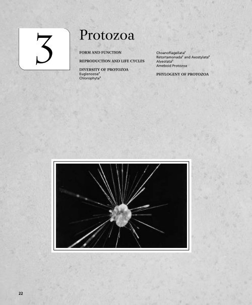
3 <strong>Protozoa</strong>FORM AND FUNCTIONREPRODUCTION AND LIFE CYCLESDIVERSITY OF PROTOZOAEuglenozoa PChlorophyta PChoanoflagellata PRetortamonada P and Axostylata PAlveolata PAmeboid <strong>Protozoa</strong>PHYLOGENY OF PROTOZOA22
24 Chapter 3 <strong>Protozoa</strong>microtubules of a mitotic spindle, which radiate from centriolesand form the mitotic apparatus (Fig. 2-2). In other protozoa,such as the spherical radiolarians and heliozoans, bundles ofmicrotubules radiate from a centroplast (an MTOC) at thecell’s center and then extend into and support a raylike projectionof the cell’s surface (axopod; Fig. 3-1D). The centroplastand its microtubules resemble the starlike asters that formaround centrioles at the poles of the mitotic spindle.Vesicles, known as alveoli, occur immediately below the cellmembrane in many protozoans, such as dinoflagellates,apicomplexans, and ciliates (together forming the Alveolata).“Empty” alveoli, like those that occur in ciliates, may be turgidand help to support the cell, but they also store Ca 2 , whichcan be released to trigger cellular responses (Fig. 3-1E).In some dinoflagellates, plates of cellulose secreted into thealveolar vesicles form a rigid endoskeleton (Fig. 3-1F).<strong>Protozoa</strong>n skeletons, like those of metazoans, can also beendo- or exoskeletons. A skeleton that forms a more or lesscomplete covering, whether internal or external, is calleda test (or a lorica, theca, or shell).The protozoan locomotor organelles may be flagella, cilia,or flowing extensions of the cell known as pseudopodia(described in Chapter 2). The undulatory waves of flagellapass from base to tip and drive the organism in the oppositedirection (Fig. 2-6). The flagella of many protozoansbear fine lateral “hairs” called mastigonemes (Fig. 3-2). Themastigonemes cause the flagellum to pull rather than pushas the flagellar waves pass from base to tip. Flagellar, ciliary,and pseudopodial specializations characterize many of theprotozoan taxa.All types of nutrition occur in protozoa. Some protozoarely on photosynthesis, others absorb dissolved organic materialfrom the environment, and many digest food particles orprey intracellularly in food vacuoles. Food enters the vacuoleby phagocytosis, often at a definite cell mouth, or cytostome.The vacuole then may be shuttled to the interior along a specializedmicrotubular tract called a cytopharynx. Macromoleculesenter by micro- and macropinocytosis, which may occurFIGURE 3-2 <strong>Protozoa</strong>: Flagellar mastigonemes. Phytoflagellatewith one short smooth flagellum and one long flagellum bearingmastigonemes.Image courtesy of BioPhoto Associates.over the entire surface of the cell. Intracellular digestion hasbeen most studied in amebas and ciliates, and, for the mostpart, it follows the general pattern described in Chapter 2.Digestive specializations of ciliates will be described later inthis chapter.Diffusion is important for internal transport in all protozoansand may be the sole mechanism in small cells. Somelarge protozoans and those with long pseudopodia have activemechanisms of internal transport. The inner, fluid cytoplasmof Paramecium circulates, via cyclosis, in a closed loop(Fig. 2-10). In forams and actinopods, bidirectional shuttlingof vesicles occurs on tracks of microtubules in the axis of eachslender pseudopodium.Most protozoans are aerobes that rely on diffusion for theuptake of oxygen and release of CO 2 . A few protozoans,however, are obligate anaerobes, especially those that live assymbionts in the digestive tract of animals. Aquatic speciesassociated with decomposing organic matter may be facultativeanaerobes, using oxygen when it is present but alsocapable of anaerobic respiration. In general, the changingavailability of food and oxygen associated with decompositionresults in a successional sequence of protozoan species.Because of their short generation time, protozoan communitystructure changes rapidly with environmental change and canbe used to monitor aquatic systems for pollution.Many freshwater protozoa osmoregulate to remove excesswater (volume regulation) and to adjust the concentrationand proportions of their internal ions (ionic regulation).Excess water enters by osmosis when the internal osmoticconcentration exceeds that of the surrounding water. Additionalwater may enter with food in vacuoles and pinocytoticvesicles. For example, an ameba fed on a protein solutionimbibes, by macropinocytosis, a quantity of water equivalent toone-third of its body volume.Osmoregulation is accomplished by active ion transport atthe cell membrane and by a system of water- and ion-pumpingorganelles called the contractile vacuole complex (Fig. 3-3).The complex is composed of a large spherical vesicle—thecontractile vacuole proper—and, surrounding it, an array ofcytoplasmic vesicles or tubules termed the spongiome. Thespongiome collects fluid from the cytoplasm and conducts itto the contractile vacuole. The contractile vacuole then contractsand discharges the fluid to the outside of the organismthrough a temporary or permanent pore. The rate ofdischarge depends on the osmotic concentration of the externalmedium. Paramecium caudatum, which lives in fresh water,can complete a cycle of vacuole filling and discharge as rapidlyas every 6 s and expel a volume equivalent to its entire bodyevery 15 min. The basis for contraction may differ amonggroups of protozoans. In dinoflagellates, a flagellar rootletbranches to form a contractile sheath around the vacuole. InParamecium, microtubules provide a scaffold around the vacuole(Fig. 3-3), but actin and myosin or elastic energy storedin the stretched vacuolar membrane may be responsible forcontraction.Freshwater protozoans excrete a hypotonic urine. Althoughthe mechanism of contractile vacuole function is not fullyunderstood, it is likely that ions are pumped from thecytoplasm into the spongiome tubules, establishing an osmoticgradient. Cytoplasmic water enters the tubules down the
Reproduction and Life Cycles 25AMicrotubulesContractile vacuole porePellicleBMitochondrionSpongiome tubulesSpongiometubulesAmpullaSpongiome vesiclesCollecting tubuleMitochondriaDCFIGURE 3-3 <strong>Protozoa</strong>: Diagram of four types of contractile vacuoles. Types A and B are from ciliates, in which thespongiome is composed of irregular, fluid-filled tubules. Actin filaments (not shown) wind around the pore andextend over the vacuole surface. A, The network of spongiome tubules empties directly into the vacuole. B, Thenetwork of irregular tubules first empties into ampullae, which dilate and then contract, discharging fluid into thevacuole, as occurs in Paramecium. C, Typical of flagellates and small amebas, the spongiome contains small vesiclesand tubules. D, Arrangement found in large amebas. (After Patterson, D. J. 1980. Contractile vacuoles and associatedstructures; their organization and function. Biol. Rev. 55:1–46. © Copyright Cambridge University Press, reprinted by permission.)osmotic gradient. As water and ions flow along the tubules,ions and perhaps other substances are selectively reabsorbedbefore the urine is discharged to the exterior. The contractilevacuole system is of no particular significance in removingmetabolic wastes, such as ammonia and CO 2 , as these simplydiffuse to the outside of the organism.REPRODUCTION AND LIFE CYCLESClonal (asexual) reproduction by mitosis occurs in mostprotozoa and is the only known mode of reproduction in somespecies. Division of the parent into two or more daughter cells iscalled fission. When this process results in two similar progenycells, it is termed binary fission; when one progeny cell is muchsmaller than the other, the process is called budding. Division ofthe parent into more than two daughter cells is known asmultiple fission. Schizogony is a specialized form of multiplefission in which repeated divisions of the nucleus precede thecell divisions. With few exceptions, clonal reproduction involvessome replication of organelles before or after fission.The mitotic division of the protozoan cell nucleus differs,in most cases, from that of an animal cell. In animal cellsundergoing mitosis, the nuclear membrane disintegratesduring mitosis as the chromosomes condense and attach to themitotic spindle, located in the cytoplasm of the cell. Because thenuclear membrane breaks down, this form of mitosis is said tohave an open spindle. Later in mitosis, after the chromosomeshave separated, a new nuclear membrane is assembled aroundeach nucleus. Among most of the protozoans described in thischapter, however, the nuclear membrane does not break downduring mitosis and the spindle forms within the nucleus itself. Asthe chromosomes separate, the intact nucleus stretches and thenconstricts, pinching off two new nuclei. <strong>Protozoa</strong>ns with thisarrangement have a closed spindle. The closed spindle isregarded as the primitive form of mitosis in eukaryotic cells.Intermediates between closed and open spindles occur in chlorophytes(Chlamydomonas, Volvox) and apicomplexans. In these taxa,the nuclear membrane remains largely intact, but breaks occurthat allow cytoplasmic spindle microtubules to enter the nucleusand attach to the chromosomes.Sexual reproduction is widespread but not universal in protozoans,and life cycles are diverse. Many well-studied protozoanslack sexual reproduction entirely. In some species this absencemay be primitive, whereas in others it may be a secondary loss.The primitive protozoan life cycle may have been sex free: a haploid(N) individual reproduced solely by fission, as in the livingkinetoplastids (Fig. 3-4A).The three general forms of sexual life cycles in protozoansare haploid dominance, diploid dominance, andhaploid-diploid codominance. A haploid-dominant life cycle
26 Chapter 3 <strong>Protozoa</strong>Individual N(Fission)ASEXUALHAPLOIDCYCLE(Fission)AIndividual NIndividual NSEXUAL CYCLEHAPLOID ADULTSWITHZYGOTIC MEIOSISIsogameteIsogameteCBIndividual 2NDIPLOIDCYCLE(Meiosis)Gamete(Meiosis)(Fertilization)Zygote2NGameteZygote2NZygote 2N(Fertilization)GameteD(Fertilization)Individual 2NHAPLOID-DIPLOIDCYCLEGameteIndividual N(Meiosis)Spore NFIGURE 3-4 <strong>Protozoa</strong>: Life cycles. N haploid,2N diploid. A, Haploid asexual life cycle: Newindividuals arise directly by fission (mitosis), asillustrated by the kinetoplastids. B, Haploiddominantlife cycle: Two N individuals mitoticallyproduce isogametes, which fuse to form a diploidzygote. The zygote then undergoes meiosis toform haploid individuals. Examples include theVolvocida, many dinoflagellates, axostylates, andapicomplexans (sporozoans). C, Diploid-dominantlife cycle: 2N individuals meiotically produceN gametes, which fuse to restore a 2N individual,as happens in some axostylates, heliozoans, manygreen algae, diatoms, and ciliates (and multicellularanimals). Ciliates, however, do not formgametes, but exchange haploid nuclei, whichfuse. D, Haploid-diploid codominant life cycle:2N individuals meiotically produce N spores thatdevelop into N individuals that mitotically formN gametes that fuse to restore the 2N individuals;includes many forams, and many algae (andmulticellular green plants).includes haploid individuals that either transform intogametes or produce them by mitosis. Fusion of the haploidgametes results in a diploid zygote that soon undergoesmeiosis to form four new haploid individuals (Fig. 3-4B).The haploid-dominant life cycle typifies apicomplexans. Ina diploid-dominant life cycle, the 2N individuals undergomeiosis to produce N gametes (or gamete nuclei), whichfuse into a 2N zygote individual (Fig. 3-4C). This type of lifecycle occurs, for example, in ciliates (and animals). In thehaploid-diploid codominant life cycle, an asexual generation(N or 2N) alternates with a sexual generation (2Nor N; Fig. 3-4D). This pattern is characteristic of forams (andplants).Encystment is characteristic of the life cycle of manyprotozoa, including the majority of freshwater species.In forming a cyst, the protozoan secretes a thickened envelopeabout itself and becomes inactive. Depending onthe species, the protective cyst is resistant to desiccationor low temperatures and encystment enables the cell topass through unfavorable environmental conditions. Thesimplest life cycle includes only two phases: an active phaseand a protective, encysted phase. However, the morecomplex life cycles are often characterized by encystedzygotes or by formation of special reproductive cysts inwhich fission, gametogenesis, or other reproductive processestake place.
Diversity of <strong>Protozoa</strong> 27<strong>Protozoa</strong> may be dispersed over long distances in either theactive or encysted stages. Water currents, wind, and mud anddebris on the bodies of waterbirds and other animals arecommon means of dispersal.DIVERSITY OF PROTOZOAEUGLENOZOA PEuglenoidea CThe euglenoid species of Peranema and Euglena are among themost familiar of all flagellates. The body is elongate with aninvagination, the reservoir, at the anterior end (Fig. 3-5). Thecytostome lies at the base of the reservoir and joins a cytopharynx.A contractile vacuole discharges into the reservoir infreshwater species, and two flagella, each bearing a row ofmastigonemes, arise from the reservoir wall.In Euglena, one flagellum is very short and terminatesat the base of the long flagellum (Fig. 3-5A). A pigmentedeyespot, or stigma, shades a photosensitive bulge, the paraflagellarbody, at the base of the long flagellum. In the colorlessheterotroph Peranema, both flagella are long, but one trailsbackward and can be used to catch food or temporarily attachto something (Fig. 3-5B). The long locomotory flagellum isthickened, up to five times the normal flagellar diameter, andstiffened along most of its length by a paraxial rod locatedto one side of the axoneme (Fig. 3-5B,C). Only the mobileterminal end of the flagellum lacks the rod.Seen in cross section, the euglenoid pellicle is thrown intorounded ridges alternating with narrow grooves, whichtogether wind helically around the cell. The ectoplasm hasa dense skeletal mesh of fibrous proteins. Pellicular microtubulessituated below this mesh may be responsible for theperistaltic movements of the cell known as euglenoid movement(or metaboly).Approximately two-thirds of the 1000 species of the marineand freshwater Euglenoidea are colorless heterotrophs andCanalParaflagellarbodyEyespotReservoirBasal bodiesPhospholipidvesiclesLocomotoryflagellumSecond (nonemergent)flagellumContractile vacuolar regionLeadingflagellumFlagellarresevoirNucleus of EuglenaAnterior end of Euglenain cytopharynx ofPeranemaRod organof cytopharynxChromosomesNuclearenvelopeNucleolusNucleusBasal bodyParamylon inpyrenoid of chloroplastNucleus of PeranemaFree paramylon granule(stored carbohydrate)Trailing flagellumChloroplastsBParaxial rodAxonemeACFIGURE 3-5 Euglenozoa: Euglenoidea. A, Structure of the photosynthetic Euglena gracilis. B, The colorlessheterotroph Peranema swallowing a Euglena. C, Cross section of leading flagellum of Peranema showingparaxial rod. (A, From Leedale, G. F., 1967, Euglenoid Flagellates. Prentice-Hall, Inc. Englewood Cliffs, N.J.; B, Modifiedafter Chen)
28 Chapter 3 <strong>Protozoa</strong>one-third are green photoautotrophs, such as the speciesEuglena. The chloroplasts of photosynthetic species containchlorophylls a and b. Photosynthetic euglenoids rotate aroundtheir longitudinal axis as they swim toward light. As long asthey maintain this orientation, the photosensitive paraflagellarbody receives constant illumination. But if they deviate fromtheir head-on approach to a light source, the rotating eyespotperiodically shades the paraflagellar body and elicits acourse correction. The heterotrophic mode of nutrition isprimitive in euglenoids. Chloroplasts were acquired secondarilywithin the taxon and independently of other photosyntheticflagellates.The green, photosynthetic species such as Euglena storefood energy as a unique starchlike carbohydrate calledparamylon. Paramylon is synthesized in a specialized region,the pyrenoid, of the chloroplast, but stored as free granules inthe cytoplasm (Fig. 3-5A). The large paramylon granules mayalso have a skeletal function, as in Cyclidiopsis acus, whose longitudinallyaligned granules form an intracellular “backbone.”The chloroplasts of euglenoids are surrounded by three membranes,not two as in green algae and plants. For this reason,euglenoids are believed to have acquired their chloroplasts byphagocytosis of an entire eukaryotic algal cell, probablya chlorophyte (discussed later), which then became an endosymbiont.If so, the outermost membrane of the euglenoidchloroplast may correspond to the cell membrane of thechlorophyte cell, or the cell membrane fused with themembrane of the phagocytic vesicle.Food for the colorless heterotrophs consists of organiccompounds absorbed from the surrounding water, bacteria,and other protozoan cells. Peranema seizes prey with a uniquerod organ associated with its cytopharynx and cytostome. Therod organ (Fig. 3-5B) consists of two stiff, parallel rods(microtubule bundles) and other intracellular structures called“vanes.” (Euglena has a rudimentary rod organ, an indication ofits heterotrophic ancestry.) Peranema feeds on a wide variety ofliving organisms, including Euglena, and the cytostome can begreatly distended to permit phagocytosis of large prey. Whilefeeding, the rod organ is protruded, attaches to the prey, andthen retracts, pulling the prey into the cytostome and cytopharynx.(Fig. 3-5B). The prey is swallowed (phagocytosed) wholeand digested in a food vacuole.Sexual reproduction has not been observed in euglenoids,but clonal reproduction occurs by longitudinal binary fission(Fig. 3-6). The two flagella and their basal bodies, as well asthe nucleus, replicate before the cell itself divides.Kinetoplastida CKinetoplastid flagellates are colorless heterotrophs. A few ofthe 600 species are free living, but most are important parasites.All share the flagellar paraxial rod with their euglenoidrelatives, but uniquely have a conspicuous mass of DNA, calleda kinetoplast, located within a single, large mitochondrion(Fig. 3-7D). Most of the kinetoplast DNA sequences code forthe morphogenesis of mitochondria. The large kinetoplastgenome probably is related to the cyclical differentiation andregression of mitochondria as parasitic kinetoplastids adapttheir energy metabolism to alternating aerobic and anaerobichost environments. The one or two flagella arise froma reservoir-like pit, which bears a cytostome that leads intoa cytopharynx. Their basal bodies are located on or near thekinetoplast mitochondrion.Species of the free-living biflagellate Bodo (Fig. 3-7D) commonlyare found in brackish and fresh water and in soil, wherethey feed on bacteria. The trypanosome kinetoplastids areCanalContractilevacuoleBasalbodyNucleusNucleolusAB C D EFIGURE 3-6 Euglenozoa: Euglenoidea. Clonal reproduction by longitudinal fission (symmetrogenic division) in Euglena. A, An interphase cellwith two flagella and two basal bodies. B, A premitotic cell: Two new basal bodies have formed adjacent to the parental basal bodies. C, Earlymitosis: The parental basal bodies separate, each becoming associated with one new basal body. All four basal bodies bear a flagellum. Elongationof the nucleolus indicates the onset of nuclear division. Unlike mitosis of animal cells, the euglenoid nuclear membrane remains intact (closedspindle) during the entire division cycle and the flagella do not regress. D, Late mitosis: Each separate pair of flagella consists of a parentaland a daughter basal body. The nucleus is dividing by constriction, the contractile vacuole has divided, and the reservoir (gullet) is undergoingdivision. E, The anterior end is dividing following duplication of organelles. (Modified and redrawn from Ratcliffe, 1927, and Triemer,www.lifesci.rutgers.edu/~triemer/flagellar_appt/flagellarapparatus.html)
Diversity of <strong>Protozoa</strong> 29Image Courtesy of S. S. Hendrix.ACytostomeBAnterior flagellum withmastigonemesFlagellar pocketUndulatingmembraneContractile vacuoleCytopharynxKinetoplastGolgi apparatusNucleusFood vacuoleMitochondrionNucleusMitochondrionKinetoplastCTrailing flagellumDFIGURE 3-7 Euglenozoa: Kinetoplastida. A, Skin lesion on a boy’s wrist, one of the symptoms of kala-azardisease caused by Leishmania. B, Life cycle of Trypanosoma brucei: The bite of a tsetse fly introduces infective-stagecells that migrate into the human’s cerebrospinal fluid to cause the lethargy associated with “sleeping sickness.”Life cycle is completed when fly takes another blood meal and ingests the parasite. C, Structure of Trypanosomabrucei. D, Bodo saltans, a free-living member of the Kinetoplastida. Only sectioned parts of the long, single loopingmitochondrion are shown. (B, From Sleigh, M. A., 1973, The Biology of <strong>Protozoa</strong>. Edward Arnold, London. p.141;C, Modified after Brooker from Farmer, J. N. 1980. The <strong>Protozoa</strong>: Introduction to Protozoology. C. V. Mosby Co., St. Louis, p. 214.)gut parasites of insects and blood parasites of vertebrates. Usuallyonly the anterior flagellum is present (Fig. 3-7C), thesecond flagellum being represented only by a basal body.Commonly, the flagellum trails and is joined to the side of thebody by an undulating membrane. The pellicle has a thick glycocalyx,whose protein composition is controlled by up to1000 genes, roughly 40% of the cell’s large nuclear genome(120 chromosomes in Trypanosoma brucei). Differential geneexpression (and protein synthesis) during the variousinfective stages changes the antigen signature of the glycocalyx,enabling the parasite to elude the host’s immune system.Species of the trypanosome genera Leishmania and Trypanosomaare agents of numerous diseases of humans anddomesticated animals in subtropical and tropical regions ofthe world. Part of the life cycle is passed within or attached togut cells of blood-sucking insects, mostly various kinds of flies,and another part of the cycle is spent in the blood plasma orin white blood cells and lymphoid cells of the vertebrate host,although other tissues may be invaded. Intracellular stagesare aflagellate, but during the life cycle, motile, extracellular,flagellated stages occur in the vertebrate bloodstream or inthe invertebrate host (Fig. 3-7B,C).Leishmania is the agent of the widespread kala-azar andrelated diseases of Eurasia, Africa, and the Americas. Theycause skin lesions (Fig. 3-7A) and interfere with immuneresponses, among other effects. Tiny biting flies known assand flies or no-see-ums (Ceratopogonidae) are the bloodsuckinginsect host of this protozoan.Chagas’s disease of tropical America, which probablyaccounted for Darwin’s chronic ill health following the voyageof the Beagle, is caused by Trypanosoma cruzi and is transmittedby blood-sucking bugs. Extensive damage to the human hostoccurs when the parasite leaves the circulatory system andinvades the liver, spleen, and heart muscles.Trypanosoma brucei rhodesiense and T. b. gambiense causeAfrican sleeping sickness and are transmitted by the tsetse fly
30 Chapter 3 <strong>Protozoa</strong>(Fig. 3-7B,C). The parasite invades the cerebrospinal fluid andbrain, producing the lethargy, drowsiness, and mental deteriorationthat mark the terminal phase of the disease. Varioustrypanosome diseases of horses, cattle, and sheep are ofconsiderable economic importance.Kinetoplastids undergo binary fission. Like euglenoids, sexualreproduction has not been observed, but its occurrence issuspected.CHLOROPHYTA PVolvocida OVolvocida is a taxon of green algae (Chlorophyta), in whichthe large, cup-shaped chloroplasts contain chlorophylls a andb and a pyrenoid that synthesizes starch (amylopectin) as afood storage product. Many chlorophytes are nonmotile marineand freshwater algae, such as the filamentous Spirogyra offresh water. The cells of Volvocida, however, are permanentlyflagellated: Each cell bears two, four, or occasionally eight flagellalacking mastigonemes. An eyespot and two contractilevacuoles also may be present. The cells are enclosed in a gelmatrix composed of glycoproteins and glycoaminoglycans andare interjoined by cytoplasmic bridges.Among the flagellated species, some are solitary, such asChlamydomonas (Fig. 3-8A), and others are colonial. Thecolonies of Gonium (Fig. 3-8B) are flat plates of 32 to 40 cells,but other genera form hollow spheres: Pandorina (16 to 32cells), Eudorina (32 cells), Pleodorina (64 to 128 cells), Volvox(2000 to 6000 cells).Chlamydomonas reproduces clonally by longitudinal binaryfission. In Volvox, only specialized, large, aflagellate cells (gonidia)are capable of asexual and sexual reproduction. Duringclonal reproduction, a gonidium undergoes multiple fissionand forms a hollow sphere within the parent colony (Fig.3-8C). The cell polarity of this sphere, however, is oppositethat of the parent—the future flagella-bearing ends of thecells face the interior of the young colony. To correct itsreversed polarity, the daughter colony inverts and reformsa sphere, now with flagella on the outer surface. The daughtercolonies usually escape by rupturing the wall of the parentcolony.The volvocids have a haploid-dominant life cycle withpostzygotic meiosis (Fig. 3-4B). In most species of Chlamydomonas,the two structurally identical cells act as gametes(isogametes), fuse, and form a zygote. Other species show thebeginnings of sex differentiation by having gametes that differslightly in size (anisogametes). In Pleodorina, the size distinctionis pronounced, but the large macrogametes still retain flagellaand are free swimming. Finally, in Volvox, true eggs and spermdevelop from gonidia at the posterior of the colony. The egg isstationary and is fertilized within the parent colony by a spermpacket released from another colony. Colonies may be eitherhermaphrodites or one or the other sex.Although closely related to plants and not to animals,Volvox nevertheless illustrates how multicellularity might haveevolved in the first animals. Beginning as a single cell, subsequentmitoses result in a symmetrical colony composed ofhundreds of cells. These cells then specialize functionally intosomatic cells and reproductive cells (gonidia).ABCCell wallNucleusChloroplastDaughter coloniesStarch granuleFIGURE 3-8 Chlorophyta: Volvocida. A, Chlamydomonas reinhardtii,a noncolonial solitary species. B, Gonium pectorale. Gonium speciesform colonies in the form of a flat, square plate in which all cells areembedded in a common gelatinous envelope. C, Volvox colonies arehollow spheres. Note daughter colonies within parent colonies. (A, FromSleigh, M. 1989. <strong>Protozoa</strong> and Other Protists. Edward Arnold, London, p.140;B, Courtesy of General Biological Supply House, Inc.)CHOANOFLAGELLATA PSurprising as it may seem, the marine and freshwaterchoanoflagellates are the sister taxon of animals (Metazoa).Both choanoflagellate and primitive monociliated animalcells bear a single flagellum, which bears a bilateral vane ofCourtesy of General Biological Supply House, Inc.
Diversity of <strong>Protozoa</strong> 31CollarTestCell properStalkACSecondaryChoanoecastageMotileProterospongiastageBMinutecellFIGURE 3-9 Choanoflagellata.Choanoflagellates have one flagellumsurrounded by a collar of microvilli.A, A stalked colonial species. The stalk is anextension of the vaselike test that surroundseach cell. B, Proterospongia, a colonialspecies with cells united in a gelatinousmatrix. C, Proterospongia choanojunctahas both a sessile and planktonic stage.(A, From Farmer, J. N. 1980. The <strong>Protozoa</strong>:Introduction to Protozoology. C. V. Mosby Co., St.Louis.; B and C, From Leadbeater, B. S. C. 1983.Life-history and ultrastructure of a new marinespecies of Proterospongia. Jour. Mar. Biol. Assoc.U.K. 63:135–160.)mastigoneme-like filaments and is surrounded by a cylindricalcollar of microvilli (Fig. 4-2A). This synapomorphy, along withsupport provided by rDNA sequences, unites the choanoflagellatesand metazoans as sister taxa in a monophyletic taxon(see Chapter 1 for cladistic terms and method).The 600 species of choanoflagellates are mostly tiny andinconspicuous, usually not in excess of 10 m in diameter(Fig. 3-9, 4-12A,B, 4-13A). While feeding, the flagellum createsa water current from which the collar filters bacteria andorganic particulates. Bacteria trapped on the collar areingested by phagocytosis.Choanoflagellates may be solitary or colonial, attached orfree swimming. Some sessile species are attached by a stalk,part of a vaselike test (Fig. 3-9A). The test is composed ofinterconnected, extracellular, siliceous rods. The individuals ofcolonial planktonic forms, such as species of Proterospongia, areunited by a jellylike extracellular matrix or by their collars (Fig.3-9B,C, 4-12A,B). In the latter case, the colony may resemblea plate, with all of the collars and flagella located on the sameside, or a sphere on which the flagellated collars radiate fromthe surface (Fig. 4-12A). The marine Proterospongia choanojunctawas found to include both a colonial planktonic stage anda solitary, aflagellate attached stage (Fig. 3-9C).RETORTAMONADA P ANDAXOSTYLATA PThese two taxa of heterotrophic flagellates have from four tothousands of flagella organized in functional groups. A few ofthe 700 species are free living (Hexamita) in anoxic habitats,but most live anaerobically in the guts of vertebrates andinsects, especially wood roaches and termites. Because theylive in oxygen-free environments, mitochondria are eitherabsent or atypical, the cells being specialized for glycolysisrather than aerobic respiration. Even when mitochondria areabsent, as in Giardia, certain mitochondrial genes and proteinsdo occur, suggesting that the lack of mitochondria issecondary rather than primary.Retortamonads, such as Giardia lamblia, have four flagella,one of which trails behind the leading three and the cell body,and lack Golgi bodies as well as mitochondria. Giardia lamblia,which can cause a bloody diarrhea, is a common intestinalparasite in the United States. It frequently occurs in toddlersand child-care workers, but also can be acquired by drinkingfrom seemingly pristine mountain streams. The axostylateTrichomonas vaginalis is a small parasite with four anteriorflagella (Fig. 3-10A) that inhabits the urogenital tract of
32 Chapter 3 <strong>Protozoa</strong>RostrumMidregionNucleusVacuole with woodchips in posteriorcytoplasmAxostylePosterior regionABFIGURE 3-10 Axostylata. A, Trichomonas vaginalis, a trichomonad parasite in the human vagina and malereproductive tract. In addition to the four anterior flagella, a trailing flagellum borders a looping undulatingmembrane. An axial skeleton, the axostyle, originates at the flagellar basal bodies, passes through the body ofthe cell, and protrudes posteriorly. B, The hypermastigid Trichonympha campanula lives in the gut of termites.(A, After Wenrich; B, from Farmer, J. N. 1980. The <strong>Protozoa</strong>: Introduction to Protozoology. C. V. Mosby Co., St. Louis. p. 266.)humans and causes a widespread sexually transmitted disease.Living tissues can be invaded and the vaginas of seriouslyinfected women produces a greenish yellow discharge.The axostylates have a bundle of microtubules called anaxostyle that extends the length of the cell. It most species, it isskeletal in function, like an intracellular backbone, but in someprimitive species, it undulates and imparts a snaky motion tothe cell. The derived axostylates, such as the hypermastigidmutualists in the gut of termites and wood roaches, have hundredsor thousands of flagella and astounding internal complexity;Trichonympha (Fig. 3-10B) is a good example. Most havea saclike or elongated body usually bearing an anterior rostrum.Axostylates lack mitochondria, but have Golgi bodies.Many termites and wood-eating cockroaches are dependenton their hypermastigids for the digestion of wood. Theflagellates, however, rely on intra- and extracellular bacteriaand spirochetes for the actual breakdown of cellulose. Thenutrients released from the wood are used by bacterium,flagellate, and insect. The termite host loses its gut mutualistswith each molt of its exoskeleton, but by licking other individuals,by rectal feeding, or by eating cysts passed in feces(in the case of roaches), a new innoculation is obtained. Inwood-eating cockroaches, the life cycles of the flagellates areclosely tied to the production of molting hormones by thelate nymphal insect.Diversity of RetortamonadaRetortamonadea C : Two or four anterior flagella, one ofwhich is associated with the cytostome, which is elongatelongitudinally as a body furrow; mitochondria are absent.Chilomastix sp. (plural, spp.) cause diarrhea in humans,poultry; Retortamonas.Diplomonadea C : Cell with eight flagella, two cytostomes,two nuclei (twinned, diplozoic cell); mitochondria areabsent. Free-living Hexamita and parasitic Giardia, with theirattachment disc and long flagellar axonemes.Diversity of AxostylataOxymonadea C : Four posterior flagella; no cytostome, mitochondria,or Golgi bodies. Undulatory axostyle, with intracellularbacteria and surface-attached spirochetes. Anaerobesin the gut of termites and wood roaches. Oxymonas,Pyrsonympha.Parabasalea C : Cells have from a few to thousands of flagellaand aggregates of large Golgi bodies (parabasal bodies).Axostyle is skeletal, single, replicated, or lost; mitochondriaare absent; gut symbionts. Trichomonadida O has two tosix flagella; a recurrent flagellum forms an undulatingmembrane; axostyle projects posteriorly to form the attachmentsite. Trichomonas and Mixotricha paradoxa in the termitegut have surface-attached spirochetes whose motion propelsthe flagellate. Hypermastigida O has many flagella at theanterior end of the cell (sometimes also elsewhere). Mosthave surface-attached symbiotic bacteria. In the gut oftermites and wood roaches. Barbulanympha, Lophomonas,Trichonympha.ALVEOLATA PThree taxa, Dinoflagellata, Ciliophora, and Apicomplexa(Sporozoa) constitute the Alveolata. Alveolates are united onthe basis of having similar ribosomal DNA sequences andpellicular alveoli.
Diversity of <strong>Protozoa</strong> 33Dinoflagellata sPApproximately one-half of the 4000 marine and freshwaterspecies of dinoflagellates have chloroplasts and are importantprimary producers, especially in the sea. The xanthophyll pigmentperidinin colors them red-brown or golden brown. Theirchloroplasts are surrounded by three membranes and havechlorophylls a and c, but lack chlorophyll b. Dinoflagellatechloroplasts are diverse, having originated as endosymbiontsfrom at least three different taxa of photosynthetic cells. Heterotrophicdinoflagellates lack plastids and are colorless. Likeeuglenoids, dinoflagellates originated as colorless heterotrophsthat independently acquired chloroplasts by endosymbiosis,probably more than once. A few dinoflagellates are endoparasitesof other protozoans, crustaceans, and fishes. The cell nucleuscontains permanently condensed (thickened) chromosomeshaving relatively small amounts of protein, and eachchromosome is permanently attached to the nuclear membrane.Typical dinoflagellates have two flagella. One is attacheda short distance behind the middle of the body, is directed posteriorly,and lies in a longitudinal groove (sulcus) (Fig. 3-11B).Its surface is smooth or it may have two rows of mastigonemes.The other flagellum is transverse and located in a groove (cingulum)that either rings the body once or forms a spiral of severalturns. The transverse flagellum, which bears a unilateralrow of mastigonemes, causes both rotation and forward movement.The longitudinal flagellum drives water posteriorlyand contributes to forward motion. The dinoflagellate contractilevacuole, called a pusule, opens to the exterior near thebases of the flagella. The pusule is surrounded by contractilemyonemes.Dinoflagellates have a complex skeleton, or theca, whichoften contains deposits of skeletal cellulose in alveoli. Wherethe theca is thin and flexible, as in the common freshwaterand marine genus Gymnodinium, the dinoflagellate is said tobe unarmored, or naked (Fig. 3-11A). Armored dinoflagellateshave a thick theca composed of a few to several plates(Fig. 3-11B) formed by cellulose-filled alveoli. Frequently thearmor is sculptured, and often long projections or winglikeextensions protrude from the body, creating bizarre shapes(Fig. 3-11C). The large, colorless, and aberrant Noctiluca(Fig. 3-11D) and many smaller species are the principal contributorsto planktonic bioluminescence. At night on a quietsea, their greenish light sparkles in the wake of a boat or asstartled fish streak away like shooting stars.Dinoflagellates are either pigmented photoautotrophs orcolorless heterotrophs, but some pigmented species exhibitboth modes of nutrition. The prey is usually captured withpseudopodia and ingested through an oral opening associatedwith the longitudinal flagellar groove. Noctiluca is a predatorthat uses a single contractile tentacle, containing myonemes,to catch prey and convey it to its cell mouth (Fig. 3-11D).Among the symbiotic dinoflagellates, the mutualistic zooxanthellaeof corals, without which the coral-reef ecosystem probablywould not exist, are primarily one dinoflagellate species,Symbiodinium microadriaticum.Myriad dinoflagellates occur in marine plankton as importantcontributors to oceanic primary production, especiallyin the tropics. Marine species of the genera Gymnodinium,Gonyaulax, and others are responsible for outbreaks of theso-called red tides (Fig. 3-11E). Under ideal environmentalconditions and perhaps with the presence of a growthpromotingsubstance, populations of certain species increaseastronomically. Red tides, however, are not always red. Thewater may be yellow, green, or brown, depending on thepredominant pigments of the blooming organisms. Concentrationsof toxic alkaloids produced by the dinoflagellates canreach such high levels that other marine life may be killed.The 1972 red tides off the coasts of New England and Floridakilled thousands of birds, fish, and other animals and wreakedhavoc on the shellfish industry by infecting clams and oystersthat fed on the dinoflagellates.Pfiesteria piscicida, the cell from Hell, is the dinoflagellateresponsible for fish kills in estuaries along the middleAtlantic and southeastern coasts of the United States. Underconditions of organic enrichment, either from human pollutionor the feces of schooling fish, the normally nontoxiccells release a waterborne toxin that causes skin lesions infish. The dinoflagellates then attack the sores and consumethe fish. Pfiesteria is a colorless heterotroph that feeds byphagocytosis on a variety of organisms. When feeding onunicellular algae, it can digest the prey-cell but retain itschloroplasts intact and then use them to provide itself withphotosynthate. The Pfiesteria life cycle includes severalstages besides the typical biflagellated planktonic cell. Theseinclude a benthic ameba and encysted stages, as well asa planktonic form that superficially resembles a heliozoan(see Heliozoa later in this chapter).Ciguatera food poisoning in humans is caused by a marinedinoflagellate that lives attached to multicellular algae. Ciguatoxinis acquired by grazing herbivorous fish that concentratethe toxin in their tissues and pass it up the food chain. Thetoxin can reach such high levels in the tissues of carnivorousfish that, when eaten by humans, it produces serious poisoningand even death. In addition to gastrointestinal symptoms suchas diarrhea and nausea, there may be respiratory problems,muscle weakness, and long-lasting, strange skin sensations.Dinoflagellates undergo longitudinal binary fission. Cysts areformed in many flagellate groups, including dinoflagellates. Inaddition to the ameboid form of Pfiesteria already noted, somedinoflagellates can adopt the form of a naked, nonflagellatedball called a palmella. Fission often transforms the unicellularpalmella into a cluster of cells. The dinoflagellates that inhabitcorals as zooxanthellae do so in the palmella stage.Ciliophora sPCiliophora is a monophyletic taxon of animated and engagingcell-organisms. Most seem like diminutive animals becauseof their sophisticated cellular organelles and the complexityof their behavior. Many animal tissues and organs, such asmuscle and gut, have analogs in the cellular anatomy ofciliates. The 8000 described species are widely distributed infresh water, the sea, and in the water film around soil particles.All ciliates are heterotrophs, but about one-third of them areecto- or endocommensals or parasites.FORM AND FUNCTIONDiverse body forms occur among the ciliates and, despite theirmotility and fixed anterior-posterior polarity, most are asymmetric.A few, however, are radially symmetric with an anterior
34 Chapter 3 <strong>Protozoa</strong>AFrom Dodge, J. D., and Lee, J. J. 1985. Dinoflagellida. In Lee, J. J. et al. (Eds.): An IllustratedGuide to <strong>Protozoa</strong>. Society for Protozoology, Lawrence, KS. p. 29ApexTransverseflagellumCingulumSulcusPlatesCingulumLongitudinalflagellumBCFlagellumDTentacleFIGURE 3-11 Alveolata: Dinoflagellata. A, The naked Gymnodinium. B, A freshwater armored species,Glenodinium cinctum. C, The armored, Ceratium. D, Noctiluca, a bioluminescent carnivore with a prehensiletentacle. Only one small flagellum occurs in an “oral” depression. E, Gonyaulax digitale, a marine species thatcauses red tides. (B, After Pennak, R. W. 1978. Freshwater Invertebrates of the United States. 2nd Edition. John Wiley andSons, New York; C, After Jorgenson; D, After Robin)EMicrograph courtesy of Dodge, J. D.mouth (Fig. 3-12). Most ciliates are solitary and motile, butsome species form colonies and are sedentary. Most ciliatesare “naked,” but tintinnids, some heterotrichs, peritrichs, andsuctorians are housed in a test of secreted organic material orof cemented foreign matter (Fig. 3-13). Ciliate cell size rangesfrom 10 m to 4.5 mm.The surface cilia are specialized into a somatic ciliature onthe general body surface and an oral ciliature associated with
Diversity of <strong>Protozoa</strong> 35Polykinetids (membranelles)CytostomeKinety“Tentacles”TestFIGURE 3-12 Alveolata: Ciliophora. Prorodon, a radially symmetricalciliate. (After Fauré-Fremiet from Corliss, 1979)FIGURE 3-13 Alveolata: Ciliophora. Tintinnopsis, a marine ciliate(tintinnid) with a test composed of foreign particles. Note conspicuouspolykinetids (membranelles) and tentacle-like organelles interspersedbetween them. (After Fauré-Fremiet from Corliss, 1979)the mouth region. Distribution of body cilia varies betweenspecies. In some, cilia cover the entire cell and are arranged inlongitudinal rows, each called a kinety (Fig. 3-12), but in morespecialized taxa the cilia are restricted to regions of the body(Fig. 3-13, 3-18).A kinety is a row of repeating kinetids, each comprisinga cilium, basal body, and associated fibers (Fig. 3-14). One ofthe fibers attached to the basal body is a striated rootlet,which is oriented anteriorly. The rootlet fibers from all basalbodies in a row may combine, like wires in a cable, to forma single kinetodesma, which runs the length of the row (Fig.3-14). The other fibers associated with each basal body areribbons of microtubules. A postciliary microtubular ribbonextends posteriorly from each basal body. A transverseCell membraneOuter alveolar membraneInner alveolarmembraneCiliumCircumciliaryspaceAlveolusAlveolarcavityKinetodesmaStriated rootletBasal bodyTrichocystFIGURE 3-14 Alveolata: Ciliophora. The pellicle of Paramecium. (After Ehret and Powers from Corliss, 1979)
36 Chapter 3 <strong>Protozoa</strong>microtubular ribbon extends from the left side of each basalbody. All kinetid fibers are thought to be skeletal in function,either for ciliary anchorage or maintenance of cell shape.The unitary kinetid described in the preceding paragraphis called a monokinetid (Fig. 3-14). In some ciliates, themonokinetid is doubled into a dikinetid and the cilia occur inpairs along the kinety. In polykinetids, multiple cilia functiontogether in a compound unit. If that unit is a tuft, it is calleda cirrus (plural, cirri) (Fig. 3-18B), and if it is a short row,then the paddlelike unit is known as a membranelle (Fig.3-18A,B,F). Kinetids typify all ciliates, even groups such as theSuctoria, which lack cilia as adults but retain the intracellularcomponents of the kinetids.The ciliate body is typically covered by a complex pellicle.Below the outer cell membrane is a single layer of small membranoussacs, the alveoli, each of which is moderately togreatly flattened (Fig. 3-1E, 3-14). Cilia emerge from betweenadjacent alveoli, as do trichocysts and other extrusomes discussedlater (Fig. 3-14). Alveoli have a skeletal function andalso store Ca 2 ions. Following an appropriate stimulation ofthe cell, these ions are released into the cytoplasm, where theycan initiate changes in ciliary beat or discharge of extrusomes.Extrusomes are secretory bodies specialized for rapidrelease at the surface of the cell. In Paramecium and otherciliates, bottle-shaped extrusomes, trichocysts, alternate withthe alveoli (Fig. 3-14). In the undischarged state, a trichocyst isperpendicular to the body surface. At discharge, the trichocystrapidly ejects a long, striated, threadlike shaft surmounted bya barb (Fig. 3-15). The shaft is not evident in the undischargedstate and probably polymerizes during discharge.Trichocysts appear to function in defense against predators.Toxicysts are extrusomes found in the pellicle of Dileptus andDidinium. A toxicyst discharges a long thread with a bulbousbase containing a toxin. Toxicysts are used for defense and forcapturing prey. They are commonly restricted to the parts ofthe ciliate body that contact prey, such as around thecytostome in Didinium or the anterior body region of Dileptus(Fig. 3-16). Mucocysts are arranged in rows like trichocystsand discharge a spray or network of mucoid filaments. Thesemay function in the formation of protective cysts or provide asticky surface for prey capture. They occur in many ciliates,including Didinium.LOCOMOTIONThe ciliates are the fastest protozoans, achieving velocities inthe range of 0.4 to 2 mm s 1 (or approximately eight bodylengthss 1 for a typical Paramecium). The fastest flagellates, onSite of toxicystsCytostomeCytopharynxMacronucleusContractile vacuolesFIGURE 3-15 Alveolata: Ciliophora. Discharged trichocysts ofParamecium (electron micrograph). Note golf-tee-shaped barb andpart of long striated shaft.By Jakus and Hall. 1946. Biol. Bull. 91:141–144.FIGURE 3-16 Alveolata: Ciliophora. Dileptus anser, a carnivorousciliate with a long row of toxicysts in front of the cytostome. (AfterSleigh, M. 1989. <strong>Protozoa</strong> and Other Protists. Edward Arnold,London. p. 198)
Diversity of <strong>Protozoa</strong> 37the other hand, reach only 0.2 mm s 1 . On average, ciliatesmove faster than flagellates because of the numerous cilia ontheir surfaces.Metachronal waves (Chapter 2 and Fig. 2-7A) pass over thesurface of active ciliates, approximately 10 waves at anymoment on the body of a Paramecium. The metachronalcoordination of cilia is thought to be controlled by watermotion. The water movement created by one cilium initiatesmovement in the next cilium, like a sequence of fallingdominoes. The kinetodesmal fibers are not regarded as aconducting system in ciliary-beat coordination.In genera such as Paramecium, the direction of the ciliaryeffective stroke is oblique to the long axis of the body(Fig. 3-17A). This causes the ciliate to swim in a spiral courseand simultaneously to rotate around its longitudinal axis. Tochange direction, Paramecium instantaneously reverses thedirection of ciliary beat, retreats, stops, turns, and then proceedsforward in a new direction (Fig. 3-17B). This turningsequence is known as an avoidance reaction. Mechanical stimulimay be detected by long, stiff, nonmotile (sensory) cilia.The direction and intensity of the beat are controlled bychanging levels of Ca 2 and K ions released from alveolarstores in the pellicle.AFIGURE 3-17 Alveolata: Ciliophora. Locomotion in Paramecium.A, Metachronal waves in Paramecium during forward swimming.Wave crests are shown by diagonal lines (dotted on ventral surface),and their direction is shown by the small solid arrows. Rotationalforward movement of the ciliate indicated by large arrow. B, Theavoidance reaction of Paramecium. When Paramecium contacts anobject, the cell membrane is depolarized, allowing an influx ofCa 2 into the cytoplasm, which causes reversal of the ciliary beat.As Ca 2 pumps are reactivated and cytoplasmic Ca 2 levels begin todrop, the ciliary beat becomes uncoordinated and the cell turns asa result. When cytoplasmic Ca 2 levels reach their normal level,forward motion resumes. The alveoli are the sites of Ca 2 uptake,storage, and release. (A, From Machemer, H. 1974. Ciliary activity andmetachronism in <strong>Protozoa</strong>. In Sleigh, M. A. (Ed.): Cilia and Flagella. AcademicPress, London. p. 224. B, After Hyman, L. H. 1940. The Invertebrates, Vol.1.McGraw-Hill Book Co., New York)BThe highly specialized stichotrichs and hypotrichs, such asUrostyla, Stylonychia, and Euplotes (Fig. 3-18A,B), have bodiesdifferentiated into distinct dorsal and ventral sides. Cilia havelargely disappeared except on localized ventral areas that bearcirri. The cilia of each cirrus are synchronized and the cirrusbeats functionally as a single large, forceful unit.Some ciliates, such as the elongate karyorelictids that livebetween sand grains on marine beaches or common sessilespecies of Vorticella or Stentor, are highly contractile and withdrawrapidly from potential predators. Contraction results fromthe shortening of striated protein fibers called myonemes.Stentor shortens its entire body with pellicular myonemes, but inVorticella and the colonial Carchesium, the myonemes extendinto the stalk as a single large, spiral fiber, the spasmoneme(Fig. 3-18C,D). This spasmoneme contracts rapidly, in a fewmilliseconds, presumably as an escape response. Re-extensionof the spasmoneme is slow and may result from the elasticrecoiling of the extracellular sheath around the stalk and thebeating of the oral cilia. Myonemes are not composed of actinand myosin, as in animal muscle, but rather of another proteincalled spasmin that requires Ca 2 , but apparently not ATP, forcontraction.NUTRITIONFree-living ciliates may be detritivores, bacteriovores, herbivores,or predators. Predators may be raptorial, actively pursuingtheir prey, or ambush predators that lie in wait for theirquarry. The predators feed on other protozoans, includingother ciliates, and even small animals such as rotifers. Manysmall ciliates move in search of food—bacteria, diatoms,detritus—and ingest it after making contact. Others, usuallylarger-bodied species, may use their body cilia to suspensionfeed on similar foods. The preoral cilia of suspension feedersis usually complex, whereas ciliates that feed by direct interceptionhave less complex oral regions.Most ciliates have a cytostome, a dedicated endocytic areaof the cell membrane that is free of cilia, infraciliature, andalveoli. In some groups the cytostome is anterior (Fig. 3-12),but in most ciliates it has been displaced more or less posteriorly(Fig. 3-16, 3-19). In its least complex form, the cytostomelies directly over a cytopharynx, a cylinder of microtubuleslocated in the cytoplasm (Fig. 3-16). Food is ingested at thecytostome by phagocytosis and the cytopharynx conveys thefood vacuole inward.The oral structures may consist solely of the cytostome andcytopharynx (Fig. 3-16, 3-18F), but in most ciliates thecytostome is preceded by a preoral chamber that aids in foodcapture and manipulation. The preoral chamber, called avestibule, may be lined only with simple cilia derived fromsomatic cilia. In other, more complex ciliates, the preoralchamber differs from a vestibule by containing compoundciliary organelles (polykinetids) instead of simple cilia and isthen designated a buccal cavity (or peristome; Fig. 3-18A,D).In Paramecium, the preoral chamber is divided into an outervestibule and an inner buccal cavity (Fig. 3-19). Thepolykinetids of its buccal cavity create a current that transportsbacteria or small protozoans into the cavity.Among predators, species of Didinium have been carefullystudied. These barrel-shaped ciliates feed on other ciliates,
38 Chapter 3 <strong>Protozoa</strong>dBPolykinetid(membranelle)CirrusBDouble row of ciliaCirriBuccal cavityMembranellesCiliarymembranePeripheral shelfBuccalcavityContractilevacuoleMacronucleusSpasmonemeAStalksheathCPolykinetids(membranelles)FIGURE 3-18 Alveolata: Ciliophora. A, Ventral view of the hypotrich Euplotes. B, Lateral view of thestichotrich Stylonychia mytilus. The arrangement of organelles on the ventral side is similar to that of Euplotes.C and D, Vorticella convallaria (Peritrichia) in contracted state (C) and extended state (D). E, Carchesiumpolypinum, a colonial peritrich similar to Vorticella. F, Stentor coeruleus (Heterotrichia). Note the largemacronucleus in Vorticella and Stentor, both of which are large cells (up to 2 mm). (A, After Pierson from Kudo.B, By Machemer, H. In Grell, K. G. 1973. Protozoology, Springer-Verlag, New York, p. 304. C, D, and F, from Sleigh, M.1989. <strong>Protozoa</strong> and Other Protists. Edward Arnold, London. pp. 211 and 213.)From Small, E. B., and Lynn, D. H. 1985. Phylum ciliophora. In Lee, J. J. et al. (Eds.): Illustrated Guide to<strong>Protozoa</strong>. Society for Protozoology, Lawrence, KS, p. 555EDCytopharynxMacronucleusF
Diversity of <strong>Protozoa</strong> 39Contractilevacuole(diastole)MacronucleusMicronucleusContractilevacuole(followingsystole)TrichocystsFoodparticlesOralgrooveVestibuleBuccal cavityCytostomeFormingfoodvacuoleCytoproctFIGURE 3-19 Alveolata: Ciliophora. Structure of Paramecium. (AfterMast from Dogiel; B, After Clakins from Hyman; C and D, from Sleigh, M. A.1973. The Biology of <strong>Protozoa</strong>. Edward Arnold Publishers, London. p. 64.Based on micrographs of Rudzinska, Bardele, and Grell.)particularly Paramecium (Fig. 3-20A). When Didinium attacksa Paramecium, it discharges toxicysts into the Paramecium andthe proboscis-like anterior end attaches to the prey throughthe terminal cytostome, which can open almost as wide as thediameter of the body. Once seized, the Paramecium is ingestedby phagocytosis.The free-living members of the Suctoria are ambushpredators that resemble tiny, carnivorous sundew plants (Fig.3-20B). Unlike other ciliates, suctorians lack cilia, except inimmature stages. Suctorians are sessile and most are attachedby a stalk to the surface of marine and freshwater invertebrates.Stiff tentacles radiate outward from the body and maybe knobbed at their tips or shaped like long, pointed spines(Fig. 3-20B). Each tentacle is supported internally by a cylinderof microtubules and bears special attachment extrusomescalled haptocysts at the tentacle tips (Fig. 3-20C,D).When prey organisms, including other ciliates, strike the tentacles,the haptocysts are discharged into the prey, anchoringit to the tentacles. The contents of the prey are then“sucked” into the tentacle, entering a long food vacuole thateventually extends into the body of the suctorian. “Suction”is actually a rapid phagocytosis, accelerated by the microtubularcylinder, which functions as a cytopharynx in the axisof each tentacle.Suspension feeders typically have a buccal cavity. Food isbrought to the body and into the buccal cavity by the compoundciliary organelles. From the buccal cavity the food particles aredriven through the cytostome and into the cytopharynx. Whenthe particles reach the cytopharynx, they are collected in a foodvacuole.CADFIGURE 3-20 Alveolata: Ciliophora. Predatory ciliates. A, Four Didinium attacking one Paramecium. B, Acineta,a suctorian. C, A single undischarged haptocyst below the surface of a tentacle cell membrane. D, Severalhaptocysts in a tentacle tip; the two lines below the haptocysts are a section through the microtubular cylinderin the tentacle. (A, After Mast from Dogiel; B, After Clakins from Hyman, 1940; C and D, From Sleigh, 1973)B
40 Chapter 3 <strong>Protozoa</strong>In the filter-feeding Peritrichia, whose members possesslittle or no body cilia, the buccal ciliary structures are highlydeveloped and are part of a disklike area at the oral end of thebody. In Vorticella, a peripheral shelf (Fig. 3-18D) closes overthe disk during retraction (Fig. 3-18C). The buccal cilia are ina groove between the edge of the disk and the peripheralshelf. These cilia form an outer membrane of fused cilia andan inner double row of unfused cilia. Both membrane andciliary rows wind counterclockwise around the margin of thedisk and then turn downward into the funnel-shaped buccalcavity (Fig. 3-18D). The inner ciliary rows generate the watercurrent, and the outer membrane acts as the filter. The food,mostly bacteria, is transported between the membrane andciliary rows into the buccal cavity.Food is ingested by phagocytosis at the cytostome and thefood vacuole is transported inward by the cytopharynx. Whenthe food vacuole reaches a certain size, it breaks free from thecytopharynx and a new vacuole forms at the cytostome.Detached vacuoles then begin a more or less circulatory movementthrough the endoplasm.Digestion follows the general pattern described in theIntroduction to <strong>Protozoa</strong>, but is peculiar in that it develops avery low initial pH. In Paramecium, following the formation ofthe food vacuole (Fig. 3-21), acidic vesicles (acidosomes) fusewith the vacuole and some cell membrane is removed. As aresult, the vacuole becomes smaller and the pH drops to 3.Lysosomes now join the vacuole, but the contents are too acidfor effective enzymatic action. For reasons still unknown, thepH rises, and at pH 4.5 to 5, digestion occurs. This is the samepH characteristic of intracellular digestion in other organismsthat have been studied. Following digestion, the waste-ladenfood vacuole moves to a fixed exocytosis site, the cytoproct,fuses with the cell membrane and expels its contents. Residualvacuolar membrane breaks up into small vesicles that move tothe region of the cytostome and then reconstitute a foodvacuole.About 15% of ciliate species are parasites, and many areecto- and endocommensals. Many suctorians are commensalsand a few are parasites. Hosts include fishes, mammals, variousinvertebrates, and other ciliates. Endosphaera, for example, isparasitic within the body of the peritrich Telotrochidium. Thecommensal hypotrich Kerona and peritrich Trichodina bothoccur on the surface of Hydra. Balantidium species areendocommensals or endoparasites in the gut of insects andmany vertebrates. Balantidium coli (Trichostomatia) occupiesthe intestine of pigs and is passed by means of cysts in thefeces. This ciliate has occasionally been found in humans,where, in conjunction with bacteria, it erodes pits in theintestinal mucosa, causing pathogenic symptoms. Othertrichostomes are mutualists in the digestive tract of ruminants.Like the flagellate symbionts of termites and roaches, some ofthem ingest and break down the cellulose of the vegetationeaten by their hosts. The products of digestion are utilized bythe host.Some ciliates harbor symbiotic algae. The most familiarof these is Paramecium bursaria, in which the endoplasm har-ELIMINATIONOF WASTEUndigestedwasteCytoproctVACUOLEFORMATIONBuccal cavityCytostomeCytopharnyxFood vacuoleVesicle addingmembraneFood particleACIDIFICATIONAcidic vesicleDIGESTIONCONDENSATIONTubular vesicleFood fragmentPinocytosisADDITION OFENZYMESLysosomeFIGURE 3-21 Alveolata: Ciliophora.Intracellular digestion in ciliates. A forming foodvacuole enlarges by fusion with small vesiclesthat add membrane to the vacuole wall. Fusionof acidic vesicles (acidosomes) causes a drop inpH. Fusion of lysosomes adds digestive enzymes.Eventually, the vacuole shrinks as it blebs offsmall vesicles to be added to a new foodvacuole.
Diversity of <strong>Protozoa</strong> 41bors green zoochlorellae. A mouthless marine species ofMesodinium has algal symbionts.EXCRETIONExcretion in ciliates is largely a matter of volume regulation.Contractile vacuoles are found in both marine and freshwaterspecies, but in the latter they discharge more frequently. Insome species a single vacuole is located near the posterior, butmany species have more than one (Fig. 3-16). In Paramecium, avacuole is located at both the posterior and anterior ends ofthe body (Fig. 3-19). The vacuoles are always associated withthe innermost region of the ectoplasm and empty throughone or two permanent pores that penetrate the pellicle. Thespongiome contains a network of irregular tubules that mayempty into the vacuole directly or by way of collecting tubules(Fig. 3-3).NUCLEAR DIMORPHISMIn contrast to most other protozoan classes, ciliates have twotypes of nuclei (heterokaryosis): a micronucleus that is inactiveexcept during cell division and houses the master copy ofthe genome, and a macronucleus whose genes are activelytranscribed for the daily synthetic activities of the cell. Eachcell typically has 1 to 20 diploid micronuclei and 1 to manypolyploid macronuclei; the numbers vary by species. Themacronucleus is sometimes called the vegetative nucleusbecause it is not essential in sexual reproduction. Instead it isnecessary for normal metabolism and the control of celldifferentiation. The macronucleus contains hundreds tothousands of times more DNA than does the micronucleusbecause of duplications following the micronuclear origin ofthe macronucleus. But many of the DNA sequences of themicronucleus (up to 98% in Stylonychia) are eliminated duringmacronucleus formation. Furthermore, macronuclearDNA is organized not in chromosomes, but rather in smallsubchromosomal or gene-sized units, some of which are amplifiedup to 1 million times. The macronuclei contain manynucleoli in which ribosomal RNA is synthesized. The amplificationof genes in the macronucleus and the multiple nucleoliprobably increase the rate of synthesis of proteins to beused in the assembly of the complex and numerous ciliateorganelles.Macronuclei may assume a variety of shapes (Fig. 3-16,3-18D,F). The large macronucleus of Paramecium is oval orbean-shaped and located just anterior to the middle of the body(Fig. 3-19). In Stentor and Spirostomum, the macronuclei are longand arranged like a string of beads (Fig. 3-18F). Not infrequently,the macronucleus is in the form of a long rod bent indifferent configurations, such as a C in Euplotes or a horseshoein Vorticella (3-18D). The unusual shape of many macronucleimay be an adaptation to reduce the diffusion distance betweenthe nucleus and the cytoplasm of these large cells.CLONAL REPRODUCTIONClonal reproduction is by binary transverse fission, with thedivision plane cutting across the kineties (Fig. 3-22A) incontrast to the longitudinal fission of flagellates (Fig. 3-6).Many sessile ciliates, for example, Vorticella, reproduce asexuallyby budding (Fig. 3-22B).AFIGURE 3-22 Alveolata: Ciliophora. A, Transverse fission, in whichthe plane of division cuts across the kineties. B, Detached bud ofDendrocometes. C, Conjugation in Vorticella. Note the small, motilemicroconjugant. (A, After Corliss, 1979; B, After Pestel from Hyman, 1940;C, After Kent from Hyman, 1940)The micronucleus divides by mitosis with a closed spindle.Division of the macronuclei is amitotic and is usually accomplishedby constriction. When several macronuclei are present,they may first combine as a single body before dividing.SEXUAL REPRODUCTIONSexual reproduction in ciliates is a direct exchange of geneswithout first packaging them in either egg or sperm cells. Toaccomplish this, two sexually compatible ciliates fuse along ashared surface, the membrane between them disappears,and a mutual exchange of genes occurs (Fig. 3-23A-F). Thisprocess is known as conjugation and the two fused ciliates arecalled conjugants. Conjugants may be blissfully fused forseveral hours. Only the micronuclei function in conjugation;the macronucleus disintegrates during the sexual process.The steps leading to the exchange of genes between the twoconjugants are fairly constant in all species. After two meioticdivisions of the micronuclei, all but one degenerate. This onethen divides, producing two haploid gametic micronuclei thatare genetically identical. One is stationary while the othermigrates into the opposite conjugant. Once the migratorynucleus arrives, it fuses with the partner’s stationary nucleus toform a 2N zygote nucleus, or synkaryon. Shortly after nuclearfusion the two ciliates separate, and each is then called anexconjugant. Each exconjugant undergoes mitotic nucleardivisions to restore the species-specific number of cell nuclei.This event usually, but not always, involves cell divisions. Forexample, in species normally with a single macronucleus anda single micronucleus, the synkaryon divides once. One of thenuclei forms a micronucleus; the other becomes the macronucleus.In this case, the normal nucleus number is restored withoutany cell divisions.But in Paramecium caudatum, which also has a single nucleusof each type, the synkaryon divides three times, producingCB
42 Chapter 3 <strong>Protozoa</strong>Macronucleus(polyploid)Micronucleus (2N)DegeneratingmacronucleusThird division(mitotic)of micronucleusMigrantmicronucleus (N)Stationary micronucleus (N)Zygotenucleus (2N)AB C D E FFIGURE 3-23 Alveolata: Ciliophora. Conjugation (sexual reproduction) in Paramecium caudatum, a specieswith one macronucleus and one micronucleus. A, Two individuals are united in conjugation. B–D, Themicronucleus of each conjugant undergoes three divisions, the first two of which (B and C) are meiotic. E,Migrant micronuclei are exchanged between conjugants. F, The migratory nucleus fuses with the stationarymicronucleus of the opposite conjugant to form a synkaryon, or “zygote nucleus.” Note that the micronuclearmembrane does not break down during meiosis (or mitosis) in Paramecium (or other ciliates).eight nuclei. Four become micronuclei and four becomemacronuclei. Three of the micronuclei degenerate. Theremaining micronucleus divides during each of the two subsequentcell divisions and each of the four resulting offspringcells receives one macronucleus and one micronucleus. Inthose species that have numerous nuclei of both types, there isno cell division; the synkaryon merely divides a sufficient numberof times to produce the appropriate number of macronucleiand micronuclei.In some of the more specialized ciliates, the conjugants are alittle smaller than nonconjugating individuals, or the two membersof a conjugating pair are of strikingly different sizes. Suchgonochoric macro- and microconjugants occur in Vorticella (Fig.3-22C) and are an adaptation for conjugation in sessile species.The macroconjugant remains attached while the small bell ofthe microconjugant breaks free from its stalk and swims about.On contact with an attached macroconjugant the two bellsadhere. A synkaryon forms only in the macroconjugant fromone gametic N nucleus contributed by each conjugant. Theconjugal bond is permanent and fatal to the microconjugant,which degenerates after contributing its gamete nucleus. In thesessile attached Suctoria, conjugation takes place between twoadjacent individuals that lean together like lovers on a parkbench.The frequency of conjugation varies from once every fewdays to not at all (or not yet observed). In some species aperiod of “immaturity,” in which only fission occurs, precedesa period during which individuals are capable of conjugation.Numerous factors, such as temperature, light, and food supply,are known to induce or influence conjugation.In some ciliates, sex is rejuvenating and necessary foradditional bouts of clonal fission. For example, some species ofParamecium are limited to only 350 clonal generations and dieout in the absence of conjugation. Sex restores asexualcapacity.Most ciliates are capable of forming resistant cysts inresponse to unfavorable conditions, such as lack of foodor desiccation. Encystment enables the species to survive coldor dry periods and provides a form for dispersal by wind orattachment to animals.DIVERSITY OF CILIOPHORAKaryorelictea C : Freshwater Loxodes and marine interstitialGeleia, Remanella, and Tracheloraphis, all highly contractile.Macronuclei and micronuclei both diploid; somaticdikinetids.Spirotrichea C : Ciliates with oral membranelles (polykinetids)that wind clockwise to the cytostome; somatic dikinetids orpolykinetids. Includes Heterotrichia sC , the contractile Blepharisma,Folliculina, Spirostomum, Stentor; Oligotrichia sC , thetintinnids, Halteria with somatic cirri; Stichotrichia sC , withventral cirri, such as the dorsoventrally flattened Stylonychia(Fig. 3-18B); Hypotrichia sC , which are flattened with cirrion the ventral surface and have postciliary microtubule(MT) ribbons, such as bacterivorous Aspidisca and Euplotes(Fig. 3-18B).Litostomatea C : Somatic monokinetids; MTs from circumcytostomaldikinetids form basketlike cytopharynx and have atransverse ribbon of MTs from ciliary basal bodies and laterallydirected kinetodesmal fibers. Includes Haptoria sC ,mostly predators with lateral, ventral, or posterior cytostomeand toxicysts, Didinium, Dileptus, Mesodinium (with endosymbioticdinoflagellates); and Trichostomatia sC , mutualists inthe gut of ruminants that assist in breakdown of cellulose,Balantidium and Entodinium.Prostomatea C : Oral region similar to that of litostomates,but some polykinetids are also present; have somaticmonokinetids with radially arranged MT ribbons and acytostome at the anterior end of the cell; toxicysts are common.Marine and freshwater Coleps, Prorodon.Phyllopharyngea C : Leaflike ribbons of MTs surrounded bylongitudinal bundles of MTs that form a basket-shapedcytopharynx (cyrtos); somatic monokinetids. Phyllopharyn-
Diversity of <strong>Protozoa</strong> 43gia sC , of which Chilodonella is flattened, ciliated ventrally, andfound in sewage; Chonotrichia sC , which are sessile, nonciliatedfilter feeders with a spiral oral end that attach to crustaceans;Suctoria sC , which are sessile, cilia-free predators withprey-catching tentacles, resemble miniature sundews andinclude Allantosoma (in horse colon), Ephelota, Heliophrya,Tokophrya. Marine and fresh water.Nassophorea C : Transverse MT ribbons tangential to thebasal bodies; well-developed kinetodesma; MT bundles forma complex, basket-shaped cytopharynx (nasse); somaticmono- or dikinetids. Peniculida O has an oral apparatus thatis an elastic slit and three oral membranelles (peniculus) onits left side and an undulating membrane on the right; anasse is absent; includes the slipper ciliate, Paramecium.Oligohymenophorea C : A few oral polykinetids, usually three,on left side of the cytostome; somatic monokinetids with MTribbons that radiate from the basal bodies. HymenostomatiasC , oral apparatus like that of Nassophorea. The bestknownciliate is the free-living Tetrahymena; Ichthyophthirius,the cause of “ich” disease of freshwater fishes; Pleuronema,Uronema. Peritrichia sC , a ciliary ring on its oral rim that windshelically counterclockwise to the cytostome and then splitsinto three membranelles; somatic cilia are reduced; oftenhave contractile stalks (or bodies) and are mostly sessile andattached, but some can detach and swim: Carchesium, Epistylis,Trichodina, Urceolaria, Vorticella.Colpodea C : Kidney-shaped cells with spiral kineties andsomatic dikinetids: Bursaria, Colpoda.The extraordinary life cycles of apicomplexans achievemind-challenging complexity in species that infect more thanone host. The basic life cycle, however, is reasonably straightforward.Its sexual and clonal stages are haploid, except forthe zygote (haploid-dominant cycle; Fig. 3-4D). The motileinfective stage is called a sporozoite. The haploid sporozoiteenters the body of the host, takes up host nutrients, grows, anddifferentiates into a gamont, or gamete-producing cell. Generally,male and female gamonts pair, become enclosed in a commonenvelope (cyst), and each produces many gametes viamultiple fission within the cyst. Once full grown, these gametesfuse to form diploid zygotes, each of which secretes a protectiveextracellular capsule and is then called a spore. Within thespore, the zygote nucleus undergoes meiosis to restore the haploidchromosome number and then mitosis to produce eightcells, which differentiate into sporozoites. The encapsulatedsporozoites are liberated from the spore after it is ingested by ahost. In this life cycle, gamogony, the production of gametes,refers to the period from the pairing of the gamonts to thefusion of gametes. Sporogony, the production of spores, refersto the period beginning with meiosis of the zygote to the differentiationof sporozoites within the spore.The basic life cycle is illustrated by the gregarine (Gregarinea)Monocystis lumbrici, which parasitizes seminal vesiclesof the earthworm, Lumbricus terrestris (Fig. 3-25). Wormsbecome infected when they ingest soil containing spores.Within the earthworm’s gizzard, the spores hatch and releaseApicomplexa sP (Sporozoa)The some 5000 species of apicomplexans are widespread andcommon parasites of such animals as worms, echinoderms,insects, and vertebrates. Depending on the species, they maybe extra- or intracellular parasites or both at different stages ofthe life cycle. Apicomplexans also are responsible for malaria,the number-one parasitic disease of humankind, as well assimilar debilitating diseases of livestock.Apicomplexans are so named because motile infectivestages (sporozoites, merozoites) bear an anterior apical complexthat attaches to or penetrates into host cells. A fullydeveloped apical complex consists of an anterior conoid, oneor two polar rings, 2 to 20 flask-shaped glandular structures(rhoptries), and numerous membranous Golgi-derived tubules(micronemes) (Fig. 3-24). The conoid is open at both endsand encircled by the polar rings, which link to subpellicularmicrotubules. The micronemes contain enzymes presumablyused for host-cell penetration, but the functions of the othercomponents are unclear. Apicomplexans lack cilia, but flagellaoccur on their microgametes. Pseudopodia also areabsent. Infective stages move by gliding, which may resultfrom microscopic undulations of the pellicle. One or morefeeding pores, called micropores, are located on the side ofthe body (Fig. 3-24). The apicomplexan pellicle consists ofthe outer cell membrane and two additional membranesbelow it. The two inner membranes are actually the outer andinner walls of a flattened alveolus, which completely enclosesthe subpellicular cytoplasm except for breaks anteriorly (apicalcomplex), laterally (micropores), and posteriorly (site ofexocytosis).AlveolusEndoplasmicreticulumPolar ringConoidRhoptries(paired organelles)MicronemeMicroporeRhoptriesGolgi bodyNucleusMitochondrionPosterior ringSite of exocytosisFIGURE 3-24 Alveolata: Apicomplexa. Lateral view of a generalizedsporozoan. The polar ring, conoid, micronemes, and rhoptries are partsof the apical complex. (From Farmer, J. N. 1980. The <strong>Protozoa</strong>: Introductionto Protozoology. C. V. Mosby Co., St. Louis. p. 360)
44 Chapter 3 <strong>Protozoa</strong>DHostcellESporozoiteFCBLOODSEMINAL VESICLEGGamontBASporeGUTSOILLMouthFUNNELSeminalvesicleKHemal systemSpermfunnelsSEMINALVESICLEGutGonoporeJIHFIGURE 3-25 Alveolata: Apicomplexa. Lifecycle of the gregarine Monocystis lumbrici,a parasite of earthworm seminal vesicles.A, Spore containing a 2N zygote, whichundergoes meiosis and then mitosis to generateN sporozoites. B, Sporozoites in the spore.C, Sporozoites emerge from the spore in thegizzard. D, Sporozoite enters sperm-formingcell in the wall of the seminal vesicle.E, Sporozoite grows at the expense of thedeveloping spermatocytes (small cells).F, Sporozoite enters the cavity of the seminalvesicle bearing remnants (tails) of aborted hostsperm and transforms into a gamont.G, Gamonts pair. H, Paired gamonts. I–K,Encysted gamonts mitotically produce microandmacrogametes. L, Gamete fusion produceszygotes, each one enclosed in a spore. (Modifiedand redrawn from Janovy, J., and Roberts, L. S. 2000.Foundations of Parasitology. 6th Ed. McGraw-HillCo., NY, 688 pp.)sporozoites that penetrate into the circulatory system, eventuallyentering the seminal vesicles. Here they penetrate andenter sperm-forming cells in the vesicle, wall, parasitizing themat the expense of the developing spermatocytes. The enlargedsporozoites then emerge from the host cells, enter the cavity ofthe vesicle, and transform into gamonts (trophozoites) approximately200 m in length. Male and female gamonts attach tothe funnels of the worm’s sperm ducts, pair, and encyst. Withinthe cyst, multiple gametes of each sex are produced. Eachgamete-pair fuses to form a zygote that becomes encapsulatedas a spore. Eventually, eight sporozoites are generated in eachspore. Either the cyst or liberated spores exit the host’s spermducts and are deposited in the soil where they await a feedingworm, the next host.Other gregarines are extracellular or intracellular parasitesof the gut and other organs of invertebrates, especially annelidsand insects. Some reach 10 mm in length. The body of a feeding-stagegregarine (trophozoite) is elongate and the anteriorpart sometimes bears hooks, one or more suckers, or a simplefilament or knob for anchoring the parasite into the host’s cells.Compared with gregarines, the malaria-causing Plasmodiumand relatives (Hematozoea and Coccidia) are small cells, andsexual reproduction typically occurs within a host cell. For agiven species, there may be only one host, as in gregarines, butmany require two hosts to complete the life cycle.These parasites add one or more rounds of multiple fission(schizogony) to the basic life cycle described above (Fig. 3-26).GamogonyGAMETESMEROZOITESFusionMerogonyMergogonyZYGOTESPOROZOITE(TROPHOZOITE)SporogonyFIGURE 3-26 Alveolata: Apicomplexa. Life cycle of coccidian andhaematozoen sporozoans. All stages are haploid except the zygote,which undergoes meiosis in the formation of spores (sporogony).The ability of merozoites to produce more merozoites (merogony)constitutes a clonal cycle within the sexual life cycle. (From Levine, N. D.1985. Phylum Apicomplexa. In Lee, J. J., et al. (Eds.): Illustrated Guide to the<strong>Protozoa</strong>. Society for Protozoology, Lawerence, KS. p. 325)
Diversity of <strong>Protozoa</strong> 45Each of these additional rounds, called merogony, results inthe production of motile, reinfective merozoites. The typicallife cycle includes a sporozoite that infects a host cell, grows,and transforms into an ameboid trophozoite. The trophozoiteundergoes merogony to form merozoites, each of whichresembles a sporozoite. The merozoites infect other hostcells in which they undergo another round of merogonyor transform into gamonts, initiating gamogony. Each femalegamont transforms into one macrogamete, but a malegamont, via multiple fission, produces many biflagellatedmicrogametes. After fertilization, the zygote undergoes sporogonyto produce sporozoites, which are encapsulated as anoocyst. This encysted zygote undergoes meiosis, then mitosis,to form several encapsulated spores. Later, sporozoites differentiatewithin each of the spores.The most notorious hematozoeans are four species of Plasmodiumthat cause malaria, one of the worst scourges ofhumankind. Originally restricted to the Old World tropics,malaria was introduced into the New World by Europeancolonists. Currently, about 300 million people (1 in 50) worldwideare believed to be infected each year, and the annualdeath rate is about 1% of those infected. Left untreated, thedisease can be long-lasting, debilitating, and fatal.Malaria has played a major but often unrecognized role inhuman history. The name means literally “bad air,” becauseoriginally the disease was thought to be caused by the fetid airof swamps and marshes. Although malaria had been recognizedsince ancient times, the causative agent was not discovereduntil 1880, when Louis Laveran, a physician with theFrench army in North Africa, identified the parasite Plasmodiumin the blood cells of a malarial patient. In 1887, RonaldRoss, a physician in the British army in India, determined thata mosquito was the vector.The malarial parasite is introduced into a human host bythe bite of Anopheles mosquitoes, which inject saliva andsporozoites into the capillaries of the skin (Fig. 3-27). Thesporozoite is carried by the bloodstream to the liver, where itinvades a liver cell and becomes a feeding trophozoite. Afterfurther development, the trophozoites reproduce clonally bymerogony to form thousands of merozoites. These merozoitesreinvade host liver cells and undergo another round of merogony.After a week or so, merozoites leave the liver cells andinvade red blood cells. Within the red blood cell the merozoitestransform into trophozoites, which increase in size andagain undergo merogony to form yet more merozoites thatreinvade other red cells. After a few days, merozoite releaseoccurs in discrete pulses as their developmental cycles becomesynchronized. The periodic release of the merozoites, alongwith cell fragments and metabolic byproducts, causes chillsand fever—the typical symptoms of malaria. Serious damageresults from the blocking of capillaries by infected and less pliablered blood cells. While in the host’s red cells, the trophozoitesphagocytose protein (hemoglobin) at their micropores.Eventually, some of the merozoites transform into gamonts(gametocytes) within the red blood cell, but these do not unitein pairs. Instead, each separately produces gametes only afterbeing ingested by the mosquito. Once the mosquito imbibesinfected blood, the gamonts are released from the red bloodcells and produce gametes in the gut lumen. After fertilization,the zygote transforms directly into a motile cell (with apicalStomach wallSporozoites in hemocoel Gamete GameteZygoteStomachRedbloodcellGametocytesSEXUAL CYCLE(Further developmentmust take placein mosquito)MerozoitesRedbloodcellIN ANOPHELINE MOSQUITOESIN HUMANSASEXUALCYCLE(Red bloodcells)MerozoitesRed blood cellFIGURE 3-27 Alveolata: Apicomplexa. Malaria: the life cycle of Plasmodium in mosquito and human.Reinvasion of liver cells in humans, as shown in this figure, does not occur in Plasmodium falciparum. (Redrawnand modified from Blacklock and Southwell)SporozoiteASEXUALCYCLE(Liver)SalivaryglandLiver cellNucleus
46 Chapter 3 <strong>Protozoa</strong>complex) that penetrates and encysts in the gut wall. Sporogonywithin the cyst eventually results in the release of thousandsof sporozoites into the mosquito’s hemocoel. The sporozoitesmigrate into the insect’s salivary glands, from which theywill be injected into the next victim of the mosquito’s bite.Related parasites (Coccidia) cause diseases in domesticatedanimals. Species of the genus Eimeria, for example, affectchickens, turkeys, pigs, sheep, and cattle.Two other taxa of spore-forming parasites, the Microsporidiaand the Myxosporidia, were formerly considered to be closerelatives of the apicomplexans. Now, the microsporidians areclassified either with the fungi or placed near the base of theeukaryotes because they lack, and presumably never had, flagella,mitochondria, and Golgi bodies. The myxosporidians,currently called myxozoans, are multicellular organisms withcnidae (stinging capsules) that are now classified with the metazoantaxon Cnidaria (corals, anemones, and jellyfishes) and aredescribed in Chapter 7.DIVERSITY OF APICOMPLEXA (SPOROZOA)Gregarinea C : Life cycle with one host; multiple fissionby both male and female gamonts; constriction separatesgamont body into anterior protomerite and posteriordeutomerite, with epimerite (such as hooks) at tip ofprotomerite; gamont cells unite (syzygy) before encystmentand move by gliding; most stages are extracellular parasitesof echinoderms, molluscs, annelids, and especially arthropods.Species identification based on epimerite structure.Gregarina, Monocystis.Coccidia C : Each macrogamont forms but one macrogamete;gamonts encyst; most species are intracellular parasites ofinvertebrates and vertebrates in one or two hosts. Cryptosporidium,Eimeria (in poultry), Haemogregarina, Toxoplasma(causes toxoplasmosis in cats and sometimes in humans).Hematozoea C : Blood parasites that alternate between vertebrate(intermediate) and arthropod (final) hosts; sporozoitesinfect vertebrates and motile micro- and macrogametes aretransferred to arthropods; gamonts do not pair or encyst.Haemoproteus, Leucocytozoon (causes turkey malaria), Plasmodium(causes malaria).AMEBOID PROTOZOAThe ameboid protozoans, traditionally placed in the taxon Sarcodina(subphylum), have flowing extensions of the body calledpseudopodia. Included here are the classroom amebas as well asmany other marine, freshwater, and terrestrial taxa withpseudopodia. Pseudopodia are used for capturing prey and, inbenthic taxa, also for locomotion. Ameboid movement may bea primitive character of eukaryotic cells. If so, it is a symplesiomorphyof the amoeboid protozoans that cannot unite themin a monophyletic taxon. Within this paraphyletic or, morelikely, polyphyletic assemblage, some subgroups are monophyleticand others are not. Taxon names with a superscriptednotation are probably monophyletic, while stand-alone namesare not.Ameboid protozoa are mostly asymmetric, but some withskeletons exhibit radial symmetry. In general, small-bodiedspecies have one nucleus whereas large species have manyand, in one taxon (forams), heterokaryotic nuclei occur, as inciliates. Ameboid protozoa have relatively few specializedorganelles and in this respect are among the simplest protozoa.The skeletal structures that occur in the majority ofspecies, however, reach a complexity and beauty that issurpassed by few other organisms. The three principal groupsof ameboid protozoa are the amebas (Caryoblasta, Heterolobosa,and Amoebozoa), the forams (Foraminiferea), and theactinopods (Radiolaria, Acantharea, and Heliozoa).AmebasAmebas (or amebae) may be naked or enclosed in a test. Thenaked amebas, which include Amoeba, live in the sea, in freshwater, and in the water film around soil particles (Fig. 3-28A).The shape, although constantly changing, is characteristic indifferent species. Some giants, such as Pelomyxa palustris orChaos carolinense, can be 5 mm in length and are multinucleatedcells. The cytoplasm in amebas is divided into a stiff,clear, external ectoplasm and a more fluid internal endoplasm(Fig. 3-28A). The pseudopodia adopt one of two generalforms. Lobopodia, which are typical of many amebas, arewide and rounded with blunt tips (Fig. 3-28A,B). They arecommonly tubular and composed of both ectoplasm andendoplasm. Filopodia, which occur in many small amebas, areslender, clear, and sometimes branched, but the branches donot interjoin extensively to form nets (Fig. 3-28C).In shelled (testate) amebas, which are largely inhabitants offresh water, damp soil, and mosses, either a radial or bilateralextracellular test is secreted by the cytoplasm. The test is anorganic matrix to which secreted siliceous elements or foreignmaterials are attached. The ameba is attached by cytoplasmicstrands to the inner wall of the test. Pseudopodia, which maybe either lobopods or filopods, protrude through an openingin the test. In Arcella (Fig. 3-29A,B), one of the most commonfreshwater amebas, the brown or straw-colored protein test hasthe shape of a squat dome with the aperture in the undersidecenter. In Euglypha the organic test bears overlapping siliceousscales (Fig. 3-29C). Difflugia has a test composed of mineralparticles that are first endocytosed, then exocytosed andembedded in the organic matrix (Fig. 3-29D).Marine species commonly lack contractile vacuoles; freshwaterspecies have one to several, and, at least in larger nakedspecies, contractile vacuoles form and fill anteriorly anddischarge at the trailing end of the cell.DIVERSITY OF AMEBASCaryoblasta P : Cell has one to several nonmotile flagella, eachwith a basal body from which MTs radiate and surround anucleus; mitochondria and Golgi bodies are absent, respiratoryorganelles are endosymbiotic bacteria. Pelomyxa palustrisis 1 to 5 mm in diameter with hundreds of nuclei and severalflagella; Mastigamoeba and Mastigella have one flagellum andone nucleus. In micro-oxic freshwater sediments. Some systematistsregard caryoblasts as premitochondrial eukaryotes,and thus among the most primitive of living protists.Heterolobosa P : Cells have an inducible flagellated stage withtwo to four functional flagella; lobopodia seem to erupt duringameboid movement. Encystment occurs under adverse
Diversity of <strong>Protozoa</strong> 47NucleusPseudopod(lobopod)EctoplasmEndoplasmContractilevacuoleAFoodvacuoleFIGURE 3-28 Amebas: Naked amebas. A, Amoeba. B,Chaos carolinense, a large, multinucleate ameba moving witha single lobopodium (the species is not strictly monopodial).C, Penardia mutabilis, a freshwater ameba with branching,anastomosing filopods. (B and C, From Bovee, E. C. 1985. Classlobosea and class filosea. In Lee, J. J., et al, (Eds.): Illustrated Guideto the <strong>Protozoa</strong>. Society for Protozoology, Lawerence, KS. pp. 162 and230)BCconditions. Freshwater and marine sediments. Naegleria (twoflagella; cause of primary amebic meningoencephalitis),Tetramitus (four flagella). Contemporary systematists includethis taxon with the flagellates.Amoebozoa: Polyphyletic taxon: cells with pseudopodia thatlack MTs. MTs are associated only with the mitotic spindle; flagellaand centrioles are absent; freshwater, marine, terrestrial,and symbiotic habitats. “Lobosea,” with lobopodia, includesthe naked (atestate) amebas—Acanthamoeba, Amoeba, Chaos,Entamoeba, Vannella—and the testate amebas—Arcella, Difflugia.“Filosea,” with rapidly forming filopodia, includes testateand atestate species: Euglypha, Gromia, Vampyrella.Foraminiferea PThe large taxon Foraminiferea (forams or foraminifers) isprimarily marine. The countless filiform pseudopodia, calledreticulopodia, actively branch and interconnect (anastomose)to form a complex threadlike mesh, usually known as a reticulopodialnetwork (Fig. 3-30B). Each reticulopodium has anaxis of microtubules that shuttles vesicles bidirectionally toand from the cell body. The abundant vesicles confer a granularappearance on the reticulopods. Locomotion in creepingforams, such as Allogromia, results from extension of the reticulopodialnetwork, anchorage on the substratum, and retractionof the net, which pulls the cell body forward. Movementof the reticulopodial net involves lengthening and shorteningof the axial microtubules.Forams construct an extracellular test of organic material,cemented foreign mineral particles, or calcium carbonatesecreted onto the organic matrix. Calcareous tests are commonand well preserved in the fossil record; 40,000 of the 45,000described species of forams are fossil species. The largestforams, members of the deep-sea Xenophyophorea, are severalcentimeters in diameter (the size of a clenched fist).A few foram species occupy a test of one chamber, but mosthave multichambered calcified tests. Multichambered foramsbegin life in a single chamber, but as the organism increases insize, reticulopods extend from the aperture of the originalchamber, arrange themselves in the appropriate shape, andsecrete the new chamber. This process continues throughout
48 Chapter 3 <strong>Protozoa</strong>Opening of testNucleusNucleusLobopodiumBContractile vacuoleALobopodiumCFIGURE 3-29 Amebas: Testate amebas. A, Arcella vulgaris, apical view. B, Side view. C, Test of Euglyphastrigosa, composed of siliceous scales and spines. D, Difflugia oblonga with test of gathered mineral particles.(A, B, and D, After Deflandre, G. 1953. In Grassé, P. Traité de Zoologie, Masson and Co., Paris. Vol. I, pt II. C, After Wailes)Dlife and results in a series of many chambers, each of whichmay be larger than the preceding one. Because the additionof new chambers follows a symmetrical pattern, the tests havea distinctive shape and arrangement of chambers (Fig. 3-30).The entire test is filled by one cell that extends from onechamber to the next. An extension of the cell from the aperturealso creates a thin layer outside the test. Reticulopodia may berestricted to the aperture or they may arise from the test layer(Fig. 3-30B). In some species they emerge through test pores,but others have blind pores that do not penetrate the test.Forams cast their extensive reticulopodial nets widely oversurfaces, into the water, or between grains of sand in search offood. The net is dynamic, with its shape and extent changingconstantly as reticulopods shorten, lengthen, fuse, and arise orregress spontaneously anywhere in the net. No crevice is toosmall to be probed by the myriad tentacles of the net. Once adiatom, bacterium, or other small prey is contacted, it adheresto a reticulopod and is transported along it, as if on an escalator,to the cell body waiting like an orb spider at the net’s hub.On reaching the cell body, food is ingested by phagocytosis.Most forams are benthic, but species of Globigerina andrelated genera are common planktonic forms. The chambers ofthese species are spherical, but spirally arranged (Fig. 3-30B,D).Planktonic forams have more delicate tests than do benthicspecies and the tests commonly bear spines, which slow the rateof sinking. The spines are so long in some species that theforam is visible to the naked eye and can be scooped with a jarby a scuba diver. A few forams are sessile. Homotrema rubrumforms large, red, calcareous tubercles about the size of a wart onthe underside of coral heads. The pink sands of the beaches ofBermuda result from the accumulation of Homotrema tests.Several forams harbor an unusual diversity of endosymbioticphotosynthetic protists—chlorophytes, diatoms, dinoflagellates,or unicellular red algae, depending on the foram.One taxon harboring zooxanthellae, the Soritidae (whichincludes mermaid’s pennies), averages about 1 cm in diameterand is common on coral reefs.Forams first appeared in the Cambrian period and havefossilized throughout geological history. Extensive accumulationsof tests occurred during the Mesozoic and early Cenozoiceras and contributed to the formation of great limestoneand chalk deposits in different parts of the world. The WhiteCliffs of Dover in England and the quarries that providedstone for the Egyptian pyramids are composed predominantlyof foram tests.Their widespread fossil occurrence and their long geologichistory make forams useful as index fossils. Because sedimentaryrock containing the same taxa of forams was deposited at thesame time, geologists use these index species to identify oilcontainingstrata. In some species of Globigerina, the coilingdirection of the test is influenced by water temperature: Lefthand(sinistral) coiling is associated with low temperatures
Diversity of <strong>Protozoa</strong> 49ReticulopodiumABCReprinted with permission from Be, A. W. H. 1968. Science, 161:881 – 884.Copyright 1968 by American Association for the Advancement of Science.EFIGURE 3-30 Foraminiferea. A, Test of the foram Rheophax nodulosa, entire and in section. B, LivingGlobigerina bulloides. C, Test of an ellipsoidinid foram, in section. D, Cleaned test of Globigerinoides sacculifer,a tropical planktonic foram with spines. E, Archaias sp., a common benthic foram of shallow tropical seas.(A, After Brady. B, drawn form a photograph in Grell, K. G. 1973. Protozoology. Springer-Verlag, Berlin, p. 285)Dwhereas right-hand (dextral) coiling is associated with high temperatures.Thus, the coiling direction of certain fossils providesa record of past cold and warm periods. The varying ratiosof oxygen isotopes in foram tests from deep-sea sediments alsoprovide clues about global temperature change and glacial iceaccumulation.DIVERSITY OF FORAMINIFEREAUntil recently, Foraminiferea was included in Granuloreticulosa,a phylum-level taxon of three major subgroups—Athalamida, Monothalamida, and Foraminiferida—all sharingthe character reticulopodia. In contrast to forams, however, theother two taxa lack an alternation of generations in their lifecycle. Athalamids lack a test and occur in fresh water;monothalamids have an organic or calcareous test of onechamber and occur primarily in fresh water, although somespecies are marine. Recent molecular studies suggest thatathalamids are forams modified for life in fresh water. Here weconsider only taxa traditionally considered to be forams.Allogromiina C : Organic test is flexible and sometimes hasattached foreign matter. Iridia, Myxotheca, Nemogullmia.Textulariina C : Organic test made rigid by adding foreignparticles. Allogromia, Ammodiscus, Astrorhiza, Clavulina,Textularia.Miliolina C : Calcareous test resembles porcelain. Amphisorus(mermaid’s penny), Pyrgo (ooze former), Quinqueloculina,Sorites.Rotaliina C : Calcareous test is glassy (hyaline) and has pores.Bulimina, Discorbis, Globigerinoides (planktonic), Homotrema,Lagena, Marginulina, Rotaliella.
50 Chapter 3 <strong>Protozoa</strong>Actinopoda PThe actinopods are primarily spherical, planktonic cells withlong, stiff, needlelike pseudopodia called axopodia thatradiate outward like spikes on a mace (Fig. 3-31, 3-32, 3-33).The axis of each axopodium contains a supportive axial rodthat is a bundle of microtubules originating in the cell body.Bidirectional shuttling of vesicles occurs in axopodia, as inreticulopodia, and they can shorten rapidly after contactingANeg./Trans. No. 318863. Courtesy Department of Library Services, American Museum of Natural HistoryFrom Cachon, J. and Cachon, M. 1985. Class Polycystinea. In Lee, J, J., et al. (Eds.): Illustrated Guide to the<strong>Protozoa</strong>. Society for Protozoology, Lawerence, KS, pp. 288 and 296BCFrom Cachon, J. and Cachon, M. 1985. Class Polycystinea. In Lee, J, J., et al. (Eds.):Illustrated Guide to the <strong>Protozoa</strong>. Society for Protozoology, Lawerence, KS, pp. 288and 296FIGURE 3-31 Radiolaria. A, Glass model of a colonial radiolarian, Trypanosphaera transformata. Notethe radiating axopodia, thick vacuolated cortex, and medulla overlaid by a skeletal grid. B, Spherical siliceoustest of Hexacontium. C, Conical test of Lamprocyclas.
Diversity of <strong>Protozoa</strong> 51Cortex“Cuticle”AxopodMedullaSpineFIGURE 3-32 Acantharea. Acanthometra, with mineralized test ofradiating strontium sulfate spines. Note extracellular cuticle enclosingcell and attached to spines. (From Farmer, J. N. 1980. The <strong>Protozoa</strong>:Introduction to Protozoology. C. V. Mosby Co., St. Louis, p. 353)and adhering to prey. The axopods are used for prey capture,flotation, locomotion, and attachment to surfaces.Most actinopods are enclosed in a perforated, organic,extracellular test (central capsule). The actinopodia andother types of pseudopodia emerge through the test pores.Unlike the pseudopodia of forams and testate amebas, however,those of actinopods permanently extend over andbeyond the test and do not withdraw into it. The nonactinopodiapseudopods, which are often reticulopodia,filopodia, or vacuolated pseudopodia (for flotation), form athick shroud over the test. This pseudopodial shroud is calledthe cortex (calymma, or ectoplasm) and the cell body is themedulla (central capsule, or endoplasm; Fig. 3-32, 3-33A,3-34, 3-35). The perforated test (capsular membrane), whenpresent, separates the medulla from the cortex. If the cortexis experimentally removed, a new one is regenerated fromthe medulla.Medulla and cortex compartmentalize the actinopod cell.The cortex encounters the external environment, capturesand digests prey, conveys nutrients to the medulla, providesflotation, and often bears photosynthetic endosymbionts. Themedulla houses the nucleus and the synthetic machinery ofthe cell, as well as nutrients held in storage.In addition to the organic test, most actinopods also have amineral skeleton, usually made of silica, that may be intracellular,extracellular, or both, according to the taxon. In heliozoans,the siliceous skeleton is apparently restricted to thecortex, whereas in acanthareans and some radiolarians, itoccurs in the medulla and cortex (Fig. 3-35).RADIOLARIA CWith a siliceous test that looks like a crystal starburst, radiolariansare among the most elegant protozoans (Fig. 3-31).Entirely marine and primarily planktonic, radiolarians arerelatively large protozoa: A few solitary species are millimetersin diameter, and some colonial species attain a length ofup to 20 cm (Collozoum). The radiolarian cell is usually sphericaland divided distinctly into medulla and cortex by a perforatedorganic test (Fig. 3-31A, 3-35C). Highly specializedAMedullaAxopodiaCortexFood vacuoleFIGURE 3-33 Heliozoa: Atestate heliozoans. A, A multinucleate heliozoan, Actinosphaerium eichorni.B, A living, sessile, stalked heliozoan. Stalk extends toward the lower right corner. Medulla, cortex, and axopodiaare visible. (A, After Doflein)BFrom Febre-Chevalier, C, 1985. Class Heliozoea, In Lee, J. J. et al. (Eds.):Illustrated Guide to the <strong>Protozoa</strong>. Society for Protozoology, Lawrence, KS. p. 307
52 Chapter 3 <strong>Protozoa</strong>AxopodiaAxopodiumMedullaSpineScaleCortexCortexMedullaABFIGURE 3-34 Heliozoa: Testate heliozoans. A, Pinaciophora fluviatilis with a test of scales. B, Heterophrysmyriopoda with cuticle-bearing spines. (A and B, After Penard from Hall)perforations (fusules) in the test allow for the passage of theaxopodia as well as vacuolated filopodia and reticulopodiathat form the cortex. The cortex, sometimes highly vacoluated(Fig. 3-31A), functions in flotation, prey capture, and intracellulardigestion as well as often bearing symbiotic dinoflagellates(zooxanthellae) or other photosynthetic protists. Themedulla, which is often vacuolated peripherally, contains oneto many nuclei and nutritional reserves, such as lipid drops(also used for buoyancy).In addition to the organic test, radiolarians also have an intracellularmineral test of silica (SiO 2 ) synthesized in the reticulopodialnetwork of the cortex and sometimes also inthe medulla. The siliceous test itself is a network of slender,interconnected rods that resembles a geodesic dome, oftenwith radiating spines (Fig. 3-31). In its more complex forms, itcan consist of two or three interconnected concentric spheresof striking symmetry and beauty. In some radiolarians thesiliceous skeleton is rudimentary or even absent, but if absent,the organic test is still present.The planktonic radiolarians display a distinct vertical stratificationfrom the ocean surface down to 5000 m depths. Atestimony to the enormous population densities of planktonicradiolarians is provided by the thick accumulation of their tests,after death, on many parts of the ocean floor. In some of theseareas, where tests account for 30% or more of sediment composition,the sediment is called radiolarian ooze. Similarly,foraminiferan ooze, from accumulated foram tests, characterizesother parts of the ocean floor. At depths below 4000 m, however,the great pressure tends to dissolve the calcareous foram tests.Diversity of RadiolariaPolycystinea O : Taxon contains the majority of familiarradiolarians. All have perforated siliceous skeletons and aresolitary and colonial species, 30 m to 2 mm in size.Collozoum, Eucoronis, Thallasicola.Phaeodarea O : Taxon of deep-sea radiolarians having asiliceous test with hollow spines and incorporated organicmatter. Central capsule has three openings, one oraland two for axopods. Yellow-brown pigment mass (phaeodium)near the oral opening. Astracantha, Coelodendrum,Phaeodina.ACANTHAREA CAcanthareans are planktonic marine actinopods that superficiallyresemble radiolarians (Fig. 3-32). The cell is a radiallysymmetric sphere from which project slender axopodia and10 to 20 stout skeletal spines. The intracellular spines unitein the center of the medulla, radiate outward, and projectbeyond the periphery of the cell. Myonemes attach to thespine bases and, on contraction, expand the cortex to the tipsof the spines. Acantharean spines are composed of strontiumsulfate and not the silica of radiolarians. Strontium sulfate issoluble in seawater and, after cell death, the skeleton soon dissolvesand leaves no trace either as a bottom ooze or in thefossil record. The cell is divided by a perforated organic testinto a medulla and vacuolated cortex. The cortex is overlaidwith a flexible extracellular cuticle. The medulla contains severalnuclei and, often, zooxanthellae.HELIOZOA CThe spherical actinopods called heliozoans (sun animalcules)occur in the sea, in still bodies of fresh water, and in mosses. Inaquatic habitats, they may be floating or, more commonly,located in bottom debris. Some benthic species are stalked(Fig. 3-33B). Numerous slender axopodia radiate stiffly from the
Diversity of <strong>Protozoa</strong> 53ReticulopodsNucleusAOrganic testBOrganic test(calcified)MedullaCortex“Cuticle”ZooxanthellaCortexMedullaSiliceous scalesSiliceoustestOrganictest(capsularmembrane)AxopodiumCentroplastCDFIGURE 3-35 Anatomy of forams, radiolarians, and heliozoans. A, A single-chambered foram;B, A multichambered foram with test pores; C, A radiolarian; D, A heliozoan. B–D, Partly hypothetical.surface of the cell (Fig. 3-33A,B), but can shorten or even “melt”as the axial microtubules depolymerize. Some heliozoan specieshave long, delicate filopodia in addition to the axopodia.A heliozoan cell, like that of other actinopods, is dividedinto a cortex of vacuolated filopods and a medulla, but anorganic test is absent between the two regions (Fig. 3-33A).Some heliozoans, however, secrete an extracellular cuticle(gel) over the surface of the cortex, as do acanthareans andperhaps some radiolarians. Discrete skeletal structures areattached to this cuticle in some species. These may be organicor siliceous spicules or incorporated foreign materials. Thediverse siliceous spicules may be scales (Fig. 3-34A), spines thatradiate from the cell surface (Fig. 3-34B), or structures of othershapes. Contractile vacuoles occur in the cortex of freshwaterspecies. The medulla contains one to many nuclei, the basesof the axial rods, and sometimes symbiotic green algae(zoochlorellae).Diversity of HeliozoaActinophryida O : Axopodial MTs originate on the nuclearmembrane and form two intertwined spirals; uni- andmultinucleate species are capable of encystment; marine,freshwater, terrestrial (peat). Actinophrys, Actinosphaerium,Camptonema.Desmothoracida O : Sessile, mostly stalked species; irregularlyarranged axopod MTs; filopodia are present. Clathrulina,Hedriocystis, Orbulinella.Ciliophryida O : Similar in form to actinophryids, but adultbears a single flagellum with pinnate mastigonemes;axopods with few MTs. Actinomonas, Ciliophrys, Pteridomonas.Taxopodida O : Bilaterally symmetric with stout siliceousspines in rosettes; axopodal MTs in a hexagonal pattern;marine. Sticholonche.Centrohelida O : Numerous slender and long axopods arisefrom a central point (centroplast); axopods bear extrusomesused in prey capture; often have surface covering ofextracellular siliceous scales or spinelets; axopod MTs inhexagonal or triangular arrays. Acanthocystis, Gymnosphaera,Hedraiophrys, Heterophrys.NutritionAmeboid protozoa are heterotrophs. Their food consists ofsmall organisms such as bacteria, algae, diatoms, protozoans,and even small multicellular animals, including rotifers and
54 Chapter 3 <strong>Protozoa</strong>Food vacuoleNucleusContractile vacuoleAroundworms (nematodes). The prey is captured by pseudopodiaand ingested by phagocytosis.In amebas, pseudopodia extend around the prey, eventuallyenveloping it completely, or the body surface invaginatesto form a food vacuole (Fig. 2-8, 2-9). In forams, heliozoans,and radiolarians, the numerous radiating pseudopodia aretraps for prey. Any organism that comes in contact with themadheres and is quickly paralyzed, presumably by toxins.Enzymes released from the pseudopods predigest prey, suchas small crustaceans, before they are phagocytosed by thecell. The axial rods of some heliozoans contract, drawingthe prey into the cortex, or the axopods may coalesce andsurround the food, forming a vacuole at the site of capture.The vacuole then moves inward. Digestion occurs in the cortexof heliozoans and radiolarians. Egestion (exocytosis) cantake place at any point on the body surface, but in crawlingamebas, wastes are usually released at the trailing end ofthe cell.Some naked amebas are parasites. The majority of theseare endoparasites in the digestive tract of annelids, insects,and vertebrates. Several species occur in the human digestivetract, but of these only Entamoeba histolytica, which isresponsible for amebic dysentery, is ordinarily pathogenic.The life cycle of these intestinal amebas is direct, and theparasites are usually transmitted from the digestive tract ofone host to that of another by means of cysts that are passedin feces.ReproductionClonal reproduction in most amebas, heliozoans, and radiolariansis by binary fission (Fig. 3-36A). In amebas with a thinorganic test, the test divides into two parts and each daughtercell forms a new half. When the test is dense and continuous,as in Arcella and Euglypha, an outgrowth of the cell emergesfrom the test aperture prior to division and secretes a new test(Fig. 3-36B). The double-test cell then divides (Fig. 3-36C).Multiple fission is common in multinucleated amebas, forams,and heliozoans. The fission products of some, such as heliozoans,may be biflagellated swarmers that disperse and transformfirst into amebas and then into the form of the parent.Division in the radiolarians is similar to that in the testateamebas: Either the test itself divides and each progeny cellforms the lacking half, or one offspring retains the test andthe other secretes a new one.Sexual reproduction has been observed infrequently inamebas. Among the heliozoans, sexual reproduction isknown in some genera, such as Actinosphaerium andActinophrys (diploid-dominant life cycle, Fig. 3-4C). Sexualreproduction in forams commonly involves an alternationof clonal and sexual generations (haploid-diploid codominance,Fig. 3-4D).BFIGURE 3-36 Amebas: Clonal reproduction. A, Fission ina naked ameba. B and C, Two stages in the division of Euglypha,a testate ameba. B, Formation of test plates on a cellular massprotruding from the aperture. C, Division of the nucleus. One ofthe daughter nuclei will move into the new cell. Note that the nuclearmembrane remains intact during mitosis. (B and C, After Sevajakovfrom Dogiel)CPHYLOGENY OF PROTOZOAThe evolutionary origin of protozoa is discussed in Chapter 2,Introduction to <strong>Protozoa</strong>. The phylogenetic relationshipsamong the protozoan taxa have been notoriously difficult tounravel, but progress is ongoing thanks to the applicationof microanatomical and molecular techniques (Fig. 3-37). Ingeneral, the ameboid taxa (and some flagellates) are thoughtto represent primitive taxa, although their interrelationshipsare presently unclear. At least five monophyletic taxahave been proposed. One of these is the Euglenozoa, whichincludes the euglenoids and kinetoplastids. Another is theChlorophyta, which includes the Volvocida, green algae,and multicellular green plants. Ciliates, dinoflagellates, andapicomplexans form another monophyletic taxon (Alveolata)based on their common possession of alveoli and mitochondriawith tubular cristae. From the perspective of animalevolution, the most interesting monophyletic taxon is theOpisthokonta, characterized by a posterior flagellum onmotile cells as well as gene-sequence similarities. Includedin this taxon are the sister taxa Choanoflagellata and Metazoa(multicellular animals) as well as Fungi and perhapsMicrosporidia. The only other opisthokont taxon is theformer protozoan group Myxosporidia, which is now includedamong the metazoans as Myxozoa.
References 55ChoanoflagellataMetazoa + MyxozoaMicrosporidiaFungiCiliophoraDinoflagellataApicomplexaVolvocida (Chlorophyta)MetaphytaOpalinataDiatomsBrown algaeChrysophytaEuglenoideaKinetoplastidaRetortamonadaAxostylataCryptophytaHaptophytaMost ameboid taxaOpisthokontaAlveolataGreen plantsStramenopilaEuglenozoa2 3456Eukaryota1FIGURE 3-37 Phylogeny of <strong>Protozoa</strong> based on gene-sequence data and morphology. Morphologicalcharacters: 1, Eukaryota: Nucleus is enclosed in a nuclear membrane. 2, Opisthokonta: Locomotoryflagellum is posterior on motile cells. 3, Alveolata: Alveoli are below the cell membrane, mitochondrialcristae are tubular. 4, Green plants: Chloroplasts are enclosed in two membranes and includechlorophylls a and b, starch is produced as a storage product, and a cellulose cell wall is present.5, Stramenopila: Flagella have three-part, tubular mastigonemes. 6, Euglenozoa: One or two anteriorflagella emerge from a pit, a paraflagellar rod is present, and mitochondrial cristae are discoid.REFERENCESGENERALAdditional references on protozoa can be found in the Referencessection of Chapters 1 and 2.Anderson, O. R. 1983. Radiolaria. Springer-Verlag, Berlin.355 pp.Anderson, O. R. 1987. Comparative Protozoology: Ecology,Physiology, Life History. Springer-Verlag, Berlin. 482 pp.Be, A. W. H. 1968. Shell Porosity of Recent PlanktonicForaminifera as a Climatic Index. Science 161:881–884.Be, A. W. H. 1982. Biology of planktonic Foraminifera. Stud.Geol. 6:51–92.Borror, A. C. 1973. <strong>Protozoa</strong>: Ciliophora. Marine Flora andFauna of the Northeastern United States. NOAA Tech.Report NMFS Circular 378. U.S. Government PrintingOffice. 62 pp.Bouck, G. B., and Ngo, H. 1996. Cortical structure and functionin euglenoids with reference to trypanosomes, ciliates, anddinoflagellates. Int. Rev. Cytol. 169:267–318.Boynton, J. E., and Small, E. B. 1984. Ciliates by the slice. Sci.Teach. Feb.:35–38.Capriulo, G. M. (Ed.): 1990. Ecology of Marine <strong>Protozoa</strong>.Oxford University Press, 366 pp.Coombs, G. H., Vickerman, K., Sleigh, M. A., and Warren, A.(Eds.): 1998. Evolutionary Relationships among <strong>Protozoa</strong>.Kluwer Academic, Dordrecht, The Netherlands. 486 pp.Corliss, J. O. 1979. The Ciliated <strong>Protozoa</strong>: Characterization,Classification, and Guide to the Literature. 2nd Edition.Pergamon Press, New York. 455 pp.Corliss, J. O. 1994. An interim utilitarian (“user-friendly”) hierarchicalclassification and characterization of the protists.Acta Protozool. 33:1–51.Elliott, A. M. (Ed.): 1973. Biology of Tetrahymena. Dowden,Hutchinson, and Ross, Stroudsburg, PA. 508 pp.Farmer, J. N. 1980. The <strong>Protozoa</strong>: Introduction to Protozoology.C. V. Mosby, St. Louis. 732 pp.Fenchel, T. 1987. Ecology of <strong>Protozoa</strong>: Biology of Free-LivingPhagotrophic Protists. Science Tech, Madison, WI. 197 pp.Galloway, J. 1987. A cause for reflection? Nature 330:204–205.(Discusses water temperature and coiling direction inforams.)Grell, K. G. 1973. Protozoology. Springer-Verlag, Berlin. 554 pp.Hammond, D. M., and Long, P. L. (Eds.): 1973. The Coccidia.University Park Press, Baltimore, MD. 482 pp.Harrison, R. W., and Corliss, J. O. (Eds.): 1991. MicroscopicAnatomy of Invertebrates. Vol. 1. <strong>Protozoa</strong>. Wiley-Liss, NewYork. 508 pp.
56 Chapter 3 <strong>Protozoa</strong>Harumoto, T., and Miyake, A. 1991. Defensive function of trichocystsin Paramecium. J. Exp. Zool. 260:84.Hausmann, K., and Hülsmann, N. 1996. Protozoology. GeorgThieme, New York. 338 pp.Haynes, J. R. 1981. Foraminifera. John Wiley and Sons, NewYork. 434 pp.Kreier, J. P. 1991–1995. Parasitic protozoa. 2nd Edition. AcademicPress, New York. (Ten volumes.)Laybourn-Parry, J. 1984. A Functional Biology of Free-Living<strong>Protozoa</strong>. University of California Press, Berkeley, CA.218 pp.Laybourn-Parry, J. 1992. <strong>Protozoa</strong>n Plankton Ecology. Chapmanand Hall, New York. 231 pp.Leadbeater, B. S. 1983. Life-history and ultrastructure of a newmarine species of Proterospongia. J. Mar. Biol. Ass. U.K.63:135–160.Lee, J. J., and Anderson, O. R. 1991. Biology of Foraminifera.Academic Press, London. 384 pp.Lee, J. J., Leedale, G. F., and Bradbury, P. (Eds.): 2000. An IllustratedGuide to the <strong>Protozoa</strong>. Vol. 1. 2nd Edition. Society ofProtozoologists, Lawrence, KS. 683 pp.Lee, J. J., Leedale, G. F., and Bradbury, P. (Eds.): 2000. An IllustratedGuide to the <strong>Protozoa</strong>. Vol. 2. 2nd Edition. Society ofProtozoologists, Lawrence, KS. 1432 pp.Leedale, G. F. 1967. Euglenoid Flagellates. Prentice-Hall, EnglewoodCliffs, NJ. 242 pp.Lynn, D. H. 1981. The organization and evolution of microtubularorganelles in ciliated protozoa. Biol. Rev.56:243–292.Margulis, L., Corliss, J. O., Melkonian, M., et al. (Eds.): 1990.Handbook of Protoctista. Jones and Bartlett, Boston.1024 pp.Noble, E. R., and Noble, G. A. 1982. Parasitology. 5th Edition.Lea & Febiger, Philadelphia. 522 pp.Ogden, C. G., and Hedley, R. H. 1980. An Atlas of FreshwaterTestate Amoebae. British Museum, Oxford University Press,Oxford. 222 pp.Patterson, D. J. 1980. Contractile vacuoles and associatedstructures: Their organization and function. Biol. Rev.55:1–46.Patterson, D. J., and Larsen, J. 1991. The Biology of Free-LivingHeterotrophic Flagellates. Oxford University Press, NewYork. 505 pp.Prescott, D. M. 1994. The DNA of ciliated protozoa. Microbiol.Rev. 58:233–267.Sarjeant, W. A. S. 1974. Fossil and Living Dinoflagellates. AcademicPress, London. 1002 pp.Sleigh, M. A. 1989. <strong>Protozoa</strong> and Other Protists. Edward Arnold,London. 342 pp.Sleigh, M. A. 1991. Mechanisms of flagellar propulsion. A biologist’sview of the relation between structure, motion andfluid mechanics. Protoplasma 164:45–53.Spoon, D. M., Chapman, G. B., Cheng, R. S., et al. 1976. Observationson the behavior and feeding mechanisms of thesuctorian Heliophyra erhardi (Reider) Matthes preying onParamecium. Trans. Am. Micros. Soc. 95:443–462.Taylor, F. J. R. (Ed.): 1987. The Biology of Dinoflagellates. BlackwellScientific, Oxford. 785 pp.Wichterman, R. 1986. The Biology of Paramecium. 2nd Edition.Plenum, New York. 599 pp.INTERNET SITESGeneralwww.mdsg.umd.edu/pfiesteria/ (Maryland Sea Grant page on Pfiesteria.)www.med.cmu.ac.th/dept/parasite/framepro.htm (Light microscopeimages of parasitic protozoa.)http://megasun.bch.umontreal.ca/protists/gallery.html (Images of freelivingprotozoa.)www.microscopy-uk.org.uk/mag/wimsmall/flagdr.html (Color imagesof living and preserved protozoa.)www.durr.demon.co.uk/ (Steve Durr’s page of color images of livingprotozoa.)www.uga.edu/~protozoa/ (Society of Protozoolgists home page.)Euglenozoahttp://bio.rutgers.edu/euglena/ (The Euglenoid Project page.)http://taxa.soken.ac.jp/WWW/PDB/Images/Mastigophora/Peranema/(Color images of living Peranema.)www.microscopyu.com/moviegallery/pondscum/protozoa/peranema/(Movies of living Peranema.)http://tryps.rockefeller.edu/crosslab_intro.html (Introduction to trypanosomesand disease.)Retortamonada and Axostylatawww.geocities.com/CollegePark/Lab/4551/ (Giardia page.)http://vm.cfsan.fda.gov/~mow/chap22.html (U.S. Food and DrugAdministration page on Giardia.)www.utoronto.ca/forest/termite/flagella.html (List of flagellate speciesfound in the gut of termites.)http://comenius.susqu.edu/bi/202/ProtistPix/parabasalotista/trichonympha.htm(Images of Trichonympha from a termite gut.)Volvocidahttp://megasun.bch.umontreal.ca/protists/chlamy/appearance.html(Image of living Chlamydomonas and other information.)http://taxa.soken.ac.jp/WWW/PDB/Images/Chlorophyta/Volvox/(Color images of living Volvox.)Choanoflagellatahttp://thalassa.gso.uri.edu/rines/ecology/choanofl.htm (Image of livingchoanoflagellates.)http://protist.i.hosei.ac.jp/taxonomy/Zoomastigophora/Choanoflagellida.html(Color images of living choanoflagellates.)Dinoflagellatawww.geo.ucalgary.ca/~macrae/palynology/dinoflagellates/dinoflagellates.html (Dinoflagellate images and information.)Ciliophorawww.uoguelph.ca/~ciliates/ (The Ciliate Resource Archive.)www.micrographia.com/specbiol/protis/cili/peri0100.htm (Color imagesof peritrichs.)http://members.magnet.at/p.eigner/ (Information on and images ofhypotrichs.)http://taxa.soken.ac.jp/WWW/PDB/images/Protista/Ciliophora.html(Color images of living ciliates.)
References 57Apicomplexawww.saxonet.de/coccidia/ (Life cycle of Eimeria.)www.biosci.ohio-state.edu/~parasite/plasmodium.html (Malaria page.)www-micro.msb.le.ac.uk/224/Bradley/Biology.html (Another malariapage with animation and images.)Amebashttp://micro.magnet.fsu.edu/moviegallery/pondscum/protozoa/amoeba/(Movies of ameboid motion.)www.microscopy-uk.org.uk/intro/illu/dark.html (Images of livingAmoeba proteus.)http://taxa.soken.ac.jp/WWW/PDB/Images/Sarcodina/ap/intactcell2.html (Images of living Amoeba.)http://taxa.soken.ac.jp/WWW/PDB/Galleries/Uruguay1999/Arcella/(Color images of living Arcella.)http://taxa.soken.ac.jp/WWW/PDB/Images/Sarcodina/Difflugia/(Images of Difflugia.)Foraminifereahttp://cushforams.niu.edu/Forams.htm (Images of living foramsand skeletons.)Radiolaria and Acanthareawww.radiolaria.org/ (Images and information on fossil radiolarians.)http://caliban.mpiz-koeln.mpg.de/~stueber/haeckel/radiolarien/index.html (Ernst Haeckel’s 1862 color illustrations of Radiolariaand Acantharea.)www.cladocera.de/protozoa/rhizopoda/imgal_radiolaria.html (Colorimage of a living colonial radiolarian.)Heliozoawww.biol.kobe-u.ac.jp/labs/suzaki/heliozoa/heliozoa-E.html (Researchpage on contraction mechanism of axopodia.)www.cladocera.de/protozoa/rhizopoda/imgal_heliozoa.html (Color imagesof living heliozoans.)www.microscopyu.com/moviegallery/pondscum/protozoa/actinophrys/(Movies of Actinophrys moving and feeding.)Phylogenyhttp://tolweb.org/tree?groupEukaryotes&contgroupLife (Pattersonand Sogin’s Tree of Life page.)