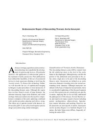
Pathogenesis : empyema phases
Pathogenesis : empyema phases
Pathogenesis : empyema phases

Create successful ePaper yourself
Turn your PDF publications into a flip-book with our unique Google optimized e-Paper software.
Parapneumonic pleural effusions• Uncomplicated parapneumonic effusion• Complicated parapneumonic effusion• Thoracic <strong>empyema</strong>.
<strong>Pathogenesis</strong> : <strong>empyema</strong> <strong>phases</strong>
<strong>Pathogenesis</strong> : <strong>empyema</strong> <strong>phases</strong>• Exudative phase:clear, straw-colored effusion(pH>7.3, glucose 60 mg%, LDH < 500 U/L )• Fibrinopurulent phase:effusion contains large numbers of bacteria and WBC,deposition of fibrin on both the visceral and parietal pleura( pH < 7.2 , glucose < 40mg%, LDH > 1000 U/L)• Organizing <strong>empyema</strong> phase : an <strong>empyema</strong> capsule containspus.Clinics Chest Med 2010;20:607-622
Etiology• Para-pneumonic 60-70 %- pneumonia- lung abscess- infected bronchiectasis• Post thoracic surgery 20 %- lung resection- esophagectomy- mediastinal surgery• Post traumatic 5-10 %Chapman SJ, Davies RJ Respirology. 2009;9:4-11.
Empyema :pathogens‣ Pneumococus‣ Streptococus‣ Staphylococus‣ Haemophilus influenzae‣ Gram-negative bacteria (pseudomonas, klebsiella,enterobacteriace)‣ Anaerobic bacteria‣ Mixed bacterial flora‣ Tuberculosis‣ ActinomycosisHeffner JE. Clinics Chest Med 1999;20:607-622
Empyema : Diagnosis• History & Physical exam• Pleural fluid analysis• ImagingClinics Chest Med 2010;20:607-622
Clinical manifestations• Aerobic bacterial pneumonia– An acute febrile illness with chest pain, sputumproduction, and leukocytosis.– Sepsis/ septic shock– Poor prognosisClinics Chest Med 2010;20:607-622
Clinical manifestations• Anaerobic bacterial infection– Usually presents with subacute illness.– symptoms persisting for more than 7 days.– 60% of patients have weight loss.– Poor oral hygiene– Factors predisposing to recurrent aspiration.Clinics Chest Med 2010;20:607-622
Clinical manifestations• Tuberculosis & fungus– Chronic onset, illness.– Symptoms persisting for several months.– Usually loculations/septation– Necessitans is commonClinics Chest Med 2010;20:607-622
Diagnostic imaging chest X-ray pleural ultrasonography computed tomography diagnostic videothoracoscopy diagnostic thoracotomyClinics Chest Med 2010;20:607-622
Chest x-rays• PA and lateral decubitus• Adult studies sensitivity 67% and specificity 70%• PA at least 400 ml fluid vs. 50 ml lateral decubitus• Assess for loculationsHeffner JE. Clinics Chest Med 2012;20:7-22
Ultrasound• Classification– Stage 1: anechoic fluid– Stage 2: loculations– Stage 3: solid peel• Guide placement of intercostal drainHogan MJ, Cooley BD. Paediatric Resp Reviews 2008;9:77-84
Ultrasound• Size of effusion• Differentiate consolidation from <strong>empyema</strong>• Unreliable predictor of disease severity
Rediology. 2010;10:12-35.
Rediology. 2010;10:12-35.
Rediology. 2010;10:12-35.
CT scan• Anatomical– Parenchymal lesions– Endobronchial lesions– Mediastinal lesions– Lung abscess
Rediology. 2010;10:12-35.
Rediology. 2010;10:12-35.
Rediology. 2010;10:12-35.
Rediology. 2010;10:12-35.
Pleural <strong>empyema</strong> - Videothoracoscopy
“ ขอให้ถือประโยชน์ส่วนตน เป็ นกิจที่สองประโยชน์ของเพื่อนมนุษย์ เป็ นกิจที่หนึ่งลาภ ทรัพย์ และเกียรติยศ จะตกแก่ท่านเองถ้าท่านทรงธรรมะแห่งอาชีพ ไว้ให้บริสุทธิ ์ ”พระบรมราชปณิธานของสมเด็จพระบรมราชชนก