PISA Evaluation of MR - Casecag.com
PISA Evaluation of MR - Casecag.com
PISA Evaluation of MR - Casecag.com

Create successful ePaper yourself
Turn your PDF publications into a flip-book with our unique Google optimized e-Paper software.
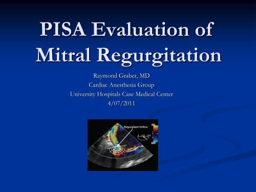
<strong>PISA</strong> <strong>Evaluation</strong> <strong>of</strong>Mitral RegurgitationRaymond Graber, MDCardiac Anesthesia GroupUniversity Hospitals Case Medical Center4/07/2011
Introduction• <strong>Evaluation</strong> <strong>of</strong> <strong>MR</strong>.• What is <strong>PISA</strong>?• Physiologic basis• Issues• How to do it with GE Vivid 7.
Zoghbi WA, et al. Re<strong>com</strong>mendations for <strong>Evaluation</strong> <strong>of</strong> the Severity <strong>of</strong> Native Valvular Regurgitation withTwo-dimensional and Doppler Echocardiography. J Am Soc Echocardiogr 2003;16:777-802.In the OR, typical methods for <strong>MR</strong> evaluation include qualitativemeasures such as color jet area, chamber size, and pulmonary vein flow.Quantitative measures include vena contracta and measures <strong>of</strong> EROAand regurgitant volume.
Regurgitant ColorDoppler FlowPattern:• Distal jet.• Vena contracta• Proximal flowconvergence
Jet Area IssuesEvaluating color jet area would seem to be an easy thing to do, butthere are multiple caveats:• Assumes that regurgitant velocities correlates with regurgitantvolumes.• Is one point in time, whereas volume includes duration <strong>of</strong> flow.• Behavior <strong>of</strong> jet depends on receiving chamber – can beconstrained by side <strong>of</strong> atrium• Jets can course outside <strong>of</strong> ultrasound plane.• Depend on machine settings.• Depends on driving pressure across the mitral valve.
Phenylephrine used to elevate BPsys by 40-50mm Hg in group A.Gain settings changed.
What is <strong>PISA</strong>?• <strong>PISA</strong> = Proximal Isovelocity Surface Area• A concept that can be used to help determinesize <strong>of</strong> a regurgitant or stenotic orifice.• Based on flow dynamics, use <strong>of</strong> aliasing andcontinuity principle.
Flow Dynamics 1• When fluid is forced from a chamber thru anorifice, the fluid accelerates towards the orifice,and velocity is greatest at the narrowest point <strong>of</strong>the orifice.
• With color Doppler, asflow accelerates towardsorifice – at some point,velocity may exceed thealiasing velocity, andcolor will reverse fromred to blue. At thishemisphere, the velocityis thus known (it equalsthe aliasing velocity).Use <strong>of</strong> Aliasing
Continuity Principle• Because <strong>of</strong> the conservation <strong>of</strong> mass principle, flowrate must remain constant along the length <strong>of</strong> a conduit(assuming the absence <strong>of</strong> any leaks or additional input)A 1 x V 1 = A 2 x V 2
Applying this to <strong>MR</strong>:Regurgitant Orifice Flow = Flow at hemisphere<strong>of</strong> color changeEROA x V orifice = A hemisphere V hemisphereV orifice =V max (by CWD)A hemisphere = 2πr 2V hemisphere = V AliasingEROA x V max = 2 πr 2 x V Aliasing
Assumptions:• Assumes accurate Doppler measurement <strong>of</strong> regurgitantvelocity.• Assumes regurgitant orifice is circular.• Assumes that the orifice is on a planar surface, and thatthe in<strong>com</strong>ing flow forms a <strong>com</strong>plete hemisphere.• Assumes single orifice.• Depends on accurate measurement <strong>of</strong> radius.• Assumes that regurgitant orifice is constant in size.
Can we measure regurgitant velocityaccurately with Doppler?
Central jet: goodCWD curve.
Eccentric jet:suboptimalCWD curve.
Are Regurgitant Orifices Circular?This 3DTEE showsa regurgitantorifice that iselongated.
Rifkin and Sharma. Alternative Isovelocity Surface Model. J AC C : Cardiovascular Imaging 2010Other mathematicalmodels beingdeveloped for <strong>PISA</strong> innon-hemisphericalorifices.
Is the orifice on a planar surface?Eccentric jets frequently don’t have a planar surface.A correction factor (a/180) can be used.(Multiply this times the calculated EROA and RV)
Is There A Single Orifice?Frequently not!
Getting an AccurateMeasurement <strong>of</strong> Radius:• Adjusting the aliasingvelocity by shifting thecolor Doppler baselinetowards the direction<strong>of</strong> flow increases themeasured radius andimproves accuracy.
Is the Regurgitant OrificeConstant In Size?Orifices can changein size over time,especially withprolapse. It isre<strong>com</strong>mended to usethe <strong>PISA</strong> radius thatcorresponds to thetime <strong>of</strong> peakregurgitant velocity.
(J Am Coll Cardiol Img 2010; 3:235– 43)How good are these measurements? Can we agree upon them?
• In the ideal situationclinicians would look at ameasure, and all would agreethat the <strong>MR</strong> was severe, oragree that it was not. Yet inthis study, was not the case.For example, looking at Jetsize in patient 1, 39% rated itas severe, and 61% rated it asnot severe!
Cardiologist Agreement• The authors defined“substantial agreement” as>80% <strong>of</strong> cardiologists were inagreement with a finding for aspecific patient.• In what % <strong>of</strong> images was theresubstantial agreement? :• Jet Area: 44%• Vena Contracta: 44%• EROA: 38%
Reasons for Variability <strong>of</strong> Assessments• > 30% variation <strong>of</strong> <strong>PISA</strong> radius during thecourse <strong>of</strong> the <strong>MR</strong> jet: 44%• > 30% variation <strong>of</strong> VC width during thecourse <strong>of</strong> the <strong>MR</strong> jet : 44%• Effective <strong>MR</strong> orifice identifiable: 44%• Eccentric were much harder to evaluatequantitativly then central jets
Their Conclusions:• The VC and <strong>PISA</strong> measurements for distinction <strong>of</strong>severe versus non-severe <strong>MR</strong> are only modestly reliableand associated with suboptimal interobserveragreement.• The presence <strong>of</strong> an identifiable effective regurgitantorifice improves reproducibility <strong>of</strong> VC and a centralregurgitant jet predicts substantial agreement amongmultiple observers <strong>of</strong> <strong>PISA</strong> assessment.
Example: <strong>PISA</strong> Step By STEP
• Use MELAX view, obtain image <strong>of</strong> <strong>MR</strong> jet thatincludes <strong>PISA</strong> shell, flow convergence and venacontracta. Also obtain image <strong>of</strong> CWD thru mitralvalve, lining up with the regurgitant jet. Note thetiming <strong>of</strong> the peak velocity <strong>of</strong> <strong>MR</strong> jet.
• Bring up the MELAX view, and scroll to image thatshows <strong>PISA</strong> shell. Ideally, this should correspond tothe time <strong>of</strong> peak <strong>MR</strong> velocity.
• Zoom to magnify the image, adjust the colorDoppler baseline towards jet direction to achievean aliasing velocity <strong>of</strong> .30-.40 m/sec.
In the Measurement menu – find <strong>PISA</strong> <strong>MR</strong> underthe <strong>PISA</strong> folder.
Turn color <strong>of</strong>f to visualize ventricular side <strong>of</strong>orifice center.
Place cursor at this location to start radius measurement.
Turn color back on, and draw radius to <strong>PISA</strong> shell.
• Bring up CWD <strong>of</strong> mitral regurg jet. Find <strong>PISA</strong> <strong>MR</strong>under the <strong>PISA</strong> folder.
• Trace <strong>MR</strong> curve. ERO and RV are calculated bythe machine.
EROA = 2 πr 2 (V Aliasing / V max )EROA = 2 (3.14)(.8 cm) 2 (.29 m/sec)/(4.35 m/sec)EROA = .269 cm 2RV = EROA x TVI mr = .269 cm 2 x 137.9 cmRV = 37.09 cm 3Note “rounding”by the machine!
Putting it All Together:• Jet Area: 7 cm 2• Vena Contracta: .3 cm• EROA: .269 cm 2• RV: 37 ml
• Jet Area: 7 cm 2• Vena Contracta: .3 cm• EROA: .269 cm 2• RV: 37 mlModerate <strong>MR</strong> (low end)
Notes:• If you are doing calculations by hand, make sureyou convert units as needed.Some machines usecm/sec, others usem/sec.GE Vivid 7: m/sec
Notes:• Use angle correction factor as needed:• Generally don’t need to correct central jets, butbe<strong>com</strong>es an issue with eccentric jets and also whenused in mitral stenosis.• Multiply machine calculated EROA and RV by(a/180) to get corrected numbers.• If doing your own calculations, use this factor onlyin the EROA calculation. Then this correctedEROA x TVI mr = corrected RV.
Modifications:Derivation <strong>of</strong> Angle Correction:• If hemisphere is not <strong>com</strong>plete because <strong>of</strong>impingement by wall or leaflet (leading to afunnel constraining flow), area <strong>of</strong> hemisphere ismodified:A hemisphere = 2πr 2 (a/180)Where a is the proximal flow convergence angleThus: EROA = 2 πr 2 (V Aliasing / V max ) (a/180)
Modifications:No CWD Measures• If you can’t get a good CWD waveform, here is amethod to estimate EROA.• Set aliasing velocity to 40 cm/sec.• Assume Vmax = 500 cm/sec (This works when LVsystolic pressure is greater than left atrial pressure byabout 100 mm Hg)EROA x V max = 2 πr 2 x V AliasingEROA = 2(3.14)r 2 (40)/(500)EROA = r 2 /2
Conclusions:• We discussed basis <strong>of</strong> <strong>PISA</strong>calculations.• Discussed pitfalls <strong>of</strong> <strong>PISA</strong>.• Showed an example how to do<strong>PISA</strong> measurements with theGE Vivid 7.
• In the end, one must integrate all the qualitativeand quantitative measures to <strong>com</strong>e up with agood <strong>MR</strong> severity assessment.“It seems that we have a lot <strong>of</strong> room forimprovement, and that current echocardiographicgrading <strong>of</strong> <strong>MR</strong> severity is more art than science.”Paul A. Grayburn, MD, Paul Bhella, MD 2010
References:• Zoghbi WA, et al. Re<strong>com</strong>mendations for <strong>Evaluation</strong> <strong>of</strong> the Severity <strong>of</strong> NativeValvular Regurgitation with Two-dimensional and Doppler Echocardiography. J AmSoc Echocardiogr 2003;16:777-802.• Lambert S. Proximal Isovelocity Surface Area Should Be Routinely Measured inEvaluating Mitral Regurgitation: A Core Review. Anesth Analg 2007;105:940 –3• Shanewise JS. PRO: Proximal Isovelocity Surface Area Should Be Routinely Measuredin Evaluating Mitral Regurgitation. Anesth Analg 2007;105:947-8• Savage RM, Konstadt S. CON: Proximal Isovelocity Surface Area Should Not BeMeasured Routinely in All Patients with Mitral Regurgitation Anesth Analg2007;105:944-6• Paul A. Grayburn, MD, Paul Bhella, MD Grading Severity <strong>of</strong> Mitral RegurgitationbyEchocardiography: Science or Art? JACC: Cardiovascular Imaging 2010. 3: 244-246• Biner S, Rafique A, Rafii F, et al. Reproducibility <strong>of</strong> proximal isovelocity surface area,vena contracta, and regurgitant jet area for assessment <strong>of</strong> mitral regurgitation severity. JAm Coll Cardiol Img 2010;3:235–43.