Chapter 7 Water Relations - Bryophyte Ecology
Chapter 7 Water Relations - Bryophyte Ecology
Chapter 7 Water Relations - Bryophyte Ecology

You also want an ePaper? Increase the reach of your titles
YUMPU automatically turns print PDFs into web optimized ePapers that Google loves.
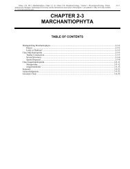
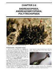
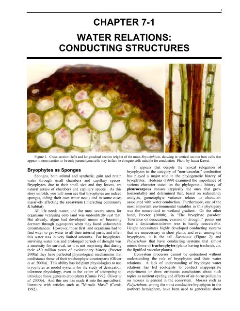
1CHAPTER 7-1WATER RELATIONS:CONDUCTING STRUCTURESFigure 1. Cross section (left) and longitudinal section (right) of the moss Bryoxiphium, showing in vertical section how cells thatappear in cross section to be only parenchyma cells may in fact be elongate cells suitable for conduction. Photo by Isawa Kawai.<strong>Bryophyte</strong>s as SpongesSponges, both animal and synthetic, gain and retainwater through small chambers and capillary spaces.<strong>Bryophyte</strong>s, due to their small size and tiny leaves, arenatural arrays of chambers and capillary spaces. As thisstory unfolds, you will soon see that bryophytes are indeedsponges, aiding their own water needs and in some casesmassively affecting the ecosystem (interacting community& habitat).All life needs water, and the most severe stress fororganisms venturing onto land was undoubtedly just that.But already, algae had developed means of becomingdormant through zygospores when they faced unfavorablecircumstances. However, those first land organisms had tofind ways to get water to all their internal parts, and oftenthis water was in very limited amounts. For bryophytes,surviving water loss and prolonged periods of drought wasa necessity for survival, so it is not surprising that duringtheir 450 million years of evolutionary history (Proctor2000a) they have perfected physiological mechanisms thatoutdistance those of their tracheophyte counterparts (Oliveret al. 2000a). This ability has led plant physiologists to usebryophytes as model systems for the study of desiccationtolerance physiology, even to the extent of attempting tointroduce those genes to crop plants (Comis 1992; Oliver etal. 2000b). And this use has made it into the agriculturalliterature with articles such as "Miracle Moss" (Comis1992).It appears that despite the typical relegation ofbryophytes to the category of "non-vascular," conductionhas played a major role in the phylogenetic history ofbryophytes. Hedenäs (1999) examined the importance ofvarious character states on the phylogenetic history ofpleurocarpous mosses (typically the ones that growhorizontally) and determined that, based on redundancyanalysis, gametophyte variance relates to charactersassociated with water conduction. Furthermore, one of themost important environmental variables in this phylogenywas the nonwetland to wetland gradient. On the otherhand, Proctor (2000b), in "The bryophyte paradox:Tolerance of desiccation, evasion of drought," points outthat a desiccation-tolerant tree is hardly conceivable.Height necessitates highly developed conducting systemsthat are unnecessary in short plants, and even among thebryophytes, it is the tall Dawsonia (Figure 2) andPolytrichum that have conducting systems that almostmimic those of tracheophytes (plants having tracheids, i.e.the lignified vascular plants).Ecosystem processes cannot be understood withoutunderstanding the role of bryophytes and their waterrelations. A lack of understanding of bryophyte waterrelations has led ecologists to conduct inappropriateexperiments or draw erroneous conclusions about suchtopics as nutrient cycling and effects of air-borne pollutantson mosses in general in the ecosystem. Mosses such asPolytrichum, among the most conductive bryophytes in thenorthern hemisphere, have been used to generalize about
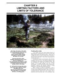
<strong>Chapter</strong> 7-1: <strong>Water</strong> <strong>Relations</strong>: Conducting Structures 3<strong>Bryophyte</strong>s are adapted to land but restricted in theirmorphology by a biochemical impasse, i.e. the inability tosynthesize lignin (Niklas 1976). Because they lack lignin,they lack the tracheids and vessels of other plants, but haveproduced instead vascular strands with similar elongateshapes. Nevertheless, they are unable to support a largestructure or great mass because they lack the strengtheningability of lignin. Because of their importance in bothstructure and physiology, water relations seem anappropriate place to start in our consideration of the limitsimposed on bryophytes, for without that understanding, wecannot understand their other limitations, nor can we fullyevaluate their ecological relationships.Conducting Structure<strong>Bryophyte</strong>s have two paths of water movement, oftenboth in the same plant: internal through a central cylinder(endohydric) and external along the surface of the leafyplant (ectohydric) (Buch et al. 1938). Thallose liverworts,Polytrichaceae, and Mniaceae represent the endohydricgroup (Buch 1945, 1947; Proctor 2000b). Ectohydricmosses typically maintain a constant internal water contentby absorbing water from the external capillary spaces asneeded (Proctor 2000b). These two modes each requiretheir own structural adaptations. Lacking lignin, xylem isnot possible. Furthermore, in the lignified vascular plants,it is the sporophyte generation that carries out organizedinternal conduction, and the gametophyte, with rareexception, does not. By contrast, in bryophytes it is theleafy gametophyte that must obtain and conduct water andnutrients about the plant, although conduction also occursin the moss sporophyte.Although the Anthocerotophyta are considered bysome to be reduced from more advanced plants, waterconductingtissue is unknown in this phylum (Ligrone et al.2000), although Hébant (1977) reported the presence ofcells resembling phloem sieve cells (leptoids?) inDendroceros. Few liverworts (Marchantiophyta) havespecialized conducting tissues in their gametophytes, andnone have them in the sporophyte. Nonetheless,conducting strands have been known since 1901 in thethallose liverwort Pallavicinia lyellii (Figure 4; Tansley &Chick 1901). As in mosses, Pallavicinia strands (Figure 5)closely resemble tracheids, with long cells, tapering ends,and obliquely oriented pits, and they likewise are dead atmaturity.Figure 5. Pallavicinia lyellii cross section of thallus.Drawing from Hébant (1977).Unlike the liverworts, mosses can have conductingcells in both generations (Ligrone et al. 2000). In the mossTakakia (a primitive moss once thought to be a liverwort;Figure 6) and some liverworts of Calobryales andPallaviciniaceae (Metzgeriales), there exist waterconductingcells with perforated walls derived fromplasmodesmatal pores (Ligrone et al. 2000), but these donot seem to be organized into a distinctive central strand(group of elongate cells forming central axis of stems andthalli of some bryophytes, usually thin-walled and oftencolored; Figure 11).Figure 4. Pallavicinia lyellii thallus. Photo by Jan-PeterFrahm.Figure 6. Cross section of stem of Takakia lepidozioides.Photo with permission from www.botany.ubc.ca/bryophyte/LAB8.htm.Kawai (1991a) describes the moss stem as having abasic structure much like that of tracheophytes (lignifiedvascular plants) with an epidermis surrounding the cortex(Figure 11, top). This basic structure describes most of thepleurocarpous mosses that move internal substances mostlyhorizontally.Among the acrocarpous mosses, more complex stemscan have a leptome (= leptom), surrounding the hydromesheath, a sterome (= sterom), and a hydrome (also knownas hadrom and hydrom) (Figure 11). The leptome iscomposed of phloem-like cells called leptoids, which aredifficult to distinguish from the cortex cells in cross
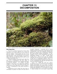
4 <strong>Chapter</strong> 7-1: <strong>Water</strong> <strong>Relations</strong>: Conducting Structuressection. However, in vertical section they resemble phloemsieve cells (Figure 12). Hydroids (Figure 12) and stereidsmake up the central strand (Zamski & Trachtenberg 1976)and are collectively called the hydrome or hydrom(Scheirer 1980) and sterome (Hébant 1977; sterom ofZamski & Trachtenberg 1976), respectively. Hydroids arewater-conducting cells similar to tracheids but lacking anyhorizontal connections (i.e. no pits). Stereids are elongate,thick-walled, slender, and fiber-like cells; they can occur inthe leaf costa (midrib-like strand; Figure 7) and stem.True perforation plates (end walls of vessels) havenot been found in Polytrichaceae (Frey & Richter 1982) ormost other mosses (Hébant 1973). Consequently, Frey andRichter (1982) set out to discover them in mosses. In thedendroid moss Canalohypopterygium tamariscinum, theyfound structures resembling perforation plates of Ephedra,although they were not numerous and were restricted inlocation to branching areas. Perhaps this type of vascularstructure permits them to be dendroid (Figure 9), lackingthe close structure needed for capillary action along thestem. Smith (1964) had already demonstrated perforationsin the conducting elements of the liverwort Symphogynacircinata. Furthermore, pits are known, particularly in endwalls, from Haplomitrium [considered to be basal to leafyliverworts (Crandall-Stotler & Stotler 2000)] and Takakia(now classified as a primitive moss in the Takakiopsida), asconfirmed by electron microscope.Figure 7. Cross section of moss leaf blade showingarrangement of broad portion (lamina), costa, and supportingstereids. Large cells in costa serve for conduction. Photo byJanice Glime.The elongated, water-conducting hydroids typicallyoccur in groups of 2-3 in bryophyte stems (Hébant 1970);they are similar to tracheids, but lack lignin and secondarywall thickenings (Taylor 1988). Consequently, hydroidsare usually thin-walled (Zamski & Trachtenberg 1976) andlack the helices and other thickenings typical of tracheids.Hydroids senesce at maturity and become dead, emptycells, like those of xylem, with slanted end walls that abuton the end wall of the next cell, as in tracheids (Richardson1981). Hydroids of Bryopsida typically lack perforations.Scheirer (1973) used Dendroligotrichum (Polytrichopsida)to demonstrate that hydrolysis leaves behind only celluloseremains of the primary walls of end walls of hydroids.Subsequent examination by electron-dense crystals ofPrussian blue on the end walls in Polytrichum communesuggests that these end walls are highly permeable (Figure8), but that substances are unable to move through thelateral walls (Scheirer & Goldklang 1977).Figure 8. Cross section of Polytrichum stem stained withaniline blue to show thin areas in end walls of cells, but these arenot in the hydrom. Photo by Isawa Kawai.Figure 9. Hypopterygium novae-seelandiae showingdendroid growth habit. Photo by Janice Glime.Although hydroids do not seem to contain true lignin,as do tracheophyte xylem cells, they do contain apolyphenolic cell wall component that functions similarlyto lignin. This compound protects the wall from hydrolyticattack and aids in internal transport of water. InRhacocarpus purpurascens, Edelman et al. (1998) foundwalls composed of "mainly lignin, hemicellulose (Hbondedto cellulose in plant cell walls), and cellulose in aratio of ca. 9:8:5." Although the resonance spectrumindicated various characteristics typical of lignin, somespecific peaks associated with known lignin compoundswere missing. Thus the question remains, is this truelignin?True leaf traces (conducting cells connecting the leafcosta to the hydrom; Figure 11) exist in somePolytrichaceae, whereas in the Mniaceae they are false leaftraces that do not connect with the leaf costa (Figure 11).The rhizome (underground, horizontal stem connectingupright plants), on the other hand, has hypodermal andradial strands but lacks leaf traces and a sterome. Thehypodermis consists of one to several layers of distinctcells just beneath the epidermis and may be thick-walled orcolored (Figure 10).Leptoids (Figure 12) are very similar to phloem sievecells, and in fact, Behnke (1975) calls them just that.Taylor (1988) considers that in some cases they are nearlyidentical to protophloem cells of certain tracheophytes.They, along with parenchyma cells, comprise the leptome(Hébant 1970, 1974; Behnke 1975; Figure 11). We know
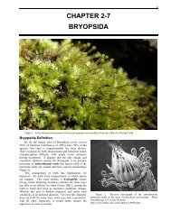
<strong>Chapter</strong> 7-1: <strong>Water</strong> <strong>Relations</strong>: Conducting Structures 5that they are typical in the Polytrichaceae, but have alsobeen found in Sphagnum, Hookeriaceae, Neckeraceae, andOrthotrichaceae (Ligrone & Duckett 1994, 1998; Duckett& Ligrone 2003). It is likely that they are much morecommon than we realize because in cross section withoutstain they appear no different from the unspecializedparenchyma cells. The leptoids are distinct in verticalsection by their elongate shape and slightly oblique endwalls (Behnke 1975). At maturity, the nucleus degenerates,as in phloem sieve cells (Richardson 1981). InPolytrichum, the leptoids are not connected end-to-end bysieve plates or pores as in tracheophytes, but by numerousplasmodesmata. However, Cortella and coworkers (1994)considered the thin areas of central strand parenchyma cellsto be primary pit fields in Hookeria lucens stems andsuggest that these cells have a conducting function.tracheophyte counterparts, they move carbohydrates andother substances away from the apex.Figure 10. Polytrichum stem cross section showinghypodermis. Photo by Isawa Kawai.Even the development of leptoids seems similar to thatof phloem sieve cells. During leptoid maturation inPolytrichaceae, ribosomes (centers of protein synthesis)disintegrate and nuclei become smaller and inactive,although they do not dissolve completely as intracheophytes; mitochondria persist. The parenchyma cellscontain starch-storing chloroplasts. As in theirFigure 11. Stem cross sections illustrating lack of centralcylinder and well-developed cylinders. Top: Trichodoncylindricus. Photo by Janice Glime. Center: Plagiomnium.Photo by Janice Glime. Bottom: Polytrichum. Photo courtesy ofIsawa Kawai.Figure 12. Cross section of Polytrichum juniperinum and longitudinal section of Atrichum undulatum stem to illustrate parts ofcentral strand (leptoids and hydroids) and stem structures. Drawings by Margaret Minahan, modified from Hébant (1977).
6 <strong>Chapter</strong> 7-1: <strong>Water</strong> <strong>Relations</strong>: Conducting StructuresThe hydroids and leptoids present interestingevolutionary implications, since it appears that they areprimitive characters that are lost in more advancedbryophyte taxa (Hébant 1970; Behnke 1975). Unlike mosttracheophytes, the mosses retain conducting cells in bothgenerations, but the haploid generation is the first to loseleptoids evolutionarily, as in Funaria (Behnke 1975).Furthermore, leptoids of setae, unlike those oftracheophytes, show less differentiation than in theirgametophytic counterparts. In the seta they are notintermixed with specialized parenchyma cells andapparently lack enlarged plasmodesmata in their end walls,as seen in gametophytes of some taxa (Hébant 1974).Except in the setae of a few species (Hébant 1974),leptoids have not been found in the arthrodontous mosses(considered more advanced) and are unknown inliverworts. To add interest to the picture, the leptoids arepresent in forms that are transitional between theparenchyma cells and the fully differentiated leptoid cells(Hébant 1974).Summary<strong>Bryophyte</strong>s often are not the nonvascular plants weonce thought them to be. They often possess hydroidsthat conduct water and leptoids that conduct sugars,arranged as in tracheophytes, with the waterconductingcells surrounded by the sugar-conductingcells. They usually have a thin cuticle, but it seems tolack wax in most cases. Rhizoids, although anchoringthe plants as do roots, typically do not serve inobtaining water.AcknowledgmentsThis chapter has benefitted from the help of BethScafone and Medora Burke-Scoll, who read the manuscriptfor clarity. Linda Luster checked the literature citations,proofread, and made glossary suggestions from alayperson's perspective.Literature CitedAnderson, L. E. and Bourdeau, P. F. 1955. <strong>Water</strong> relations intwo species of terrestrial mosses. <strong>Ecology</strong> 36: 206-212.Behnke, H.-D. 1975. Phloem tissue and sieve elements in algae,mosses and ferns. In: Aronoff, S., Dainty, J., Gorham, P.R., Srivastava, L. M., and Swanson, C. A. (eds.). PhloemTransport. Plenum Press, N. Y., pp. 187-210.Buch, H. 1945. Über die Wasser- und Mineralstoffversorgungder Moose (Part 1). Soc. Sci. Fenn., Comment. Biol. 9(16):1-44.Buch, H. 1947. Über die Wasser- und Mineralstoffversorgungder Moose. (Part 2). Soc. Sci. Fenn., Comment. Biol. 9(20):1-61.Buch, H., Evans, A. W., and Verdoorn, F. 1938. A preliminarycheck list of the Hepaticae of Europe and America (North ofMexico). Ann. Bryol. 10: 3-8.Comis, D. 1992. Miracle moss: Add water and watch it grow.Agric. Res. 40(6): 10-11.Cortella, A., Ron, E., Estébanez, B., and Alfayate, C. 1994. Onthe occurrence of primary pit field cells in the caulidia ofHookeria lucens (Hedw.) Sm. (Bryopsida, Bryophyta). J.Hattori Bot. Lab. 77: 287-294.Crandall-Stotler, B. and Stotler, R. E. 2000. Morphology andclassification of the Marchantiophyta. In: Shaw, A. J. andGoffinet, B. (eds.). <strong>Bryophyte</strong> Biology, CambridgeUniversity Press, Cambridge, UK, pp. 21-70.Duckett, J. G. and Ligrone, R. 2003. What we couldn't havedone if we'd stayed in Europe: Selection and serendipity inthe Southern Hemisphere!. Bull. Brit. Bryol. Soc. 80: 19-21.Edelmann, H. G., Neinhuis, C., Jarvis, M., Evans, B., Fischer, E.,and Barthlott, W. 1998. Ultrastructure and chemistry of thecell wall of the moss Rhacocarpus purpurascens(Rhacocarpaceae): A puzzling architecture among plants.Planta 206: 315-321.Frey, W. and Richter, U. 1982. Perforierte Hydroiden beiLaubmoosen? J. Hattori Bot. Lab. 51: 51-60.Gray, J. 1985. The microfossil record of early land plants:Advances in understanding of early terrestrialization, 1970-1984. Phil. Trans. Roy. Soc. Lond. B 309: 167-195.Hébant, C. 1970. A new look at the conducting tissues ofmosses (Bryopsida): Their structure, distribution andsignificance. Phytomorphology 20: 390-410.Hébant, C. 1973. Diversity of structure of the water-conductingelements in liverworts and mosses. J. Hattori Bot. Lab. 37:229-234.Hébant, C. 1974. The phloem (leptome) of bryophytes. In:Aronoff, S., Dainty, J., Gorham, P. R., Srivastava, L. M.,and Swanson, C. A. (eds.). Phloem Transport. PlenumPress, N. Y., pp. 211-215.Hébant, C. 1977. The Conducting Tissues of <strong>Bryophyte</strong>s. J.Cramer, Lehre, Germany, 157 pp. + 80 Plates.Hedenäs, L. 1999. How important is phylogenetic history inexplaining character states in pleurocarpous mosses? Can. J.Bot. 77: 1723-1743.Kawai, I. 1971a. Systematic studies on the conducting tissue ofthe gametophyte in Musci (2). On the affinity regarding theinner structure of the stem in some species of Dicranaceae,Bartamiaceae (sic), Entodontaceae, and Fissidentaceae.Ann. Rept. Bot. Gard., Fac. Sci. Kanazawa Univ. 4: 18-40.Kawai, I. 1971b. Systematic studies on the conducting tissue ofthe gametophyte in Musci (3). On the affinity regarding theinner structure of the stem in some species of Thuidiaceae.Sci. Rept. Kanazawa Univ. 16(1): 21-60.Kawai, I. 1971c. Systematic studies on the conducting tissue ofthe gametophyte in Musci (4). On the affinity regarding theinner structure of the stem in some species of Mniaceae.Sci. Rept. Kanazawa Univ. 16(2): 83-111.Kawai, I. 1976. Systematic studies on the conducting tissue ofthe gametophyte in Musci (6). On the essential coordinationamong the anatomical characteristics of the stem in somespecies of Hypnaceae. Sci. Rept. Kanazawa Univ. 21(1):47-124.Kawai, I. 1977a. Die systematische Forschung auf Grund derZellteilungsweise für die <strong>Bryophyte</strong>n II. DieZellteilungsweisen der Gametophyten in derLebensgeschichte (1). Climacium. Sci. Rept. KanazawaUniv. 22: 45-90.Kawai, I. 1977b. Systematic studies on the conducting tissue ofthe gametophyte in Musci (7). On the essential coordinationamong the anatomical characteristics of the stems in thesome species of Isobryales. Sci. Rept. Kanazawa Univ.22(2): 197-305.Kawai, I. 1978. Systematic studies on the conducting tissue ofthe gametophyte in Musci (8). On the essential coordinationamong the anatomical characteristics of the stems in somespecies of Amblystegiaceae. Sci. Rept. Kanazawa Univ.23(2): 93-117.Kawai, I. 1979. Systematic studies on the conducting tissue ofthe gametophyte in Musci (9). On regularity among
<strong>Chapter</strong> 7-1: <strong>Water</strong> <strong>Relations</strong>: Conducting Structures 7anatomical characteristics of stems in some species ofDicranaceae. Sci. Rept. Kanazawa Univ. 24(1): 13-43.Kawai, I. 1980a. Anatomical characteristics of stems in somespecies of Dicranaceae. Proc. Bryol. Soc. Japan 2: 126.Kawai, I. 1980b. Systematic studies on the conducting tissue ofthe gametophyte in Musci (11). Anatomical characteristicsof stems in some species of Leucobryaceae. Sci. Rept.Kanazawa Univ. 25(1): 31-42.Kawai, I. 1981. Systematic studies on the conducting tissue ofthe gametophyte in Musci (10). Organization of the stemand its origin. Hikobia (Suppl.) 1: 29-33.Kawai, I. 1982. Systematic studies on the conducting tissue ofthe gametophyte in Musci (12). Anatomical characteristicsof stems in some species of Bartramiaceae. Sci. Rept.Kanazawa Univ. 26: 31-50.Kawai, I. 1989. Systematic studies on the conducting tissues ofthe gamestophyte [sic] in Musci: XVI. <strong>Relations</strong>hipsbetween the anatomical characteristics of the stem and theclassification system. Asian J. Plant Sci. 1: 19-52.Kawai, I. 1991a. Systematic studies on the conducting tissue ofthe gametophyte in Musci (18). On the relationship betweenthe stem and the rhizome. Ann. Rept. Bot. Gard., Fac. Sci.Kanazawa Univ. 14: 17-25.Kawai, I. 1991b. Systematic studies on the conducting tissue ofthe gametophyte in Musci (19). <strong>Relations</strong>hips between thestem and seta in some species of Polytrichaceae, Bryaceae,Mniaceae, Bartramiaceae and Dicranaceae. Sci. Rept.Kanazawa Univ. 36(1): 1-19.Kawai, I. and Ikeda, K. 1970. Systematic studies on theconducting tissue of the gametophyte in Musci. (1) On theaffinity regarding the conducting tissue of the stem in somespecies of Polytrichaceae. Sci. Rept. Kanazawa Univ. 15(2):71-98.Kawai, I. and Ochi, H. 1987. Systematic studies on theconducting tissues of the gametophyte in Musci (15).<strong>Relations</strong>hips between the taxonomic system and anatomicalcharacteristics of stems in some species of Bryaceae. Sci.Rept. Kanazawa Univ. 32(1): 1-67.Kawai, I., Yoshitake, S., and Yamazaki, M. 1985. Systematicstudies on the conducting tissue of the gametophyte in Musci(13). Anatomy of the stem through analysis of pigmentdeposition in Polytrichum commune Hedw. and Pogonatumcontortum (Brid.) Lesq. Sci. Rept. Kanazawa Univ. 30: 47-53.Kawai, I., Yoshitake, S., and Yamamoto, E. 1986. Systematicstudies on the conducting tissue of the gametophyte in Musci(14). Anatomy of the stems of Rhizogonium, Mnium, andFissidens. Sci. Rept. Kanazawa Univ. 21(1,2): 31-42.Ligrone, R. and Duckett, J. G. 1994. Cytoplasmic polarity andendoplasmic microtubules associated with the nucleus andorganelles are ubiquitous features of food conducting cells inbryalean mosses (Bryophyta). New Phytol. 127: 601-614.Ligrone, R. and Duckett, J. G. 1998. The leafy stems ofSphagnum (Bryophyta) contain highly differentiatedpolarized cells with axial arrays of endoplasmicmicrotubules. New Phytol. 140: 567-579.Ligrone, R., Duckett, J. G., and Renzaglia, K. S. 2000.Conducting tissues and phyletic relationships of bryophytes.Philosoph. Trans. Roy. Soc. London B 355(1398): 795-813.Niklas, K. J. 1976. Plant evolution and the reciprocity model.Ann. Bot. 40: 1255-1264.Oliver, M. J., Tuba, Z., and Mishler, B. D. 2000a. The evolutionof vegetative desiccation tolerance in land plants. PlantEcol. 151: 85-100.Oliver, M. J., Velten, J., and Wood, A. J. 2000b. <strong>Bryophyte</strong>s asexperimental models for the study of environmental stresstolerance: Tortula ruralis and desiccation-tolerance inmosses. Plant Ecol. 151: 73-84.Proctor, M. C. F. 2000a. Mosses and alternative adaptation tolife on land. New Phytol. 148: 1-6.Proctor, M. C. F. 2000b. The bryophyte paradox: Tolerance ofdesiccation, evasion of drought. Plant Ecol. 151: 41-49.Puckett, K. J. 1988. <strong>Bryophyte</strong>s and lichens as monitors of metaldeposition. In: Nash, T. H. III. and Wirth, V. (eds.),Lichens, <strong>Bryophyte</strong>s and Air Quality. Biblioth. Lichenol.30: 231-267.Richardson, D. H. S. 1981. The Biology of Mosses. John Wiley& Sons, Inc., N. Y., 220 pp.Ron, E. and Kawai, I. 1990. Systematic studies on theconducting tissue of the gametophyte in Musci (17). On therelationships between the stem and the rhizome (forecast).Ann. Rept. Bot. Gard., Fac. Sci. Kanazawa Univ. 13: 15-18.Scheirer, D. C. 1973. Hydrolysed walls in the water-conductingcells of Dendroligotrichum (Bryophyta): Histochemistryand ultrastructure. Planta 115: 37-46.Scheirer, D. C. 1980. Differentiation of bryophyte conductingtissues: Structure and histochemistry. Bull. Torrey Bot.Club 107: 298-307.Scheirer, D. C. and Goldklang, I. J. 1977. Pathway of watermovement in hydroids of Polytrichum commune Hedw.(Bryopsida). Amer. J. Bot. 64: 1046-1047.Smith, J. L. 1964. <strong>Water</strong> conducting system of Symphogyna.Nature 202: 617.Tansley, A. G. and Chick, E. 1901. Notes on the conductingtissue system in the Bryophyta. Ann. Bot. 15: 1-39.Taylor, T. N. 1988. The origin of land plants: Some answers,more questions. Taxon 37: 805-833.Zamski, E. and Trachtenberg, S. 1976. <strong>Water</strong> movement throughhydroids of a moss gametophyte. Israel J. Bot. 25: 168-173.
8 <strong>Chapter</strong> 7-1: <strong>Water</strong> <strong>Relations</strong>: Conducting Structures