LABOULBENIALES - Agentschap voor Natuur en Bos
LABOULBENIALES - Agentschap voor Natuur en Bos
LABOULBENIALES - Agentschap voor Natuur en Bos

You also want an ePaper? Increase the reach of your titles
YUMPU automatically turns print PDFs into web optimized ePapers that Google loves.
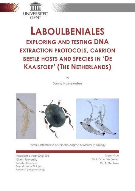
<strong>LABOULBENIALES</strong>EXPLORING AND TESTING DNAEXTRACTION PROTOCOLS, CARRIONBEETLE HOSTS AND SPECIES IN „DEKAAISTOEP‟ (THE NETHERLANDS)byDanny Haelewaters© February 2011, Faculty of Sci<strong>en</strong>ces – Research group MycologyAll rights reserved. This master thesis contains confid<strong>en</strong>tial information andconfid<strong>en</strong>tial research results that are property to the UG<strong>en</strong>t. The cont<strong>en</strong>ts ofthis master thesis may under no circumstances be made public, nor completeor partial, without the explicit and preceding permission of the UG<strong>en</strong>trepres<strong>en</strong>tative, i.e. the supervisor. The thesis may under no circumstances becopied or duplicated in any form, unless permission granted in writt<strong>en</strong> form.Any violation of the confid<strong>en</strong>tial nature of this thesis may impose irreparabledamage to the UG<strong>en</strong>t. In case of a dispute that may arise within the contextof this declaration, the Judicial Court of G<strong>en</strong>t only is compet<strong>en</strong>t to be notified.Pictures front page: (from left to right) Hesperomyces viresc<strong>en</strong>s from Harmoniaaxyridis (specim<strong>en</strong> 1c, thallus 1) collected in „De Kaaistoep‟ (Tilburg, TheNetherlands), new for the mycoflora of The Netherlands; picture by DannyHaelewaters (2010). Carrion beetle Saprinus semistriatus; picture by UdoSchmidt (2008). Laboulb<strong>en</strong>ia flagellata from Agonum assimile (specim<strong>en</strong> DH4,thalli G and H) collected at Schellandpolderdijk (Hing<strong>en</strong>e, Belgium); picture byDanny Haelewaters (2009).
PART II<strong>LABOULBENIALES</strong>: EXPLORING AND TESTINGDNA EXTRACTION PROTOCOLS311. INTRODUCTION 321.1. DIFFICULTIES FOR DNA EXTRACTION OF <strong>LABOULBENIALES</strong> 321.2. GENBANK 321.2.1. THE GENBANK SEQUENCE DATABASE 321.2.2. THE SEARCH FOR SEQUENCES OF <strong>LABOULBENIALES</strong>/LABOULBENIOMYCETES INGENBANK 322. MATERIALS & METHODS 342.1. FUNGUS, HOST AND ORIGIN 342.2. MORPHOLOGICAL PROTOCOL AND DNA EXTRACTION 342.2.1. INTRODUCTION 342.2.2. PROTOCOL I (BASED ON WEIR & BLACKWELL, 2001b) 342.2.3. PROTOCOL II (BASED ON WEIR & BLACKWELL, 2001a) 352.2.4. PROTOCOL III: PUREGENE KIT A 352.2.5. PROTOCOL IV: DNEASY EXTRACTION KIT 352.2.6. PROTOCOL V: DIRECT PCR 362.3. PCR AMPLIFICATION 362.3.1. ITS 362.3.2. POST PCR 372.4. SEQUENCING AND SEQUENCE ANALYSIS 372.4.1 SEQUENCING 372.4.2 SEQUENCE ANALYSIS 373. RESULTS 393.1. DNA EXTRACTION 393.2. AMPLIFICATION 393.2.1. PRIMER PAIR ITS5/ITS4-A 393.2.2. PRIMER PAIR ITS5/ITS2 404. DISCUSSION 415. CONCLUSION AND SUGGESTIONS 44PART III<strong>LABOULBENIALES</strong> OF CARRION BEETLES:A PRELIMINARY STUDY451. INTRODUCTION 461.1. FORENSIC ENTOMOLOGY 461.2. ANIMAL CADAVERS 461.2.1. SOURCES OF BIODIVERSITY 461.2.2. THE „ROTTING‟ OF A CADAVER 461.2.2.1. The fresh stage 471.2.2.2. The bloat stage 471.2.2.3. The early decomposition 47P a g e | 2
PART IVPRELIMINARY CHECKLIST OF <strong>LABOULBENIALES</strong> IN„DE KAAISTOEP‟771. INTRODUCTION 781.1. NATURAL LANDSCAPE DE KAAISTOEP 781.1.1. DESCRIPTION OF THE SITE 781.1.2. BIODIVERSITY IN DE KAAISTOEP 781.1.2.1. Plants and fungi in De Kaaistoep 791.1.2.2. Animals in De Kaaistoep 791.2. <strong>LABOULBENIALES</strong> IN THE NETHERLANDS 791.2.1. CONTRIBUTIONS OF MIDDELHOEK 791.2.2. CONTRIBUTIONS OF BOEDIJN 812. MATERIALS & METHODS 822.1. HOSTS 822.1.1. COLLECTION OF HOSTS 822.1.1.1. Pitfall traps 822.1.1.2. Window traps 822.1.1.3. Bands and rings 822.1.1.4. Malaise trap 832.1.1.5. Light traps 832.1.2. IDENTIFICATION OF HOSTS 832.2. <strong>LABOULBENIALES</strong> 832.2.1. SCREENING FOR AND IMBEDDING OF <strong>LABOULBENIALES</strong> 832.2.2. IDENTIFICATION OF <strong>LABOULBENIALES</strong> 842.3. PRESENTING RESULTS 843. RESULTS 863.1. PARASITE-HOST LIST 863.2. HOST- PARASITE LIST 864. DESCRIPTION OF THE SPECIES AND DISCUSSIONS 874.1. LABOULBENIA CALATHI T. MAJEWSKI 874.2. LABOULBENIA EUBRADYCELLI HULDÉN 884.3. LABOULBENIA PEDICELLATA THAXT. 894.4. LABOULBENIA VULGARIS PEYR. 914.5. HAPLOMYCES TEXANUS THAXT. 924.6. HESPEROMYCES VIRESCENS THAXT. 934.7. RHACHOMYCES LASIOPHORUS (THAXT.) THAXT. 944.8. STICHOMYCES CONOSOMATIS THAXT. 954.9. STIGMATOMYCES MAJEWSKII H.L. DAINAT, MANIER & BALAZUC 965. CONCLUSION AND SUGGESTIONS 98PART VREFERENCES99P a g e | 4
ACKNOWLEDGEMENTSThe work pres<strong>en</strong>ted in this master thesis is the result of many years of biologicalinterest, not just the few that constituted my studies.I am very grateful to my supervisor, Annemieke Verbek<strong>en</strong>, for what she hastaught me throughout my studies. Her <strong>en</strong>thusiasm and interest for mycologyplayed a huge role in me choosing my study path. Her expertise andknowledge have taught me more than I could ever have expected.I would also like to thank my second supervisor, André De Kesel, for teachingme all about the fascinating Laboulb<strong>en</strong>iales. His passion has inspired me tofinish this work and ev<strong>en</strong> start new chapters.My thanks go out to Paul Van Wielink and <strong>Natuur</strong>museum Brabant (Tilburg) forgiving me the chance to work in a group of professional <strong>en</strong>tomologists. Theid<strong>en</strong>tification of carrion beetles would not have be<strong>en</strong> so pleasant at any otherplace. Special thanks for involving me in the project of „De Kaaistoep‟.I wish to thank Dirk Raes (<strong>Ag<strong>en</strong>tschap</strong> <strong>voor</strong> <strong>Natuur</strong> <strong>en</strong> <strong>Bos</strong>) and Yves Braet andSofie Vanpoucke (National Institue for Criminalistics and Criminology) forinvolving me in the project Dood doet Lev<strong>en</strong> and for the possibility to work atthe laboratories of the National Institute for Criminalistics and Criminology.I have to thank Jorinde Nuytinck, Kobeke Van de Putte, Luc Crêvecoeur, MarcDelbol, Jan Willem van Zuijl<strong>en</strong>, Hans Huijbregts, Pedro Crous, Walter Rossi andMeredith Blackwell for sharing their expertise with me. More than once, onlysmall issues were bothering me. However, without their help I was naught.I cannot forget my fri<strong>en</strong>ds who have supported me throughout difficult times,who were always there wh<strong>en</strong> I needed a word. Special thanks to Stev<strong>en</strong>“Sev<strong>en</strong>less” Haes<strong>en</strong>donckx (doing my very best to see you becoming doctor),Evelyn Theuninck (we will meet again in Chantemerle-lès-Grignan), LiesbethVerlind<strong>en</strong> (for some Mazurka distraction), Lindsay De Decker (loving you, in anasexual way, although… ^^), Frederik Byttebier (you gave me inspiration and –rec<strong>en</strong>tly – a new dream to live for), Annemieke Krammer (dreaming aboutcandles, reincarnation, and lots – LOTS – of paper), Merel Van D<strong>en</strong> Broucke(my best witch fri<strong>en</strong>d), Eveli<strong>en</strong> De Wilde (for giving me a home) and all fri<strong>en</strong>dsof the G<strong>en</strong>tse Biologische Kring (I will never forget you).I will always be indebted to my par<strong>en</strong>ts, who have supported me at all times.Thanks dad;-) And mum, I hope that – wherever you are – you can be proud.Your belief in me, ev<strong>en</strong> now, was the outstanding factor always helping me toget through.P a g e | 5
SAMENVATTINGINLEIDINGLaboulb<strong>en</strong>iales zijn obligaat ectoparasitaire ascomycet<strong>en</strong> die <strong>voor</strong>kom<strong>en</strong> opArthropoda, meestal insect<strong>en</strong>. Er zijn meer dan 2000 soort<strong>en</strong> beschrev<strong>en</strong> in141 g<strong>en</strong>era. Laboulb<strong>en</strong>iales hebb<strong>en</strong> ge<strong>en</strong> mycelium; ze vorm<strong>en</strong> kleine thalli,opgebouwd uit e<strong>en</strong> receptaculum met perithecia, aanhangsels <strong>en</strong> antheridia.Laboulb<strong>en</strong>iales zijn gastheerspecifiek. Het parasiter<strong>en</strong> van e<strong>en</strong> bepaaldegastheersoort is afhankelijk van zowel de eig<strong>en</strong>schapp<strong>en</strong> van het integum<strong>en</strong>t,het microklimaat ter hoogte van het oppervlak van de gastheer <strong>en</strong> debeschikbaarheid van nutriënt<strong>en</strong> als de eig<strong>en</strong> <strong>voor</strong>keur<strong>en</strong> <strong>voor</strong> e<strong>en</strong> bepaaldmilieu. Er zijn ge<strong>en</strong> vrijlev<strong>en</strong>de lev<strong>en</strong>sstadia. Enkel seksuele reproductie isgek<strong>en</strong>d. Aandacht wordt besteed aan de morfologie, de classificatie <strong>en</strong> deecologie, met bijzondere aandacht <strong>voor</strong> de verschill<strong>en</strong>de vorm<strong>en</strong> vanspecificiteit van Laboulb<strong>en</strong>iales.BESTUDEERDE ONDERWERPEN EN CONCLUSIESSpecies-gerelateerde taxonomische problem<strong>en</strong> bij Laboulb<strong>en</strong>iales kunn<strong>en</strong><strong>en</strong>kel sluit<strong>en</strong>d bestudeerd word<strong>en</strong> door het combiner<strong>en</strong> van klassiekemorfologische method<strong>en</strong> <strong>en</strong> moleculaire protocoll<strong>en</strong>. Vier DNAextractieprotocoll<strong>en</strong>werd<strong>en</strong> getest; WEIR & BLACKWELL (2001a) <strong>en</strong> WEIR &BLACKWELL (2001b) vormd<strong>en</strong> de basis <strong>voor</strong> twee protocoll<strong>en</strong>, de ander<strong>en</strong> war<strong>en</strong>gebaseerd op de Qiag<strong>en</strong> Pureg<strong>en</strong>e Kit A <strong>en</strong> de Qiag<strong>en</strong> Dneasy Plant Mini Kit.Bij het vijfde extractieprotocol werd<strong>en</strong> thalli zonder <strong>en</strong>ige modificatiegeamplificeerd.Er werd<strong>en</strong> ge<strong>en</strong> sequ<strong>en</strong>ties van Laboulb<strong>en</strong>iales gevond<strong>en</strong>; we kreg<strong>en</strong> <strong>en</strong>kel temak<strong>en</strong> met contaminant<strong>en</strong> uit verschill<strong>en</strong>de klass<strong>en</strong>. Één contaminant waszelfs e<strong>en</strong> basidiomyceet. Op basis van deze resultat<strong>en</strong> kan word<strong>en</strong>geconcludeerd dat de beschrijving<strong>en</strong> in de reeds vermelde refer<strong>en</strong>ties (WEIR &BLACKWELL 2001a, 2001b) volledig noch betrouwbaar zijn.Suggesties <strong>voor</strong> verder onderzoek word<strong>en</strong> gegev<strong>en</strong>.Gericht onderzoek naar Laboulb<strong>en</strong>iales op kadaverkevers werd tot op hed<strong>en</strong>nog niet verricht. In dit werk werd<strong>en</strong> in het Zoniënwoud (België) kevers(Coleoptera) bemonsterd in de directe omgeving van kadavers; dit gebeurdein het kader van het project Dood doet Lev<strong>en</strong> (<strong>Ag<strong>en</strong>tschap</strong> <strong>voor</strong> <strong>Natuur</strong> <strong>en</strong><strong>Bos</strong>). Additioneel materiaal werd bezorgd door het Nationaal Instituut <strong>voor</strong>Criminalistiek <strong>en</strong> Criminologie. Alle kevers werd<strong>en</strong> geïd<strong>en</strong>tificeerd <strong>en</strong>gescre<strong>en</strong>d op de aanwezigheid van Laboulb<strong>en</strong>iales.In deze studie werd<strong>en</strong> ge<strong>en</strong> Laboulb<strong>en</strong>iales gevond<strong>en</strong>. Mogelijkerwijs hebb<strong>en</strong>Laboulb<strong>en</strong>iales door hun eig<strong>en</strong> milieu<strong>voor</strong>keur<strong>en</strong> moeilijkhed<strong>en</strong> om zich opkadaverkevers te ontwikkel<strong>en</strong>. Kadaverkevers beschikk<strong>en</strong> namelijk oververschill<strong>en</strong>de soort<strong>en</strong> habitats.P a g e | 6
Voorlopig is Asaphomyces tubanticus (Middelh. & Boel<strong>en</strong>s) Scheloske de <strong>en</strong>igebek<strong>en</strong>de soort van Laboulb<strong>en</strong>iales die systematisch <strong>voor</strong>komt opkadaverkevers. Deze soort werd al waarg<strong>en</strong>om<strong>en</strong> in België (RAMMELOO, 1986;DE KESEL & RAMMELOO, 1992; DE KESEL, 1997). Rhadinomyces pallidus Thaxt. werdook reeds geobserveerd op kadaverkevers (MAJEWSKI, 2003), maar is nog nietbek<strong>en</strong>d in België.Suggesties <strong>voor</strong> verder onderzoek word<strong>en</strong> gegev<strong>en</strong>.In Nederland is er sinds de jar<strong>en</strong> ‟40 (BOEDIJN, 1923; MIDDELHOEK, 1941, 1942,1943a, 1943b, 1943c, 1943d, 1945, 1947a, 1947b, 1949) ge<strong>en</strong> onderzoek meerverricht naar Laboulb<strong>en</strong>iales. „De Kaaistoep‟ wordt beschouwd als e<strong>en</strong>hotspot van biodiversiteit in Nederland <strong>en</strong> le<strong>en</strong>t zich dan ook uitstek<strong>en</strong>d <strong>voor</strong>gericht onderzoek naar Laboulb<strong>en</strong>iales. In dit werk werd<strong>en</strong> insect<strong>en</strong> uit decollectie van het <strong>Natuur</strong>museum Brabant (Tilburg, Nederland), verzameld inDe Kaaistoep, gescre<strong>en</strong>d op de aanwezigheid van Laboulb<strong>en</strong>iales.Deze bijdrage resulteerde in neg<strong>en</strong> soort<strong>en</strong> Laboulb<strong>en</strong>iales, waarvan zessoort<strong>en</strong> nieuw zijn <strong>voor</strong> Nederland. Het gaat om Laboulb<strong>en</strong>ia calathi T.Majewski, Laboulb<strong>en</strong>ia eubradicelli Huldén, Hesperomyces viresc<strong>en</strong>s Thaxt.,Rhachomyces lasiophorus (Thaxt.) Thaxt., Stichomyces conosomatis Thaxt. <strong>en</strong>Stigmatomyces majewskii H.L. Dainat, Manier & Balazuc.We verwacht<strong>en</strong> dat er nog meer nieuwe soort<strong>en</strong> <strong>voor</strong> Nederland zull<strong>en</strong>word<strong>en</strong> ontdekt in De Kaaistoep, naarmate het onderzoek zich meer opLaboulb<strong>en</strong>iales zal richt<strong>en</strong>.P a g e | 7
PART IGENERAL INTRODUCTIONTHESIS OUTLINEAIMSP a g e | 8
1. <strong>LABOULBENIALES</strong>1.1. DEFINITION OF <strong>LABOULBENIALES</strong>Laboulb<strong>en</strong>iales include over 2000 species in 141 g<strong>en</strong>era (SANTAMARIA, 1998; KIRKet al., 2001; WEIR & BLACKWELL, 2005) of obligate, ectoparasitic fungi, which liveassociated with arthropods, mostly true insects. Laboulb<strong>en</strong>iales have nomycelium; their thalli are small and of determinate growth, bearing antheridiaand perithecia on a receptacle with app<strong>en</strong>dages (TAVARES, 1985). Only sexualstages are known. Laboulb<strong>en</strong>iales are host-specific (THAXTER, 1896; SCHELOSKE,1969; TAVARES, 1985; MAJEWSKI, 1994; DE KESEL, 1996, 1997).The knowledge on Laboulb<strong>en</strong>iales is rather rec<strong>en</strong>t and has increased slowly.Large parts of their biodiversity are still unknown and many questions stillunresolved.1.2. HISTORICAL BACKGROUNDIn the 1840s, two Fr<strong>en</strong>ch <strong>en</strong>tomologists, Joseph Alexandre Laboulbéne andAuguste Rouget, did the earliest observations on Laboulb<strong>en</strong>iales resulting in afirst publication on the Laboulb<strong>en</strong>iales (ROUGET, 1850, ref. in TAVARES, 1985). Atfirst, the structures on Brachinus (Coleoptera, Carabidae) were thought to besupernumerary ant<strong>en</strong>nal segm<strong>en</strong>ts. ROUGET (1850, ref. in TAVARES, 1985)suggested that they were living organisms. The recognition as fungi came in1852 and the g<strong>en</strong>us Laboulb<strong>en</strong>ia was raised within the Pyr<strong>en</strong>omycetes – anext<strong>en</strong>sive group of Ascomycetes (ROBIN, 1853). Later on, specim<strong>en</strong>s onwingless dipteran parasites of bats were still mistak<strong>en</strong>ly described asArthrorhynchus, an acanthocephalan worm, by KOLENATI (1857).MAYR (1853) thought that the „hairlike structures‟ on Nebria (Coleoptera,Carabidae) were chitinous. Suggesting that they were outgrowths of theinsect‟s integum<strong>en</strong>t, he noticed differ<strong>en</strong>ces betwe<strong>en</strong> those structures onyounger and older beetles. On older individuals, there was always a variableswelling on the hair, whereas no such swelling was pres<strong>en</strong>t on the hair ofyounger beetles.PEYRITSCH (1871) made ext<strong>en</strong>sive observations on the structure anddevelopm<strong>en</strong>t of the housefly Laboulb<strong>en</strong>ia (L. muscae Peyr.). PEYRITSCH (1871)redescribed Arthrorhynchus as Laboulb<strong>en</strong>ia nycteribiae Peyr. He laterestablished the family Laboulb<strong>en</strong>iaceae in the Ascomycetes (PEYRITSCH, 1873)and laid a solid foundation for our understanding of the biology ofLaboulb<strong>en</strong>iales (PEYRITSCH, 1875), based on observations of laboratory coloniesof houseflies and studies of Laboulb<strong>en</strong>iales in the field.A systematic study of the Laboulb<strong>en</strong>iaceae was begun by Roland Thaxter in1890, wh<strong>en</strong> he published his first in a series of papers describing hundreds ofnew species. In 1896, THAXTER‟S first monographic volume on theLaboulb<strong>en</strong>iaceae appeared. It was particularly a summary upon all previouswork (TAVARES, 1985). The monograph of Thaxter – five volumes, published in1896, 1908, 1924, 1926 and 1931 – constituted the basis for later studies of thegroup. Unfortunately Thaxter died only one year after the publication of theP a g e | 9
fifth volume, therefore the sixth volume – which was meant to be a synthesis –was never prepared. More than 40 years study resulted in the description of103 g<strong>en</strong>era, approximately 1260 species and 13 varieties (BENJAMIN, 1971). Upto now, about two thirds of all the known species have be<strong>en</strong> described byThaxter (HULDÉN, 1983).THAXTER (1896) first separated the family of the Laboulb<strong>en</strong>iaceae into two„groups‟ (Table I below): the Endog<strong>en</strong>ae and the Exog<strong>en</strong>ae (based on thedevelopm<strong>en</strong>t of spermatia in the antheridia). The Endog<strong>en</strong>ae included twoorders: the Laboulb<strong>en</strong>ieae (with simple antheridia; 15 g<strong>en</strong>era) and thePeyritschielleae (with compound antheridia; 11 g<strong>en</strong>era). Those orders wouldcorrespond to tribes in modern classification systems. In the second volumeTHAXTER (1908) replaced the terms Endog<strong>en</strong>ae and Exog<strong>en</strong>ae by the subordinalnames Laboulb<strong>en</strong>iinae and Ceratomycetinae (Table I below); the majorsubdivisions from the former group Endog<strong>en</strong>ae were substituted by families,Laboulb<strong>en</strong>iaceae and Peyritschiellaceae. Thaxter did not recognize aseparate family within the Ceratomycetinae. The name Ceratomycetaceae –now universally accepted – first has be<strong>en</strong> introduced by MAIRE in 1916.Volume I Family Laboulb<strong>en</strong>iaceaeTable I: Classification in Thaxter‟s monographs I and II„Group‟ Endog<strong>en</strong>ae Exog<strong>en</strong>aeOrder Laboulb<strong>en</strong>ieae PeyritschielleaeVolume II Order Laboulb<strong>en</strong>ialesSuborder Laboulb<strong>en</strong>iinae CeratomycetinaeFamily Laboulb<strong>en</strong>iaceae PeyritschiellaceaeP a g e | 10After Thaxter‟s death in 1932, various authors contributed to the knowledge bypublishing some short papers and/or describing new species and g<strong>en</strong>era:Spegazzini, Picard, Maire, Chatton, Cépède, Giard and Istvánffi. The majorityof the numerous publications that have appeared were regional studies,collection reports and rather short (TAVARES, 1985; DE KESEL, 1997). However,wh<strong>en</strong> considered together, they have added ext<strong>en</strong>sive data on thedistribution of these fungi.Some researchers suggested a relationship betwe<strong>en</strong> red algae andLaboulb<strong>en</strong>iales and thought that Dikaryomycota derived from red algae, withthe Laboulb<strong>en</strong>iales as an intermediate step KARSTEN, 1869; SACHS, 1874). Sexualreproductive structures, including a highly differ<strong>en</strong>tiated trichogyne and theabs<strong>en</strong>ce of mycelium, are unusual ascomycete characteristics andsuperficially resemble floridean algal features (BLACKWELL, 1994). CÉPÈDE (1914)believed the Laboulb<strong>en</strong>iales as a whole occupied a special place among thefungi; he placed them in the Phycascomycetes, a name he proposedbecause of the superficial similarities both to the Ascomycetes and to theRhodophyta (red algae). Until g<strong>en</strong>eticists unquestionably proved thatAscomycetes derived from Chytridiomycetes-like ancestors, this “Red Algal (orFloridean) Hypothesis” persisted (JAMES et al., 2006). The analysis of JAMES et al.(2006) suggests that several clades of the Chytridiomycota (true fungi, stillproducing zoospores) form a paraphyletic assemblage at the base of the
Fungi, consist<strong>en</strong>t with the view that the pres<strong>en</strong>ce of flagella is an ancestralcharacter state in the Fungi.The first observation of a member of the Laboulb<strong>en</strong>iales in Belgium took place by Cépède andBondroit, in October 1910. It concerned a Laboulb<strong>en</strong>ia rougetii Mont. & C.P. Robin, parasitizingBrachynus crepitans (COLLART, 1947).1.3. MORPHOLOGY OF THE THALLUS (TA VA R E S, 1985; MA J EWSKI, 1994)Figure I: Thallus of Laboulb<strong>en</strong>ia calathi (from Calathus melanocephalus, specim<strong>en</strong> 3a, thallus 1).Abbreviations: f foot, I + II + III cells of primary axis of the receptacle, IV + V additional cells of thereceptacle, andr androstichum (complex of cells III-V), cell e insertion cell, ia inner app<strong>en</strong>dage, oaouter app<strong>en</strong>dage, VI stalk cell of the perithecium, VII secondary stalk cell, s stalk, p perithecium.Scale bar = 50 µm. Picture by Danny Haelewaters (2010).1.3.1. INTRODUCTIONThe thallus of the Laboulb<strong>en</strong>iales develops from a bicellular ascospore, by arestricted number of mitotic divisions, therefore resulting in a thallus with arestricted number of cells. The primary septum separates the larger andsmaller cell of the ascospore. It is oft<strong>en</strong> detected by its thickness and colour(slightly dark<strong>en</strong>ed).The main axis of the thallus is formed by the receptacle, which is attached atthe host‟s integum<strong>en</strong>t by means of a foot. Both the receptacle and the footoriginate from the larger – lower – cell of the ascospore. Additional divisions ofsome receptacle cells account for the perithecia, the only spore formingstructures within the Laboulb<strong>en</strong>iales (no asexual spore formation pres<strong>en</strong>t). Thesmaller cell of the ascospore produces the primary app<strong>en</strong>dage system – this isa prolongation of the receptacle axis. On the primary app<strong>en</strong>dage, or on itsbranches, there is formation of antheridia, which produce spermatia.Thalli have a bilateral symmetry. This kind of symmetry occurs in many groupsof animals, but only exceptionally in plants and fungi.P a g e | 11
1.3.3. THE PERITHECI UMAscospores originate in the perithecium. TAVARES (1967, 1985) used peritheciumdevelopm<strong>en</strong>t and external structure of the perithecial wall to describe highertaxa. The perithecium is formed by divisions of one of the primary orsecondary receptacle cells. This gives at once the differ<strong>en</strong>tiation betwe<strong>en</strong>primary and secondary perithecium. In species without secondarily dividedreceptacle cells, the perithecium arises by divisions of cell II.We can distinguish the stalk and the perithecium:The stalk comprises two cells, the stalk cell (cell VI) and the secondary stalkcell (cell VII). The stalk cell is usually clearly visible; it subt<strong>en</strong>ds the basalcells of the perithecium (m, n and n’). Cell VII is directly derived from thestalk cell and is situated abaxially.The perithecium is more or less elongated and narrowed distally.Sometimes it is clearly differ<strong>en</strong>tiated into a rounded v<strong>en</strong>ter and a narrowneck (apex), terminating in an ostiole. The apex can rarely beasymmetric, so that the ostiole is situated subapical. The perithecial wallcells surrounding the ostiole oft<strong>en</strong> form distinct lips.The perithecium always consists of a well determined number of cells.Exceptions on this are g<strong>en</strong>era in Herpomycetidae and Ceratomycetidae. Thelowest cells of the perithecium, just above the stalk cell and secondary stalkcell, are the basal cells. Usually, there are three such cells: the cell m is formedfrom cell VI, the cells n and n’ originate from cell VII. Divisions of cells n and n’result in three vertical rows of perithecial wall cells. The fourth row is formed bydivisions of cell m.The perithecial wall is double, i.e. composed of an interior and exterior part.The number and shape of the external wall cells plays an important role intaxonomy. The cells of the external wall are arranged in four vertical rows. Inprimitive g<strong>en</strong>era, the number of cells in each of these rows is large; in othergroups, such as the Laboulb<strong>en</strong>iaceae, the vertical rows are composed of 4-5superposed cells. In contrast to the primitive g<strong>en</strong>era, the cells in theLaboulb<strong>en</strong>iaceae are usually unequal in height, with the lower ones beingelongated and the apical ones being short<strong>en</strong>ed.The basal part of the interior part of the perithecium contains the ascogonium.After fertilization through the trichogyne, it undergoes divisions resulting in oneto numerous ascog<strong>en</strong>ous cells. The ascog<strong>en</strong>ous cells give rise to new ascicontaining ascospores. The ascus wall is very thin; it disintegrates while still inthe perithecium during spore maturation.The trichogyne is an external thin app<strong>en</strong>dage-like outgrowth of the youngperithecium. It is the place through which fertilization proceeds, a process inwhich differ<strong>en</strong>t steps are recognized. At first, the trichophoric cell connectsthe trichogyne with the ascogonium within the perithecium. At an early stageof developm<strong>en</strong>t, the perithecium is equipped with a trichogyne that receivesspermatia from the antheridia (TAVARES, 1985; DE KESEL, 1997). The next step inthe fertilization process is the fusion betwe<strong>en</strong> the nuclei of the spermatium andthe ascogonium. The trichogyne deteriorates early after fertilization, except forthe basal part which oft<strong>en</strong> remains as a more or less promin<strong>en</strong>t scar on thesurface of the mature perithecium.P a g e | 13
Figure II: Thallus of Laboulb<strong>en</strong>ia calathi (from Calathus melanocephalus, specim<strong>en</strong> 3c, thallus 1),showing the trygogyne (arrow). Scale bar = 50 µm. Picture by Danny Haelewaters (2010).1.3.4. THE APPENDAGESThe upper – smaller – cell of the germinating ascospore produces the primaryapp<strong>en</strong>dage system only. The primary app<strong>en</strong>dage is usually a directcontinuation of the receptacle axis. Its upper part sometimes carries a spine,the remains of the pointed spore apex. Ext<strong>en</strong>sion of cytoplasma intoapp<strong>en</strong>dage branches takes place just below the spore apex, which is spinose.The failure of the spore apex to elongate directly upwards suggests that thespinose tip has a wall incapable of expansion. In Laboulb<strong>en</strong>ia andStichomyces, on the contrary, no trace of the spore apex remains at maturity.The upper cell of the ascospore elongates directly upwards in these twog<strong>en</strong>era.Primary app<strong>en</strong>dages pres<strong>en</strong>t differ<strong>en</strong>t ways of appearance. In some g<strong>en</strong>erathey consist of only one or two cells. Ext<strong>en</strong>sive primary app<strong>en</strong>dage systemsoccur in Laboulb<strong>en</strong>ia and some other g<strong>en</strong>era. In Dipodomyces monstruosusand – probably – Chaetarthriomyces flexatus the primary app<strong>en</strong>dagebecomes aborted.The basal cell of the primary app<strong>en</strong>dage of Laboulb<strong>en</strong>ia, also called theinsertion cell (cell e), is dark<strong>en</strong>ed and flatt<strong>en</strong>ed. This insertion cell gives rise toand supports the outer and inner app<strong>en</strong>dages. The outer (abaxial)app<strong>en</strong>dage is simple or branched but usually sterile, whereas the inner(adaxial) app<strong>en</strong>dage is shorter and bears flask shaped antheridia. The outerapp<strong>en</strong>dage may break off as the thallus develops.Little is known about the function of the sterile app<strong>en</strong>dages. It is suggestedthat they play a role in water balance of the thallus (DE KESEL, 1996). However,no specific research has yet be<strong>en</strong> done in this direction.The secondary app<strong>en</strong>dages are less diverse. They include all app<strong>en</strong>dages,derived from the lower spore cell. Secondary app<strong>en</strong>dages can be simple orbranched, and sterile or bearing antheridia. In some g<strong>en</strong>era, the secondaryapp<strong>en</strong>dage is two-celled. The distal cell is separated from the basal one by ablack septum and oft<strong>en</strong> breaks off.P a g e | 14
The id<strong>en</strong>tity of app<strong>en</strong>dages, together with the way of reg<strong>en</strong>erating in maturethalli, is very important wh<strong>en</strong> describing species.1.3.5. THE ANTHERIDIAIn the primitive repres<strong>en</strong>tatives of the Laboulb<strong>en</strong>iales, spermatia areg<strong>en</strong>erated exog<strong>en</strong>ously. However, for many primitive species, the manner offormation of spermatia has not yet be<strong>en</strong> observed. Exog<strong>en</strong>ous spermatialproduction is found mainly in species associated with aquatic hosts (WEIR &BLACKWELL, 2005).In the major part of the order, spermatia are <strong>en</strong>dog<strong>en</strong>ously formed within thespecialized antheridial cells. Antheridia can be found on both the primary andsecondary app<strong>en</strong>dages. They oft<strong>en</strong> occur as an individual cell, usually with asl<strong>en</strong>der neck functioning as a discharge tube. In Laboulb<strong>en</strong>ia,Stigmatomyces, and many other g<strong>en</strong>era, the antheridia are rather short andstout, with slightly narrowed necks. Herpomyces has very sl<strong>en</strong>der antheridia,which are taller than those in Laboulb<strong>en</strong>ia and others. In some g<strong>en</strong>era theapp<strong>en</strong>dage cells function as antheridia, with only the discharge tube beingfree (e.g. Chaetarthriomyces).In some species, old antheridia proliferate into sterile branchlets.frequ<strong>en</strong>tly happ<strong>en</strong>s in many members of the g<strong>en</strong>us Laboulb<strong>en</strong>ia.ThisCompound antheridia only occur in repres<strong>en</strong>tatives of the subfamiliesPeyritschielloideae and Monoicomycetoideae. Antheridial cells are arrangedso that the spermatia are released into a „chamber‟ (an intercellular space)with only one common exit. In the Monoicomycetoideae, the antheridia arerounded distally, with an indistinguishable pore. Compound antheridia with anelongated neck occur in the Peyritschielloideae. This shows that compoundantheridia originated more than once, as suggested by FAULL (1911).BENJAMIN (1971) wrote that of the 115 g<strong>en</strong>era of Laboulb<strong>en</strong>iales th<strong>en</strong>recognized, 25 g<strong>en</strong>era had no known male sexual structures. Somedescriptions „with reservation‟ have be<strong>en</strong> made.These three ways of spermatia formation – exog<strong>en</strong>ously, <strong>en</strong>dog<strong>en</strong>ously insimple antheridia, <strong>en</strong>dog<strong>en</strong>ously in compound antheridia – were used byTHAXTER (1896, 1908) as main criterion in the classification of the orderLaboulb<strong>en</strong>iales.1.3.6. THE ASCOSPORESCharacteristics are: hyaline, elongate, spindle-shaped, surrounded by a thinmucilaginous <strong>en</strong>velope which makes them adhesive, immobile and relativelylarge (20-80 μm). The ascospores of Laboulb<strong>en</strong>iales are exclusively spread bythe activities of the host (DE KESEL, 1993; DE KESEL, 1996; DE KESEL, 1997) andtransmitted directly or indirectly (cfr. 1.6.1. TRANSMISSION OF SPORES).Ascospores are formed in the perithecium in such way that their larger cell isdirected upwards, and thus the first to be released from the ostiole.P a g e | 15
Figure III: Detail of thallus ofLaboulb<strong>en</strong>ia pedicellata (fromBembidion guttula, specim<strong>en</strong> 12a,thallus 2), showing the peritheciumwith ascospores. Scale bar = 20µm. Picture by Danny Haelewaters(2010).Adher<strong>en</strong>ce to the host surface is beingfacilitated by the sticky <strong>en</strong>velope. Sporesalways consist of two unequal parts. Thelarger cell initiates the parasitic contactby forming the foot (also the receptacleoriginates from this cell). A septumseparates the basal cell from the smallerapical one. As m<strong>en</strong>tioned before, thesmaller cell produces the app<strong>en</strong>dagesystem.[For detailed information about the ontog<strong>en</strong>y of theLaboulb<strong>en</strong>iales, one is referred to TAVARES (1985), DEKESEL (1989) and WEIR & BEAKES (1996), whoext<strong>en</strong>sively described the <strong>en</strong>tire developm<strong>en</strong>t (fromascospore to mature thallus).]1.4. CLASSIFICATION OF THE <strong>LABOULBENIALES</strong>1.4.1. POSI TION AMONG THE FUNGIThere has be<strong>en</strong> much debate about the group the Laboulb<strong>en</strong>iales belong to.They have be<strong>en</strong> considered to be acanthocephalans, basidiomycetes,zygomycetes, as well as ascomycetes. However, molecular analysis by WEIR &BLACKWELL (2001b) strongly supports the hypothesis that the Laboulb<strong>en</strong>iales-Pyxidiophora clade belongs within the Ascomycota. Dep<strong>en</strong>ding on theauthor, this Laboulb<strong>en</strong>iales-Pyxidiophora clade is treated as a separate class(Laboulb<strong>en</strong>iomycetes) or as a subclass (Laboulb<strong>en</strong>iomycetidae) (cfr. Table IIbelow). Within the Ascomycota, the order of the Laboulb<strong>en</strong>iales can becharacterized by the abs<strong>en</strong>ce of mycelium, abs<strong>en</strong>ce of anamorphic stage,obligate parasitism on Arthropoda and individualized thick-walled thalliproducing only two-celled ascospores (BARR, 1983). Since their discovery, thereis agreem<strong>en</strong>t that these fungi form a separate group within the phylog<strong>en</strong>etictree. Especially the nearly invariable spore morphology suggests that thegroup is monophyletic (BENJAMIN, 1973); this monophyly is confirmed byphylog<strong>en</strong>etic analysis (WEIR & BLACKWELL, 2001b) (cfr. Figure IV below).Table II: Classification of the Laboulb<strong>en</strong>iales among the Fungi, according to KIRK et al. (2001) versusHIBBETT et al. (2007) and LUMBSCH & HUHNDORF (2007).Kingdom FungiKingdom FungiSubkingdom DikaryaPhylum AscomycotaPhylum AscomycotaSubphylum PezizomycotinaClass Laboulb<strong>en</strong>iomycetesClass AscomycetesSubclass Laboulb<strong>en</strong>iomycetidaeOrder Laboulb<strong>en</strong>ialesOrder PyxidiophoralesOrder Laboulb<strong>en</strong>ialesOrder PyxidiophoralesKIRK et al., 2001 HIBBETT et al., 2007; LUMBSCH & HUHNDORF, 2007P a g e | 16
Figure IV: Position of Laboulb<strong>en</strong>iomycetes among the Fungi, from The Tree of Life Web Project(SPATAFORA, 2007).1.4.2. CLASSIFICATI ON OF THE <strong>LABOULBENIALES</strong>“The structure of the perithecium and the relatively greater complication inthe g<strong>en</strong>eral structure of both sexes (of Herpomyces) might be assumed toplace it higher in the scale than either Amorphomyces or Dioicomyces,although the occurr<strong>en</strong>ce of a series of forms on Blattidae, which are supposedto be repres<strong>en</strong>tatives of one of the most anci<strong>en</strong>t types of true insects, mightperhaps have be<strong>en</strong> expected to be correlated with a more primitive type inthe parasite. But although the unisexual forms with simple antheridia might forsome reasons be assumed to be the more primitive, the pres<strong>en</strong>t g<strong>en</strong>us isdistinguished by a far more complicated structure than the other unisexualforms of the same type, and Amorphomyces still remains… by far the simplestin structure of all the Laboulb<strong>en</strong>iales.”– Roland Thaxter, 1908The order of the Laboulb<strong>en</strong>iales pres<strong>en</strong>tly comprises over 2000 species in 143g<strong>en</strong>era (SANTAMARIA, 1998; KIRK et al., 2001; WEIR & BLACKWELL, 2005; LUMBSCH &HUHNDORF, 2007), and it is expected that this number will increase by futurestudies. THAXTER (1896, 1908) used the differ<strong>en</strong>t ways of spermatia formation –exog<strong>en</strong>ously, <strong>en</strong>dog<strong>en</strong>ously in simple antheridia, <strong>en</strong>dog<strong>en</strong>ously in compoundantheridia – as main criterion in the classification.P a g e | 17
Table III: Classification of the Laboulb<strong>en</strong>iales, comparison betwe<strong>en</strong> THAXTER (1908) and TAVARES (1985).THAXTER‟S classification (1908) TAVARES‟ classification (1985)Suborder Laboulb<strong>en</strong>iineae (Endog<strong>en</strong>ae)Suborder HerpomycetinaeFamily Laboulb<strong>en</strong>iaceaeTribe HerpomyceteaeTribe AmorphomyceteaeTribe StigmatomyceteaeTribe IdiomyceteaeTribe TeratomyceteaeTribe CorethromyceteaeTribe Laboulb<strong>en</strong>ieaeTribe RhachomyceteaeTribe ClematomyceteaeTribe CompsomyceteaeTribe ChaetomyceteaeTribe EcteinomyceteaeTribe MisgomyceteaeFamily PeyritschiellaceaeTribe DimorphomyceteaeTribe RickieaeTribe PeyritschielleaeTribe EnarthromyceteaeTribe HaplomyceteaeSuborder Ceratomycetineae (Exog<strong>en</strong>ae)Tribe CeratomyceteaeTribe ZodiomyceteaeFamily HerpomycetaceaeTribe HerpomyceteaeSuborder Laboulb<strong>en</strong>iinaeFamily CeratomycetaceaeSubfamily TettigomycetoideaeSubfamily CeratomycetoideaeTribe ThaumasiomyceteaeTribe CeratomyceteaeSubtribe HelodiomycetinaeSubtribe CeratomycetinaeTribe DrepanomyceteaeFamily EuceratomycetaceaeFamily Laboulb<strong>en</strong>iaceaeSubfamily ZodiomycetoideaeSubfamily Laboulb<strong>en</strong>ioideaeTribe CompsomyceteaeSubtribe CompsomycetinaeSubtribe KainomycetinaeTribe HydrophilomycetaeTribe CoreomyceteaeTribe TeratomyceteaeSubtribe TeratomycetinaeSubtribe RhachomycetinaeSubtribe ChaetomycetinaeSubtribe FilariomycetinaeSubtribe SmeringomycetinaeSubtribe ScelophoromycetinaeSubtribe HisteridomycetinaeSubtribe RhipidiomycetinaeSubtribe AmphimycetinaeSubtribe AsaphomycetinaeTribe Laboulb<strong>en</strong>ieaeSubtribe Laboulb<strong>en</strong>iinaeSubtribe MisgomycetinaeSubtribe ChitonomycetinaeSubtribe ChaetarthriomycetinaeSubtribe StigmatomycetinaeSubtribe AmorphomycetinaeTribe EuphoriomyceteaeSubtribe EuphoriomycetinaeSubtribe AporomycetinaeSubfamily PeyritschielloideaeTribe PeyritschielleaeSubtribe PeyritschiellinaeSubtribe MimeomycetinaeSubtribe EnarthromycetinaeSubtribe DiandromycetinaeTribe DimorphomyceteaeTribe HaplomyceteaeSubtribe HaplomycetinaeSubtribe KleidiomycetinaeSubfamily MonoicomycetoideaeP a g e | 18This way of separating groups in the Laboulb<strong>en</strong>iales was universally accepted,until TAVARES (1967, 1985) applied a new way for classification using peritheciumdevelopm<strong>en</strong>t and perithecial wall structure as characteristics. Table III pres<strong>en</strong>tsthe comparison betwe<strong>en</strong> THAXTER (1908) and TAVARES (1985).
LUMBSCH & HUHNDORF (2007) distinguish four families within the Laboulb<strong>en</strong>iales, asalready recognized by TAVARES (1985):Ceratomycetaceae 12 g<strong>en</strong>era;Euceratomycetaceae 5 g<strong>en</strong>era;Herpomycetaceae 1 g<strong>en</strong>us;Laboulb<strong>en</strong>iaceae 125 g<strong>en</strong>era.1.5. HOSTS1.5.1. GENERALLaboulb<strong>en</strong>iales are found on theintegum<strong>en</strong>t of living arthropods,mostly true insects. Table IV shows thedistribution of arthropod hosts beingparasitized by Laboulb<strong>en</strong>iales. Themajority of the Laboulb<strong>en</strong>ialesparasitize repres<strong>en</strong>tatives of thesubphylum Hexapoda, oft<strong>en</strong>Coleoptera (beetles). About 80% ofthe described species ofLaboulb<strong>en</strong>iales parasitize beetles(WEIR & BLACKWELL, 2005).The vast majority of the beetle hostsare repres<strong>en</strong>tatives of the twofamilies Carabidae (ground beetles)and Staphylinidae (rove beetles).Also other coleopteran families arebeing parasitized in minor degree,among them Alexiidae, Anthicidae,Apotomidae, Byrrhidae, Catopidae,Cerambycidae, Chrysomelidae,Ciidae, Clambidae, Cleridae,Coccinellidae, Corylophidae, Cryptophagidae,Cucujidae, Dryopidae,Table IV: Distribution of arthropod hostsparasitized by Laboulb<strong>en</strong>iales, persubphylum.Phylum ArthropodaSubphylum CheliceriformesClass ChelicerataSubclass ArachnidaOrder AcariSubphylum MyriapodaClass DiplopodaSubclass ChilognathaOrder CallipodidaOrder JulidaOrder SphaerotheriidaOrder SpirostriptidaSubphylum HexapodaClass PterygotaSubclass ExopterygotaOrder HemipteraOrder MallophagaOrder ThysanopteraOrder DictyopteraOrder OrthopteraOrder DermapteraOrder IsopteraSubclass EndopterygotaOrder Hym<strong>en</strong>opteraOrder DipteraOrder ColeopteraDytiscidae, Elateridae, Endomychidae, Erotylidae, Gyrinidae, Haliplidae,Heteroceridae, Histeridae, Hydra<strong>en</strong>idae, Hydrophilidae, Lathridiidae,Leiodidae, Limnichidae, Microsporidae, Mycetophagidae, Nitidulidae,Passalidae, Phalacridae, Ptiliidae, Scarabaeidae, Scydma<strong>en</strong>idae, Silphidae,Silvanidae and T<strong>en</strong>ebrionidae and Zopheridae (HINCKS, 1960; SCHELOSKE, 1969;MAJEWSKI, 1994, 2003; SANTAMARÍA et al., 1991; DE KESEL, 1997).[Name changes and updates on taxonomy were reviewed using VORST (2010). For example, manylaboulb<strong>en</strong>ialean species have be<strong>en</strong> recorded on members of the former family Pselaphidae,nowadays regarded as a subfamily Pselaphinae within the Staphylinidae.]P a g e | 19
Laboulb<strong>en</strong>iales only occur on adult hosts. BENJAMIN (1971) reports a fewcontrary studies, in which has be<strong>en</strong> observed that repres<strong>en</strong>tatives of theLaboulb<strong>en</strong>iales can grow also on immature stages. Herpomyces stilopygae,for example, grows on nymphs as well as on adults of their host (Blattaori<strong>en</strong>talis, suborder Blattodea, order Dictyoptera). However, infection is lessint<strong>en</strong>se on nymphs and disappears completely with the molt (RICHARDS & SMITH,1955).1.5.2. ORDER COLEOPTERAThe Coleoptera form the most diverse group of extant Metazoa. Beetles arevirtually everywhere; they show an extreme diversity both ecologically andmorphologically. Their size ranges from tiny animals of nearby 0,3 mm(Ptiliidae) to „giants‟ of almost 18 cm (Titanus giganteus, the giant Amazonianlonghorn beetle) (PONS et al., 2010). The order consists of 370.000 speciesspread over 166 families (BURNIE, 2001), and is the largest of all insect orders. InWest-Europe, 5.000 species have be<strong>en</strong> described.Coleopterans have two pairs of wings, of which the first pair is hard or leathery.The first wings – called elytra – touch each other dorsally. The second pair ofwings is membranous and lies folded up underneath the elytra. Sometimes thesecond pair of wings is not pres<strong>en</strong>t or reduced (brachypterous); some speciesev<strong>en</strong> have no wings at all (apterous).Mouth parts of coleopterans are always biting. This is a physical constraint;beetles, however, have invaded all available biotopes (including the sea) andop<strong>en</strong>ed up all possible food sources. The order comprises herbivores,detritivores, predators and parasites. Many species play a negative role forman: cockshafers and many other species harm our crops. Beetles, on theother hand, can be very useful allies in the battle against other threat<strong>en</strong>inginsects. In particular lady birds, that eat gre<strong>en</strong>flies, are important. All mouthparts, including the biting mandibles, are well developed. However, thisdoesn‟t mean that beetles are restricted to solid food; many species soak theirfood with digestive fluid before taking it up. Other species lick nectar, whilelarvae of ev<strong>en</strong> other species suck liquid food with their tube-like mandibles.The order of the Coleoptera consists of three suborders: Adephaga,Polyphaga and Archostemmata. The latter is not repres<strong>en</strong>ted in Europe. Thesuborder of the Adephaga is the more primitive suborder and consists ofcarnivorous species. The name Adephaga refers to the habit to run after food(Latin ad = [go] to). The suborder of the Polyphaga is the larger one, and – ascan be expected (Polyphaga means „eating lots of things‟) comprises manyincoher<strong>en</strong>t repres<strong>en</strong>tatives. The phylog<strong>en</strong>y of this group is far from beingelucidated. Ev<strong>en</strong> after 20 years of research, there still are large and ratherheterog<strong>en</strong>eous superfamilies in the suborder Polyphaga.The family of the Staphylinidae (suborder Polyphaga) is parasitized by thegreatest number of g<strong>en</strong>era of the Laboulb<strong>en</strong>iales (49 g<strong>en</strong>era; TAVARES, 1979).The family of the Carabidae (suborder Adephaga) is parasitized by 15 g<strong>en</strong>era(TAVARES, 1979). Also the family of the Hydrophilidae is parasitized by 15g<strong>en</strong>era, but by a much smaller number of species (TAVARES, 1979).P a g e | 20
Substrate is the intermediate factor in the case of indirect (self-)transmission(cfr. Figure V below, right). The indirect transmission of L. slack<strong>en</strong>sis is stronglyaffected by the age of the spores (DE KESEL, 1995b).It has be<strong>en</strong> experim<strong>en</strong>tally prov<strong>en</strong> by DE KESEL (1995b) that the differ<strong>en</strong>cebetwe<strong>en</strong> direct and (lower) indirect transmission with L. slack<strong>en</strong>sis is due to thelow output of ascospores towards the <strong>en</strong>vironm<strong>en</strong>t. Spores ooze out of theperithecium and form adher<strong>en</strong>t thread-like structures, favouring the return tothe host (direct self-transmission) or a co-habitant with whom contact wasmade (direct transmission).Also the biology of the host is of great significance: Clivina fossor, host forLaboulb<strong>en</strong>ia clivinalis Thaxt., leads a mainly subterranean way of life. Thereforethe importance of indirect transmission must not be underestimated (DE KESEL,1995a).1.6.2. MODEL OF ISLAND BIOGEOGRAPHY (MACA R T H U R & WI L S O N, 1967)The host populations of Laboulb<strong>en</strong>iales are probably similar to islands in themodel of Island Biogeography (DE KESEL, 1996, 1997): the diverg<strong>en</strong>ce of hostpopulations will lead to diverg<strong>en</strong>ce of the parasites by isolation of g<strong>en</strong>e pools(reinforced ecological isolation).The great diversity in ecological adaptations of the Laboulb<strong>en</strong>iales within thefamily of the Carabidae is probably closely related to the differ<strong>en</strong>t modes ofbehavior, habitat prefer<strong>en</strong>ces and physiological requirem<strong>en</strong>ts of the carabidhosts. Specialization of the Laboulb<strong>en</strong>iales is the result of 1/ obligateectoparasitism, 2/ host specificity and 3/ dep<strong>en</strong>d<strong>en</strong>ce on a specific<strong>en</strong>vironm<strong>en</strong>t.1.6.3. DISTRIBUTION PATTERNS (D E K E S E L, 1997)There are two distribution patters of Laboulb<strong>en</strong>iales:One specific host group parasitized by a large number of unrelatedparasite g<strong>en</strong>era Staphylinidae;Many related parasite taxa parasitizing a single host family Carabidae.The adaptive radiation of the Laboulb<strong>en</strong>ia g<strong>en</strong>us within each tribe of thefamily of the Carabidae is indicated by the significant relationship betwe<strong>en</strong>the number of host species in each Carabidae-tribe and the number ofLaboulb<strong>en</strong>ia species parasitizing hosts of that tribe.In g<strong>en</strong>eral, it is likely that most epidemiological traits of host-parasiterelationships are controlled by complex interactions betwe<strong>en</strong> the bothg<strong>en</strong>otypes (GHxGP interactions; VALE et al., 2008). Moreover, ev<strong>en</strong>phylog<strong>en</strong>etic correspond<strong>en</strong>ce betwe<strong>en</strong> parasite and host can be important.A history of parallel diversification betwe<strong>en</strong> parasite and host has tak<strong>en</strong> careof concordant (~ congru<strong>en</strong>t) phylog<strong>en</strong>ies betwe<strong>en</strong> the both (as suggested byFUTUYAMA, 2005). Although no study has yet be<strong>en</strong> conducted in whichspecifically phylog<strong>en</strong>ies were compared, the above seems obvious: adaptiveradiation of Laboulb<strong>en</strong>iales dep<strong>en</strong>ds upon the number of available hosts inthe host taxon.P a g e | 22
An all-inclusive discussion on the parallels betwe<strong>en</strong> host phylog<strong>en</strong>y andLaboulb<strong>en</strong>iales phylog<strong>en</strong>y based alone on the host-parasite list is unrealistic.DE KESEL (1997) suggests that that the latter can be used in host cladistics, ifcombined with morphological, physiological, etc. data from the hosts.1.6.4. DETERMINING FACTORSAll factors with some influ<strong>en</strong>ce on the occurr<strong>en</strong>ce and growth of thalli can besummarized in two groups: the host (1) and the (host‟s) <strong>en</strong>vironm<strong>en</strong>t. The lattercategory can be classified once more into abiotic (2) and biotic (3)<strong>en</strong>vironm<strong>en</strong>tal factors (DE KESEL, 1991; DE KESEL, 1993).Abiotic factors can be temperature, day l<strong>en</strong>gth, humidity, substratecharacteristics, … Among biotic factors are beetle behavior (reproduction,activity), population d<strong>en</strong>sity and population structure. Abiotic factors alwaysstrongly influ<strong>en</strong>ce biotic factors.Observations of Clivina fossor during a complete year-cycle (DE KESEL, 1995a)suggest that the increasing temperature, host activity and the specificmicrohabitat selection of the host in spring <strong>en</strong>hances the thallus d<strong>en</strong>sity ofparasitizing Laboulb<strong>en</strong>ia clivinalis. DE KESEL (1995b, 1996) proved forLaboulb<strong>en</strong>ia slack<strong>en</strong>sis that the indirect transmission (1.6.1. TRANSMISSION OFSPORES) is not affected by the substrate.Artificial infections with L. slack<strong>en</strong>sis showed that the parasite is pot<strong>en</strong>tiallyplurivorous on Carabidae (cfr. 1.7.3.2. Host specificity). The involvem<strong>en</strong>t of L.slack<strong>en</strong>sis in an intimate association is determined by several factors, includingits own <strong>en</strong>vironm<strong>en</strong>tal prefer<strong>en</strong>ces, characteristics of the host integum<strong>en</strong>t, themicroclimate in the host‟s surface and the availability of nutri<strong>en</strong>ts (DE KESEL,1996). Successful establishm<strong>en</strong>t of the parasite requires both the pres<strong>en</strong>ce of apot<strong>en</strong>tial host and favorable <strong>en</strong>vironm<strong>en</strong>tal conditions for the fungus.[More detailed information on the ecology of Laboulb<strong>en</strong>iales can be found in SCHELOSKE, 1969 andDE KESEL, 1997.]1.7. PATHOGENICITYP a g e | 231.7.1. ARE <strong>LABOULBENIALES</strong> REAL P ARASITES?Undoubtedly, the Laboulb<strong>en</strong>iales are obligate ectoparasites of their arthropodhosts (SCHELOSKE, 1969; BENJAMIN, 1971; MAJEWSKI, 1994). In nature, they neveroccur apart from hosts, and attempts to grow members of the Laboulb<strong>en</strong>ialesin ax<strong>en</strong>ic culture failed at early stages (BENJAMIN, 1971; WEIR & BLACKWELL,2001a). WHISLER (1967) did succeed in early developm<strong>en</strong>t of the thalli to the 20-cell stage. To this <strong>en</strong>d, ascospores from Fanniomyces ceratophorus (Whisler) T.Majewski were placed on a medium consisting mainly of brain-heart infusionagar. However, no developm<strong>en</strong>t of the perithecial phase was initiated.Laboulb<strong>en</strong>iales cannot survive without nutri<strong>en</strong>ts of their host; there is noevid<strong>en</strong>ce of the host receiving something in exchange. Thus, most probably,they are parasites. However, since no precise experim<strong>en</strong>tal data on th<strong>en</strong>utrition of the Laboulb<strong>en</strong>iales is available (BENJAMIN, 1971; DE KESEL, 1997),things remain unclear. It could be a comm<strong>en</strong>sal or mutualistic relationship(dep<strong>en</strong>ding on how much the host would attain in return).
CAVARA (1899, ref. in BENJAMIN, 1971) suggested that Laboulb<strong>en</strong>iales was ableto retrieve nutri<strong>en</strong>ts from the <strong>en</strong>vironm<strong>en</strong>t by means of the trigogyne or sterileapp<strong>en</strong>dages. SPEGAZZINI (1917), inspired by the abs<strong>en</strong>ce of visible damage onthe host cuticle, suggested nearly the same idea: the Laboulb<strong>en</strong>iales retrievingtheir food from the <strong>en</strong>vironm<strong>en</strong>t, ev<strong>en</strong> without app<strong>en</strong>dages, as there aremany species. However, these are hypotheses; more research is needed in thisarea. DE KESEL (1996) assumes that the app<strong>en</strong>dages play a role in the waterbalance of the thallus.1.7.2. ATTA CHMENT TO THE HOS TFigure VI: Detail of thallus ofLaboulb<strong>en</strong>ia calathi (from Calathusmelanocephalus, specim<strong>en</strong> 3a,thallus 1), showing the foot (arrow).Scale bar = 20 µm. Picture by DannyHaelewaters (2010).Thalli of nearly all species of theLaboulb<strong>en</strong>iales are attached to theirhost through the modified lowermostpart of the basal cell of the receptacle,the foot (cfr. Figure VI). In most species,the foot is black. The developm<strong>en</strong>t ofthe foot occurs very early in thallusontog<strong>en</strong>y, starting with a thick<strong>en</strong>ingmucilaginous sheath at one side of theascospore, probably as the firstdiffer<strong>en</strong>tiation of the basal cell;observed by TAVARES (1985) and DE KESEL(1989).Most Laboulb<strong>en</strong>iales do not p<strong>en</strong>etrateinto living tissue of their host, but make contact through integum<strong>en</strong>t pores(TAVARES, 1985).In a few g<strong>en</strong>era (e.g. Arthrorhynchus), the foot is abs<strong>en</strong>t and the basal cellp<strong>en</strong>etrates into the host hemocoel through well developed haustoria(MAJEWSKI, 1994). Herpomyces species make contact with the host by manyhaustoria of the secondary receptacle, p<strong>en</strong>etrating only into the epidermalcell layer. Also the species without any visible haustoria probably take upnutri<strong>en</strong>ts from the host hemolymph (SCHELOSKE, 1969).P a g e | 241.7.3. SPECIFICITY OF THE <strong>LABOULBENIALES</strong>1.7.3.1. Position specificityLaboulb<strong>en</strong>ia truncata Thaxt. and L. perp<strong>en</strong>dicularis Thaxt. are located onrestricted areas of the body of their host, Bembidion picipes (= positionspecificity): L. truncata on the tarsi of the forelegs of males, L. perp<strong>en</strong>dicularisbetwe<strong>en</strong> the coxae of the forelegs of female beetles. Both species alsodisplay a marked similarity of structure (BENJAMIN & SHANNOR, 1952). Whetherboth species are two differ<strong>en</strong>t taxa or growth forms of one species will have tobe studied by using both morphological and molecular methods.Laboulb<strong>en</strong>iales can exhibit great ph<strong>en</strong>otypic plasticity. Growth position oft<strong>en</strong>leads to the developm<strong>en</strong>t of totally differ<strong>en</strong>t morphs on one host (BENJAMIN &SHANOR, 1952; BENJAMIN, 1971; SCHELOSKE, 1976; HULDÉN, 1985, ref. in DE KESEL, 1997;MAJEWSKI, 1994; DE KESEL & VAN DEN NEUCKER, 2005; DE KESEL & WERBROUCK, 2008).Many of these morphs have be<strong>en</strong> considered as differ<strong>en</strong>t taxa. BENJAMIN &SHANOR (1952) suggest that position specificity may be the result of particular
PART II<strong>LABOULBENIALES</strong>:EXPLORINGANDTESTINGDNA EXTRACTION PROTOCOLSP a g e | 31
1. INTRODUCTION1.1. DIFFICULTIES FOR DNA EXTRACTION OF <strong>LABOULBENIALES</strong>Molecular studies of Laboulb<strong>en</strong>iales are chall<strong>en</strong>ging for the following reasons(WEIR & BLACKWELL, 2001a):The microscopic size of the thalli (l<strong>en</strong>gth range [35 μm – 2 mm], on average200-300 μm) (SCHELOSKE, 1969; HULDÉN, 1983; SANTAMARÍA, 1998);Thalli require micromanipulation techniques in order to remove them fromtheir hosts;Thalli are heavily melanized and difficult to break op<strong>en</strong>;The outer <strong>en</strong>velope comprises an adhesive mucilaginous compon<strong>en</strong>tinterfering with c<strong>en</strong>trifugation;Recalcitrance to ax<strong>en</strong>ic culture.Many differ<strong>en</strong>t approaches did not succeed in releasing DNA. Differ<strong>en</strong>tapproaches were used, most without any success: prolonged boiling of thalli(HANSON, 1992), microwave treatm<strong>en</strong>t (GOODWIN & LEE, 1993) and immersion inliquid nitrog<strong>en</strong> (HAUGLAND et al., 1999). Also direct addition of intact thalli toPCR mastermix was unsuccessful (e.g. pers. comm. DE KESEL, 2009).Beyond this, extra problems arose wh<strong>en</strong> working with hosts that are dried orpreserved in ethanol (95 %) (WEIR & BLACKWELL, 2001a).1.2. GENBANK1.2.1. THE GENBANK SEQUENCE DATABAS EDatabases form the basis for most applications in bioinformatics. The G<strong>en</strong>Banksequ<strong>en</strong>ce database is an op<strong>en</strong> access, annotated collection of all publiclyavailable DNA sequ<strong>en</strong>ces. G<strong>en</strong>Bank continues to grow at an expon<strong>en</strong>tial rate– in February 2008, there were approximately 82.853.685 sequ<strong>en</strong>ces inG<strong>en</strong>Bank; in October 2010 this number was already increased to 125.764.384(GENBANK RELEASE NOTES, 2010). This database is part of the InternationalNucleotide Sequ<strong>en</strong>ce Database Collaboration, which comprises the DDBJ, theEMBL, and G<strong>en</strong>Bank at the National C<strong>en</strong>ter for Biotechnology Information(NCBI). These three institutes exchange data on a daily basis.1.2.2. THE SEARCH FOR SEQUEN CES OF <strong>LABOULBENIALES</strong>/LABOUL-BENIOMYCETES IN GENBANKG<strong>en</strong>Bank only includes 21 repres<strong>en</strong>tatives of the order of the Laboulb<strong>en</strong>iales,shown in Table VII below.Of the 27 sequ<strong>en</strong>ces in G<strong>en</strong>Bank, categorized within the classLaboulb<strong>en</strong>iomycetes, 19 are the same as with the „Laboulb<strong>en</strong>iales‟ search.This could be the result of wrongly classified species. However, thisinconsist<strong>en</strong>cy can also be the consequ<strong>en</strong>ce of the two differ<strong>en</strong>t ways ofclassifying Laboulb<strong>en</strong>iales. The remaining 8 sequ<strong>en</strong>ces are shown in Table VIIbelow.P a g e | 32
Table VII: Overview of all sequ<strong>en</strong>ces in G<strong>en</strong>Bank, found with both the „Laboulb<strong>en</strong>iales‟ search (first 21sequ<strong>en</strong>ces) and the „Laboulb<strong>en</strong>iomycetes‟ search (last 8 sequ<strong>en</strong>ces). Abbreviations: bp number ofbase pairs, rRNA ribosomal RNA, SSU small subunit, LSU large subunit, TEF1- translation elongationfactor-1 , RPB2 RNA polymerase II second largest subunit.Sequnce l<strong>en</strong>gthSpeciesLocusAccession Publication[bp]Laboulb<strong>en</strong>iales sp. LM68 18S rRNA 1022 EF060444.1 (a)Rickia passalina 18S rRNA 490 AF432129.1 bCeratomyces mirabilis 18S rRNA 515 AF431764.1 bRhadinomyces pallidus 18S rRNA 515 AF431763.1 bCorethromyces bicolor 18S rRNA 502 AF431762.1 bCorethromyces sp. AW-2001 18S rRNA 515 AF431761.1 bBotryandromyces ornatus 18S rRNA 516 AF431760.1 bStigmatomyces rugosus 18S rRNA 514 AF431759.1 bStigmatomyces scaptomyzae 18S rRNA 518 AF431759.1 bStigmatomyces hydreliae 18S rRNA 516 AF431757.1 bRhachomyces philonthinus 18S rRNA 513 AF431756.1 bZodiomyces vorticellarius SSU rRNA 1083 AF407577.1 cStigmatomyces limnophorae SSU rRNA 1028 AF407576.1 cHesperomyces coccinelloides SSU rRNA 1038 AF407575.1 cHesperomyces viresc<strong>en</strong>s LSU rRNA 409 AF298235.1 dStigmatomyces protrud<strong>en</strong>s LSU rRNA 401 AF298234.1 dHesperomyces viresc<strong>en</strong>s SSU rRNA 1085 AF298233.1 dStigmatomyces protrud<strong>en</strong>s SSU rRNA 1081 AF298232.1 dPyxidiophora sp. IMI-1989 SSU rRNA 1103 AF313769.1 (e)Kathistes calyculata SSU rRNA 1089 AF313768.1 (e)Kathistes analemnoides SSU rRNA 1096 AF313767.1 (e)Pyxidiophora arvern<strong>en</strong>sis is. AFTOL-ID 2197 TEF-1 678 FJ238412.1 (f)Pyxidiophora arvern<strong>en</strong>sis is. AFTOL-ID 2197 RPB2 857 FJ238377.1 (f)Pyxidiophora arvern<strong>en</strong>sis is. AFTOL-ID 2197 28S rRNA 1080 FJ176894.1 (f)Pyxidiophora arvern<strong>en</strong>sis is. AFTOL-ID 2197 18S rRNA 1478 FJ176839.1 (f)Termitaria snyderi is. DAH 14 18S rRNA 1046 AY212812.1 gLaboulb<strong>en</strong>iopsis termitarius is. DAH 18 18S rRNA 998 AY212810.1 gPyxidiophora sp. 03 18S rRNA 1064 AY212811.1 gPyxidiophora arvern<strong>en</strong>sis18S rRNA +type I intron424 U43339.1 (h)[(a) MAHDI & DONACHIE, 2006 – unpublished; b WEIR & HUGHES, 2002; c WEIR & BLACKWELL, 2001b; d WEIR & BLACKWELL,2001a; (e) WEIR & BLACKWELL, 2000 – unpublished; (f) SCHOCH, 2008 – unpublished; g HENK, WEIR & BLACKWELL, 2003; (h)Jones & Blackwell, 1995 – unpublished.]P a g e | 33
2. MATERIALS & METHODS2.1. FUNGUS, HOST AND ORIGINCarabid hosts of Laboulb<strong>en</strong>ia species were collected in November 2008 and2009 (July – December) by means of hand capturing. All collecting sites aresituated in Hing<strong>en</strong>e (Belgium, Prov. Antwerp<strong>en</strong>, N51°06‟ – E4°12‟), atSchellandpolderdijk along the river Schelde.The collecting sites can be categorized as alluvial areas (polders). These areasare not flooded daily, being protected by dykes; they have fine alluvial soilsthat can become relatively dry in summer. The sampled alluvial areas consistmostly of poplar plantations or mixed forests with mainly Alnus, Fraxinus,Quercus and Acer.2.2. MORPHOLOGICAL PROTOCOL AND DNA EXTRACTION2.2.1. INTRODUCTIONThe hosts were narcotized in a pot filled with chloroform gas (insects had nocontact with liquid chloroform). Scre<strong>en</strong>ing of the hosts was done by using bothbinocular and stereomicroscope (50x). Separation of infected and uninfectedhosts had to be done with precision. Carabid hosts were id<strong>en</strong>tified to specieslevel, using the id<strong>en</strong>tification key of BOEKEN (1987).From this point, differ<strong>en</strong>t strategies were tried out, in order to improve themethodology. The protocols will be discussed below, in chronological order.2.2.2. PROTOCOL I (BASED ON WEIR & BLACKWELL, 2001b)Thalli were aseptically removed from each host using a sterile micropin andtransferred to a 2 µL drop of Milli-Q H2O on a sterile microscope slide. An 18 x18 mm coverslip was placed over the thallus. Hosts were observed using aNikon SMZ800 stereoscopic zoom microscope (binocular); a Nikon Eclipse E600research microscope was used for visual inspection of the thalli. Thalli wereid<strong>en</strong>tified to species level using MAJEWSKI (1994), and photographed with NikonDigital Camera DXM1200. After photographing, the thalli were crushedbetwe<strong>en</strong> the two slides. A razor blade was used to remove the coverslip.Visual inspection was needed in order to find the thalli on the coverslip or themicroscopic slide. The crushed material was hydrated in 20 μL of extractsolution (cfr. Figure VIII for composition).99 µL Milli-Q H2O + 1 µL Triton (100%) 100 µL 1% Triton deterg<strong>en</strong>t1 µL 1% Triton + 19 µL Milli-Q H2O 20 µL extract solutionFigure VIII: Composition of extract solution.P a g e | 34The thalli were transferred into 2mL tubes using a sterile micropin. Glass beads(0,25-0,50 mm in diameter) were added to the tube. The cont<strong>en</strong>t of the tubewas additionally crushed in a bead beater (3 x 90 sec; 30x/s) (RETSCH Mixer MillMM200, Haan, Germany). C<strong>en</strong>trifugation took place for 2 minutes at 14.000rcf. 30 µL Milli-Q H2O was added to the tube, whereupon the tube was placed
in a heating block for 5 minutes at 100° C. Storage for short periods took placeat 2 – 7° C; for longer periods at -20° C.2.2.3. PROTOCOL II (BASED ON WEIR & BLACKWELL, 2001a)One thallus or a group of thalli inserted at the same place (two to nine) wereaseptically removed from each host using a sterile micropin and transferred toa 2 µL drop of 0.1xTE buffer (10 mM Tris.Cl, 0.1 mM EDTA, pH 8.0) on a sterilemicroscope slide. An 18 x 18 mm coverslip was placed over the thallus/thalli.Hosts were observed using a Nikon SMZ800 stereoscopic zoom microscope(binocular); a Nikon Eclipse E600 research microscope was used for visualinspection of the thalli. Thalli were id<strong>en</strong>tified to species level using MAJEWSKI(1994), and photographed with Nikon Digital Camera DXM1200. Afterphotographing, the thalli were crushed betwe<strong>en</strong> the two slides. The slide wasimmediately placed on a bed of dry ice (Ice Man BVBA, Bottelare, Belgium)and allowed to freeze. A razor blade was used to remove the coverslip. Visualinspection was needed in order to find the thalli on the coverslip or themicroscopic slide. 2 μL of the extract solution (cfr. Figure IX for composition)was pipetted on top of the crushed material.99 µL Milli-Q H2O + 1 µL Triton (100%) 100 µL 1% Triton deterg<strong>en</strong>t2 µL 1% Triton + 18 µL 0.1xTE buffer 20 µL extract solutionFigure IX: Composition of extract solution.This material was again allowed to freeze on the bed of dry ice. Wh<strong>en</strong> theextraction solution started to defrost, after removal from the dry ice, thethallus/thalli were transferred into a 2 mL tube, together with the remaining 18μL of extract solution. An additional 30 μL of 0.1xTE buffer was added,whereupon the tube was placed in a heating block for 15 minutes at 60° C.Storage took place at -20° C.2.2.4. PROTOCOL III: PUREGENE KIT ADNA was extracted using Qiag<strong>en</strong>‟s Pureg<strong>en</strong>e Kit A (Qiag<strong>en</strong>, The Netherlands)and the provided docum<strong>en</strong>ts at Gh<strong>en</strong>t University, research group Mycology.Qiag<strong>en</strong>, G<strong>en</strong>tra Pureg<strong>en</strong>e Handbook (2007).2.2.5. PROTOCOL IV: DNEASY EXTRACTION KITDNA was extracted using Qiag<strong>en</strong>‟s DNeasy Plant Mini Kit (Qiag<strong>en</strong>, TheNetherlands) and the provided docum<strong>en</strong>ts at Gh<strong>en</strong>t University, research groupMycology. The elution of DNA for all specim<strong>en</strong>s was performed in duplicatewith 100 µL AE buffer. For every specim<strong>en</strong>, two elutions were carried out.In order to overcome ev<strong>en</strong>tual contaminations, single hosts were preced<strong>en</strong>tlyplaced in a ultrasonic bath for 5 minutes.Qiag<strong>en</strong>, DNeasy Plant Mini and DNeasy Plant Maxi Handbook (2004).P a g e | 35
2.2.6. PROTOCOL V: DIRECT PCROne thallus or a group of thalli inserted at the same place (two to nine) wereaseptically removed from each host using a sterile micropin, crushed andwithout any modification transferred into a 2 mL tube and used directly in PCRamplification.This protocol, however, provides more risk for contamination. Therefore, singlehosts were placed in a ultrasonic bath for 5 minutes.2.3. PCR AMPLIFICATION2.3.1. ITSThe internal transcribed spacer (ITS) region of the nuclear rRNA was amplifiedby PCR using the primer sets ITS5/ITS4A and ITS5/ITS2.Table VIII: Characteristics of primers used in this study: F forward, R reverse, Tm melting temperature (in°C), Ta optimal annealing temperature (in °C), #bp number of base pairs, GC perc<strong>en</strong>tage of GC bases, Rrefer<strong>en</strong>ce).Primer name Sequ<strong>en</strong>ce F/R Tm Ta #bp GC RITS5 5‟-GGAAGTAAAAGTCGTAACAAGG-3‟ F 63 58 22 40,9 aITS4-A 5‟-CGCCGTTACTGGGGCAATCCCTG-3‟ R 68 63 23 65,2 bITS2 5‟-GCTGCGTTCTTCATCGATGC-3‟ R 62 57 20 55,0 a[a WHITE et al., 1990; b LARENA et al., 1999.]PCR micture (cfr. Table IX) consisted of CoralLoad PCR Buffer II (10x), MgCl2 (25mM), dNTP (10 mM), forward + reverse primer (10 µM), Taq polymerase (5 u/µL),H2O MilliQ and DNA extract. The total volume was 50 µL. All PCR reactionscontained 5 µl DNA extract. The next protocol was applied:Table IX: Overview of PCR compon<strong>en</strong>ts, amplification protocol.Chemical Refer<strong>en</strong>ce Conc<strong>en</strong>tration Volume/reaction [µl]PCR buffer II Qiag<strong>en</strong> Hild<strong>en</strong>, Germany 10x 5MgCl2 Qiag<strong>en</strong> Hild<strong>en</strong>, Germany 25 mM 0,5dNTP Epp<strong>en</strong>dorf Hamburg, Germany 10 mM 1Forward primer Invitrog<strong>en</strong> TM Merelbeke, Belgium 10 µM 1Reverse primer Invitrog<strong>en</strong> TM Merelbeke, Belgium 10 µM 1Taq polymerase Qiag<strong>en</strong> Hild<strong>en</strong>, Germany 5 u/µL 0,3H2O MilliQ / 36,2DNA extract / 5TOTAL 50The amplification was conducted un der the following conditions: an initiald<strong>en</strong>aturation step at 94° C for 10 minutes, 35 cycli of d<strong>en</strong>aturation (at 94° C for30 seconds), primer annealing (at 55° C for 30 seconds) and ext<strong>en</strong>sion (at 70°C for 45 seconds), and a final ext<strong>en</strong>sion step at 70° C for 10 minutes. Hereafter,the temperature was held constant at 20° C until PCR products were tak<strong>en</strong> outof the thermocycler.Only with the direct PCR protocol (2.2.7. PROTOCOL V: DIRECT PCR), the thallus ora group of thalli inserted at the same place were directly added to the PCRreaction.P a g e | 36
2.3.2. POST PCRPCR products were analyzed by electrophoresis on 1% agarose gels in 1xTAEbuffer (Qiag<strong>en</strong>, The Netherlands). PCR products were run by 120 mV forapproximately 30 minutes using a 50 to 2000 bp molecular weight marker(Merck NV, Overijse, Belgium). After electrophoresis, gels were stained withethidium bromide (EtBr) and viewed under UV light (254-365 nm).2.4. SEQUENCING AND SEQUENCE ANALYSIS2.4.1 SEQUENCI NGThe obtained PCR products were purified using ExoSAP (USB ® , USA). The DNAsequ<strong>en</strong>cing reactions were performed with the ABI PRISM ® BigDyeTMTerminators v3.1 Cycle Sequ<strong>en</strong>cing Kit using the same primers on an ABI PRISM ®3130xl DNA Sequ<strong>en</strong>cer.2.4.2 SEQUENCE ANALYSISThe g<strong>en</strong>erated chromatograms were visualized using the programSequ<strong>en</strong>cher ® 4.9 (G<strong>en</strong>e Codes Corporation, Michigan, USA). These peak plotswere controlled and modifications were submitted manually.The modified sequ<strong>en</strong>ces were compared with DNA databank G<strong>en</strong>Bank. Theaim was to consider similarities with species already pres<strong>en</strong>t in G<strong>en</strong>Bank (1.2.2.THE SEARCH FOR SEQUENCES OF <strong>LABOULBENIALES</strong>/LABOUL-BENIOMYCETES IN GENBANK).Therefore, the program BLAST (Basic Local Alignm<strong>en</strong>t Search Tool) wasdeveloped. BLAST is supported by the National C<strong>en</strong>ter for BiotechnologyInformation (NCBI) and can be found at the following link:http://www.ncbi.nlm.nih.gov/BLAST.The BLASTn search <strong>en</strong>ging was used, which is suitable for queries of 3000 or lessnucleotides in particular. All default algorithm parameters have be<strong>en</strong> used,except the “low complexity regions”. With this filter working, sequ<strong>en</strong>ces withlong series of A‟s or T‟s or repeating sequ<strong>en</strong>ces – such as GTGTGTGT – will berecognized and deleted. This function is used to exclude false similarities, butalso “real” similarities are deleted, leading to a false top 50 of similarsequ<strong>en</strong>ces in G<strong>en</strong>Bank.Each matching sequ<strong>en</strong>ce resulting from the BLASTn search is proved with:Accession number;Description;Max score;Total score;Query coverage;E value;Max id<strong>en</strong>tity.The accession number contains the link to view the <strong>en</strong>tire sequ<strong>en</strong>ce. The (max+ total) score is a number that counts for both the id<strong>en</strong>tities and gaps of thecompared sequ<strong>en</strong>ces. The query coverage is the perc<strong>en</strong>tage of the queryl<strong>en</strong>gth that is included in the aligned sequ<strong>en</strong>ces. The E(xpect) value describesthe number of matches a particular sequ<strong>en</strong>ce would be having by randomP a g e | 37
P a g e | 38chance. Thus, the lower the E value, the more significant the similarity of thequery sequ<strong>en</strong>ce to another. The max id<strong>en</strong>tity is the highest perc<strong>en</strong>tageid<strong>en</strong>tity for an aligned sequ<strong>en</strong>ce to the query sequ<strong>en</strong>ce (oft<strong>en</strong> referred to as„% similarity‟).
3. RESULTS3.1. DNA EXTRACTIONDNA was extracted from 48 specim<strong>en</strong>s. Hereafter, DNA was used for PCRreaction; except for the direct PCR protocol in which the thallus or groups ofthalli directly were transferred into the 2 mL tubes. With every DNA extractionprotocol, a negative control was included to check for contaminations duringthe extraction. The negative controls were included in the PCR reactions.3.2. AMPLIFICATION3.2.1. PRIMER PAIR ITS5/ITS4-AThe adapted ITS sequ<strong>en</strong>ces of specim<strong>en</strong>s labo1111 [immature thallus, DH 3B],flag1126 [thallus DH5A], flag1129 [thallus DH 5E], flag1131 [immature thalli, DH11A + 11B], labo1132 [thallus DH 11E], coll1133 [thallus DH 11F], labo1136[immature thallus, DH 12A], flag1137 [two thalli, DH 13A + 13B] and flag1141[thallus DH 13J] match for 99% with Davidiella macrospora and for 98% withDavidiella tassiana. Davidiella is classified within the class of theDothideomycetes (Ascomycota).The adapted ITS sequ<strong>en</strong>ce of specim<strong>en</strong> flag1115 [two thalli, DH 3H + 3I)matches for 99% with Cladosporium, a member of the Dothideomycetes(Ascomycota).The adapted ITS sequ<strong>en</strong>ce of specim<strong>en</strong> flag1146 [two thalli, DH 15A + 15B]matches with several organisms for 97 to 99%: Cladosporium (Ascomycota,Dothideomycetes), Davidiella (Ascomycota, Dothideomycetes), Choiromyces(Ascomycota, Pezizomycetes) and Dioscorea (Spermatophyta).The adapted ITS sequ<strong>en</strong>ce of specim<strong>en</strong> flag1152 [thallus DH 16C] matches for99% with Pichia sp., for 93% with Pichia castillae and media and for 90% withPichia stipitis (but with query coverage of 100%). Furthermore, the sequ<strong>en</strong>cematches for 91% with Candida ferm<strong>en</strong>ticar<strong>en</strong>s and for 90-92% withDebaryomyces. All hits – Pichia, Candida, Debaryomyces – belong to theSaccharomycetes (Ascomycota).Table X: Top matches with G<strong>en</strong>Bank for specim<strong>en</strong>s amplified using primers ITS5 andITS4-A. Specim<strong>en</strong>s id<strong>en</strong>tified only to g<strong>en</strong>us level were immature.Specim<strong>en</strong> Species Protocol L<strong>en</strong>gth Comparison G<strong>en</strong>Bank Refer<strong>en</strong>celabo1111 Laboulb<strong>en</strong>ia sp. I 595flag1115 Laboulb<strong>en</strong>ia flagellata I 598flag1126 Laboulb<strong>en</strong>ia flagellata III 608flag1129 Laboulb<strong>en</strong>ia flagellata III 608labo1131 Laboulb<strong>en</strong>ia sp. IV 608labo1132 Laboulb<strong>en</strong>ia sp. IV 607Davidiella macrosporaDavidiella tassianaCladosporium sp.Cladosporium cladosporioidesDavidiella macrosporaDavidiella tassianaDavidiella macrosporaDavidiella tassianaDavidiella macrosporaDavidiella tassianaDavidiella macrosporaDavidiella tassiana-a-b-a-a-a-aP a g e | 39
coll1133 Laboulb<strong>en</strong>ia collae IV 608labo1136 Laboulb<strong>en</strong>ia sp. V 608flag1137 Laboulb<strong>en</strong>ia flagellata V 608flag1141 Laboulb<strong>en</strong>ia flagellata V 609flag1146 Laboulb<strong>en</strong>ia flagellata II 505flag1152 Laboulb<strong>en</strong>ia flagellata II 691Davidiella macrosporaDavidiella tassianaDavidiella macrosporaDavidiella tassianaDavidiella macrosporaDavidiella tassianaDavidiella macrosporaDavidiella tassianaCladosporium cladoporioidesCladosporium sp.Choiromyces v<strong>en</strong>osusDavidiella tassianaDioscorea polystachyaCladosporium ossifragaPichia sp.Pichia castillaePichia mediaPichia stipitisCandida ferm<strong>en</strong>ticar<strong>en</strong>sDebaryomyces hans<strong>en</strong>iiDebaryomyces pseudopolymorphus-a-a-a-a-cde-f-ggh-i-[a SIMON & WEISS, 2008; b BRAUN et al, 2003; c WIRSEL et al., 2002; d FERDMAN et al., 2005; e BUKOVSKA et al., 2010; fSCHUBERT et al., 2007; g VILLA-CARVAJAL et al., 2006; h JEFFRIES et al., 2007; i ARTEAU et al., 2010.]3.2.2. PRIMER PAIR ITS5/ITS2The adapted ITS sequ<strong>en</strong>ce of specim<strong>en</strong> flag1151 [thallus, DH 16B) matches for92% with Myc<strong>en</strong>a amabilissima (Basidiomycota, Agaricomycetes).Table XI: Top matches with G<strong>en</strong>Bank for specim<strong>en</strong>s amplified using primers ITS5 and ITS2Specim<strong>en</strong> Species Protocol L<strong>en</strong>gth Comparison G<strong>en</strong>Bank Refer<strong>en</strong>ceflag1151 Laboulb<strong>en</strong>ia flagellata II 359 Myc<strong>en</strong>a amabilissima a[a MATHENY et al., 2006.]P a g e | 40
4. DISCUSSIONMolecular techniques are used to study symbionts and parasites, includingplant pathog<strong>en</strong>s and lich<strong>en</strong>ized and mycorrhizal fungi. However,Laboulb<strong>en</strong>iales show particular difficulties wh<strong>en</strong> it comes to DNA extraction:their microscopic size, the finite number of thick-walled cells in the thallus andfrequ<strong>en</strong>tly heavily melanized regions, thus difficult to break op<strong>en</strong>. Therefore,attempts to amplify ITS sequ<strong>en</strong>ces of Laboulb<strong>en</strong>iales failed. Manycontaminations were found instead.For all 48 specim<strong>en</strong>s, ITS amplification following the differ<strong>en</strong>t protocols (WEIR &BLACKWELL, 2001a; WEIR & BLACKWELL, 2001b; 2.2. MORPHOLOGICAL PROTOCOL ANDDNA EXTRACTION) was not successful in releasing laboulb<strong>en</strong>ialean DNA. For 12specim<strong>en</strong>s, amplification with ITS5/ITS4-A led to sequ<strong>en</strong>ces that could be usedfor comparison with G<strong>en</strong>Bank. Matches were obtained from differ<strong>en</strong>tascomycotan classes: Dothideomycetes, Pezizomycetes and Saccharomycetes.For one specim<strong>en</strong> [flag1146], there ev<strong>en</strong> was a 99% similarity matchwith Dioscorea polystachya, a member of the Archaeplastida (while fungi areOpisthokonta). One specim<strong>en</strong> amplified using ITS5/ITS2 [flag1151] matched inG<strong>en</strong>Bank with Myc<strong>en</strong>a amabilissima (Basidiomycota, Agaricomycetes). Thephylog<strong>en</strong>y of the most common resulting matches in G<strong>en</strong>Bank is pres<strong>en</strong>ted inFigure X below.SpermatophytaAscomycota, DothideomycetesAscomycota, PezizomycetesBasidiomycota, AgaricomycetesAscomycota, SaccharomycetesFigure X: Phylog<strong>en</strong>y of the most common resulting matches in G<strong>en</strong>Bank, based on 18S rDNA. Thephylog<strong>en</strong>etic tree is the result of a neighbor joining analysis of a dataset with 15 sequ<strong>en</strong>ces.Sequ<strong>en</strong>ces (with accession number betwe<strong>en</strong> brackets) are (from top to bottom) Dioscoreapolystachya (FJ860063), Cladosporium sp. (GU214631), Davidiella tassiana (EU343349),Cladosporium cladoporioides (AY251074), Cladosporium ossifragi (EF679382), Davidiella macrospora(EU167591), Choiromyces v<strong>en</strong>osus (AF435827), Myc<strong>en</strong>a amabilissima (DQ490644), Pichia sp.(HM627157), Candida ferm<strong>en</strong>ticar<strong>en</strong>s (FM178353), Pichia castillae (DQ409168), Pichia media(DQ409170), Pichia stipitis (CP000497), Debaromyces hans<strong>en</strong>ii (GQ458025) and Debaromycespseudopolymorphus (EF198011).P a g e | 41
Cladosporium (Ascomycota, Dothideomycetes) is one of the largest, mostheterog<strong>en</strong>eous g<strong>en</strong>era of Hyphomycetes, including <strong>en</strong>dophytic, fungicolous,human pathog<strong>en</strong>ic, phytopathog<strong>en</strong>ic and saprobic species (CROUS et al.,2007). Davidiella is the teleomorph. It seems that incorrectly id<strong>en</strong>tifiedsequ<strong>en</strong>ces are pres<strong>en</strong>t in the G<strong>en</strong>Bank database, as suggested by BROCK et al.(2009). Blasting the resulting match Dioscorea polystachya (FJ860063) givesDavidiella tassiana (99% id<strong>en</strong>tity), Cladosporium cladosporioides (99% id<strong>en</strong>tity)and Cladosporium sp. (99% id<strong>en</strong>tity) as most common matches, alongDioscorea alata and other Dioscorea polystachya specim<strong>en</strong>s (from the sameresearch group). It is suggested that those sequ<strong>en</strong>ces of Dioscoreapolystachya with accession numbers FJ860063, FJ860073, FJ860075, FJ860081and FJ860096 and Dioscorea alata with accession numbers FJ860064 andFJ860066 are incorrectly id<strong>en</strong>tified.The amplification with ITS5/ITS2 led to useful sequ<strong>en</strong>ces for only on specim<strong>en</strong>[flag1151]. The resulting ITS sequ<strong>en</strong>ce shows, compared with the G<strong>en</strong>Bankdatabase, high similarity with Myc<strong>en</strong>a amabilissima, a member ofbasidiomycotan Agaricomycetes.Amplification using ITS5/ITS2 led to shorter sequ<strong>en</strong>ces than amplication withITS5/ITS4-A, since with ITS2 as reverse primer only the ITS1 region will beamplified (cfr. Figure XI) (WHITE et al., 1990).Figure XI: Schematic overview of the <strong>en</strong>tire internal transcribed spacer region (ITS), flanking DNA andprimer positions (not drawn to scale).Protocol II was suggested to give consist<strong>en</strong>t results (WEIR & BLACKWELL, 2001a).Yet, it may be stated that the protocol outline was not very clearly described,in both WEIR & BLACKWELL (2001a) and WEIR & BLACKWELL (2001b) on whichprotocol I of this research was based. We tried to communicate with Alex Weirand his stud<strong>en</strong>ts for additional information on the protocols but got no reply.WEIR & HUGHES (2002) suggest that the DNA extraction protocol described inWEIR & BLACKWELL (2001a) is reliable. However, since 2002, no other studies wereperformed using this protocol. The curr<strong>en</strong>t research suggests that there is a lotof work in finding a real reliable and feasible DNA extraction protocol and PCRamplification procedure for Laboulb<strong>en</strong>iales.The DNA extraction protocol using Qiag<strong>en</strong>‟s Pureg<strong>en</strong>e Kit A (protocol III) wasunsuccessful, as the protocol using Qiag<strong>en</strong>‟s DNeasy Plant Mini Kit.Direct addition of thalli to the PCR reaction (protocol V) was unsuccessful,confirming previous reports (WEIR & BLACKWELL, 2001a).P a g e | 42
Suggestions are giv<strong>en</strong> regarding the DNA extraction protocol forLaboulb<strong>en</strong>iales.At first, after narcotizing in a pot filled with chloroform gas, infected hostsshould be put in a glass tube filled with EDTA (Ethyl<strong>en</strong>ediaminetetra-aceticacid), a dival<strong>en</strong>t cation chelating ag<strong>en</strong>t, and placed in an ultrasonic bath for5 minutes. EDTA has be<strong>en</strong> shown to inhibit Candida albicans (Ascomycota,Saccharomycetes) through its inhibitory effect on filam<strong>en</strong>tation and hyphaldevelopm<strong>en</strong>t (RAMAGE et al., 2007). The ultrasonic bath provides a superficialphysical purification of the hosts: high frequ<strong>en</strong>cy sound waves are used toagitate; cavitation bubbles induced by the agitation act on contaminants.Hereafter, the hosts should by washed with Milli-Q H2O whereupon they shouldbe put in a new glass tube filled with mannose and placed in an ultrasonicbath for 5 minutes. LEMAIRE et al. (2004) provide strong evid<strong>en</strong>ce that mannoseis an antagonist of the G protein-coupled receptor Gpr1 in Saccharomycescerevisiae (Ascomycota, Saccharomycetes). Gpr1 is important for thedetection of glucose and the detection of low sucrose conc<strong>en</strong>trations (duringperiods of famine). This s<strong>en</strong>sor system has a great importance for the survivalof C. cerevisiae.It is important not to forget that the success of molecular research not onlydep<strong>en</strong>ds on DNA extractions. It might be that our PCR amplificationprocedure was unsuccessful. Therefore, the design of laboulb<strong>en</strong>ialean-specificprimer should be investigated.All together, the suggestions regarding both the DNA extraction protocol andthe PCR amplification procedure are subject for a doctoral study.The success of incorporating Laboulb<strong>en</strong>iales into a molecular phylog<strong>en</strong>y isindicated to be dep<strong>en</strong>d<strong>en</strong>t on clean and pure cultures of taxa. The followingsuggestions are interesting topics for further research:The nutri<strong>en</strong>ts Laboulb<strong>en</strong>iales derive from their hosts are unknown (BENJAMIN,1971; DE KESEL, 1997). Labelling techniques of hosts using chemicals thatremain detectable in the parasite should be used.In order to be able to work with clean and pure cultures of taxa, a mediumin which Laboulb<strong>en</strong>iales can form stable populations should be created.Stable populations of Laboulb<strong>en</strong>iales have successful spore transmission,thallus developm<strong>en</strong>t and spore production.The study of Laboulb<strong>en</strong>iales should make a shift towards the biochemicalresearch: What are the nutri<strong>en</strong>ts that the parasites derive from their hosts?How important are the sterile app<strong>en</strong>dages to the water balance of thethalli and what are the consequ<strong>en</strong>ces on the developm<strong>en</strong>t of the abovem<strong>en</strong>tioned medium?P a g e | 43
5. CONCLUSION AND SUGGESTIONSMany attempts to amplify ITS sequ<strong>en</strong>ces of Laboulb<strong>en</strong>iales failed, also theprotocols used in the curr<strong>en</strong>t research. Contaminations have be<strong>en</strong> found, likeothers did, belonging to Ascomycota, Dothideomycetes (Davidiella),Saccharomycetes (Pichia, Candida, Debaryomyces) and Basidiomycota,Agaricomycetes (Myc<strong>en</strong>a).It is suggested that the protocol outline in both WEIR & BLACKWELL (2001a) andWEIR & BLACKWELL (2001b) is incomplete and unreliable.Differ<strong>en</strong>t suggestions are giv<strong>en</strong>:Infected hosts should be treated with EDTA and mannose, as additionalpurification steps;Laboulb<strong>en</strong>ialean-specific primers should be investigated;A medium should be created in which Laboulb<strong>en</strong>iales can form stablepopulations;Biochemical research should be done of Laboulb<strong>en</strong>iales in order to giveanswers about the app<strong>en</strong>dages and water balance of the thalli.P a g e | 44
PART III<strong>LABOULBENIALES</strong>OFCARRION BEETLES:A PRELIMINARY STUDYP a g e | 45
1. INTRODUCTION1.1. FORENSIC ENTOMOLOGYFor<strong>en</strong>sic sci<strong>en</strong>ce is a multidisciplinary field, comprising differ<strong>en</strong>t kinds ofapproaches (MILDENHALL et al., 2006). One field of for<strong>en</strong>sic sci<strong>en</strong>ce is that offor<strong>en</strong>sic <strong>en</strong>tomology, combining the study of insects and other arthropods withthe criminal investigation. Wh<strong>en</strong> for<strong>en</strong>sic <strong>en</strong>tomology is involved, the insectsbecome part of the evid<strong>en</strong>ce.The major use of for<strong>en</strong>sic <strong>en</strong>tomology is to estimate the time of death or thepost mortem interval (PMI) of a corpse, based on the insects invading thedead body. Both the time it has tak<strong>en</strong> for the insects to reach the body forlaying eggs and the rate of developm<strong>en</strong>t need to be considered wh<strong>en</strong>estimating a (minimum) PMI.Species id<strong>en</strong>tification is the most crucial elem<strong>en</strong>t in for<strong>en</strong>sic <strong>en</strong>tomology.Species that appear almost the same to the naked eye may have differ<strong>en</strong>tgrowth rates, behaviours and habitat prefer<strong>en</strong>ces. Members of the ordersDiptera and Coleoptera are vital in for<strong>en</strong>sic <strong>en</strong>tomology (cfr. Table XII).Major fly familiesCalliphoridaeSarcophagidaeMuscidaePiophilidaeScathophagidaeSepsidaeSphaeroceridaeStratiomyidaePhoridaePsychodidaeTable XII: Important fly and beetle families for for<strong>en</strong>sic <strong>en</strong>tomology.Major beetle families(order Diptera)(order Coleoptera)SilphidaeDermestidaeStaphylinidaeHisteridaeCleridaeTrogidaeScarabaeidaeNitidulidae1.2. ANIMAL CADAVERS1.2.1. SOURCES OF BIODI VERSI TYAn animal consists for the greater part of carbohydrates, lipids and proteins.Wh<strong>en</strong> it dies, these high quality nutri<strong>en</strong>ts become available for otherorganisms. The cadaver offers a temporary living for many invertebrates.Animal cadavers repres<strong>en</strong>t the most important source of biodiversity (pers.comm. DIRK RAES, 2010). MELIS et al. (2004) suggest to leave cadavers in foreststo better stimulate a natural <strong>en</strong>vironm<strong>en</strong>t, since they have a significantecological impact.1.2.2. THE „ROTTING‟ OF A CADA VER (SC H I L T H UI Z E N & V A L L E N D U U K, 1998)The cadaver of an animal undergoes quick and considerable changes. This„rotting‟ takes place in a few stages:P a g e | 46
Figure XII: Stages of the „rotting‟ of differ<strong>en</strong>t cadavers. Top left: fresh stage. Top middle, right: earlydecomposition. Bottom left: late decomposition. Bottom middle, right: dry stage. Pictures by<strong>Ag<strong>en</strong>tschap</strong> <strong>voor</strong> <strong>Natuur</strong> <strong>en</strong> <strong>Bos</strong> (2008) and Danny Haelewaters (2010).1.2.2.1. The fresh stageThe fresh stage begins immediately after the cessation of the heart. Thecadaver looks completely normal (cfr. Figure XII, top left); there‟s not yet any(bacterial) decomposition. During this stage, which lasts one to two days, thefirst flies and staphylinids start to <strong>en</strong>croach upon the cadaver. These beetlesare saprophagous, feeding on decaying and dead organic matter.Oxyg<strong>en</strong> pres<strong>en</strong>t in the body is quickly depleted by the aerobic organismsfound within. This creates the ideal <strong>en</strong>vironm<strong>en</strong>t for the anaerobic organisms,transforming carbohydrates, lipids and proteins to yield organic acids andgases. This process of microbial proliferation is referred to as putrefication andleads to the second stage.1.2.2.2. The bloat stageThis second stage is characterisized by bacterial decomposition, thus gaseswithin the cadaver are formed giving the cadaver a swoll<strong>en</strong> appearance anda strong sc<strong>en</strong>t. As the pressure of the gases within the cadaver increases,fluids are forced to escape from natural orifices such as the nose, mouth andanus. This pressure may also cause rupturing of the skin. Fly eggs and younglarvae (maggots) are pres<strong>en</strong>t. Maggot feeding, together with theaccumulation of gases, will lead to post-mortem ruptures of the skin, furtherallowing fluids to escape into the surrounding <strong>en</strong>vironm<strong>en</strong>t. Also the hair willdetach from the skin, due to maggot activity. The first carrion beetles arecoming to the cadaver: silphids, histerids and large staphylinids.1.2.2.3. The early decompositionThis stage is characterized by great mass losses, resulting from the feeding ofthe maggots and the purging of fluids into the surrounding <strong>en</strong>vironm<strong>en</strong>t, theso-called cadaver decomposition island (CDI) (CARTER et al., 2007). Th<strong>en</strong>umber of maggots is <strong>en</strong>ormous: the whole cadaver can be filled withmaggots (cfr. Figure XII, top middle, right).P a g e | 47
Differ<strong>en</strong>t groups of carrion beetles attain their maximum d<strong>en</strong>sities. The actualdecomposition of a corpse is a bacterial process which requires oxyg<strong>en</strong>. Onlywh<strong>en</strong> the cadaver is riddled by the activity of insects (maggots), oxyg<strong>en</strong> isable to reach the interior; aerobic microorganisms can do their work anddecomposition begins. The internal temperature increases significantly abovethe ambi<strong>en</strong>t temperature (MELIS et al., 2004), therefore creating amicroclimatic patch. The cadaver is starting to look very dirty now. Body fluidsare abundantly released, the skin detaches.1.2.2.4. The late decompositionIn this stage, all the meat has gone and the skin has completely released. Themaggots are migrating away from the cadaver to pupate (cfr. Figure XII,bottom left). Large staphylinids predate on the remaining maggots and largescav<strong>en</strong>gers – fox, wild boar, badger, rav<strong>en</strong> – appear around the cadaver.After three to nine weeks, the cadaver is largely desintegrated.The CDI will display an increase of soil carbon and nutri<strong>en</strong>ts.1.2.2.5. The dry stageThe last stage begins wh<strong>en</strong> the last maggot leaves the cadaver. All thatremains is hard, dry skin, cartilage and bones (cfr. Figure XII, bottom right).Some specialized fungi, such as hoof fungus (Onyg<strong>en</strong>a equina), can <strong>en</strong>d upupon the remains. Until now, little is known about this. The remaining bonesare a rich source of calcium and phosphorus for animals.If all soft tissue has be<strong>en</strong> removed from the cadaver, it will be referred to ascompletely skeletonized.Dep<strong>en</strong>ding on the temperature, humidity, body volume and activity ofscav<strong>en</strong>gers, the „rotting‟ process takes several weeks to many months.Until now, few is known about possible prefer<strong>en</strong>ces of beetles associated withdecomposing cadavers of specific species. More research and moreover acomparative study betwe<strong>en</strong> carrion beetles upon cadavers of deer, foxes,pigs and other species is needed.It is worth studying cadavers, at least if no artificial interv<strong>en</strong>tions occur: thebody should remain as intact as possible, free from any human disturbance.This way large numbers of insects and scav<strong>en</strong>gers can be observed and animpression of the natural succession of species can be obtained (VAN WIELINK,2004).1.2.3. PROJECT DOOD DOET LEVENIn june 2008, <strong>Ag<strong>en</strong>tschap</strong> <strong>voor</strong> Nauur <strong>en</strong> <strong>Bos</strong> started the project Dood doetLev<strong>en</strong> in the Sonian Forest, in addition to a similar project in the naturallandscape Ooijpolder (Nijmeg<strong>en</strong>, The Netherlands). Rec<strong>en</strong>tly, the NationalInstitute for Criminalistics and Criminology (Brussels, Belgium) became a partnerof the project.P a g e | 48
Wh<strong>en</strong> an animal dies in an accid<strong>en</strong>t, Dirk Raes, conservator in the SonianForest, is being informed by the police or fire departm<strong>en</strong>t. The animal isplaced in the forest on a spot that is not accessible to the public. Dirk is theonly one who knows exactly where the cadavers are situated. Nearby eachcadaver a (hidd<strong>en</strong>) camera is placed, recording every movem<strong>en</strong>t. Maggotsand beetles are collected by both hand capturing and simple pitfall traps andsubsequ<strong>en</strong>tly id<strong>en</strong>tified up to species level.There is still some taboo around dead animals in forests. However, a deadanimal gives life: flies are the first ones to approach the cadaver, they lay eggsand thus give rise to maggots, which in addition attract beetles (pers. comm.DIRK RAES, 2010). Next to these smaller fauna, there are the foxes using thebones to sharp<strong>en</strong> their teeth and the buzzards eating some last pieces ofmeat. This way cadavers accounts for a welcome meal and/or resid<strong>en</strong>ce fornatural scav<strong>en</strong>gers.The project Dood doet Lev<strong>en</strong> in the Sonian Forest has three int<strong>en</strong>tions:To study the insect, beetle and mammal species pres<strong>en</strong>t upon and/or in th<strong>en</strong>eighborhood of cadavers;To describe and analyse the curr<strong>en</strong>ts status of biodiversity, following theEuropean “Regional Nature Conservation Objectives” project (directive92/43/EEG);To make the public familiar with the pres<strong>en</strong>ce of dead animals in nature.1.3. DESCRIPTION OF COLLECTING SITES1.3.1. SONIAN FOREST (DUTCH: ZONIËNWOUD)The Sonian Forest (N50°46‟ – E4°25‟) is a 4.421 ha forest, situated southeast ofBrussels. The forest lies in the Flemish towns Sint-G<strong>en</strong>esius-Rode, Hoeilaart,Overijse and Tervur<strong>en</strong>, the Brussels-capital region towns Ukkel, Watermaal-<strong>Bos</strong><strong>voor</strong>de, Oudergem and Sint-Pieters-Woluwe, and the Walloon townsTerhulp<strong>en</strong> and Waterloo.The Sonian Forest is part of the scattered remains of the anci<strong>en</strong>t SilvaCarbonaria. The first m<strong>en</strong>tion of the Sonian Forest dates from the early MiddleAges (Soniaca Silva) (DUVIVIER, 1862). At the beginning of the 19th c<strong>en</strong>tury, theforest had a surface of 10.000 ha (pres<strong>en</strong>t: 4.421 ha). The loss of forest areahas be<strong>en</strong> caused by large-scale deforestation during the first years of theBelgian indep<strong>en</strong>d<strong>en</strong>cy (1830), rec<strong>en</strong>t landscape developm<strong>en</strong>ts and theconstruction of racecourses, infrastructure, roads and railways.The forest consists mainly of European beeches (Fagus sylvatica) and oaks(Quercus sp.). Most of the trees are over 200 years old. The forest wasoriginally inhabited by 46 differ<strong>en</strong>t mammal species. Of those, sev<strong>en</strong>disappeared due to human influ<strong>en</strong>ce and impoverishm<strong>en</strong>t of the ecosystem:brown bear, wolf, hazel dormouse, red deer, badger and hare.1.3.2. MEERDAAL FOREST (DUTCH: MEERDAALWOUD)Together with the nearby Heverlee Wood, Meerdaal Forest (N50°48‟ – E4°42‟)repres<strong>en</strong>ts a clear <strong>en</strong>vironm<strong>en</strong>tal beacon lying south of Leuv<strong>en</strong> city. Both, aswell as the Sonian Forest, are remnants of the anci<strong>en</strong>t Silva Carbonaria.P a g e | 49
The 1.319 ha Meerdaal Forest has be<strong>en</strong> cut off from the smaller Heverlee Wood(650 ha) by the high way E40 (Brussels – Germany). This high way has resultedin the establishm<strong>en</strong>t of the association „Vri<strong>en</strong>d<strong>en</strong> van Heverleebos <strong>en</strong>Meerdaalwoud‟.The main tree species are:Beech (Fagus sylvatica) (31%);Scots pine (Pinus sylvestris);English or pedunculate oak (Quercus robur);Northern red oak (Quercus rubra).In op<strong>en</strong> areas, there is a lot of brack<strong>en</strong> (Pteridium aquilinum) and birch (Betulasp.).The shrub layer is abs<strong>en</strong>t. The soil vegetation is scattered and the herb layer ismainly constituted by brack<strong>en</strong> (Pteridium aquilinum), blackberry (Rubusfructicosus), lily-of-the-Valley (Convallaria maialis) and May Lily (Maianthemumbifolium).P a g e | 50
2. MATERIALS & METHODS2.1. SONIAN FOREST(PR O J E C T DOOD DOET LEVEN)Five cadavers of deer (Capreolus capreolus (Linnaeus)) were placed atdiffer<strong>en</strong>t sites by Dirk Raes, conservator in the Sonian Forest:Site 1 (ZO1): Site „oef<strong>en</strong>r<strong>en</strong>baan‟. This site is an op<strong>en</strong> beech forest.Site 2 (ZO2): Reserve Joseph Zwa<strong>en</strong>epoel, Haras area. This part is an halfop<strong>en</strong>oak forest.Site 3 (ZO3): Arboretum Gro<strong>en</strong><strong>en</strong>daal, location „educatief pad‟.Site 4 (ZO4) consists of beeches.Site 5 (ZO5): Reserve Joseph Zwa<strong>en</strong>epoel, Haras area. This part is an halfop<strong>en</strong>oak forest, with solitary dead – and banded – beeches.The cadavers were attached with one or more metal pins in the ground. Thecadaver at site 5 was ev<strong>en</strong> placed in a metal cage, so the fox could not dragthe cadaver away.The sites were inspected at 19/05 (site 1 and 2), 16/06 (site 1, 2 and 3), 5/08 (site4) and 13/08/10 (site 5). Inspection consisted at first of the observation withouttouching the cadaver. After about t<strong>en</strong> minutes, the legs and head were liftedand ev<strong>en</strong> the whole cadaver was replaced (in order to collect thecoleopterans underneath the cadaver).Five or six simple pitfall traps were positioned in a circle around each cadaver.All pitfall traps were made of flower pots, of about 10 cm wide and 8 cmdeep. Those were emptied while inspecting the cadavers. All insects weredirectly put into 90% ethanol for storage.Id<strong>en</strong>tification of beetlesId<strong>en</strong>tification of all beetles was done up to species level or at least g<strong>en</strong>us level,using appropriate id<strong>en</strong>tification keys:BOEKEN (1987): Id<strong>en</strong>tification of Carabidae;FREUDE et al. (1964, 1974): Id<strong>en</strong>tification of Staphylinidae;FREUDE et al. (1979): Id<strong>en</strong>tification of Cleridae;JANSSENS (1960): Id<strong>en</strong>tification of Geotrupidae;SCHILTHUIZEN & VALLENDUUK (1998): Id<strong>en</strong>tification of Histeridae and Silphidae.Any name changes and updates on taxonomy concerning coleopterans werereviewed using VORST (2010).The material is deposited at the private collection of the author (Chantemerlelès-Grignan,France).Scre<strong>en</strong>ing for Laboulb<strong>en</strong>ialesAll beetles were scre<strong>en</strong>ed for the pres<strong>en</strong>ce of Laboulb<strong>en</strong>iales by using astereomicroscope (50x).P a g e | 51
2.2. MEERDAAL FOREST(DA TA NA TI O NA L INSTI TUTE FOR CRIMI NA LI S TI C S A ND CRI MI NO LOGY)In a period of almost three years (from August 2003 to March 2006), 13cadavers of the domestic pig (Sus scrofa Linnaeus) were exposed in a specificsite in the Meerdaal Forest, having the following characteristics:Sandy soil covered with beeches (Fagus sylvatica);Op<strong>en</strong> areas with brack<strong>en</strong> (Pteridium aquilinum) and birch (Betula sp.).The sites were inspected at differ<strong>en</strong>t mom<strong>en</strong>ts and in differ<strong>en</strong>t intervals: initiallyinspection occurred every day. After two weeks, the cadavers wereinspected only twice a week. After four to five weeks in total, the cadaver waschecked once per month for the pres<strong>en</strong>ce of coleopterans.Inspection happ<strong>en</strong>ed at first without touching the cadaver. After about t<strong>en</strong>minutes, the legs and head were lifted and ev<strong>en</strong> the whole cadaver wasreplaced (in order to collect the coleopterans underneath the cadaver).G<strong>en</strong>erally, the cadavers were examined on all of their sides in order to collectlarvae and adults of insects (coleopterans and dipterans; the latter notincluded in this research). Most of the collecting happ<strong>en</strong>ed by capturing byhand, although also some pitfall traps were used. Important for theinterpretation of the results is that only the insects with for<strong>en</strong>sic interest werecollected. All insects were directly put into 90% ethanol for storage.Id<strong>en</strong>tification of beetlesId<strong>en</strong>tification of all beetles was done up to species level or at least g<strong>en</strong>us level,using appropriate id<strong>en</strong>tification keys:BOEKEN (1987): Id<strong>en</strong>tification of Carabidae;FREUDE et al. (1964, 1974): Id<strong>en</strong>tification of Staphylinidae;FREUDE et al. (1966): Id<strong>en</strong>tification of Cerambycidae and Chrysomelidae;FREUDE et al. (1967): Id<strong>en</strong>tification of Nitidulidae;FREUDE et al. (1969): Id<strong>en</strong>tification of Scraptiidae, Scarabaeidae andT<strong>en</strong>ebrionidae;FREUDE et al. (1971): Id<strong>en</strong>tification of Ptiliidae;FREUDE et al. (1979): Id<strong>en</strong>tification of Cantharidae, Melyridae, Cleridae,Elateridae, Buprestidae, Dryopidae and Dermestidae;FREUDE et al. (1981, 1983): Id<strong>en</strong>tification of Curculionidae;JANSSENS (1960): Id<strong>en</strong>tification of Geotrupidae;KLAUSNITZER (1997): Id<strong>en</strong>tification of Silphidae larvae;SCHILTHUIZEN & VALLENDUUK (1998): Id<strong>en</strong>tification of Histeridae and Silphidae;ZEEGERS & HEIJERMAN (2008): Id<strong>en</strong>tification of Cerambycidae;Any name changes and updates on taxonomy concerning coleopterans werereviewed using VORST (2010).The material is deposited at the collection of the National Institute ofCriminalistics and Criminology (Brussels, Belgium).Scre<strong>en</strong>ing for Laboulb<strong>en</strong>ialesAll beetles were scre<strong>en</strong>ed for the pres<strong>en</strong>ce of Laboulb<strong>en</strong>iales by using astereomicroscope (50x).P a g e | 52
3. RESULTS3.1. SONIAN FOREST3.1.1. GENERALDuring the five collecting mom<strong>en</strong>ts (May 19 to August 13 2010), a total of 224coleopterans were captured by means of pitfall traps and hand capturing. Allof them were id<strong>en</strong>tified to species level. An overview of the number ofspecim<strong>en</strong>s and the number of species per family is giv<strong>en</strong> in Table XIII below.The column BE / NL in Table XIII repres<strong>en</strong>ts the number of known species perfamily in Belgium and The Netherlands. A total of 4163 species of coleopteransare recorded in The Netherlands, spread over 96 families (VORST, 2010).Table XIII: Overview of Coleoptera in the Sonian Forest, found upon cadavers of deer.The right column shows the number of species known in Belgium (BE) and TheNetherlands (NL), based on (1) LUC CRÊVECOEUR (pers. comm., 2011), (2) BELGIAN SPECIES LIST(2011) and (3) VORST (2010).Family # specim<strong>en</strong>s # species BE / NLCarabidae 22 4 390 (2) / 372 (3)Cleridae 1 1 12 (2) / 12 (3)Geotrupidae 122 1 8 (2) / 7 (3)Histeridae 33 3 62 (2) / 64 (3)Silphidae 37 7 21 (2) / 21 (3)Staphylinidae 9 6 1060 (1) / 1057 (3)TOTAL 224 22 1553 / 1533Laboulb<strong>en</strong>iales on carrion beetlesAll of these specim<strong>en</strong>s have be<strong>en</strong> scre<strong>en</strong>ed for the pres<strong>en</strong>ce ofLaboulb<strong>en</strong>iales; no infections were found.Explanation of results:The families are placed in alphabetical order, as well as the subfamilies withineach family, the g<strong>en</strong>era within each (sub)family and the species within eachg<strong>en</strong>us.An overview of the collected families is giv<strong>en</strong>. Per family the number ofspecim<strong>en</strong>s, of species, and of species known from Belgium / The Netherlands ism<strong>en</strong>tioned. For each family an overview is giv<strong>en</strong> on the collected species. Foreach species the number of specim<strong>en</strong>s and the collecting period ism<strong>en</strong>tioned, as well as the specific collecting area.For all families an introduction is giv<strong>en</strong>.The differ<strong>en</strong>t sites at the Sonian Forest are abbreviated as follows: AG, site 3(Arboretum Gro<strong>en</strong><strong>en</strong>daal); JZH, site 2 and 5 (Reserve Joseph Zwa<strong>en</strong>epoel,Haras area); OR, site 1 („oef<strong>en</strong>r<strong>en</strong>baan‟); 4, site 4.P a g e | 533.1.2. FAMILY CARABIDAECarabids, also knows as ground beetles, have long legs and typically largeeyes. This is a large family, with more than 40.000 species worldwide, of which
approximately 2.700 are found in Europe. Most species are carnivorous andactively hunt for any invertebrate prey. Only a few species are specialized, likeCalosoma upon caterpillars, Cychrus upon snails, some Dyschirius species uponstaphilinids and Lebia upon larvae of leaf beetles (family Chrysomelidae).Most carabids are nocturnal (Luff, 1978; SABO & POWER, 2002).Coleoptera undergo a complete metamorphosis: egg – larva – pupa - adult.Most species finish the developm<strong>en</strong>t from egg to adult within one season andhibernate as adults. Members of this family can grow quite old, oft<strong>en</strong> a fewyears.Table XIV: Family Carabidae from cadavers in the Sonian Forest.Order ColeopteraSuborder AdephagaSuperfamily CaraboideaFamily Carabidae LatreilleSubfamily CarabinaeSpecies # specim<strong>en</strong>s Location DateCollectionrefer<strong>en</strong>ceCarabus Linnaeusauronit<strong>en</strong>s Fabricius 1 JZH 19/05/10 ZO2.5nemoralis Müller 1 JZH 19/05/10 ZO2.6violaceus Linnaeus 194JZHJZH05/08/1013/08/1013/08/10ZO4.5ZO5.4ZO5.9Subfamily HarpalinaeSpecies # specim<strong>en</strong>s Location DateCollectionrefer<strong>en</strong>cePterostichus Bonelliniger (Schaller) 1 JZH 13/08/10 ZO5.10Order ColeopteraSuborder PolyphagaInfraorder CucujiformiaSuperfamily CleroideaFamily Cleridae LatreilleSubfamily KorynetinaeNecrobia Olivier3.1.3. FAMILY CLERIDAEMembers of this family, commonly referred to as checkered beetles, have avarierty of habitats and feeding prefer<strong>en</strong>ces. Most g<strong>en</strong>era are predaceousand feed on other beetles and larvae. However, others are scav<strong>en</strong>gers orev<strong>en</strong> poll<strong>en</strong> feeders. Larvae of clerids are predaceous and feed vigorouslybefore pupation.Some species are occasionally found on cadavers in the late decomposition /dry stage.Species # specim<strong>en</strong>s Location DateTable XV: Family Cleridae from cadavers in the Sonian Forest.Collectionrefer<strong>en</strong>ceviolacea (Linnaeus) 1 OR 16/06/10 ZO1.11P a g e | 54
Order ColeopteraSuborder PolyphagaInfraorder ScarabaeiformiaSuperfamily ScarabaeoideaFamily Geotrupidae LatreilleGeotrupes Latreille3.1.4. FAMILY GEOTRUPIDAEGeotrupidae include about 620 species distributed in temperate, subtropicaland Asian-tropical regions. Most excavate burrows in which they lay theireggs. Eggs are laid in or upon the provision mass and buried; the developinglarvae feed upon the provisions. A few species communicate by stridulation:rubbing body parts together to make sounds.Members of this family are typically detrivores (feeding on decomposingorganic matter); also occasionally coprophagous.Table XVI: Family Geotrupidae from cadavers in the Sonian Forest.Species # specim<strong>en</strong>s Location Datestercorarius (Linnaeus) 122JZHJZHOR419/05/1019/05/1005/08/1013/08/10Collectionrefer<strong>en</strong>ceZO1.4ZO2.1ZO4.3ZO5.6, ZO5.11, ZO5.123.1.5. FAMILY HISTERIDAEHisterids are small beetles, rarely over 10 mm in l<strong>en</strong>gth, commonly known asclown beetles. Their body form is typically rounded or ovoid. Histerids will hideunder the cadaver in the soil during the day and come out at night to feed.Both adults and larvae feed on maggots. Some species have developed anext<strong>en</strong>sive specialization. For example, Hister helluo only occurs upon alderwh<strong>en</strong> the larvae of the alder leaf beetle (Agelastica alni, familyChrysomelidae) host and feed on the leafs.Order ColeopteraSuborder PolyphagaInfraorder StaphyliniformiaSuperfamily HisteroideaFamily Histeridae Gyll<strong>en</strong>haalSubfamily HisterinaeMargarinotus MarseulTable XVII: Family Histeridae from cadavers in the Sonian Forest.Species # specim<strong>en</strong>s Location Datestriola (Sahlberg) 6ORAG19/05/1016/06/10Collectionrefer<strong>en</strong>ceZO1.1ZO3.1ASubfamily SaprininaeSpecies # specim<strong>en</strong>s Location DateCollectionrefer<strong>en</strong>ceHypocaccus Thomsonrugifrons Paykull 1 AG 16/06/2010 ZO3.1BSaprinus Erichsonsemistriatus (Scriba) 26ORJZH19/05/201019/05/2010ZO1.1ZO2.3P a g e | 55
Order ColeopteraSuborder PolyphagaInfraorder StaphyliniformiaSuperfamily StaphylinoideaFamily Silphidae LatreilleSubfamily NicrophorinaeNicrophorus Fabricius3.1.6. FAMILY SILPHIDAESilphids are also knows as burying beetles. They are large, striking beetles,colored black with oft<strong>en</strong> a red or orange marking on the elytra. Silphid adultsmainly consume maggots; the larvae feed on decaying organic material.Silphids are considered to be important to for<strong>en</strong>sic <strong>en</strong>tomologists: since theyare found on cadavers, they can be used to help estimating a post morteminterval (PMI) (CARVALHO et al., 2000).Members of the g<strong>en</strong>us Nicrophorus are unusual among the Coleoptera inexhibiting bipar<strong>en</strong>tal care of their offspring.Table XVIII: Family Silphidae from cadavers in the Sonian Forest.Species # specim<strong>en</strong>s Location Datehumator (Gleditsch) 24JZH05/08/1013/08/10Collectionrefer<strong>en</strong>ceZO4.1ZO5.3investigator Zetterstedt 1 JZH 13/08/10 ZO5.5BSubfamily SilphinaeNecrodes LeachOiceoptoma Leachvespilloides Herbst 10JZHJZH19/05/1013/08/10Species # specim<strong>en</strong>s Location Datelittoralis (Linnaeus) 11thoracicum Linnaeus 5ORJZHJZH4JZH19/05/1013/08/1019/05/1005/08/1013/08/10ZO2.2ZO5.5ACollectionrefer<strong>en</strong>ceZO1.3ZO5.8ZO2.4ZO4.4ZO5.14Thanatophilus Leachrugosus (Linnaeus) 1 OR 19/05/10 ZO1.7sinuatus (Fabricius) 3 JZH 13/08/10 ZO5.13.1.7. FAMILY STAPHYLINIDAEStaphylinids, rove beetles, are a very large family with over 46.000 species inthousands of g<strong>en</strong>era. They are primarily distinguished by their short elytra,leaving more than half of the abdom<strong>en</strong> exposed. The family Staphylinidae isvery old, with fossils from 200 million years old.One can expect that such a large family exposes great variation. Indeed,there is considerable variation among the species concerning size, form andhabitat. Sizes range from 1 to 35 mm. The form g<strong>en</strong>erally is elongated, withsome staphylinids having an ovoid shape. Members of the family are knownfrom every type of (coleopteran) habitat. Most staphylinids are predators ofinvertebrates, mostly insects.P a g e | 56
Order ColeopteraSuborder PolyphagaInfraorder StaphyliniformiaSuperfamily StaphylinoideaFamily Staphylinidae LatreilleSubfamily AleocharinaeTable XIX: Family Staphylinidae from cadavers in the Sonian Forest.Species # specim<strong>en</strong>s Location DateOxypoda MannerheimSubfamily OxytelinaeCollectionrefer<strong>en</strong>cesp. 1 AG 16/06/10 ZO3.4Species # specim<strong>en</strong>s Location DateCollectionrefer<strong>en</strong>ceAnotylus Thomsonsculpturatus (Grav<strong>en</strong>horst) 1 OR 19/05/10 ZO1.2Subfamily StaphylininaeSpecies # specim<strong>en</strong>s Location DateCollectionrefer<strong>en</strong>ceCreophilus Samouellemaxillosus (Linnaeus) 2 OR 19/05/10 ZO1.5Philonthus Steph<strong>en</strong>spolitus (Linnaeus) 2 JZH 19/05/10 ZO2.8succicola Thomson 1 JZH 19/05/10 ZO2.7At<strong>en</strong>uicornis Rey 2 JZH 19/05/10 ZO2.7B3.2. MEERDAAL FOREST3.2.1. GENERALDuring all collecting mom<strong>en</strong>ts (August 27 2003 to March 31 2006), a total of 205Coleoptera were captured by pitfall traps and hand capturing. The pitfallsresulted in a great number of Geotrupidae, Carabidae and some big sizedStaphylinidae. All of the collected Coleoptera were id<strong>en</strong>tified to species level.An overview of the number of specim<strong>en</strong>s and the number of species perfamily is giv<strong>en</strong> in Table XX below.The column BE / NL in Table XX repres<strong>en</strong>ts the number of known species perfamily in Belgium and The Netherlands. A total of 4163 species of coleopteransare recorded in Netherlands, spread over 96 families (VORST, 2010).Table XX: Overview of Coleoptera in the Meerdaal Forest, found upon cadavers of pig.The right column shows the number of species known in Belgium (BE) and TheNetherlands (NL), based on (1) LUC CRÊVECOEUR (pers. comm., 2011), (2) MARC DELBOL (pers.comm., 2011), (3) BELGIAN SPECIES LIST (2011) and (4) VORST (2010).Family # specim<strong>en</strong>s # species BE / NLBuprestidae 1 1 23 (3) / 28 (4)Cantharidae 1 1 51 (3) / 50 (4)Carabidae 2 2 390 (3) / 372 (4)Cerambycidae 3 3 127 (3) / 88 (4)Chrysomelidae 3 1 334 (1) / 315 (4)Cleridae 9 4 12 (3) / 12 (4)Curculionidae 4 3 677 (2) / 539 (4)P a g e | 57
Dermestidae 8 2 23 (3) / 22 (4)Elateridae 14 7 73 (3) / 76 (4)Geotrupidae 4 1 8 (3) / 7 (4)Histeridae 44 6 62 (3) / 64 (4)Melyridae 2 2 11 (3) / 28 (4)Nitidulidae 15 3 92 (3) / 87 (4)Ptiliidae 1 1 45 (3) / 59 (4)Scarabaeidae 6 3 87 (3) / 93 (4)Scraptiidae 6 3 15 (3) / 15 (4)Silphidae 66 8 21 (3) / 21 (4)Staphylinidae 13 5 1060 (1) / 1057 (4)T<strong>en</strong>ebrionidae 4 2 41 (1) / 47 (4)TOTAL 205 57 2944 / 2980Laboulb<strong>en</strong>iales on carrion beetlesAll of these specim<strong>en</strong>s have be<strong>en</strong> scre<strong>en</strong>ed for the pres<strong>en</strong>ce ofLaboulb<strong>en</strong>iales; no infections were found.Explanation of results:The families are placed in alphabetical order, as well as the subfamilies withineach family, the g<strong>en</strong>era within each (sub)family and the species within eachg<strong>en</strong>us.An overview of the collected families is giv<strong>en</strong>. Per family the number ofspecim<strong>en</strong>s, of species, and of species known from Belgium / The Netherlands ism<strong>en</strong>tioned. For each family an overview is giv<strong>en</strong> on the collected species. Foreach species the number of specim<strong>en</strong>s and the collecting period ism<strong>en</strong>tioned, as well as the specific collecting area.For all families that were not yet discussed in 3.1. SONIAN FOREST an introduction isgiv<strong>en</strong>.3.2.2. FAMILY BUPRESTIDAEBuprestidae are a family of beetles, referred to as jewel beetles. Theiridesc<strong>en</strong>ce common to these beetles is due to physical iridesc<strong>en</strong>ce in whichmicroscopic texture in the cuticle reflects specific frequ<strong>en</strong>cies of light inparticular directions. Adult buprestids are oft<strong>en</strong> found on plants and trees. Thelarvae dig tunnels in the wood. Therefore, the larvae have a strongly <strong>en</strong>largedand broad<strong>en</strong>ed prothorax, <strong>en</strong>abling them to maneuver in their tunnels. Jewelbeetles are mostly found on beech, birch, oak, bramble bushes, wiillows androses.Table XXI: Family Buprestidae from cadavers in the Meerdaal Forest.Order ColeopteraSuborder PolyphagaInfraorder ElateriformiaSuperfamily BuprestoideaFamily Buprestidae LEACHSubfamily AgrinilinaeCollectionSpecies # specim<strong>en</strong>s Location Daterefer<strong>en</strong>ceAgrilus Curtisangustulus (Illiger) 1 Cadaver 5 2004 NICC2P a g e | 58
Order ColeopteraSuborder PolyphagaInfraorder ElateriformiaSuperfamily ElateroideaFamily Cantharidae ImhoffSubfamily CantharinaeCantharis Linnaeus3.2.3. FAMILY CANTHA RIDAESoldier beetles are highly desired by gard<strong>en</strong>ers as biological control ag<strong>en</strong>ts ofa number of pest insects. The larvae feed on soft bodied insects, most ofwhich are pests (caterpillars, grasshoppers, aphids, …).The adults are predators of aphids. They can also feed on nectar and poll<strong>en</strong>;for that reason cantharids are minor pollinators.Table XXII: Family Cantharidae from cadavers in the Meerdaal Forest.Species # specim<strong>en</strong>s Location DateCollectionrefer<strong>en</strong>cesp. 1 Cadaver 6 13/05/04 C6 13.5BJ3.2.4. FAMILY CARABIDAETable XXIII: Family Carabidae from cadavers in the Meerdaal Forest.Order ColeopteraSuborder AdephagaSuperfamily CaraboideaFamily Carabidae LatreilleSubfamily CicindelinaeSpecies # specim<strong>en</strong>s Location DateCollectionrefer<strong>en</strong>ceCicindela Linnaeuscampestris Linnaeus 1 Cadaver 1 29/04/04 C1 29.IV.04 1Subfamily TrechinaeSpecies # specim<strong>en</strong>s Location DateCollectionrefer<strong>en</strong>ceAsaphidion des Gozisflavipes (Linnaeus) 1 Cadaver 6 19/05/04 C6 19.5.04BJ3.2.5. FAMILY CERAMBYCIDAEThe members of the Cerambycidae repres<strong>en</strong>t a great variety, with bodyl<strong>en</strong>gths ranging from 2,5 mm (Cyrtinus sp.) to more than 17 cm (Titanusgiganteus). They are referred to as longhorn(ed) beetles, as they possessextremely long ant<strong>en</strong>nae. Some species, however, have quite short ant<strong>en</strong>nae.There are only few truly defining characteristics for the family as a whole;therefore, the family is taxonomically difficult and the relationships betwe<strong>en</strong>the differ<strong>en</strong>t lineages are barely understood.P a g e | 59
Table XXIV: Family Cerambycidae from cadavers in the Meerdaal Forest.Order ColeopteraSuborder PolyphagaInfraorder CucujiformiaSuperfamily ChrysomeloideaFamily Cerambycidae LatreilleSubfamily CerambycinaeSpecies # specim<strong>en</strong>s Location DateCollectionrefer<strong>en</strong>ceClytus Laichartingarietis (Linnaeus) 1 Cadaver 4 13/05/04 C4 13.V.04 2Pyrrhidium Fairmairesanguineum (Linnaeus) 1 Cadaver 3 19/05/04 C3 19.5.4BJ 1Subfamily LepturinaeSpecies # specim<strong>en</strong>s Location DateCollectionrefer<strong>en</strong>ceSt<strong>en</strong>urella Villiersmelanura (Linnaeus) 1 Cadaver 5 19/07/04 C5 19.7.04 13.2.6. FAMILY CHRYSOMELIDAEThe family of leaf beetles is one of the largest phytophagous – plant-eating –Coleoptera. The tarsal formula is 5-5-5, but appears to be 4-4-4. Both adultsand larvae feed on differ<strong>en</strong>t plant tissues. Many species are pests ofagriculture, e.g. the Colorado potato beetle (Leptinotarsa decemlineata) andthe common asparagus beetle (Crioceris asparagi).Table XXV: Family Chrysomelidae from cadavers in the Meerdaal Forest.Order ColeopteraSuborder PolyphagaInfraorder CucujiformiaSuperfamily ChrysomeloideaFamily Chrysomelidae LatreilleSubfamily CriocerinaeCollectionSpecies # specim<strong>en</strong>s Location Daterefer<strong>en</strong>ceLilioceris Reittermerdigea (Linnaeus) 3Cadaver 2Cadaver 7Cadaver 729/04/0426/04/0405/05/04C2 29.IV.04C7 26.IV.04NICC4Order ColeopteraSuborder PolyphagaInfraorder CucujiformiaSuperfamily CleroideaFamily Cleridae LatreilleSubfamily KorynetinaeKorynetes HerbstP a g e | 603.2.7. FAMILY CLERIDAETable XXVI: Family Cleridae from cadavers in the Meerdaal Forest.Species # specim<strong>en</strong>s Location Datecaeruleus (De Geer) 4Cadaver 4Cadaver 1019/05/0431/03/06Collectionrefer<strong>en</strong>ceC4 19.5.04BJC10 31.III.06
Necrobia Olivierruficollis (Fabricius) 1 Cadaver 4 04/11/03 C4 4.XI.03Brufipes (De Geer) 2Cadaver 1Cadaver 509/09/0304/11/03C1 9.IX.03AC5 4.XI.03violaceus (Linnaeus) 2 Cadaver 7 26/05/04 C7 26/05/04 33.2.8. FAMILY CURCULI ONIDAESnout beeltes <strong>en</strong>compass the largest animal family, with over 40.000 species.Characteristics are the distinctive long snout and g<strong>en</strong>iculate ant<strong>en</strong>nae withsmall clubs.Curculionids feed on plants; most species are associated with a narrow rangeof hosts, in many cases a single species. The reproduction of these beetlesoccurs in one single leaf, from deposition of the eggs (in the midrib, exceptRhamphus, whose eggs are deposited in the leaf blade) to pupation of thelarvae and maturation.Table XXVII: Family Curculionidae from cadavers in the Meerdaal Forest.Order ColeopteraSuborder PolyphagaInfraorder CucujiformiaSuperfamily CurculionoideaFamily Curculionidae LatreilleSubfamily BrachycerinaeSpecies # specim<strong>en</strong>s Location DateCollectionrefer<strong>en</strong>ceSitona Germarlineatus (Linnaeus) 1 Cadaver 2 16/04/04 C2 16.4.04Strophosoma Billbergcaptitatum (De Geer) 1 Cadaver 6 26/05/04 C6 26.5.4BJ 2melanogrammum (Forster) 2Cadaver 5Cadaver 6200426/05/04NICC2C6 26.5.4BJ 23.2.9. FAMILY DERMESTIDAEAdult skin beetles have round oval shaped bodies covered in scales or setae.The (usually) clubbed ant<strong>en</strong>nae fit into deep grooves.Dermestids have a wide variety of habitats and most species are scav<strong>en</strong>gersof animal or plant material, such as:Animal cadavers;Feathers;Dead insects;Poll<strong>en</strong>;Nests of mammals, birds, bees or wasps;Egg cases of mantids (Dutch: bidsprinkhan<strong>en</strong>) (Thaumaglossa sp.);Pests of grain (Trogroderma sp.).Dermestids are important in for<strong>en</strong>sic <strong>en</strong>tomology, since the arrival of specificspecies to carrion occurs in a predictable succession. Dermestes maculatus isknown to arrive five to elev<strong>en</strong> days after death (RICHARDS & GOFF, 1997).Members of this family are also used in museums to clean animal skeletons, asthey eat all the flesh of skulls and bones.P a g e | 61
Table XXVIII: Family Dermestidae from cadavers in the Meerdaal Forest.Order ColeopteraSuborder PolyphagaInfraorder <strong>Bos</strong>trichiformiaSuperfamily <strong>Bos</strong>trichoideaFamily Dermestidae LatreilleSubfamily DermestinaeSpecies # specim<strong>en</strong>s Location DateCollectionrefer<strong>en</strong>ceDermestes Linnaeushaemorrhoidalis Küster 5 Cadaver 1 2003 NICC XIVmurinus Linnaeus 3Cadaver 4Cadaver 629/04/0405/05/04C4 29.IV.04C6 5.V.043.2.10. FAMILY ELATERIDAEThe Elateridae (click beetles) constitute a cosmopolitan, diverse and speciesrichfamily of beetles. The larvae of some elaterids are important in agricultureas rhizophagous species feeding on underground parts of differ<strong>en</strong>t plants, andin silvicultural contexts as saproxylic species feeding on wood decayingorganisms or as predators in woodland <strong>en</strong>vironm<strong>en</strong>ts (MAJKA & JOHNSON, 2008).Much remains to be learned about the Elateridae. Diverse g<strong>en</strong>era such asAmpedus, Limonius, Cardiophorus, and Dalopius are still in need of revisionarystudy, and phylog<strong>en</strong>etic study for most taxa is lacking.Table XXIX: Family Elateridae from cadavers in the Meerdaal Forest.Order ColeopteraSuborder PolyphagaInfraorder ElateriformiaSuperfamily ElateroideaFamily Elateridae LeachSubfamily ElaterinaeSpecies # specim<strong>en</strong>s Location DateCollectionrefer<strong>en</strong>ceAgriotes Eschscholtzlineatus (Linnaeus) 2 Cadaver 1 26/04/04 C1 26.IV.04pallidulus (Illiger) 1 Cadaver 4 13/05/04 C4 13.V.04 4pilosellus (Schönherr) 2Cadaver 1Cadaver 429/04/0405/05/04C1 29.IV.04 2C4 5.V.04sputator (Linnaeus) 2 Cadaver 4 13/05/04 C4 13.V.04 3Subfamily D<strong>en</strong>ticollinaeSpecies # specim<strong>en</strong>s Location DateCollectionrefer<strong>en</strong>ceAnostirus Thomsonpurpureus (Poda) 1 Cadaver 3 26/04/04 C3 26.IV.04Athous Eschscholtzhaemorrhoidalis (Fabricius) 1 Cadaver 6 07/04/04 C6 7.4.4BJ Bvittatus (Fabricius) 5Cadaver 5Cadaver 613/05/0407/04/04C5 13/5BJ 1C6 7.4.4BJ BP a g e | 62
Order ColeopteraSuborder PolyphagaInfraorder ScarabaeiformiaSuperfamily ScarabaeoideaFamily Geotrupidae LatreilleGeotrupes Latreille3.2.11. FAMILY GEOTRUPIDAETable XXX: Family Geotrupidae from cadavers in the Meerdaal Forest.Species # specim<strong>en</strong>s Location Datestercorarius (Linnaeus) 4Cadaver 5Cadaver 7Cadaver 8200413/05/0428/07/04Collectionrefer<strong>en</strong>ceNICC3C7 13.5BJ 2C8 28/7/04Order ColeopteraSuborder PolyphagaInfraorder StaphyliniformiaSuperfamily HisteroideaFamily Histeridae Gyll<strong>en</strong>haalSubfamily HisterinaeHister Linnaeus3.2.12. FAMILY HISTERIDAETable XXXI: Family Histeridae from cadavers in the Meerdaal Forest.Species # specim<strong>en</strong>s Location DateMargarinotus (MARSEUL)unicolor Linnaeus 5purpurasc<strong>en</strong>s (Herbst) 2striola (Sahlberg) 3Cadaver 2Cadaver 7Cadaver 8Cadaver 8Cadaver 2Cadaver 4Cadaver 2Cadaver 804/08/0413/05/0404/08/0428/07/0404/08/0416/04/0404/08/0404/08/04Collectionrefer<strong>en</strong>ceC2 4.8.4BJC7 13.V.04C7 4.8.04BJC8 28.VII.04 A2C2 4.8.4BJC4 16.4.04C2 28/7/04C8 4.8.04BJ Av<strong>en</strong>tralis (Marseul) 1 Cadaver 2 04/08/04 C2 4.8.4BJSubfamily SaprininaeSpecies # specim<strong>en</strong>s Location DateCollectionrefer<strong>en</strong>ceHypocaccus Thomsondimidiatus dimidiatus (Illiger) 3 Cadaver 8 04/08/04 C8 4.8.04BJ ASaprinus Erichsonsemistriatus (Scriba) 30Cadaver 1Cadaver 1Cadaver 6Cadaver 8Cadaver 827/08/0309/09/0307/04/0404/08/0428/07/04C1 27.VIII.03C1 9.IX.03 CC6 7.4.4BJ DC8 4.8.04BJ AC8 28.VII.04 A13.2.13. FAMILY MELYRIDAESoft-winged flower beetles possess soft wing-covers, which are posteriorlybroader than towards the anterior. Both the head and pronotum are ratherwide. Members of this family are variously colored; the body is oft<strong>en</strong> coveredwith fine hairs.P a g e | 63
Adults prey on other flower-visiting insects, or may feed on poll<strong>en</strong>. The larvaeare carnivorous, or scav<strong>en</strong>ge for dead animal material.Table XXXII: Family Melyridae from cadavers in the Meerdaal Forest.Order ColeopteraSuborder PolyphagaInfraorder CucujiformiaSuperfamily CleroideaFamily Melyridae LeachSubfamily DasytinaeSpecies # specim<strong>en</strong>s Location DateCollectionrefer<strong>en</strong>ceDasytes Paykullcaeruleus (De Geer) 1 Cadaver 5 05/05/04 C5 5.V.04 2Subfamily MalachinaeSpecies # specim<strong>en</strong>s Location DateCollectionrefer<strong>en</strong>ceMalachius Fabriciusbipustulatus (Linnaeus) 1 Cadaver 5 2004 NICC23.2.14. FAMILY NITIDULIDAESap beetles are small (2 to 3 mm in l<strong>en</strong>gth) and colored black or brown. Somespecies have red spots on the elytra. Nitidulids belong to the Coleoptera thatare mostly found on flowers. However, the differ<strong>en</strong>t species live in quitediffer<strong>en</strong>t habitats and therefore maintain differ<strong>en</strong>t lifestyles (DE OUDE, 1999).Being such a diverg<strong>en</strong>t group, Nitidulidae exhibit one of the most diversefeeding strategies among Coleoptera. They feed on flowers, but also ondecaying vegetation, carrion, dung, fungi and insects.Table XXXIII: Family Nitidulidae from cadavers in the Meerdaal Forest.Order ColeopteraSuborder PolyphagaInfraorder CucujiformiaSuperfamily CucujoideaFamily Nitidulidae LatreilleSubfamily MeligethinaeSpecies # specim<strong>en</strong>s Location DateCollectionrefer<strong>en</strong>ceMeligethes Steph<strong>en</strong>s(cf.) a<strong>en</strong>eus (Fabricius) 1 Cadaver 6 20/04/04 C6 20.4.04Subfamily NitidulinaeOmosita Erichsoncarinulatus Förster 12Cadaver 5Cadaver 519/05/0419/05/04Species # specim<strong>en</strong>s Location Datecolon (Linnaeus) 2Cadaver 1Cadaver 809/09/0304/08/04C5 19.5.4BJ 1C5 19.5.4BJ 3Collectionrefer<strong>en</strong>ceC1 9.IX.03 BC8 4.8.4BJ CP a g e | 64
Order ColeopteraSuborder PolyphagaInfraorder StaphyliniformiqSuperfamily StaphylinoideaFamily Ptiliidae HeerSubfamily PtiliinaePtiliolum Flach3.2.15. FAMILY PTILIIDAEThe family of feather-winged beetles is a highly diverse yet poorly known groupof minute beetles with narrow and feather-like hindwings. Adult ptiliids areextremely small, ranging from 0,4 to 1,3 mm in l<strong>en</strong>gth; the largest tropicalg<strong>en</strong>era may reach 2,0 mm in l<strong>en</strong>gth. The members are only reliably id<strong>en</strong>tifiedon the basis of the female g<strong>en</strong>ital (CUPPEN & VORST, 2002). Compared to theadults, eggs of feather-winged beetles are very large. Due to this, only oneegg at a time is developed and laid. Some g<strong>en</strong>era are known to beparth<strong>en</strong>og<strong>en</strong>etic (Acrotrichis, Bambara, Ptiliopycna).Curr<strong>en</strong>tly there are 630 described species in about 85 g<strong>en</strong>era, but there still is alot of work to do: large numbers of specim<strong>en</strong>s in collections are waiting to bedescribed; the true number of species is consequ<strong>en</strong>tly highly underestimated.Both the adults and larvae are found together in diverse habitats includingmoist leaf litter, mammal nests, rotting cacti, ant and termite colonies, deadtrees, sand, and others containing rotting organic material. Some species feedon the spores of particular fungi.Table XXXIV: Family Ptiliidae from cadavers in the Meerdaal Forest.Species # specim<strong>en</strong>s Location DateCollectionrefer<strong>en</strong>cesp. 1 Cadaver 1 19/07/04 C1 19.7.4BJ3.2.16. FAMILY SCARABAEIDAEScarabs (Dutch: bladsprietkevers) have stout-shaped bodies, many of themwith bright metallic colors. They possess distinctive, clubbed ant<strong>en</strong>naecomposed af specific plates. Those „lamellae‟ can be compressed into a ballor unfolded in order to s<strong>en</strong>se odors. The front legs of many species arebroad<strong>en</strong>ed and adapted for digging.The scarabid family comprises over 30.000 species. The larvae, called grubs,are sluggish, c-shaped, pale yellow or white, with a well-developed head andthoracic legs. There are two feeding strategies:The larvae feed on roots, sap and decaying wood while the adults eatleaves and flowers;Both the larvae and adults feed on carrion, dung, skin and feathers.Most adults are nocturnal, although some g<strong>en</strong>era are only active during theday. The grubs mostly live underground or under debris; they are neverexposed to sunlight.P a g e | 65
Table XXXV: Family Scarabaeidae from cadavers in the Meerdaal Forest.Order ColeopteraSuborder PolyphagaInfraorder ScarabaeiformiaSuperfamily ScarabaeoideaFamily Scarabaeidae LatreilleSubfamily AphodiinaeSpecies # specim<strong>en</strong>s Location DateCollectionrefer<strong>en</strong>ceAphodius Illigersp. 1 Cadaver 7 26/04/04 C7 26.IV.04sp. 1 Cadaver 3 18/09/04 Cochon 3 18/09 Coléosp. 4 Cadaver 5 14/10/03 C5 14.X.2003Order ColeopteraSuborder PolyphagaInfraorder CucjiformiaSuperfamily T<strong>en</strong>ebionoideaFamily Scraptiidae MulsantSubfamily AnaspidinaeAnaspis Geoffroy3.2.17. FAMILY SCRAPTIIDAEThe family Scraptiidae repres<strong>en</strong>ts a group of Coleoptera with uncertainaffinities (LESAGE, 1991). Species of this family have previously be<strong>en</strong> placed infamilies as Morbillidae, Melandryidae or Anthicidae; they are now consideredto be a closely related but separate family.Adults are oft<strong>en</strong> found on flowers; the larvae occur in decaying wood or in leaflitter.Table XXXVI: Family Scraptiidae from cadavers in the Meerdaal Forest.Species # specim<strong>en</strong>s Location DateCollectionrefer<strong>en</strong>cesp. 1 Cadaver 5 19/05/04 C5 19.5.4BJ 2sp. 4 Cadaver 5 26/05/04 C5 26.5.04BJ 1sp. 1 Cadaver 6 19/05/04 C6 19.5.04BJOrder ColeopteraSuborder PolyphagaInfraorder StaphyliniformiaSuperfamily StaphylinoideaFamily Silphidae LatreilleSubfamily NicrophorinaeNicrophorus Fabricius3.2.18. FAMILY SILPHIDAETable XXXVII: Family Silphidae from cadavers in the Meerdaal Forest.Species # specim<strong>en</strong>s Location Datehumator (Gleditsch) 8Cadaver 6Cadaver 8Cadaver 9Cadaver 1107/04/0404/08/0410/10/0531/03/06Collectionrefer<strong>en</strong>ceC6 7.4.4BJ AC8 4.8.4BJ BC9 10.X.2005C11 31/III/06investigator Zetterstedt 1 Cadaver 8 04/08/04 C8 4.VIII.04P a g e | 66
Subfamily SilphinaeNecrodes Leachvespilloides Herbst 2Cadaver 4Cadaver 529/09/0319/07/04Species # specim<strong>en</strong>s Location Datelittoralis (Linnaeus) 38Cadaver 3Cadaver 4Cadaver 4Cadaver 7Cadaver 809/03/0408/10/0307/07/0426/05/0404/08/04C4 29.IX.03C5 19.7.04 2Collectionrefer<strong>en</strong>ceC3 09/03 Larves ColéoC4 8.X.03 larvesC4 7.7.04BJC7 26/05/04 4C8 4.8.04BJ BOiceoptoma Leachthoracicum Linnaeus 1 Cadaver 6 07/04/04 C6 7.4.4BJ CThanatophilus Leachrugosus (Linnaeus) 2sinuatus (Fabricius) 14Cadaver 3Cadaver 7Cadaver 4Cadaver 5Cadaver 5Cadaver 7Cadaver 719/05/0426/05/0413/05/0413/05/0416/04/0413/05/0426/05/04C3 19.5.4BJ 2C7 26/05/04 2C4 13.V.04 1C5 13/5BJ 2C5 16.4.04C7 13.5BJ 1C7 26/05/04 1 + 23.2.19. FAMILY STAPHYLINIDAETable XXXVIII: Family Staphylinidae from cadavers in the Meerdaal Forest.Order ColeopteraSuborder PolyphagaInfraorder StaphyliniformiaSuperfamily StaphylinoideaFamily Staphylinidae LatreilleSubfamily OmaliinaeOmalium Grav<strong>en</strong>horstSpecies # specim<strong>en</strong>s Location DateSubfamily Oxytelinaerivulare (Paykull) 7Cadaver 1 + 4Cadaver 9200318/11/05Species # specim<strong>en</strong>s Location DateCollectionrefer<strong>en</strong>ceNICCXIIC9 18.XI.05Collectionrefer<strong>en</strong>ceAnotylus Thomsonrugosus (Fabricius) 1 Cadaver 4 14/10/03 C4* 14.X.2003Subfamily PaederinaeSpecies # specim<strong>en</strong>s Location DateCollectionrefer<strong>en</strong>ceLathrobium Grav<strong>en</strong>horstLathrobium s. str. 1 Cadaver 7 26/04/04 C7 26.IV.04Subfamily StaphylininaeSpecies # specim<strong>en</strong>s Location DateCollectionrefer<strong>en</strong>ceP a g e | 67
Creophilus Samouellemaxillosus (Linnaeus) 3Cadaver 4Cadaver 7Cadaver 826/09/0326/05/0404/08/04C4 26.IX.03C7 26/05/04 1C8 4.8.4BJ APhilonthus Steph<strong>en</strong>st<strong>en</strong>uicornis Rey 1 Cadaver 3 26/04/04 C3 26.IV.043.2.20. FAMILY TENEBRIONIDAEThis family displays a broad range of variability, in both shape and size. Manymembers have black elytra. Typical is that the tarsi of the hind pair have foursegm<strong>en</strong>ts, while the tarsi of fore and mid legs possess five segm<strong>en</strong>ts (tarsalformula: 5-5-4). They eat both fresh and decaying vegetation. Majorpredators include birds, rod<strong>en</strong>ts, sunspiders and lizards.Both the adults and larvae feed on dead organic material and carrion. A wellknownspecies is the mealworm beetle (T<strong>en</strong>ebrio molitor), which is a pest inrepositories of flour and grain. The larvae of this species, the so-calledmealworms, are used as a food source for reptile, fish and birds.Table XXXIX: Family T<strong>en</strong>ebrionidae from cadavers in the Meerdaal Forest.Order ColeopteraSuborder PolyphagaInfraorder CucjiformiaSuperfamily T<strong>en</strong>ebionoideaFamily T<strong>en</strong>ebrionidae LatreilleSubfamily LagriinaeSpecies # specim<strong>en</strong>s Location DateCollectionrefer<strong>en</strong>ceLagria Fabriciushirta (Linnaeus) 1 Cadaver 5 2004 NICC2Nalassus Mulsantlaevioctostriatus (Goeze) 3Cadaver 5Cadaver 1205/05/042005C5 5.V.04 1NICC1P a g e | 68
4. DISCUSSION4.1. METHODSAlthough for<strong>en</strong>sic <strong>en</strong>tomology is used to aid murder investigations, only fewrec<strong>en</strong>t literature results on observing cadavers is available. However, theknowledge obtained from this kind of research (in combination with dataconcerning the developm<strong>en</strong>t of the differ<strong>en</strong>t species) is necessary in order toestimate the (minimum) post mortem interval (PMI) and consequ<strong>en</strong>tly the timeof death (ASISTM, 2009).For<strong>en</strong>sic <strong>en</strong>tomology is g<strong>en</strong>erally a field conc<strong>en</strong>trating on Diptera. Manycoleopterans also have for<strong>en</strong>sic applications, but are oft<strong>en</strong> neglected byfor<strong>en</strong>sic <strong>en</strong>tomology researchers (MIDGLEY, 2007). It is important to realize thatthe collection of Coleoptera is both semi-qualitative and semi-quantitative,since they run away with the least interfer<strong>en</strong>ce (VAN WIELINK, 2004). Also theweather has some great influ<strong>en</strong>ce on decomposition and thus the populationsof insects. In this research, for both the experim<strong>en</strong>ts in the Sonian Forest andthe Meerdaal Forest no details concerning the weather have be<strong>en</strong>considered.4.2. IDENTIFICATION OF BEETLESThe id<strong>en</strong>tification of a few specim<strong>en</strong>s took place only to g<strong>en</strong>us level. This holdsfor Anaspis (Scraptiidae), Aphodius (Scarabaeidae), Cantharis (Cantharidae),Oxypoda (Staphylinidae) and Ptiliolum (Ptiliidae). Id<strong>en</strong>tification to species levelrequires the fullest understanding of the respective g<strong>en</strong>era, including specificshapes of the male reproductive system, minor differ<strong>en</strong>ces betwe<strong>en</strong> tarsalsegm<strong>en</strong>ts, … For example, the g<strong>en</strong>us Aphodius comprises 86 species in 27subg<strong>en</strong>era, classified according the shape of the hind tarsi, the shape of thehead and the morphology of the male reproductive system.One of the problems for for<strong>en</strong>sic investigators is the accurate id<strong>en</strong>tification ofthe larvae collected from a corpse. For this research, the id<strong>en</strong>tification ofSilphidae larvae was based on the id<strong>en</strong>tification key in KLAUSNITZER (1997).However, according to this id<strong>en</strong>tification key some characteristics of thelarvae corresponded with Necrodes, other with Thanatophilus. Ev<strong>en</strong> more, thewhole table of Silphidae larvae in KLAUSNITZER (1997) is problematic (pers.comm. HANS HUIJBREGTS, 2010). The larvae were finally determined as Necrodes(littoralis), based on their size. (The studied larvae were larger than mostThanatophilus adults.)The id<strong>en</strong>tification of Thanatophilus species is not evid<strong>en</strong>t. Thanatophilusrugosus is recognizable by its wrinkled elytra. Both T. dispar and T. sinuatus aredifficult to distinguish. Since T. dispar is rare (since 1960 only two observations inThe Netherlands; no observations found in Belgium), we could suggest ourspecim<strong>en</strong>s to be T. sinuatus. Not the id<strong>en</strong>tification key (SCHILTHUIZEN &VALLENDUUK, 1998) but the species-specific shape of the sev<strong>en</strong>th tergite (i.e. withsemicircular notch = T. sinuatus) was conclusive.P a g e | 69
4.3. <strong>LABOULBENIALES</strong> OF CARRION BEETLESThe discussion regarding Laboulb<strong>en</strong>iales of carrion beetles is not that simple.No specific research on Laboulb<strong>en</strong>iales of carrion beetles has be<strong>en</strong> done sofar. Still, there are some reports of Laboulb<strong>en</strong>iales on carrion beetles. MAJEWSKI(2003) m<strong>en</strong>tions sev<strong>en</strong> species:Asaphomyces tubanticus on Catops coracinus, C. fuscus, C. westii,Nemadus colonoides, Sciodrepoides fumatus, S. watsoni (Catopidae): 9records;Dimeromyces balazucii on Scaphidema metallicum (T<strong>en</strong>ebrionidae): 1record;Ecteinomyces trichopterophilus on Acrotrichis and Baeocrara (Ptiliidae): 4records;Laboulb<strong>en</strong>ia notiophili on Nothiophilus (Carabidae): 1 record on beetlecollected upon cadavers; accid<strong>en</strong>tal;Laboulb<strong>en</strong>ia pseudomasei on Patrobus and Pterostichus (Carabidae): 1record on beetle collected upon cadavers; accid<strong>en</strong>tal;Rhachomyces philonthinus on Philonthus and Spatulonthus (Staphylinidae):4 records;Siemaszkoa valida on Pt<strong>en</strong>idium nitidum (Ptiliidae) : 1 record.The hosts of Asaphomyces tubanticus (Middelh. & Boel<strong>en</strong>s) Scheloske, allmembers of the Catopidae (Coleoptera), occur in cadavers (and also oft<strong>en</strong> infungal fruiting bodies) (MAJEWSKI, 2003). Consequ<strong>en</strong>tly, this species is until nowconsidered the only species systematically occurring on carrion beetles. A.tubanticus has already be<strong>en</strong> observed in Belgium, on Catops fuscus, C.longulus, C. marginicollis and C. nigricans (RAMMELOO, 1986; DE KESEL &RAMMELOO, 1992; DE KESEL, 1997). In the curr<strong>en</strong>t research, however, no Catopsspecies have be<strong>en</strong> found.Both Dimeromyces balazucii W. Rossi & Cesari and Siemaszkoa valida T.Majewski have not yet be<strong>en</strong> recorded in Belgium.In this work, no species of Laboulb<strong>en</strong>iales have be<strong>en</strong> recorded on theexamined insects. Based on the curr<strong>en</strong>t results (3.1. SONIAN FOREST and 3.2.MEERDAAL FOREST) and on SCHELOSKE (1969), DE KESEL & HAGHEBAERT (1991), DE KESEL(1997) and MAJEWSKI (2003) a parasite-host list for Laboulb<strong>en</strong>iales on carrionbeetles in Belgium can be drawn (cfr. Table XL below). Only the hosts inSCHELOSKE (1969) and MAJEWSKI (2003) that were also observed in the curr<strong>en</strong>tresearch were selected. Thereafter the species of Labulb<strong>en</strong>iales on those hostsalready pres<strong>en</strong>t in Belgium were excluded using DE KESEL & HAGHEBAERT (1991)and DE KESEL (1997).Table XL: Parasite-host list for Laboulb<strong>en</strong>iales on carrion beetles in Belgium, based onthe results of the curr<strong>en</strong>t research and on SCHELOSKE (1969), DE KESEL & HAGHEBAERT (1991),DE KESEL (1997) and MAJEWSKI (2003). Species in bold have not yet be<strong>en</strong> recorded inBelgium.Parasite HostEuzodiomyces lathrobii Thaxt.Peyritschiella dubia (Thaxt.) I.I. Tav.Rhachomyces pilosellus (C.P. Robin) Thaxt.Lathrobium brunnipes, fovulum, elongatum,geminum, longulumPhilonthus politusLathrobium fulvip<strong>en</strong>ne, geminumP a g e | 70
Peyritschiella princeps (Thaxt.) I.I. Tav.Peyritschiella protea Thaxt.Rhadinomyces cristatus (Thaxt.)Rhadinomyces pallidus Thaxt.Symplectromyces vulgaris (Thaxt.) Thaxt.Philonthus politusAnotylus rugosusLathrobium brunnipes, castaneip<strong>en</strong>ne, fulvip<strong>en</strong>ne,geminumLathrobium brunnipesPhilonthus sp.Lathrobium brunnipes has be<strong>en</strong> recorded several times in Belgium (C RÊVECOEURet al., 2004), also parasitized ones: infections with Euzodiomyces lathrobii andRhadinomyces cristatus (D E K ESEL & H AGHEBAERT , 1991; D E K ESEL , 1997). However,no Lathrobium brunnipes specim<strong>en</strong>s infected with Rhadinomyces pallidushave yet be<strong>en</strong> found in Belgium.Important to m<strong>en</strong>tion is that cadavers are ephemeral substrates (cfr. 1.2.2. T HE„ ROTTING ‟ OF A CADAVER ). The time for coleopterans to arrive at the cadaver,develop and form new g<strong>en</strong>erations is not long <strong>en</strong>ough in order to form stablepopulations.The CDI (cfr. 1.2.2.3. The early decomposition) of the late decomposition stagedisplays increasing soil carbon and nutri<strong>en</strong>ts compared to the earlydecomposition stage. This CDI composition thus changes during the differ<strong>en</strong>tstages of the „rotting‟ of a cadaver. This effect may exacerbate the inability todevelop stable beetle populations. The species of beetles in the studied areasbelong to differ<strong>en</strong>t ph<strong>en</strong>ology and g<strong>en</strong>eration groups, which means that thespecies composition is able to change during the differ<strong>en</strong>t stages ofdecomposition. Species characteristic of the bloat stage are replaced byother species in the early decomposition and ev<strong>en</strong> others in the latedecomposition.A number of these coleopterans has also be<strong>en</strong> observed in plant remains,rotting fruiting bodies of fungi, animal and human feces, decomposing wood,all ephemeral substrates. This is indeed the case for the species ofLaboulb<strong>en</strong>iales on carrion beetles reported by M AJEWSKI (2003).Carrion beetles have all kind of differ<strong>en</strong>t habitats (see above), includinghibernation sites, suggesting the difficulty for Laboulb<strong>en</strong>iales to adapt tocarrion beetles, since Laboulb<strong>en</strong>iales have their own <strong>en</strong>vironm<strong>en</strong>talprefer<strong>en</strong>ces (DE KESEL, 1996).4.4. SPECIES WORTH MENTIONINGThe carrion beetle Necrodes littoralis has be<strong>en</strong> suggested to occur upon acadaver for only a short time (VAN WIELINK, 2004). However, observations at theMeerdaal Forest are inconsist<strong>en</strong>t with this hypothesis. Necrodes littoralis hasbe<strong>en</strong> recorded upon cadaver 4 in October 2003 (19 larvae) and July 2004 (1adult specim<strong>en</strong>). According to SCHILTHUIZEN & VALLENDUUK (1998) this speciesoccurs on cadavers of rabbit, dog, goat, deer, sheep, fish and chick<strong>en</strong>.The curr<strong>en</strong>t research adds cadavers of pig to that list, regarding the records inthe Meerdaal Forest. The adults of Necrodes littoralis are nocturnal. Collectionof coleopterans happ<strong>en</strong>ed during daylight, which can be the explanation ofthe fact that only one adult specim<strong>en</strong> was found upon cadaver 4.P a g e | 71
The knowledge about the prefer<strong>en</strong>ces of carrion beetles is limited. However,the suggestion that the species of the g<strong>en</strong>us Nicrophorus can breed only in thepres<strong>en</strong>ce of rod<strong>en</strong>t carrion (MELIS et al., 2004) is rejected since a total of 24repres<strong>en</strong>tatives of Nicrophorus are collected in both the Sonian and MeerdaalForest (N. humator, N. investigator, N. vespilloides). Our results are confirmedby VAN WIELINK (2004), who m<strong>en</strong>tions Nicrophorus upon cadavers of fox anddeer.The following observed species are not (yet) pres<strong>en</strong>t in the BELGIAN SPECIES LISTdatabase (2011):Pyrrhidium sanguineum (Cerambycidae Meerdaal Forest, C3 19.5.4BJ 1)Dermestes haemorrhoidales (Dermestidae Meerdaal Forest, NICC XIV)Dasytes caeruleus (Melyridae Meerdaal Forest, C5 5.V.04 2)Pyrrhidium sanguineum and Dasytes caeruleus, however, have already be<strong>en</strong>observed in Belgium by Hugo Raemdonck (unpublished data). Pyrrhidiumsanguineum is ev<strong>en</strong> suggested to be a common species in Belgium (MUYLAERT,1984). Dermestes haemorrhoidales is known in sev<strong>en</strong> of the twelve provincesin The Netherlands, also those bordering Belgium; it has also be<strong>en</strong> frequ<strong>en</strong>tlyobserved in De Kaaistoep (VORST, 2010; pers. comm. PAUL VAN WIELINK, 2011). Itis unlikely that this species would be new for the Belgian beetle fauna; morelikely is the suggestion that the BELGIAN SPECIES LIST (2011) is underprovided, ev<strong>en</strong>far from complete.26 specim<strong>en</strong>s of Saprinus semistriatus (Histeridae) have be<strong>en</strong> observed oncadavers of deer in the Sonian Forest, 30 specim<strong>en</strong>s on cadavers of pigs in theMeerdaal Forest. Consequ<strong>en</strong>tly the suggestion that S. semistriatus is verycommon on all cadavers and habitats (SCHILTHUIZEN & VALLENDUUK, 1998;TROUKENS, 2005) is confirmed.Repres<strong>en</strong>tatives of Buprestidae, Cantharidae, Cerambycidae, Chrysomelidae,Curculionidae, Elateridae, Scraptiidae – 7,5% of all specim<strong>en</strong>s collected at thecadavers – were most likely randomly pres<strong>en</strong>t. The other species are reportedto be specifically adapted to carrion, to consume any kind of decayingmaterial (adults and/or larvae) or to be carnivorous.4.5. DIFFERENCES BETWEEN SONIAN FOREST AND MEERDAALFORESTA total of 79 species from 19 coleopteran families were found: 22 in the SonianForest and 57 in the Meerdaal Forest. Although the total of specim<strong>en</strong>s in bothforests was nearly the same, the number of repres<strong>en</strong>ted families in theMeerdaal Forest was significantly higher (cfr. Table XLI below).P a g e | 72
Table XLI: Comparison betwe<strong>en</strong> Sonian Forest and Meerdaal Forest.Sonian Forest (deer)Meerdaal Forest (pig)Family # specim<strong>en</strong>s # species # specim<strong>en</strong>s # speciesBuprestidae - - 1 1Cantharidae - - 1 1Carabidae 22 4 1 1Cerambycidae - - 3 3Chrysomelidae - - 3 1Cleridae 1 1 9 4Curculionidae - - 4 3Dermestidae - - 8 2Elateridae - - 14 7Geotrupidae 122 1 4 1Histeridae 33 3 44 6Melyridae - - 2 2Nitidulidae - - 15 3Ptiliidae - - 1 1Scarabaeidae - - 6 3Scraptiidae - - 6 3Silphidae 37 7 66 8Staphylinidae 9 6 13 5T<strong>en</strong>ebrionidae - - 4 2TOTAL 224 22 205 57Notably more Geotrupes specim<strong>en</strong>s have be<strong>en</strong> recorded in the Sonian Forest.However, wh<strong>en</strong> recorded that much in the Sonian Forest, one should expectthem to also be pres<strong>en</strong>t in large numbers in the Meerdaal Forest, since theytypically feed on decomposing organic matter.Also noteworthy is the differ<strong>en</strong>ce in specim<strong>en</strong>s of carabids: 22 in the SonianForest against only 1 in the Meerdaal Forest.However, in the Meerdaal Forest only the Coleoptera with for<strong>en</strong>sic applicabilitywere collected: many Carabus violaceus and Geotrupes species wereobserved but not collected (YVES BRAET, pers. comm., 2011). Therefore, th<strong>en</strong>umber of specim<strong>en</strong>s collected in each forest cannot be compared since theprotocols were differ<strong>en</strong>t. It is suggested to replicate this research, focusing onthe following issues:(1) A single protocol should be used in both the Sonian Forest and MeerdaalForest;(2) Three plots should be studied in each forest, with all plots in the samevegetation type: a plot with deer cadaver, a plot with pig cadaver and acontrol plot;(3) Regarding the influ<strong>en</strong>ce during the weather (cfr. 4.1. WEATHER), allobservations should be done in the same season, under similar weatherconditions (temperature, humidity), at the same time in the course of aday.Adapting Table XLI following Table XII (in 1.1. FORENSIC ENTOMOLOGY) results in atable only comprising carrion beetles, thus with for<strong>en</strong>sic applicability:P a g e | 73
Table XLII: Comparison of carrion beetle families betwe<strong>en</strong> Sonian Forest and MeerdaalForest.Sonian Forest (deer)Meerdaal Forest (pig)Family # specim<strong>en</strong>s # species # specim<strong>en</strong>s # speciesCleridae 1 1 9 4Dermestidae - - 8 2Histeridae 33 3 44 6Nitidulidae - - 15 3Scarabaeidae - - 6 3Silphidae 37 7 66 8Staphylinidae 9 6 13 5TOTAL 80 17 161 31No Trogidae have be<strong>en</strong> recorded, although those beetles have be<strong>en</strong>described as being frequ<strong>en</strong>tly pres<strong>en</strong>t in the proximity of cadavers. DUCHATELET (1986) estimates the number of Western European species on twelve.Four species are pres<strong>en</strong>t in Belgium, only one of which in the locality of Brussels(TROUKENS, 2007).Since all Coleoptera with for<strong>en</strong>sic significance were collected in both theSonian Forest and Meerdaal Forest, it is no problem to compare the results ofthe two forests with each other. One should not forget, however, that thereare also other factors to be tak<strong>en</strong> into account: soil, forest composition,season, temperature, humidity, ...Reducing the original table of Coleoptera emphasizes ev<strong>en</strong> more thediffer<strong>en</strong>ces betwe<strong>en</strong> the two collecting areas. Half of the families with for<strong>en</strong>sicimportance pres<strong>en</strong>t in the Meerdaal Forest have not be<strong>en</strong> recorded at all inthe Sonian Forest.A very important factor determining insect succession is the geographicalregion where the cadaver is found (ASISTM, 2009). Both the Sonian Forest andthe Meerdaal Forest are remnants of the anci<strong>en</strong>t Silva Carbonaria. However,the soil in the Sonian Forest has some typical characteristics.Soil evolution processes, dating from the time that the climate in these regionswas similar to that in Siberia or northern Canada nowadays, <strong>en</strong>ded some10.000 years ago, along with the <strong>en</strong>ding of the Weichselian glaciation. The soilin the Sonian Forest has never be<strong>en</strong> disturbed by man, resulting in apreservation of the unique soil features. These features include tongue-shapedspots at a depth of 30 to 120 cm. Soil studies have shown that these ferruginousspots are very old bursts, the soil in betwe<strong>en</strong> being very compact (DE MEYER &LANGOHR, 1984; SANDERS et al., 1985). This means that the root system of treescan only grow superficially, in the humus layer, having both physiological andphysical consequ<strong>en</strong>ces. All minerals and water available is absorbed by thetrees (physiological root competition). In addition, this superficial root carpetcauses a (physical) lack of space for the evolution of other root systems.The upper 30 cm of the soil in the Sonian Forest displays a dehydration andreduction of all biological processes. In the parts with beech forest, the uppersoil layer, consisting of decomposing leaves, is very thick, since beech leavesP a g e | 74
decompose slowly. The humus layer, only 3 to 5 cm thick, lies b<strong>en</strong>eath andconsists of organic material, with lots of roots running through (LANGOHR &CUYCKENS, 1985). This thin, neutral humus layer indicates the poor soil fauna, assuggested by the results of this research (3.1.1. GENERAL, Table XIII).The Sonian Forest consists for almost 80% of beeches; in the Meerdaal Forest,this number is 31% (1.3. DESCRIPTION OF COLLECTING SITES). Beeches allow a smallamount of light to p<strong>en</strong>etrate to the bottom and therefore, beech forest faunais poor in species. The Meerdaal Forest is a mixed forest, offering a variedfauna (and flora) (VANAUTGAERDEN, 2005).The differ<strong>en</strong>ces in Coleoptera composition betwe<strong>en</strong> both forests, will also bedep<strong>en</strong>d<strong>en</strong>t on the cadavers themselves, as suggested by EL-KADY (1999) andVAN WIELINK (2004): cadaver species (body composition, e.g. fat ratio, musclemass) and size can have effects on decomposition rate, species compositionand insect succession.Comparing the results of the Sonian Forest with a study in De Kaaistoep (cfr.Part IV, 1.1. NATURAL LANDSCAPE DE KAAISTOEP) leads to the comparison of insectcomposition upon the same cadaver species (deer) in differ<strong>en</strong>t habitats. Theconclusion of vegetation and soil type affecting the species of insects in th<strong>en</strong>eighborhood of the cadavers, as suggested by the results of the curr<strong>en</strong>tresearch and VAN WIELINK (2004), is confirmed.P a g e | 75
5. CONCLUSION AND SUGGESTIONSNo species of Laboulb<strong>en</strong>iales have be<strong>en</strong> recorded in this research. Havingtheir own restricted <strong>en</strong>vironm<strong>en</strong>tal prefer<strong>en</strong>ces, Laboulb<strong>en</strong>iales may havedifficulties to adapt to carrion beetles, that instead have all kind of differ<strong>en</strong>thabitats. More research is needed to confirm this hypothesis.Until now, Asaphomyces tubanticus is considered the only species ofLaboulb<strong>en</strong>iales systematically occurring on carrion beetles, only parasitizingmembers of the Catopidae. Based on the curr<strong>en</strong>t results, there is a pot<strong>en</strong>tialfor finding Rhadinomyces pallidus on Lathrobium brunnipes in Belgium. Thehosts are pres<strong>en</strong>t, also infected ones, parasitized by others than Rhadinomycespallidus; up to now the latter have not yet be<strong>en</strong> recorded.Both Dimeromyces balazucii and Siemaszkoa valida, also previously collectedon carrion beetles, have not yet be<strong>en</strong> recorded in Belgium.Our research has resulted in the following secondary conclusions andsuggestions:Carrion has long be<strong>en</strong> known to <strong>en</strong>tomologists as rich sources for insectspecies. Surprisingly few g<strong>en</strong>eral studies of the faunal succession onindividual cadavers have be<strong>en</strong> made. Yet, faunal inv<strong>en</strong>tory data aredesirably linked to the ecological coher<strong>en</strong>ce of the system studied.Cadavers have a significant ecological role.In the curr<strong>en</strong>t experim<strong>en</strong>tal design there are too many uncertainties andinequalities in order to compare carrion beetle fauna. It is suggested toperform a follow-up research, in which three plots will be studied in both theSonian and Meerdaal Forest, taking the effects of the weather intoaccount.The suggestion of Necrodes littoralis occuring upon a cadaver for only ashort time is rejected. Also the hypothesis that species of the g<strong>en</strong>usNicrophorus can breed only in the pres<strong>en</strong>ce of rod<strong>en</strong>t carrion is rejected.P a g e | 76
PART IVPRELIMINARY CHECKLISTOF <strong>LABOULBENIALES</strong>IN„DE KAAISTOEP‟P a g e | 77
1. INTRODUCTION1.1. NATURAL LANDSCAPE DE KAAISTOEP1.1.1. DESCRIPTION OF THE SI TEThe natural landscape De Kaaistoep lies immediately west of the urban areaof Tilburg, province Noord-Brabant, in the south of The Netherlands. It belongsto the TWM Grond<strong>en</strong> BV (Tilburgsche Waterleiding Maatschappij). Since 1994,the agricultural area has be<strong>en</strong> transformed into a more natural landscape(VAN WIELINK, 1999).Figure XIII: The study area in the western part of De Kaaistoep, bounded by the A58motorway. Numbers 1-4 indicate ponds. © Topografische Di<strong>en</strong>st.The western part of De Kaaistoep, the actual research site, consists of op<strong>en</strong>grassland on poor sandy soil interrupted by straight rows of deciduous treesand shrubs. Four artificial small ponds were dug in 1994 and a big one ofabout 1 ha (Prikv<strong>en</strong>) in 1998 (cfr. Figure XIII). Other measures were the plantingof indig<strong>en</strong>ous trees, the removal of exotic ones, such as the American blackcherry (Prunus serotina), and mowing of the grasslands in autumn. In thewinter of 2005/2006, the canalized brook at the eastern side (Oude Leij) waspartially restored.P a g e | 781.1.2. BI ODI VERSITY IN DE KAAISTOEPSince 1995, the pres<strong>en</strong>ce of all kind of organisms, plants, fungi and animals, inDe Kaaistoep has be<strong>en</strong> studied. Considering all plants and animals that havebe<strong>en</strong> found in De Kaaistoep and id<strong>en</strong>tified, there is a total of nearly 7.000species (VAN WIELINK, 2009), of which 75% are animals. De Kaaistoep can beconsidered a hotspot in The Netherlands.
1.1.2.1. Plants and fungi in De KaaistoepThe number of species of plants and fungi found and id<strong>en</strong>tified in DeKaaistoep is pres<strong>en</strong>ted in Table XLIII below.Total (species)Tracheophytes 423 35Bryophyta 156 7Algae 93 -Fungi * 940 54Lich<strong>en</strong>s 70 2TOTAL FLORA 1.682 98* Only macrofungi have be<strong>en</strong> considered.Table XLIII: Overview of plants and fungi in De Kaaistoep.PlantsRed List (species)1.1.2.2. Animals in De KaaistoepThe number of animals in De Kaaistoep is very high, although large groups ar<strong>en</strong>ot – or hardly – considered. De Kaaistoep has more than 5.000 id<strong>en</strong>tifiedspecies, of which almost 90% are insects (cfr. Table XLIV).In De Kaaistoep, int<strong>en</strong>sive <strong>en</strong>tomological research has be<strong>en</strong> done and is stillgoing on. Work has to be done concerning mites, harvestm<strong>en</strong>, crustaceans,collembolans, annelids and mollusks.In The Netherlands, some 1.200 species of arachnids, 1.000 species of crustaceans, 196 species ofcollembolans, 180 species of annelids and 200 species of mollusks are known.Total (species)Mammals 27 4Birds 180 31Reptiles 1 1Amphibians 8 2Fish 14 -Insects ± 4.500 43Spiders 250 -Crustaceans 10 1C<strong>en</strong>tipedes + millipedes 17 -Collembolans 2 -Annelids 2 -Mollusks 17 -TOTAL FAUNA 5.028 82Table XLIV: Overview of animals in De Kaaistoep.AnimalsRed List (species)1.2. <strong>LABOULBENIALES</strong> IN THE NETHERLANDS1.2.1. CONTRIBUTIONS OF MIDDELHOEKOnly few researchers contributed to the knowledge of Laboulb<strong>en</strong>iales fromThe Netherlands. The first observations were made by Prof. Dr. de Meyere(1904, unpublished) and the first paper was from BOEDIJN (1923). He focusedmainly on the developm<strong>en</strong>t of Stigmatomyces baerii Karst<strong>en</strong> (cfr. 1.2.2.CONTRIBUTIONS OF BOEDIJN).P a g e | 79
In the 40s, Middelhoek studied and described differ<strong>en</strong>t species ofLaboulb<strong>en</strong>iales in The Netherlands (MIDDELHOEK 1941, 1942, 1943a, 1943b,1943c, 1943d, 1945, 1947a, 1947b, 1949). An overview of all species ofLaboulb<strong>en</strong>iales known in The Netherlands is giv<strong>en</strong> in Table XLV.Table XLV: Laboulb<strong>en</strong>iales reported from The Netherlands (based on MIDDELHOEK, 1943a, 1943b, 1947,1949).Species Host Location YearAsaphomyces tubanticus (Middelh. & Boel<strong>en</strong>s)ScheloskeCatops nigricans Enschede 1946Cantharomyces ori<strong>en</strong>talis Speg. Catops nigricans Enschede 1946Corethromyces stilici Thaxt.Eumonoicomyces sp.Euzodiomyces lathrobii Thaxt.Stilicus rufipesStilicus rufipesOxytelus inustusLathrobium elongatumLathrobium geminumLathrobium laevip<strong>en</strong>neH<strong>en</strong>geloNieuw Zuid-BevelandZuid-LimburgOost-KapelleLonnekerSchin op Geul194119411939194019381943Haplomyces texanus Thaxt. Bledius fracticornis D<strong>en</strong>ekamp 1943Idiomyces peyritschii Thaxt. Deleaster dichrous Voll<strong>en</strong>hov<strong>en</strong> 1941Laboulb<strong>en</strong>ia barbara Middelh. & Boel<strong>en</strong>sPhilonthus punctusPhilonthus punctusEysd<strong>en</strong>Zuid-BevelandLaboulb<strong>en</strong>ia b<strong>en</strong>jaminii Balazuc ex Santam. Badister bipustulatus Oost-Kapelle 1943Laboulb<strong>en</strong>ia cristata Thaxt.Paederus ripariusPaederus ripariusPaederus ripariusH<strong>en</strong>geloNieuw St. Joosland-19431939193819411937Laboulb<strong>en</strong>ia elegans Thaxt. Harpalus a<strong>en</strong>eus Zuid-Limburg 1936Laboulb<strong>en</strong>ia filifera Thaxt. Harpalus a<strong>en</strong>eus - -Laboulb<strong>en</strong>ia flagellata Peyr.Laboulb<strong>en</strong>ia pedicellata Thaxt.Laboulb<strong>en</strong>ia subterranea Thaxt.Laboulb<strong>en</strong>ia vulgaris Peyr.Acupalpus flavicollisAgonum fuliginosumAgonum marginatumAgonum moestumHarpalus a<strong>en</strong>eusPseudophonus griseusBembidion lunulatumBembidion ustulatumDyschirius sp.Dyschirius sp.Stilicus orbiculatusStilicus rufipesAsaphidion flavipesAtheta xanthopusBembidion assimileBembidion minimumBembidion normannumBembidion testaceumBembidion ustulatumDyschirius globosusDyschirius salinusDyschirius salinusDev<strong>en</strong>terOost-KapelleHilversumOost-KapelleOverijsselBaarnWalcher<strong>en</strong>D<strong>en</strong>ekampD<strong>en</strong>ekampLutterzandH<strong>en</strong>geloNieuw Zuid-BevelandLeid<strong>en</strong>HouthemBevelandNieuw St. JooslandAr<strong>en</strong>dskerkeBunde JulD<strong>en</strong>ekampNieuw St. JooslandNieuw St. JooslandNieuw St. Joosland1947194319431943194719471943193819381939194119411938194319431941194519321939194019411943P a g e | 80
Mimeomyces zeelandicus Middelh. & Boel<strong>en</strong>s Heterothops binotata Nieuw St. Joosland 1942Dyschirius luedersi Nieuw St. Joosland 1941Misgomyces dyschirii Thaxt.Dyschirius politusLochem 1942Monoicomyces californicus Thaxt. Oxytelus sculpturatus - 1939Atheta amicula Nieuw St. Joosland 1942Atheta gagatinaBeesterzwaag 1938Monoicomyces homalotae Thaxt.Atheta triangulum Nieuw St. Joosland 1941Atheta xanthopusHouthem 1943Monoicomyces nigresc<strong>en</strong>s Thaxt. Atheta luteipes Oost-Kapelle 1942Philonthus albipesMiddelburg 1940Peyritschiella furcifera (Thaxt.) I.I. Tav.Philonthus rectangulus Nieuw St. Joosland 1942Philonthus cephalotus Dri<strong>en</strong>e 1945Peyritschiella princeps (Thaxt.) I.I. Tav.Philonthus sordidusH<strong>en</strong>gelo 1941Oxytelus rugosusBaarn 1936Peyritschiella protea Thaxt.Oxytelus rugosusAbcoude 1942Philonthus albipesMiddelburg 1940Philonthus cephalotus Ip<strong>en</strong>rode 1934Peyritschiella vulgata (Thaxt.) I.I. Tav.Philonthus cephalotus H<strong>en</strong>gelo 1940Philontus sordidusH<strong>en</strong>gelo 1943Philonthus cru<strong>en</strong>tatusPhilonthus fimetariusPhilonthus marginatusRhachomyces philonthinus Thaxt. Philonthus marginatusPhilonthus marginatusPhilonthus variansPhilonthus variansOost-KapelleH<strong>en</strong>geloBemel<strong>en</strong>Gulp<strong>en</strong>Valk<strong>en</strong>burgH<strong>en</strong>geloAmsterdam1941194119431943194319411943Rhadinomyces pallidus Thaxt. Lathrobium elongatum Sever<strong>en</strong> 1942Quedius mesomelinus Dri<strong>en</strong>e 1940Quedius mesomelinus Borghaar<strong>en</strong> 1943Symplectromyces vulgaris (Thaxt.) Thaxt.Quedius mesomelinus Grot Sint-Pieter 1943Quedius mesomelinus Maastricht 1943Teratomyces philonthi Thaxt. Philonthus trossulus Beesterzwaag 1938Middelhoek described several species of Laboulb<strong>en</strong>iales that were new for TheNetherlands. He also described three species new for sci<strong>en</strong>ce: Laboulb<strong>en</strong>iabarbara Middelh. & Boel<strong>en</strong>s on Philonthus punctus (Grav<strong>en</strong>horst),Mimeomyces zeelandicus Middelh. & Boel<strong>en</strong>s on Heterothops binotata(Grav<strong>en</strong>horst) and Barbariella tubantica Middelh. & Boel<strong>en</strong>s, curr<strong>en</strong>tlyAsaphomyces tubanticus (Middelh. & Boel<strong>en</strong>s) Scheloske, on Catops nigricans(Sp<strong>en</strong>ce)(MIDDELHOEK, 1943a, 1949; INDEX FUNGORUM, 2010).1.2.2. CONTRIBUTIONS OF BOEDIJNBoedijn studied species of Laboulb<strong>en</strong>iales on Fannia (Homalomyia) canicularis(Linnaeus). He suggested that the fungus repres<strong>en</strong>ted in his drawings isStigmatomyces baerii H. Karst. (1869) (BOEDIJN, 1923). However, the host of thisfungus, Fannia canicularis, is everywhere else parasitized by another species ofLaboulb<strong>en</strong>iales, i.e. Stigmatomyces ceratophorus Whisler, curr<strong>en</strong>tlyFanniomyces ceratophorus (Whisler) T. Majewski (INDEX FUNGORUM, 2010).P a g e | 81
2. MATERIALS & METHODS2.1. HOSTS2.1.1. COLLECTION OF HOSTSInsect species were collected from fall 1995 until November 2010 by the insectresearch group of the Koninklijke Nederlandse <strong>Natuur</strong>historische Ver<strong>en</strong>iging(KNNV), division Tilburg. All collecting sites were situated in De Kaaistoep(Tilburg, The Netherlands, N51°33‟ – E5°01‟). Some characteristics of DeKaaistoep are discussed in 1.1.1. DESCRIPTION OF THE SITE.Several sampling methods were used: pitfall traps, window traps, bands andrings, malaise traps and by hand.2.1.1.1. Pitfall trapsFrom April 8, 200 until May 22, 2001, pitfall traps were functioning around twooaks in the western part of De Kaaistoep. The pitfall traps were placed in threecircles of four pitfalls each. One circle as close as possible to the base of thestem, the second circle at 1,5-2 m distance and the third about 6 m away fromthe stem.All pitfall traps were made of 1,5 L cups, placed in PVC tubes, of 11 cm wideand 15 cm deep. They were filled with 4% formaldehyde and a few drops ofdeterg<strong>en</strong>t. Hardboard plates covered the pitfalls at a height of at least 2 cmabove the op<strong>en</strong>ing. The pitfalls were emptied every two weeks.Occasionally, a pitfall trap was damaged by vehicles or lifted by moles.2.1.1.2. Window trapsWindow traps were used to capture flying insects. Transpar<strong>en</strong>t acrylate scre<strong>en</strong>smeasuring 1 x 2 m each were placed 1,25 to 2,25 m above the soil. A PVCgutter, 20 cm wide and 7 cm deep, sampled the falling insects. Water with 4%formaldehyde and a few drops of deterg<strong>en</strong>t was used as conservation liquid.The gutter was emptied by a tube with a tap, on which a piece of nylonstocking was bound. The liquid was reused as many times as possible. Insectswere collected from both sides of the window trap. The window traps wereinspected and emptied every week.Storm winds were able to break the window to pieces. Especially the upperscre<strong>en</strong> was prone to wind damage.2.1.1.3. Bands and ringsBands, made of brown paper, were attached to oaks. After some experim<strong>en</strong>ts,a simple strip of packing paper of about 60 cm wide was chos<strong>en</strong>. The paperwas longitudinally rumpled and wrapped around the stem. Insects hiding inand underneath the paper were easily collected by carefully op<strong>en</strong>ing andshedding the paper bit by bit over a white plastic tray. These bands wereattached to the trees about 1,60 m to 7 m above the soil. The bands wereinspected every 6-8 weeks. Sometimes, they were destroyed by strong windsand ev<strong>en</strong> jackdaws (Corvus monedula). If wet, the bands could not beinspected; this happ<strong>en</strong>ed only occasionally.P a g e | 82
Two kinds of pipes were used as stem embracing rings. One was made ofrather thick rubber (inner width 3 cm), cut longitudinally and attached to thestem of the tree. The op<strong>en</strong> side was directed outwards and the side thatcontacted the tree was kitted. A T-pipe with a bottle containing 70% ethanolwas attached to an op<strong>en</strong>ing and served as a collecting device. The other ringwas made in the same way but a smaller pipe was used (1 cm diameter) andthe op<strong>en</strong> side was turned towards the stem.2.1.1.4. Malaise trap (VAN ZUIJLEN et al., 1990)The Malaise trap consisted of a black t<strong>en</strong>t with fine meshes (mesh size ± 1 mm).The t<strong>en</strong>t was placed in this way that the highest ridge (1,9 m) with thecollecting device pointed as much as possible to the sun. The op<strong>en</strong>ings, eachwith a surface of about 2 m 2 , were pointed to south-(south)east and north-(north)west. The collecting device contained a 70% ethanol solution.Malaise traps work with very simple principles: all insects fly along the bothsides of the fine meshed dark net; they creep, th<strong>en</strong> run or fly to the highestpoint. Also in the ridge, they force to get to the highest and brightest point, inthe south, and <strong>en</strong>d up in the collecting device (OWEN, 1983).The Swedish <strong>en</strong>tomologist R<strong>en</strong>e Malaise introduced this trap in 1937.2.1.1.5. Light trapsAs a light source four lamps of 500 Watt each (Philips ML and Osram HWL, colortemperature 3700 and 4100 K) were used. Two lamps were at the top cornersand two in the middle of a vertical white polyester scre<strong>en</strong> of 3,5 m wide and1,9 m high. The lights were always switched on at nightfall. Basically all insects(except Diptera) were id<strong>en</strong>tified on the scre<strong>en</strong> and if necessary collectedmanually and either killed with ethyl acetate or directly put into 70% alcoholfor storage.2.1.2. IDENTIFICATI ON OF HOS TSId<strong>en</strong>tification of hosts was done at the <strong>Natuur</strong>museum Brabant up to specieslevel or at least g<strong>en</strong>us level, using appropriate determining keys:BOEKEN (1987): Id<strong>en</strong>tification of Carabidae;FREUDE et al. (1964, 1974): Id<strong>en</strong>tification of Staphylinidae;Any name changes and updates on taxonomy concerning Coleoptera werereviewed using VORST (2010).2.2. <strong>LABOULBENIALES</strong>P a g e | 832.2.1. SCREENING FOR AND IMB EDDING OF <strong>LABOULBENIALES</strong>Wh<strong>en</strong> infected hosts were found in the collection of De Kaaistoep, they weretransferred to separate vials (Epp<strong>en</strong>dorfer or similar, with 70% ethanol) andbrought to the National Botanic Gard<strong>en</strong> of Belgium for preparation andmounting.Thalli were removed from the hosts and mounted in perman<strong>en</strong>t microscopeslides. As a result, two collections were formed: a collection of insect hosts (in70% ethanol) and a perman<strong>en</strong>t collection of Laboulb<strong>en</strong>iales slides. Everysingle host specim<strong>en</strong> received a unique number and is stored separately. Asmany slides as possible were made from each infected host. Each slide hasbe<strong>en</strong> labeled with the host‟s number, followed by a unique character (a, b, c).
Scre<strong>en</strong>ing of the insects was done with a stereomicroscope at 50x. Thalli wereremoved and mounted on perman<strong>en</strong>t slides following the protocol fromBENJAMIN (1971) and DE KESEL (1998). The latter uses the AraGly embeddingmedium, composed of:5 mL lactic acid;60 mL aq.dist;35 g purified Arabic gum;30 mL glycerin;5 g ph<strong>en</strong>ol chrystals;0,1 g cotton blue.Upon each slide, a thin layer medium was placed. The thalli were positioned inthe medium, and giv<strong>en</strong> some time (minutes) to dry. Hereafter, a drop of theAraGly medium was placed upon the thalli, followed by the cover slips. Thisway, the briefly fixed-dried Laboulb<strong>en</strong>iales remained better in place whilefinishing the slide. The microscope slide collection (numbers <strong>en</strong>ding with –a) isdeposited at the Herbarium of the National Botanic Gard<strong>en</strong> of Belgium;numbers <strong>en</strong>ding with –b and –c are kept at respectively the Herbarium of<strong>Natuur</strong>museum Brabant (Tilburg, The Netherlands) and the private collection ofthe author (Chantemerle-lès-Grignan, France). All infected insects are kept at<strong>Natuur</strong>museum Brabant (Tilburg, The Netherlands).Photographs of intact specim<strong>en</strong>s were tak<strong>en</strong> some 24 hours after mountingusing an Olympus BX51 light microscope with digital camera and AnalySIS Fiveimaging software (Soft Imaging System GmbH). Measurem<strong>en</strong>ts were tak<strong>en</strong>from calibrated photographs of adult thalli; i.e. thallus l<strong>en</strong>gth, receptaclel<strong>en</strong>gth, perithecium l<strong>en</strong>gth and width and l<strong>en</strong>gth of inner/outer app<strong>en</strong>dages.2.2.2. IDENTIFICATI ON OF <strong>LABOULBENIALES</strong>Preparation and id<strong>en</strong>tification of the specim<strong>en</strong>s was done by André De Kesel.Id<strong>en</strong>tification of the thalli was done up to species level, using appropriateid<strong>en</strong>tification keys and/or original descriptions:DAINAT et al. (1974) : Id<strong>en</strong>tification of Stigmatomyces;DE KESEL (1998): Id<strong>en</strong>tification of Laboulb<strong>en</strong>ia;DE KESEL (2002): Id<strong>en</strong>tification of Rhachomyces;MAJEWSKI (1994);SANTAMARIA (1998): Id<strong>en</strong>tification of Laboulb<strong>en</strong>ia;SANTAMARIA (2003): Id<strong>en</strong>tification of Haplomyces and Hesperomyces;THAXTER (1896): Id<strong>en</strong>tification of Haplomyces.2.3. PRESENTING RESULTSBoth a parasite-host and a host-parasite list are giv<strong>en</strong>. In the parasite-host listall hosts are recorded under the parasite. These have be<strong>en</strong> arranged inalphabetical order. In the host-parasite list all recorded hosts are arranged ina systematic order. The nom<strong>en</strong>clature of hosts follows VORST (2010)(Carabidae, Staphylinidae, Coccinellidae).P a g e | 84
The descriptions of the taxa are based on DAINAT et al. (1974), MAJEWSKI (1994)and DE KESEL (1997); measurem<strong>en</strong>ts were tak<strong>en</strong> from the specim<strong>en</strong>s of thecurr<strong>en</strong>t study. Values betwe<strong>en</strong> round brackets are extremes giv<strong>en</strong> by MAJEWSKI(1994) and DE KESEL (1997). Some values are pres<strong>en</strong>ted betwe<strong>en</strong> squarebrackets, repres<strong>en</strong>ting exceptional or abnormal measurem<strong>en</strong>ts compared toother studied thalli of the concerning species. Additional characteristics andremarks are added with refer<strong>en</strong>ce to the specim<strong>en</strong>s of the curr<strong>en</strong>t study.Species marked with (*) were cited by MIDDELHOEK (1943).P a g e | 85
3. RESULTS3.1. PARASITE-HOST LISTA Total of nine species of Laboulb<strong>en</strong>iales were found on 10 differ<strong>en</strong>t hosts. Allhosts were Coleoptera, except Drosophila subobscura (Diptera,Drosophilidae). Six laboulb<strong>en</strong>ialean species are new for The Netherlands.Fungus Host Collection refer<strong>en</strong>ceLaboulb<strong>en</strong>ia calathi Calathus melanocephalus Haelewaters 3a, 3b, 3cLaboulb<strong>en</strong>ia eubradycelli Bradycellus harpalinus Haelewaters 8a, 8b, 8cBradycellus verbasciHaelewaters 7a, 7b, 7cLaboulb<strong>en</strong>ia pedicellata (*) Bembidion guttula Haelewaters 12a, 12bLaboulb<strong>en</strong>ia vulgaris (*) Bembidion properans Haelewaters 4a, 4b, 4cHaplomyces texanus (*) Bledius gallicus Haelewaters 6a, 6b, 6c, 6dHesperomyces viresc<strong>en</strong>s Harmonia axyridis Haelewaters 1a, 1b, 1cRhachomyces lasiophorus Anthracus consputus Haelewaters 2a, 2bStichomyces conosomatis Sepedophilus nigrip<strong>en</strong>nis Haelewaters 5a, 5bStigmatomyces majewskii Drosophila subobscura Haelewaters 9a, 9b, 9cHaelewaters 10a, 10b, 10c3.2. HOST- PARASITE LISTHostDipteraDrosophilidae*DrosophilinaeDrosophila subobscura CollinColeopteraCarabidae*TrechinaeBembidion guttula (Fabricius)Bembidion properans (Steph<strong>en</strong>s)*HarpalinaeAnthracus consputus (Duftschmid)Bradycellus harpalinus (Serville)Bradycellus verbasci (Duftschmid)Calathus melanocephalus (Linnaeus)ColeopteraStaphylinidae*TachyporinaeSepedophilus nigrip<strong>en</strong>nis (Steph<strong>en</strong>s)*OxytelinaeBledius gallicus (Grav<strong>en</strong>horst)ColeopteraCoccinellidae*CoccinellinaeHarmonia axyridis (Pallas)FungusStigmatomyces majewskii H.L. Dainat, Manier &BalazucLaboulb<strong>en</strong>ia pedicellata Thaxt.Laboulb<strong>en</strong>ia vulgaris Peyr.Rhachomyces lasiophorus (Thaxt.) Thaxt.Laboulb<strong>en</strong>ia eubradycelli HuldénLaboulb<strong>en</strong>ia eubradycelli HuldénLaboulb<strong>en</strong>ia calathi T. MajewskiStichomyces conosomatis Thaxt.Haplomyces texanus Thaxt.Hesperomyces viresc<strong>en</strong>s Thaxt.P a g e | 86
4. DESCRIPTION OF THE SPECIESAND DISCUSSIONS4.1. LABOULBENIA CALATHI T. MAJEWSKIFigure XIV: Thallus of Laboulb<strong>en</strong>ia calathi (from Calathus melanocephalus, specim<strong>en</strong> 3a, thalli 2, 3and 4). Scale bar = 100 µm. Picture by Danny Haelewaters (2010).Thallus pigm<strong>en</strong>ted, dark olive-brownish, (230-)200-330(-420) µm long.Receptacle 128-230(-300) µm long, cells I and II up to two times longer thanbroad, cell II to two times longer than cell I, cells III and IV isodiametric or up to1,5 times longer than broad, cell V small, oval. Insertion cell dark, constricted;outer app<strong>en</strong>dage up to 175(260) µm long, always simple, composed ofelongated cells; inner app<strong>en</strong>dage composed of a small basal cell and twobranches not exceeding the top of the perithecium or a little longer,terminated in young thalli by clusters of antheridia; in older thalli someantheridia may proliferate into short branchlets. Stalk cell of perithecium moreor less elongated, perithecium (95-)97-134(-140) x (35-)38-60(-73) µm, ovate orelongated, distinctly dark<strong>en</strong>ed, nearly free, with subapical black<strong>en</strong>ing, the topdirected upwards, with slightly protruding posterior lips. Ascospores two-celled,(53-)(-63) x (3-)(-4) µm.Studied material [Coleoptera, Carabidae, Harpalinae]:Calathus melanocephalus (Linnaeus):NETHERLANDS Tilburg, Kaaistoep. Pitfall trap 1, 7-14.x.2010.Haelewaters 3a, 3b, 3c.Specificity and geographical distribution:So far on species of the g<strong>en</strong>us Calathus Bonelli in Poland (MAJEWSKI, 1994) andBelgium (DE KESEL, 1997).Remarks:Some thalli display an irregular, rough surface upon the receptacle, mainlycells I and II. The older thalli show more irregularities than the young,immature thalli.One thallus has a receptacle of 128 µm long. The shortest receptaclerecorded by MAJEWSKI (1994) and DE KESEL (1997) measures 140 µm.Laboulb<strong>en</strong>ia calathi is new for the mycoflora of The Netherlands.P a g e | 87
Figure XV: Detail of thallus of Laboulb<strong>en</strong>ia calathi (from Calathus melanocephalus, specim<strong>en</strong> 3a,thallus 1), showing the striation on cell I and granulation. Scale bar = 20 µm. Picture by DannyHaelewaters (2010).Discussion:Cells I and II of some, not all adult thalli of Laboulb<strong>en</strong>ia calathi show somestriation (on cell I) and granulation, which has not yet be<strong>en</strong> described for thisspecies. This may suggest that L. calathi, as other species of Laboulb<strong>en</strong>ia, hasa certain amount of variability.4.2. LABOULBENIA EUBRADYCELLI HULDÉNFigure XVI: Thallus of Laboulb<strong>en</strong>ia eubradycelli (from Bradycellus harpalinus, specim<strong>en</strong> 8a, thallus 2).Scale bar = 100 µm. Picture by Danny Haelewaters (2010).P a g e | 88Thallus (162-)294-372(-460) µm long, yellow-brownish, perithecium andandrostichum dark<strong>en</strong>ed, except cell V. Receptacle (95-)213-286(-365) µmlong, cell I sl<strong>en</strong>der, elongated, distally yellowish pigm<strong>en</strong>ted, cell II broader in itsdistal part, up to eight times longer than broad, septum II/VI oblique, muchlonger than septum II/III, cells III and IV about 1,5 times longer than wide,dark<strong>en</strong>ed, cell IV distinctly cuneiform, cell V small, obtriangular. Insertion celldark, thick, constricted; outer app<strong>en</strong>dage up to 252 [420] µm long, simple,composed of elongated cells; inner app<strong>en</strong>dage short, composed of a small
asal cell and two short branches terminated by one or two antheridia. Stalkcell of perithecium flatt<strong>en</strong>ed, situated obliquely, or elongated, perithecium(75-)95-136(-145) x (32-)40-55(-78) µm, about 2/3 free, ovate, slightlyasymmetrical, with a more convex posterior margin, distinct subapical spotsand promin<strong>en</strong>t rounded posterior lips. Ostiolum mostly abaxially ori<strong>en</strong>ted; theperithecium than is asymmetrical.Studied material [Coleoptera, Carabidae, Harpalinae]:Bradycellus harpalinus (Serville):Tilburg TWM, Kaaistoep-west, 128.8-394.6. Licht, 31.viii.08.Haelewaters 8a, 8b, 8c.Bradycellus verbasci (Duftschmid):Tilburg TWM, Kaaistoep-west, 128.8-394.6. Licht, 6.viii.08.Haelewaters 7a, 7b, 7c.Specificity and geographical distribution:So far on species of the g<strong>en</strong>us Bradycellus Erichson and TrichocellusGanglbauer in many European countries, on Madeira Island, and in Mexico(MAJEWSKI, 1994). Type on Bradycellus caucasicus (Chaudoir) in Finland.Remarks:DE KESEL (1997) m<strong>en</strong>tions that thallus l<strong>en</strong>gth varies dep<strong>en</strong>ding on the speciesof host. Thalli of of Bradycellus harpalinus and B. verbasci (180-420 µm) differsignificantly from those of B. ruficollis and Trichocellus placidus (160-200 µm).Laboulb<strong>en</strong>ia eubradycelli is new for the mycoflora of The Netherlands.4.3. LABOULBENIA PEDICELLATA THAXT.Figure XVII: Thallus of Laboulb<strong>en</strong>ia pedicellata (from Bembidion guttula, specim<strong>en</strong> 12a, thallus 2).Scale bar = 100 µm. Picture by Danny Haelewaters (2010).Thallus (110-)542-572(-590)[830] µm long, yellow-brownish, perithecium andupper receptacle dark<strong>en</strong>ed. Receptacle (80-)447-477(-500)[725] µm long,stout or sl<strong>en</strong>der, variable in l<strong>en</strong>gth; cell I cuniform, at least 2 times longer thanbroad, cell II stout, always longer than cell I, 2-10 times longer than broad orsl<strong>en</strong>der, cylindrical, oft<strong>en</strong> narrowed and nearly hyaline in its upper half, cell IIIP a g e | 89
isodiametric or slightly elongate, rarely up to 2 times longer than broad, cells IVand V similar in l<strong>en</strong>gth, cell IV isodiametric, cell V usually narrower, septum IV/Vis either vertical or slightly oblique. Insertion cell slightly constricted, dark; basalcell of outer app<strong>en</strong>dage externally inflated, subt<strong>en</strong>ding a ramified branch, itsbranchlets numerous, hyaline, the posterior branchlet (the primary axis) withblack septum and external black<strong>en</strong>ing in lower part, fragile; basal cell of innerapp<strong>en</strong>dage smaller than the outer one, subt<strong>en</strong>ding several ramifiedbranchlets directed upwards; some of the branchlets may proliferate fromantheridia visible in very young thalli on short inner branchlets; branchlets inmature thalli usually do not exceed the top of the perithecium; only theprimary branch of the outer app<strong>en</strong>dage – if preserved – is distinctly longer.Stalk cell of perithecium isodiametric or slightly elongated, perithecium [50-](80-)125-130(-140) x [28-](35-)61-78(-80) µm, ovate, about 2/3-3/4 free, withsubapical dark<strong>en</strong>ing and somewhat protruding posterior lips. Spores (40-)(-52)x (2-)(-5) µm.Studied material [Coleoptera, Carabidae, Trechinae]:Bembidion guttula (Fabricius):Tilburg TWM, Kaaistoep-west, 128.8-394.6. Licht, 4.vii.09.Haelewaters 12a, 12b.Specificity and geographical distribution:On many species of the g<strong>en</strong>us Bembidion Latreille in a broad s<strong>en</strong>se, and onrepres<strong>en</strong>tatives of the g<strong>en</strong>us Dyschirius Bonelli, Pogonus Dejean andPterostichus Bonelli. Type on Bembidion sp., USA.Remarks:One thallus shows a swoll<strong>en</strong> cell VII.Discussion:One thallus of Laboulb<strong>en</strong>ia pedicellata displays a swoll<strong>en</strong> cell VII, which canbe added to the other known characteristics of variability of L. pedicellata(MAJEWSKI, 1994), i.e. the l<strong>en</strong>gth of cells I + II, narrowing of cell II, color andsculpture of the receptacle and app<strong>en</strong>dage structure. According to HULDÉN(1983), in the typical long forms, cell II is constricted in its upper part. However,the specim<strong>en</strong>s in this study do not display any constriction of cell II in the longforms. These observations fit in the morphological variation in L. pedicellata assuggested by TAVARES (1985), MAJEWSKI (1994) and DE KESEL (1997). Anomaliesoccur, such as compartm<strong>en</strong>tation in cell II.HULDÉN (1985, ref. in DE KESEL, 1997) distinguishes five groups within Laboulb<strong>en</strong>iapedicellata, based on the shape of cell II, the perithecium and the size of thespores. Compared to the characteristics described by Huldén, the specim<strong>en</strong>sof L. pedicellata of De Kaaistoep belong to morph-group I. This morph-grouphas – until now – only be<strong>en</strong> described for specim<strong>en</strong>s attached to the femur ofthe forelegs, while the specim<strong>en</strong>s of this research were tak<strong>en</strong> from thepronotum and prosternum. Moreover, this morph-group was not yet observedon species on Bembidion guttula.P a g e | 90
4.4. LABOULBENIA VULGARIS PEYR.Figure XVIII: Thallus of Laboulb<strong>en</strong>ia vulgaris (from Bembidion properans, specim<strong>en</strong> 4c, thallus 1).Scale bar = 50 µm. Picture by Danny Haelewaters (2010).Thallus (180-)199-215(-480) µm long, yellow-brownish, more or less dark<strong>en</strong>ed formost of its l<strong>en</strong>gth, oft<strong>en</strong> olivaceous. Receptacle stout, wedge-shaped orelongated, (120-)135-156(-380) µm long; cell I 2-3 times longer than broad, cellII sometimes flatt<strong>en</strong>ed or isodiametric, but usually elongated, up to four timeslonger than broad and oft<strong>en</strong> thinner in the middle part, cells III and IVisodiametric or elongated especially in sl<strong>en</strong>der thalli, usually of the same size,septum IV/V does not make contact with cell III, cell V obtriangular, half aslong as cell IV but oft<strong>en</strong> longer, less oft<strong>en</strong> distinctly shorter. Insertion cell dark,slightly constricted, outer app<strong>en</strong>dage up to 240 µm long, simple or – moreoft<strong>en</strong> – divided on the third cell, seldom on the second one, the lower twosepta are dark<strong>en</strong>ed, slightly constricted, the cells of the outer app<strong>en</strong>dageelongated except the two or three basal cells which are short<strong>en</strong>ed in manythalli; inner app<strong>en</strong>dage consisting of small basal cell and two short antheridialbranchlets, antheridia not proliferating, the basal septum of each branchletoft<strong>en</strong> dark<strong>en</strong>ed. Stalk cell of perithecium flatt<strong>en</strong>ed or isodiametric, rarelyelongated; perithecium (100-)111(-160) x (35-)41-49(-70) µm, half free, ovate,straight or slightly b<strong>en</strong>t outwards and protruding, rounded posterior lips.Ascospores two-celled, (45-)(-58) x (2,5-)(-3,5) µm.Studied material [Coleoptera, Carabidae, Trechinae]:Bembidion properans (Steph<strong>en</strong>s):NETHERLANDS, Tilburg, Kaaistoep. Pitfall trap 1, 8-15.iv.2010.Haelewaters 4a, 4b, 4c.Specificity and geographical distribution:Laboulb<strong>en</strong>ia vulgaris is widespread in Europe, Africa and Asia, it is also found inboth Americas. Laboulb<strong>en</strong>ia vulgaris occurs most oft<strong>en</strong> on species of theg<strong>en</strong>us Bembidion Latreille in a broad s<strong>en</strong>se, less oft<strong>en</strong> on other carabids: onTrechus Schell<strong>en</strong>berg and related g<strong>en</strong>era, on Trechoblemus Ganglbauer,Deltomerus Motschulsky and exceptionally Nebria Latreille.Discussion:Laboulb<strong>en</strong>ia vulgaris is morphologically very variable. This had led toconfusions and a relatively high number of synonyms. This variation primarilyP a g e | 91
occurs in the lower receptacle (cell I and cell II), the outer app<strong>en</strong>dage andthe degree of dark<strong>en</strong>ing. Damage of the app<strong>en</strong>dage followed byreg<strong>en</strong>eration leads to aberrant thalli. In addition, complete thalli are ratherscarce.4.5. HAPLOMYCES TEXANUS THAXT.Figure XIX: Thallus of Haplomyces texanus (from Bledius gallicus, specim<strong>en</strong> 6b, thallus 1). Scale bar= 50 µm. Picture by Danny Haelewaters (2010).P a g e | 92Thallus (240-)279-667 µm, clear brownish-yellow, v<strong>en</strong>ter of perithecium andantheridium amber-yellow. Receptacle (30-)84-104 µm long, cell I ofreceptacle 2-2,5 times longer than broad, narrow, cell II nearly isodiametric,black<strong>en</strong>ed to a greater or lesser ext<strong>en</strong>t, cell III slightly elongated, black<strong>en</strong>edwith the exception of the anterior margin, rarely cells II and III withoutblack<strong>en</strong>ing; cell III supporting a compound antheridium which is 34-38(-70) µmlong, 1,5-2 times longer than broad. Stalk cell of perithecium elongated,hyaline, 62-265 µm long; secondary stalk cell and basal cells more or lesselongated, perithecium (100-)152-333 x (45-)54-83(-85) µm, with inflated v<strong>en</strong>ter,tapering gradually to a blunt apex with minute teeth.Studied material [Coleoptera, Staphylinidae, Oxytelinae]:Bledius gallicus (Grav<strong>en</strong>horst):Tilburg TWM, Kaaistoep-west, 128.8-394.6. Licht, 24.vii.08.Haelewaters 6a, 6b, 6c, 6d.Specificity and geographical distribution:Only on species of the g<strong>en</strong>us Bledius Samouelle in several European countriesand in the USA where this species was described (type on Bledius rubiginosusErichson).Discussion:Haplomyces texanus displays distinctive variation in the black<strong>en</strong>ing of cell II.Specim<strong>en</strong>s attached to the abdom<strong>en</strong> and femur of the second leg of Blediusgallicus have a clearly black<strong>en</strong>ed cell II. Specim<strong>en</strong>s attached to thepronotum of Bledius gallicus on the other hand have a hyaline cell II is hyaline;these specim<strong>en</strong>s show a darker ring betwe<strong>en</strong> cells I and II, which has neverbe<strong>en</strong> described before suggesting a position specific characteristic ofHaplomyces texanus.
4.6. HESPEROMYCES VIRESCENS THAXT.Figure XX: Thallus of Hesperomyces viresc<strong>en</strong>s (from Harmonia axyridis, specim<strong>en</strong> 1a, thallus 1).Scale bar = 50 µm. Picture by Danny Haelewaters (2010).P a g e | 93Thallus (210-)274-286(-375) µm, hyaline to yellowish. Receptacle (60-)76-88 µmlong, with cell I triangular to rhomboidal, longer than broad, cell II slightlylonger than broad, rhomboidal to trapezoidal, cell III slightly shorter than cell II,almost isodiametric, slightly inflated dorsally. Primary app<strong>en</strong>dage (48-)50-53(-58) µm long, consisting of (3-)4 superposed cells. Basal cell 1,5 times longerthan broad, longer than each of the remaining cells of the app<strong>en</strong>dage, thirdand fourth cells bearing one and two antheridia respectively. Antheridia withoutwardly curved effer<strong>en</strong>t necks. Stalk cell of perithecium elongated, slightlybroad<strong>en</strong>ed distally; perithecium (140-)181-201(-253) x (50-)52-54(-81) µm,asymmetric, fusiform, broadest near the middle, and th<strong>en</strong> gradually taperingtowards a short, broad, indistinct neck, and a subacute, lobulated,asymmetrical apex. Septa betwe<strong>en</strong> the horizontal tiers of wall cells marked byconstrictions. The perithecial tip area includes two short lower lobes, twounicellular elongated upper lobes and two promin<strong>en</strong>t lips surrounding theostiole; the lower lobes are minute whereas the upper lobes are finger-like andusually curved outwards; the tips of the upper lobes exceed the perithecialapex height. Ascospores two-celled, (55-)(-70) µm long.Studied material [Coleoptera, Coccinellidae, Coccinellinae]:Harmonia axyridis (Pallas):Tilburg TWM, Kaaistoep-west, 128.8-394.6. Licht, 6.viii.08.Haelewaters 1a, 1b, 1c.Harmonia axyridis (Pallas):Tilburg TWM, Kaaistoep-west, 128.8-394.6. Licht, 31.viii.08.Haelewaters 11a, 11b, 11c.Specificity and geographical distribution:On Coccinellidae of the g<strong>en</strong>us Adalia Mulsant, Chilocorus Leach, HarmoniaMulsant, Hippodamia Dejean, Propylea Mulsant, Psyllobora Dejean, in severalEuropean countries, and in Africa, Asia, America and Oceania (De Kesel,1997).Remarks:Hesperomyces viresc<strong>en</strong>s is new for the mycoflora of The Netherlands.
Discussion:Two species of the g<strong>en</strong>us Hesperomyces occur in Europe, i.e. H. viresc<strong>en</strong>s andthe much less common H. coccinelloides (SANTAMARÍA, 2003). The specim<strong>en</strong>sfound in De Kaaistoep belong to H. viresc<strong>en</strong>s, they were found on Harmoniaaxyridis, an invasive coccinelid species.H. axyridis is native to northeastern Asia, including Taiwan, China, Korea,Japan, southern Siberia, Ryukyu Islands and Bonin Islands, and Australia(MANNIX, 2001). It was id<strong>en</strong>tified as a biocontrol ag<strong>en</strong>t for aphids and scaleinsects, and has be<strong>en</strong> introduced in the United States and Europe. H. axyridis isa g<strong>en</strong>eralistic species, less demanding for <strong>en</strong>vironm<strong>en</strong>tal factors, therefore itcauses a threat to all native species and biodiversity (ADRIAENS & GYSELS, 2002).For the time being, nothing is known about the range and worldwidedistribution of Hesperomyces viresc<strong>en</strong>s on Harmonia axyridis.4.7. RHACHOMYCES LASIOPHORUS (THAXT.) THAXT.Figure XXI: Thallus of Rhachomyces lasiophorus (from Anthracus consputus, specim<strong>en</strong> 2a, thallus 1).Scale bar = 100 µm. Picture by Danny Haelewaters (2010).Thallus (180-)202(-220)[480] µm long, usually straight, pale brownish,app<strong>en</strong>dages dark brown. Receptacle axis below the perithecium (60-)78(-290) µm long, consisting of 8-18 cells which are isodiametric, dark<strong>en</strong>ed in thelower part, slightly wid<strong>en</strong>ing distally; no proliferating branches visible.App<strong>en</strong>dages of type A brown-black, up to 221 [300] µm long, but in maturethalli oft<strong>en</strong> brok<strong>en</strong> off, only in the lower part of the receptaculum; type Bapp<strong>en</strong>dages up to 112 µm long, abundantly along the whole receptacle axisand around the stalk cell of the perithecium (cell VI), consist of 4-5 cells whichare nearly equal in l<strong>en</strong>gth, the distal cell is somewhat longer and slightlyswoll<strong>en</strong> in the lower part; type C app<strong>en</strong>dages sparsely dispersed on the mainaxis, forming a small group near the base of the perithecial stalk cell, up to 30P a g e | 94
µm long, two-celled. Stalk cell of perithecium short, perithecium (110-)(-140) x(45-)(-60) µm, elliptical or long ovate, constricted and dark<strong>en</strong>ed subapically.Studied material [Coleoptera, Carabidae, Harpalinae]:Anthracus consputus (Duftschmid):Tilburg TWM, Kaaistoep-west, 128.8-394.6. Licht, 6.viii.08.Haelewaters 2a, 2b.Specificity and geographical distribution:Mainly on species of the g<strong>en</strong>us Acupalpus Latreille, also on Agonum Bonelli,Atranus Leconte, Badister Schell<strong>en</strong>berg, Baudia Ragusa and Dromius Bonelli inEurope (France, Italy, Germany, Poland), Asia (Korea) and North America(USA) (MAJEWSKI, 1994). Type on Atranus pubesc<strong>en</strong>s Dejean, USA.Remarks:Rhachomyces lasiophorus is new for the mycoflora of The Netherlands.Discussion:Anthracus consputus in this study is host for Rhachomyces lasiophorus. R.lasiophorus only parasitizes Acupalpus species; it is the only repres<strong>en</strong>tative ofthe g<strong>en</strong>us Rhachomyces parasitising Acupalpus species (MAJEWSKI, 1994).Anthracus is a subg<strong>en</strong>us of Acupalpus (pers. comm. PAUL VAN WIELINK, 2010).Concerning to DE KESEL (1997), thalli of Rhachomyces lasiophorus occur onmesothorax, metathorax and legs. In this study, specim<strong>en</strong>s of R. lasiophorushave be<strong>en</strong> collected from the elytra. The hosts of R. lasiophorus are typical forbanks of stagnant water, swamps and marshes (DESENDER et al., 1995), acommon habitat at De Kaaistoep.4.8. STICHOMYCES CONOSOMATIS THAXT.Figure XXII: Thallus of Stichomyces conosomatis (from Sepedophilus nigrip<strong>en</strong>nis, specim<strong>en</strong> 5b, thallus1). Scale bar = 50 µm. Picture by Danny Haelewaters (2010).Thallus (195-)263-295(-315) µm long, brown-yellowish, body of the peritheciumand app<strong>en</strong>dage dark<strong>en</strong>ed. Receptacle 67-95(-120) µm long, cell I about twotimes longer than broad, all superposed cells of the receptacle-app<strong>en</strong>dageaxis isodiametric or slightly elongated; cell II giving rise to one or twoperithecia, cell III to perithecia or – seldom – to antheridial branchlets, or sterile.App<strong>en</strong>dage axis consisting of 1-4 usually elongated cells, gradually smallertowards the <strong>en</strong>d of the axis; the distal cell continuing into antheridial or sterileprimary branch, the other axis cells separating corner cells; antheridialP a g e | 95
anches (15-)34-38(-40) µm long, terminated by 2-5 antheridia which mayproliferate into short branchlets. Perithecium one per thallus, less frequ<strong>en</strong>tly 2-4on both sides of the receptacle; stalk cell as well as secondary stalk cell andone of the basal cells elongated, perithecium (75-)126-130(-152) x (30-)40-43(-45) µm, sl<strong>en</strong>der, with slightly differ<strong>en</strong>tiated neck tapering to narrow, distal partof stalk cell and secondary stalk cell hyaline, rounded apex. Ascospores twocelled,(32-)(-45) x (2-)(-3) µm.Studied material [Coleoptera, Staphylinidae, Tachyporinae]:Sepedophilus nigrip<strong>en</strong>nis (Steph<strong>en</strong>s):Tilburg, TW Kaaistoep. Potval, 13-27.1.2001.Haelewaters 5a, 5b.Specificity and geographical distribution:On species of the g<strong>en</strong>us Sepedophilus Gistel in USA, Algeria, Great Britain,Belgium, Poland, Spain and Japan.Remarks:Stichomyces conosomatis is new for the mycoflora of The Netherlands.Discussion:Most thalli of Stichomyces conosomatis have only one perithecium and none,one or two perithecial primordia. Both TAVARES (1985) and MAJEWSKI (1994)describe the developm<strong>en</strong>t of secondary perithecia upon the cell above cell II.Since cell III basically never bears perithecia (cfr. Part I, 1.3.2. THE RECEPTACLE),this cell should be considered as cell II‟. The thalli studied in this research haveonly perithecia(l primordial) upon cell II, consist<strong>en</strong>t with the Belgian material(DE KESEL, 1997).4.9. STIGMATOMYCES MAJEWSKII H.L. DAINAT, MANIER &BALAZUCFigure XXIII: Thallus of Stigmatomyces majewskii (from Drosophila subobscura, specim<strong>en</strong> 10a, thallus1). Scale bar = 100 µm. Picture by Danny Haelewaters (2010).P a g e | 96Thallus (320-)250-258(-372) µm long, hyaline to brownish-yellow. Receptacle133-148 µm long, sl<strong>en</strong>der, with cell II always shorter than the cell I; cell I long,narrower at the base, remarkably divided in two: granular protoplasmaticmass in the upper part, optically empty hyaline mass in the lower part.App<strong>en</strong>dage (41-)51-76 µm long, its axis consisting of four cells, of which the first
one is sterile; sev<strong>en</strong> large antheridia, biseriate (2x3 + 1 at the top). Cellularsupport of app<strong>en</strong>dage very long, swoll<strong>en</strong> below the insertion of theapp<strong>en</strong>dage; this swelling and the basal cell of the axis of the app<strong>en</strong>dagecolored brown. Perithecium having two parts: middle part and neck: middlepart protruded, yellow-brownish, 86-153 µm, neck large, almost square, mostlyas long as the rest of the perithecium, conical tip, with four regular lips, 78-163µm.Studied material [Diptera, Drosophilidae]:Drosophila subobscura Collin:NETHERLANDS, Tilburg, Kaaistoep-West. Beer trap, Brand, 19-26.viii.2008.Haelewaters 9a, 9b, 9c.Drosophila subobscura Collin:NETHERLANDS, Tilburg, Kaaistoep-West. Beer trap, Brand, 19-26.viii.2008.Haelewaters 10a, 10b, 10c, 10d.Specificity and geographical distribution:On species of the g<strong>en</strong>us Drosophila Fallèn: on Drosophila obscura Fallèn andDrosophila subobscura Collin in France (DAINAT et al., 1974), on Drosophilaobscura Fallèn and Drosophila rufifrons Loew in Austria (ERHARD, 2001).Remarks:Two giant species were recorded in the curr<strong>en</strong>t research, one thallus of 491µm long; another thallus was 643 µm long.Stigmatomyces majewskii is new for the mycoflora of The Netherlands.Discussion:Stigmatomyces majewskii was distinguished from closely related S.<strong>en</strong>tomophilus based on the following features:Perithecia of S. <strong>en</strong>tomophilus with a neck that is much longer as the restof the perithecium;Ap<strong>en</strong>dages of S. <strong>en</strong>tomophilus brown and consisting of three cells.Cell I is not remarkably divided in two. In some thalli, the granularprotoplasmatic mass in cell I is abundantly pres<strong>en</strong>t, much more than thehyaline mass. Stigmatomyces majewskii displays morphological variation, assuggested before by ERHARD (2002) (based on the number of antheridia).P a g e | 97
5. CONCLUSION AND SUGGESTIONSIn The Netherlands a total of 35 species of Laboulb<strong>en</strong>iales have be<strong>en</strong>recorded up to now, starting with the very first observation in the 1940s. Thehosts repres<strong>en</strong>t 5 families, 11 subfamilies, 24 g<strong>en</strong>era and 57 species. Until now,no Dutch insect collections have be<strong>en</strong> scre<strong>en</strong>ed by mycologists in search forLaboulb<strong>en</strong>iales, except (partly) the insect collection at the <strong>Natuur</strong>museumBrabant (Tilburg). There is no doubt that a systematic study of insect collectionsof The Netherlands will result in a considerable increase of the number of bothhosts and parasites.Since 1995, the pres<strong>en</strong>ce of all kind of organisms in De Kaaistoep has be<strong>en</strong>int<strong>en</strong>sively studied. Our short prospection has resulted in nine species ofLaboulb<strong>en</strong>iales, six of which are new for The Netherlands: Laboulb<strong>en</strong>ia calathi,Laboulb<strong>en</strong>ia eubradycelli, Hesperomyces viresc<strong>en</strong>s, Rhachomyceslasiophorus, Stichomyces conosomatis and Stigmatomyces majewskii. Thelatter is rare; it has be<strong>en</strong> reported just twice, i.e. from France (type, DAINAT etal., 1974) and Austria (ERHARD, 2001).Yet, other species will be discovered in De Kaaistoep, since the observednumber of species in an area dep<strong>en</strong>ds on the diversity of habitats in that areaand perhaps ev<strong>en</strong> more the int<strong>en</strong>sity of searching for new species. In DeKaaistoep, more research should be done in order to find Laboulb<strong>en</strong>iales:capturing of insects and scre<strong>en</strong>ing for Laboulb<strong>en</strong>iales. However, these firstresults confirm the <strong>en</strong>tomological and mycological value of the naturelandscape De Kaaistoep. They also emphasize the persist<strong>en</strong>t need forinv<strong>en</strong>tory.P a g e | 98
PART VREFERENCESP a g e | 99
P a g e | 100ADRIAENS, T. & GYSELS, J. (2002) Veelkleurig Aziatisch lieveheersbeestje Harmoniaaxyridis, van biologische bestrijder tot pestsoort? <strong>Natuur</strong>.focus 1 (4): 148-152.ARTEAU, M., LABRIE, S. & ROY, D. (2010) Terminal-restriction fragm<strong>en</strong>t l<strong>en</strong>gthpolymorphism and automated ribosomal interg<strong>en</strong>ic spacer analysis profiling offungal communities in Camembert cheese. International Dairy Journal 20 (8):545-554.BAJERLEIN, D. & BŁOSZYK, J. (2004) Phoresy of Uropodina orbicularis (Acari:Mesostigmata) by beetles (Coleoptera) associated with cattle dung in Poland.European Journal of Entomology 101: 185-188.BARR, M.E. (1983) The ascomycete connection. Mycologia 75 (1): 1-13.BELGIAN SPECIES LIST (2011) World-wide electronic publication.http://www.species.be (08/01/2011)BENJAMIN, R.K. (1971) Introduction and Supplem<strong>en</strong>t to Roland Thaxter’sContribution towards a Monograph of the Laboulb<strong>en</strong>iaceae. BibliothecaMycologica 80: 1-155.BENJAMIN, R.K. (1973) Laboulb<strong>en</strong>iomycetes. In: AINSWORTH, G.C., SPARROW, F.K. &SUSSMAN, A.S. The Fungi, an Advanced Treatise, Vol. IVa, A Taxonomic Reviewwith Keys; Ascomycetes and Fungi Imperfecti. Academic Press, New York(USA): 223-246.BENJAMIN, R.K. & SHANOR, L. (1952) Sex of host specificity and position specificityof certain species of Laboulb<strong>en</strong>ia on bembidion picipes. American Journal ofBotany 39 (2): 125-131.BLACKWELL, M. (1994) Minute mycological mysteries: the influ<strong>en</strong>ce of arthropodson the lives of fungi. Mycologia 86 (1): 1-17.BOEDIJN, K. (1923) On the developm<strong>en</strong>t of Stigmatomyces. Mededeling<strong>en</strong> vande Nederlandse Mycologische Ver<strong>en</strong>iging 13: 91-97.BOEKEN, M. (1987) De loopkevers (Cicindelidae <strong>en</strong> Carabidae) van Nederland.Jeugdbondsuitgeverij, Utrecht (The Netherlands): 1-155.BRAUN, U., CROUS, P.W., DUGAN, F., GROENEWALD, J.Z. & DE HOOG, G.S. (2003)Phylog<strong>en</strong>y and taxonomy of Cladosporium-like hyphomycetes, includingDavidiella g<strong>en</strong>. nov., the teleomorph of Cladosporium s. str. MycologicalProgress 2 (1): 3-18.BROCK, P.M., DÖRING, H. & BIDARTONDO, M.I. (2009) How to know unknown fungi:the role of a herbarium. New Phytologist 181: 719-724.BUKOVSKA, P., JELINKOVA, M., HRSELOVA, H., SYKOROVA, Z. & GRYNDLER, M. (2010)Terminal restriction fragm<strong>en</strong>t l<strong>en</strong>gth measurem<strong>en</strong>t errors are affected mainlyby fragm<strong>en</strong>t l<strong>en</strong>gth, G+C nucleotide cont<strong>en</strong>t and secondary structure meltingpoint. Journal of Microbiological Methods 82 (3): 223-228.BURNIE, D. (2005) Animal (2nd edition). Dorling Kindersley Limited, London (UK):1-624.CARTER, D.O., YELLOWLEES, D. & TIBBETT, M. (2007) Cadaver decomposition interrestrial ecosystems. Naturwiss<strong>en</strong>schaft<strong>en</strong> 84: 12-14.
CARVALHO, L.M.L., THYSSEN, P.J., LINHARES, A.X. & PALHARES, F.A.B. (2000) A Checklistof Arthropods Associated with Pig Carrion and Human Corpses in SoutheasternBrazil. Memórias do Instituto Oswaldo Cruz 95 (1): 135-138.CAVARA, F. (1899) Di una nuova Laboulb<strong>en</strong>iacea Ricka wasmannii. Malpighia13: 173-187. [Not se<strong>en</strong>, cited in BENJAMIN, 1971.]CÉPÈDE, C. (1914) Étude des Laboulbéniacées Europé<strong>en</strong>nes. Laboulb<strong>en</strong>iaBlanchardi n. sp. et son parasite Fusarium Laboulb<strong>en</strong>iae n. sp. Archives deParasitologie 16: 373-403.COLLART, A. (1947) A la découverte des Laboulbéniales. Bulletin et Annales de laSociété Entomologique de Belgique 83: 21-35.CRÊVECOEUR, L., VAN DE KERCKHOVE, P., VANDEKERKHOVE, K. (2004) De keverfaunavan het Jong<strong>en</strong>bos (Kortessem Vliermaalroot). Rapport<strong>en</strong> van het instituut <strong>voor</strong>bosbouw <strong>en</strong> wildbeheer - sectie bosbouw, 2004 (1). Instituut <strong>voor</strong> <strong>Bos</strong>bouw <strong>en</strong>Wildbeheer, Geraardsberg<strong>en</strong> (Belgium): 1-28.CROUS, P.W., BRAUN, U., SCHUBERT, K. & GROENEWALD, J.Z. (2007) DelimitingCladosporium from morphologically similar g<strong>en</strong>era. Studies in Mycology 58: 33-56.CUPPEN, J.G.M. & VORST, O. (2002) Ptinella d<strong>en</strong>ticollis nieuw <strong>voor</strong> België(Coleoptera: Ptiliidae). Phegea 30 (4): 187-190.DAINAT, H., MANIER, J.-F. & BALAZUC, J. (1974) Stigmatomyces majewskii n. sp.,Stigmatomyces papuanus THAXTER 1901, Laboulbéniales parasites de diptèresacalyptérés. Bulletin de la Société Mycologique de France 90 (3) : 171-178.DE KESEL, A. (1989) Ontog<strong>en</strong>y of Laboulb<strong>en</strong>ia slack<strong>en</strong>sis Picard & Cépède(Ascomycetes). Bulletin de la Société Royale de Botanique de Belgique 122 :37-46.DE KESEL, A. (1991) Laboulb<strong>en</strong>iales (Ascomycetes). Antwerpse MycologischeKring 91 (2): 41-51.DE KESEL, A. (1993) Relations betwe<strong>en</strong> host population d<strong>en</strong>sity and sporetransmission of Laboulb<strong>en</strong>ia slack<strong>en</strong>sis (Ascomycetes, Laboulb<strong>en</strong>iales) fromPogonus chalceus (Coleoptera, Carabidae). Belgian Journal of Botany 126 (2):155-163.DE KESEL, A. (1995a) Population dynamics of Laboulb<strong>en</strong>ia clivinalis Thaxter(Ascomycetes, Laboulb<strong>en</strong>iales) and sex-related thallus distribution its hostClivina fossor (Linnaeus, 1758) (Coleoptera, Carabidae). Bulletin et Annales dela Société Entomologique de Belgique 131: 335-348.DE KESEL, A. (1995b) Relative importance of direct and indirect infection in thetransmission of Laboulb<strong>en</strong>ia slack<strong>en</strong>sis (Ascomycetes, Laboulb<strong>en</strong>iales). BelgianJournal of Botany 128 (2): 124-130.DE KESEL, A. (1996) Host specificity and habitat prefer<strong>en</strong>ce of Laboulb<strong>en</strong>iaslack<strong>en</strong>sis. Mycologia 88 (4) : 565-573.DE KESEL, A. (1997) Contributions towards the study of the specificity ofLaboulb<strong>en</strong>iales (Fungi, Ascomycetes), with particular refer<strong>en</strong>ce to theP a g e | 101
transmission, habitat prefer<strong>en</strong>ce and host-range of Laboulb<strong>en</strong>ia slack<strong>en</strong>sis.PhD Thesis, Antwerp University, Departm<strong>en</strong>t of Biology: 1-124.DE KESEL, A. (1998) Id<strong>en</strong>tificatie <strong>en</strong> gastheerspectrum van het g<strong>en</strong>usLaboulb<strong>en</strong>ia in België (Ascomycetes, Laboulb<strong>en</strong>iales). Sterbeeckia 18: 13-31.DE KESEL, A. (2002) Het g<strong>en</strong>us Rhachomyces (Ascomycetes, Laboulb<strong>en</strong>iales) inBelgië. Sterbeeckia 21/22: 74-84.DE KESEL, A. (2005) Variabiliteit van insectparasiter<strong>en</strong>de Laboulb<strong>en</strong>ia flagellataPeyr. (Ascomycetes, Laboulb<strong>en</strong>iales) in België. Jaarboek van de Vlaamse-Mycolog<strong>en</strong>-Ver<strong>en</strong>iging 9: 17-22.DE KESEL, A. & HAGHEBAERT, G. (1991) Laboulb<strong>en</strong>iales (Ascomycetes) of BelgianStaphylinidae (Coleoptera). Bulletin de la Société Royale Belge d'Entomologie127: 253-270.DE KESEL, A. & RAMMELOO, J. (1992) Checklist of the Laboulb<strong>en</strong>iales(Ascomycetes) of Belgium. Belgian Journal of Botany 124 (2): 204-214.DE KESEL, A. & VAN DEN NEUCKER, T. (2005) Morphological variation in Laboulb<strong>en</strong>iaflagellata (Ascomycetes, Laboulb<strong>en</strong>iales). Belgian Journal of Botany 138 (2):165-172.DE KESEL, A. & WERBROUCK, T. (2008) Belgian records of Laboulb<strong>en</strong>iales fromaquatic insects. Sterbeeckia 28: 48-54.DE MEYER, H. & LANGOHR, R. (1983) Het Zoniënwoud, of de m<strong>en</strong>selijke invloed opde natuur. Wielewaal, 50: 357-365.DE OUDE, J.E. (1999) Naamlijst van de glanskevers van Nederland <strong>en</strong> hetomligg<strong>en</strong>de gebied (Coleoptera : Nitidulidae & Brachypteridae). NederlandseFaunistische Mededeling<strong>en</strong> 8: 11-32.DESENDER, K.D., MAES, D., MAELFAIT, J.-P. & VAN KERCKVOORDE, M. (1995) E<strong>en</strong>gedocum<strong>en</strong>teerde Rode Lijst van zandloopkevers <strong>en</strong> loopkevers vanVlaander<strong>en</strong>. Mededeling<strong>en</strong> van het Instituut <strong>voor</strong> <strong>Natuur</strong>behoud 1: 1-208.DUVIVIER, C. (1862) La forêt charbonnière : Silva Carbonaria. Revue d‟histoire etd‟archéologie 3 : 1-26.DU CHATELET, G. (1986) Guide des Coléoptères d’Europe. Delachaux & Niestlé,Neuchâtel (Switzerland): 1-479.ERHARD, C. (2001) The coccinellid parasite Hesperomyces viresc<strong>en</strong>s and furtherspecies of the order Laboulb<strong>en</strong>iales (Ascomycotina) new to Austria. Annal<strong>en</strong>des Naturhistorisch<strong>en</strong> Museums in Wi<strong>en</strong> 103: 599-603.FAULL, J.H. (1911) The cytology of the Laboulb<strong>en</strong>iales. Annals of Botany 25: 649-654.FERDMAN, Y., AVIRAM, S., ROTH-BEJERANO, N., TRAPPE, J.M. & KAGAN-ZUR, V. (2005)Phylog<strong>en</strong>etic studies of Terfezia pfeilii and Choiromyces echinulatus (Pezizales)support new g<strong>en</strong>era for southern African truffles: Kalaharituber andEremiomyces. Mycological Research 109 (2): 237-245.P a g e | 102
P a g e | 103FREUDE, H., HARDE, K.W. & LOHSE, G.A. (1964) Die Käfer Mitteleuropas. Band 4.Staphylinidae I (Micropeplinae bis Tachyporinae). Goecke & Evers Verlag,Krefeld (Germany): 1-264.FREUDE, H., HARDE, K.W. & LOHSE, G.A. (1966) Die Käfer Mitteleuropas. Band 9.Cerambycidae, Chrysomelidae. Goecke & Evers Verlag, Krefeld (Germany): 1-299.FREUDE, H., HARDE, K.W. & LOHSE, G.A. (1967) Die Käfer Mitteleuropas. Band 7.Clavicornia. Goecke & Evers Verlag, Krefeld (Germany): 1-310.FREUDE, H., HARDE, K.W. & LOHSE, G.A. (1969) Die Käfer Mitteleuropas. Band 8.Teredilia, Heteromera, Lamellicornia. Goecke & Evers Verlag, Krefeld(Germany): 1-388.FREUDE, H., HARDE, K.W. & LOHSE, G.A. (1971) Die Käfer Mitteleuropas. Band 3.Adephaga 2, Palpicornia, Histeroidae, Staphylionoidea 1. Goecke & EversVerlag, Krefeld (Germany): 1-365.FREUDE, H., HARDE, K.W. & LOHSE, G.A. (1974) Die Käfer Mitteleuropas. Band 5.Staphylinidae II (Hypocyphtinae und Aleocharinae), Pselaphidae. Goecke &Evers Verlag, Krefeld (Germany): 1-381.FREUDE, H., HARDE, K.W. & LOHSE, G.A. (1979) Die Käfer Mitteleuropas. Band 6.Diversicornia. Goecke & Evers Verlag, Krefeld (Germany): 1-367.FREUDE, H., HARDE, K.W. & LOHSE, G.A. (1981) Die Käfer Mitteleuropas. Band 10.Bruchidae, Anthribidae, Scolytidae, Platypodidae, Curculionidae. Goecke &Evers Verlag, Krefeld (Germany): 1-310.FREUDE, H., HARDE, K.W. & LOHSE, G.A. (1983) Die Käfer Mitteleuropas. Band 11.Rhynchophora (Schluß). Goecke & Evers Verlag, Krefeld (Germany): 1-342.FUTUYAMA, D.J. (2005) Evolution. Sinauer Associates, Inc., Massachusetts (USA): 1-603.GENBANK RELEASE NOTES (2010) NCBI-G<strong>en</strong>Bank Flat File Release 180.0.ftp://ftp.ncbi.nih.gov/g<strong>en</strong>bank/gbrel.txt (28/10/2010)GOODWIN, D.C. & LEE, S.B. (1993) Microwave miniprep of total g<strong>en</strong>omic DNAfrom fungi, plants, protists and animals for PCR. BioTechniques 15: 438-444.HAUGLAND, R.A., HECKMAN, J.L. & WYMER, L. (1999) Evaluation of differ<strong>en</strong>t methodsfor the extraction of DNA from fungal conidia by quantitative competitive PCRanalysis. Journal of Microbiological Methods 37: 165-176.HENK, D.A., WEIR, A. & BLACKWELL, M. (2003) Laboulb<strong>en</strong>iopsis termitarius, anectoparasite of termites newly recognized as a member of theLaboulb<strong>en</strong>iomycetes. Mycologia 95 (4): 561-564.HENSON, J.M. (1992) DNA hybridization and polymerase chain reaction (PCR)tests for id<strong>en</strong>tification of Gaeumannomyces, Phialophora and Magnoportheisolates. Mycological Research 96: 629-636.HIBBETT, D.S., BINDER, M., BISCHOFF, J.F., BLACKWELL, M., CANNON, P.F., ERIKSSON, O.E.,HUHNDORF, S., JAMES, T., KIRK, P.M., LUECKING, R., LUMBSCH, H.T., LUTZONI, F., MATHENY,P.B., MCLAUGHLIN, D.J., POWELL, M.J., REDHEAD, S., SCHOCH, C.L., SPATAFORA, J.W.,
STALPERS, J.A., VILGALYS, R., AIME, M.C., APTROOT, A., BAUER, R., BEGEROW, D., BENNY,G.L., CASTLEBURY, L.A., CROUS, P.W., DAI, Y.-C., GAMS, W., GEISER, D.M., GRIFFITH,G.W., GUEIDAN, C., HAWKSWORTH, D.L., HESTMARK, G., HOSAKA, K., HUMBER, R.A., HYDE,K.D., IRONSIDE, J.E., KOLJALG, U., KURTZMAN, C.P., LARSSON, K.-H., LICHTWARDT, R.,LONGCORE, J., MIADLIKOWSKA, J., MILLER, A., MONCALVO, J.-M., MOZLEY-STANDRIDGE, S.,OBERWINKLER, F., PARMASTO, E., REEB, V., ROGERS, J.D., ROUX, C., RYVARDEN, L.,SAMPAIO, J.P., SCHUESSLER, A., SUGIYAMA, J., THORN, R.G., TIBELL, L., UNTEREINER, W.A.,WALKER, C., WANG, Z., WEIR, A., WEISS, M., WHITE, M.M., WINKA, K., YAO, Y.-J. &ZHANG, N. (2007) A higher level phylog<strong>en</strong>etic classification of the Fungi.Mycological Research 111 (5): 509-547.HINCKS, W.D. (1960) Notes on the Laboulb<strong>en</strong>iales. The Naturalist 85: 97-102.HULDÉN, L. (1983) Laboulb<strong>en</strong>iales (Ascomycetes) of Finland and adjac<strong>en</strong>t partsof the U.S.S.R. Karst<strong>en</strong>ia 23 (2): 31-136.HULDÉN, L. (1985) Floristic notes on Palaearctic Laboulb<strong>en</strong>iales (Ascomycetes).Karst<strong>en</strong>ia 25 (1): 1-16. [Not se<strong>en</strong>, cited in DE KESEL, 1997.]INDEX FUNGORUM (2010) Names Record.http://www.indexfungorum.org/ (01/12/2010)JAMES, T.Y., KAUFF, F., SCHOCH, C.L., MATHENY, P.B., HOFSTETTER, V., COX, C.J., CELIO, G.,GUEIDAN, C., FRAKER, E., MIADLIKOWSKA, J., LUMBSCH, H.T., RAUHUT, A., REEB, V., ARNOLD,A.E., AMTOFT, A., STAJICH, J.E., HOSAKA, K., SUNG, G.-H., JOHNSON, D., O‟ROURKE, B.,CROCKETT, M., BINDER, M., CURTIS, J.M., SLOT, J.C., WANG, Z., WILSON, A.W., SCHÜßLER,A., LONGCORE, J.E., O‟DONNELL, K., MOZLEY-STANDRIDGE, S., PORTER, D., LETCHER, P.M.,POWELL, M.J., TAYLOR, J.W., WHITE, M.M., GRIFFITH, G.W., DAVIES, D.R., HUMBER, R.A.,MORTON, J.B., SUGIYAMA, J., ROSSMAN, A.Y., ROGERS, J.D., PFISTER, D.H., HEWITT, D.,HANSEN, K., HAMBLETON, S., SHOEMAKER, R.A., KOHLMEYER, J., VOLKMANN-KOHLMEYER, B.,SPOTTS, R.A., SERDANI, M., CROUS, P.W., HUGHES, K.W., MATSUURA, K., LANGER, E.,LANGER, G., UNTEREINER, W.A., LÜCKING, R., BÜDEL, B., GEISER, D.M., APTROOT, A.,DIEDERICH, P., SCHMITT, I., SCHULTZ, M., YAHR, R., HIBBETT, D.S., LUTZONI, F., MCLAUGHLIN,D.J., SPATAFORA, J.W. & VILGALYS, R. (2006) Reconstructing the early evolution ofFungi using a six-g<strong>en</strong>e phylog<strong>en</strong>y. Nature 443: 818-822.JANSSENS, A. (1960) Faune de Belgique. Insectes : Coléoptères lamellicornes.Institut Royal des Sci<strong>en</strong>ces Naturelles de Belgique, Bruxelles (Belgium): 1-411.JEFFRIES, T.W., GRIGORIEV, I.V., GRIMWOOD, J., LAPLAZA, J.M., AERTS, A., SALAMOV, A.,SCHMUTZ, J., LINDQUIST, E., DEHAL, P., SHAPIRO, H., JIN, Y.S., PASSOTH, V. & RICHARDSON,P.M. (2007) G<strong>en</strong>ome sequ<strong>en</strong>ce of the lignocellulose-bioconverting and xyloseferm<strong>en</strong>tingyeast Pichia stipitis. Nature Biotechnology 25 (3): 319-326.KIRK, P.M., CANNON, P.F., DAVID, J.C. & STALPERS, J.A. (2001) Ainsworth and Bisby’sDictionary of the Fungi (9th Edition). CABI Publishing, Wallingford, Oxon (UK): 1-650.KLAUSNITZER, B. (1997) Die Larv<strong>en</strong> der Käfer Mitteleuropas. Band 4. Polyphaga Teil3. Gustav Fischer Verlag, J<strong>en</strong>a (Germany): 1-370.KOLENATI, F.A. (1857) Epizoa der Nycteribi<strong>en</strong>. Wi<strong>en</strong>er EntomologischeMonatschrift 1: 66-69.P a g e | 104
LANGOHR, R. & CUYCKENS, G. (1985) E<strong>en</strong> bos op lem<strong>en</strong> voet<strong>en</strong>. Bodem <strong>en</strong> reliëf inhet Zoniënwoud: unieke getuig<strong>en</strong>! NATUURreservat<strong>en</strong> 85: 132-139.LARENA, I., SALAZAR, O., GONZALEZ, V., JULIAN, M.C. & RUBIO, V. (1999) Design of aprimer for ribosomal DNA internal transcribed spacer with <strong>en</strong>hanced specificityfor ascomycetes. Journal of Biotechnology 75: 187-194.LEMAIRE, K., VAN DE VELDE, S., VAN DIJCK, P. & THEVELEIN, J.M. (2004) Glucose andSucrose Act as Agonist and Mannose as Antagonist Ligands of the G Protein-Coupled Receptor Gpr1 in the Yeast Saccharomyces cerevisiae. MolecularCell 16 (10): 293-299.LESAGE, L. (1991) Family Scraptiidae: scraptiid beetles. In: BOUSQUET, Y. Checklistof Beetles of Canada and Alaska. Agriculture Canada. Publication 1861/E,Ottawa (Canada): 1-430.LUFF, M.L. (1978) Diel activity patterns of some field Carabidae.Entomology 3 (1): 53-62.EcologicalLUMBSCH, H.T. & HUHNDORF, S.M. (2007) Outline of Ascomycota – 2007. Myconet13: 1-58.MACARTHUR, R.H. & WILSON, E.O. (1967) The Theory of Island Biogeography.Princeton University Press, Princeton, New Jersey (USA): 1-203.MAIRE, R. (1916) Deuxième Contribution à l’étude des Laboulbéniales del’Afrique du Nord. Bulletin de la Societé d‟Histoire Naturelle de l‟Afrique duNord 7: 6-39.MAJEWSKI, T. (1994) The Laboulb<strong>en</strong>iales of Poland. Polish Botanical Studies 7: 1-466.MAJEWSKI, T. (2003) Distribution and ecology of Laboulb<strong>en</strong>iales (Fungi,Ascomycetes) in the Białowieża forest and its western foreland. Phytoco<strong>en</strong>osis15. Supplem<strong>en</strong>tum Cartographiae Geobotanicae 16 Warszawa-Białowieża: 1-144.MAJKA, C.G. & JOHNSON, P.J. (2008) The Elateridae (Coleoptera) of the MaritimeProvinces of Canada: faunal composition, new records and taxonomicchanges. Zootaxa 1811: 1-33.MANNIX, L. (2001) Harmonia axyridis, a new biological control… or a new insectpest?http://www.colostate.edu/Depts/Entomology/courses/<strong>en</strong>507/papers_2001/mannix.htm (07/01/2011)MATHENY, P.B., CURTIS, J.M., HOFSTETTER, V., AIME, M.C., MONCALVO, J.M., GE, Z.W.,SLOT, J.C., AMMIRATI, J.F., BARONI, T.J., BOUGHER, N.L., HUGHES, K.W., LODGE, D.J.,KERRIGAN, R.W., SEIDL, M.T., AANEN, D.K., DENITIS, M., DANIELE, G.M., DESJARDIN, D.E.,KROPP, B.R., NORVELL, L.L., PARKER, A., VELLINGA, E.C., VILGALYS, R. & HIBBETT, D.S.(2006) Major clades of Agaricales: a multilocus phylog<strong>en</strong>etic overview.Mycologia 98 (6): 982-995.MAYR, G. (1853) Abnorme Haargebilde an Nebri<strong>en</strong> und einige Pflanz<strong>en</strong> Krains.Verhandlung<strong>en</strong> des zoologisch-botanisch<strong>en</strong> Vereins in Wi<strong>en</strong> 2 : 75-77.P a g e | 105
MELIS, C., TEURLINGS, I. LINNELL, J.D.C., ANDERSEN, R. & BORDONI, A. (2004) Influ<strong>en</strong>ce ofa deer carcass on Coleopteran diversity in a Scandinavian boreal forest: apreliminary study. European Journal of Wildlife Research 50: 146-149.MIDDELHOEK, A. (1941) Dichomyces princeps Thaxter. Fungus 12: 56-57.MIDDELHOEK, A. (1942) E<strong>en</strong> nieuwe Laboulb<strong>en</strong>iaceae <strong>voor</strong> ons land. Fungus 13:52-53.MIDDELHOEK, A. (1943a) Laboulb<strong>en</strong>iaceae in Nederland. NederlandsKruidkundig Archief 53: 86-115.MIDDELHOEK, A. (1943b) Parasitaire keverschimmels uit Zuid-Limburg.<strong>Natuur</strong>historisch Maandblad 32: 58-60.MIDDELHOEK, A. (1943c) Enige nieuwe Laboulb<strong>en</strong>iales <strong>voor</strong> ons land. Fungus 14:57-59.MIDDELHOEK, A. (1943d) Enige nieuwe Laboulb<strong>en</strong>iales <strong>voor</strong> ons land (vervolg).Fungus 14: 71-72.MIDDELHOEK, A. (1945) Twee keverschimmels op e<strong>en</strong> gastheer. Fungus 16: 6-8.MIDDELHOEK, A. (1947a) Laboulb<strong>en</strong>iaceae in Nederland II. NederlandsKruidkundig Archief 54: 232-239.MIDDELHOEK, A. (1947b) Wij <strong>en</strong> de keverschimmels. Natura 44: 89-93.MIDDELHOEK, A. (1949) Laboulb<strong>en</strong>iaceae in Nederland III. NederlandsKruidkundig Archief 56: 249-260.MIDGLEY, J.M. (2007) Aspects on the thermal ecology of six species of carcassbeetles in South Africa. Master Thesis, Rhodes University: 1-68.MILDENHALL, D.C., WILTSHIRE, P.E.J. & BRYANT, V.M. (2006) For<strong>en</strong>sic palynology: Whydo it and how it works. For<strong>en</strong>sic Sci<strong>en</strong>ce International 163: 163-172.NATIONAL CENTER FOR BIOTECHNOLOGY INFORMATION (2009) G<strong>en</strong>Bank Overview.http://www.ncbi.nlm.nih.gov/G<strong>en</strong>bank/ (18/12/2009)NOORDIJK, J., KLEUKERS, R.M.J.C., VAN NIEUKERKEN, E.J. & VAN LOON, A.J. (2010) DeNederlandse biodiversiteit. Nederlandse Fauna, deel 10. Nederlands C<strong>en</strong>trum<strong>voor</strong> Biodiversiteit Naturalis & European Invertebrate Survey, Leid<strong>en</strong> (TheNetherlands): 1-510.OWEN, D.F. (1983) A hole in a t<strong>en</strong>t or how to explore insect abundance anddiversity. In: GUPTA, V.K. (ed.) Studies on the Hym<strong>en</strong>optera. Contributions of theAmerican Entomological Institute 20: 33-47.PEYRITSCH, J. (1871) Über einige Pilze aus der Familie der Laboulb<strong>en</strong>i<strong>en</strong>.Sitzungsberichte der Kaiserlich<strong>en</strong> Akademie der Wiss<strong>en</strong>schaft (Wi<strong>en</strong>),Mathematisch-naturwiss<strong>en</strong>schaftliche Classe 64: 441-458.PEYRITSCH, J. (1873) Beiträge zur K<strong>en</strong>ntniss der Laboulb<strong>en</strong>i<strong>en</strong>. Sitzungsberichteder Kaiserlich<strong>en</strong> Akademie der Wiss<strong>en</strong>schaft (Wi<strong>en</strong>), Mathematischnaturwiss<strong>en</strong>schaftlicheClasse 68: 227-254.P a g e | 106
P a g e | 107PEYRITSCH, J. (1875) Über Vorkomm<strong>en</strong> und Biologie von Laboulb<strong>en</strong>iace<strong>en</strong>.Sitzungsberichte der Kaiserlich<strong>en</strong> Akademie der Wiss<strong>en</strong>schaft (Wi<strong>en</strong>),Mathematisch-naturwiss<strong>en</strong>schaftliche Classe 72: 377-385.PONS, J., RIBERA, I., BERTRANPETIT, J. & BALKE, M. (2010) Nucleotide substitution ratesfor the full set of mitochondrial protein-coding g<strong>en</strong>es in Coleoptera. MolecularPhylog<strong>en</strong>etics and Evolution 56: 796-807.RAMAGE, G., WICKES, B.L. & LÓPEZ-RIBOT, J.L. (2007) Inhibition on Candida albicansbiofilm formation using dival<strong>en</strong>t cation chelators (EDTA). Mycopathologia 164:301-306.RAMMELOO, J. (1986) Asaphomyces tubanticus (Laboulb<strong>en</strong>iaceae,Ascomycetes), responsable d’une maladie vénéri<strong>en</strong>ne de Catops(Catopidae, Coleopterae). Dumortiera 34-35 : 19-22.RICHARDS, A.G. & SMITH, M.N. (1955) Infections of cockroaches with Herpomyces(Laboulb<strong>en</strong>iales). I. Life history studies. The Biological Bulletin 108: 206-218.RICHARDS, E.N. & GOFF, M.L. (1997)Arthropod succession on exposed carrion inthree contrasting tropical habitats on Hawaii Islands, Hawaii. Journal ofMedical Entomology 34: 328-339.ROBIN, C.P. (1853) Histoire Naturelle des végétaux Parasites qui Croiss<strong>en</strong>t surl’Homme et sur les Animaux Vivants. J.-P. Baillière, Paris (France): 1-702.ROUGET, A. (1850) Notice sur une production parasite observée sur le Brachinuscrepitans. Annales de la Société Entomologique de France (Paris) 8. [Notse<strong>en</strong>, cited in TAVARES, 1985.]SABO, J.L. & POWER, M.E. (2002) River-watershed exchange: effects of riverinesubsidies on riparian lizards and their terrestrial prey. Ecology 83 (7): 1860-1869.SACHS, J. (1874) Lehrbuch der Botanik (4th Edition). Wilhelm Engelmann, Leipzig(Germany): 1-928.SANDERS, J., LANGOHR, R. & CUYCKENS, G. (1985) Bodems <strong>en</strong> reliëf in het Zoniënbos.Inleiding tot e<strong>en</strong> excursie. De Aardrijkskunde 2: 87-133.SANTAMARÍA, S. (1998) Laboulb<strong>en</strong>iales, I. Laboulb<strong>en</strong>ia. Flora Mycologica Iberica4 : 1-186.SANTAMARÍA, S. (2003) Laboulb<strong>en</strong>iales, II. Acompsomyces-Ilyomyces. FloraMycologica Iberica 5: 1-344.SANTAMARÍA, S., BALAZUC, J. & TAVARES, I.I. (1991) Distribution of the EuropeanLaboulb<strong>en</strong>iales (Fungi, Ascomycotina). An annotated list of species. Treballsde l'Institut Botanic de Barcelona 14: 5-123.SCHELOSKE, H.-W. (1969) Beiträge zur Biologie, Ökologie und Systematik derLaboulb<strong>en</strong>iales (Ascomycetes) unter besondere Berücksichtigung des Parasit-Wirt-Verhältnisses. Parasitologische Schrift<strong>en</strong>reihe 19 : 1-176.SCHELOSKE, H.-W. (1976) Eusynaptomyces b<strong>en</strong>jaminii, spec. Nova, (Ascomycetes,LAboulb<strong>en</strong>iales) und seinde Anpassung<strong>en</strong> an das Fortpflanzungsverhalt<strong>en</strong>seines Wirtes Enochrys testaceus (Coleoptera, Hydrophilidae). PlantSystematics and Evolution 126: 267-285.
SCHILTHUIZEN, M. & VALLENDUUK, H. (1998) Kevers op kadavers. Wet<strong>en</strong>schappelijkeMededeling KNNV nr. 222. KNNV, Utrecht (The Netherlands): 1-146.SCHUBERT, K., GROENEWALD, J.Z., BRAUN, U., DIJKSTERHUIS, J., STARINK, M., HILL, C.F.,ZALAR, P., DE HOOG, G.S. & CROUS, P.W. (2007) Biodiversity in the Cladosporiumherbarum complex (Davidiellaceae, Capnodiales), with standardisation ofmethods for Cladosporium taxonomy and diagnostics. Studies in Mycology 58:105-156.SIMON, U.K. & WEISS, M. (2008) Intrag<strong>en</strong>omic variation of fungal ribosomal g<strong>en</strong>esis higher than previously thought. Molecular Biology & Evolution 25 (11): 2251-2254.SPATAFORA, J. (2007) The Tree of Life Web Project.http://tolweb.org/ (16/12/2009)TAVARES, I.I. (1967) A new basis for classification of the Laboulb<strong>en</strong>iales.American Journal of Botany 54: 648.TAVARES, I.I. (1979) The Laboulb<strong>en</strong>iales and their arthropod hosts. In: BATRA, L.R.Insect-fungus symbiosis, nutrition, mutualism, and comm<strong>en</strong>salism. John Wiley &Sons, New York (USA): 229-258.TAVARES, I.I. (1985) Laboulb<strong>en</strong>iales (Fungi, Ascomycetes). Mycologia Memoir 9 :1-627.THAXTER, R. (1896) Contribution towards a monograph of the Laboulb<strong>en</strong>iaceae.Memoirs of the American Academy of Arts and Sci<strong>en</strong>ces 12: 187-429.THAXTER, R. (1908) Contribution towards a monograph of the Laboulb<strong>en</strong>iaceae.Part II. Memoirs of the American Academy of Arts and Sci<strong>en</strong>ces 13: 217-469.TROUKENS, W. (2005) Spiegelkevers aan de Westrand van Brussel (Coleoptera:Histeridae). Phegea 33 (4): 138-144.TROUKENS, W. (2007) Bladsprietkevers (Coleoptera: Scarabaeoidea) aan dewestrand van Brussel. Phegea 35 (4): 147-159.VALE, P.F., SALVAUDON, L., KALTZ, O. & FELLOUS, S. (2008) The role of the <strong>en</strong>vironm<strong>en</strong>tin the evolutionary ecology of host parasite interactions. Meeting report, Paris,5th December, 2007. Infection, G<strong>en</strong>etics and Evolution 8: 302-305.VANAUTGAERDEN, J. (2005) Meerdaalwoud.http://home.scarlet.be/vanautgaerd<strong>en</strong>/meerdaalwoud.html (09/01/2011)VAN WIELINK, P. (1999) De Kaaistoep onder de loep. Natura 96: 35-39.VAN WIELINK, P. (2004) Kadavers in De Kaaistoep: de natuurlijke successie vankevers <strong>en</strong> andere insect<strong>en</strong> in e<strong>en</strong> vos <strong>en</strong> e<strong>en</strong> ree. Entomologische Bericht<strong>en</strong> 64(2): 34-50.VAN WIELINK, P. (2009) Biodiversiteit in De Kaaistoep. In: CRAMER, T. & VAN WIELINK,P. De Kaaistoep. Verslag 2009, 15e onderzoeksjaar. KNNV, Tilburg (TheNetherlands): 9-20.VAN ZUIJLEN, J.W.A., PEETERS, T.M.J., VAN WIELINK, P.S., VAN ECK, A.P.W. & BOUVY,E.H.M. (1990) Brand-stof. E<strong>en</strong> inv<strong>en</strong>tarisatie van de <strong>en</strong>tomofauna van hetP a g e | 108
<strong>Natuur</strong>reservaat ‘De Brand’ in 1990. Insect<strong>en</strong>werkgroep KNNV-afdeling Tilburg,Noordbrabants <strong>Natuur</strong>museum, Tilburg (The Netherlands): 1-228.VILLA-CARVAJAL, M., QUEROL, A. & BELLOCH, C. (2006) Id<strong>en</strong>tification of species inthe g<strong>en</strong>us Pichia by restriction of the internal transcribed spacers (ITS1 and ITS2)and the 5.8S ribosomal DNA g<strong>en</strong>e. Antonie Van Leeuw<strong>en</strong>hoek 90 (2): 171-181.VORST, O. (2010) Catalogus van de Nederlandse kevers (Coleoptera).Monografieën van de Nederlandse Entomologische Ver<strong>en</strong>iging No. 11.Nederlandse Entomologische Ver<strong>en</strong>iging, Amsterdam (The Netherlands): 1-317.WEIR, A. & BEAKES, G.W. (1996) Correlative light- and scanning electronmicroscope studies on the developm<strong>en</strong>tal morphology of Hesperomycesviresc<strong>en</strong>s. Mycologia 88 (5): 677-693.WEIR, A. & BLACKWELL, M. (2001a) Extraction and PCR amplification of DNA fromminute ectoparasitic fungi. Mycologia 93 (4): 802-806.WEIR, A. & BLACKWELL, M. (2001b) Molecular data support the Laboulb<strong>en</strong>iales asa separate class of Ascomycota, Laboulb<strong>en</strong>iomycetes. Mycological Research105 (10): 1182-1190.WEIR, A. & BLACKWELL, M. (2005) Fungal Biotrophic Parasites of Insects and OtherArthropods. In: VEGA, F.E. & BLACKWELL, M. Insect-Fungal Associations: ecologyand evolution. Oxford University Press, New York (USA): 119-145.WEIR, A. & HUGHES, M. (2002) The taxonomic status of Corethromyces bicolorfrom New Zealand, as inferred from morphological, developm<strong>en</strong>tal, andmolecular studies. Mycologia 94 (3): 483-493.WHISLER, H.C. (1968) Experim<strong>en</strong>tal studies with a new species of Stigmatomyces(Laboulb<strong>en</strong>iales). Mycologia 60: 65-75.WHITE, T.J., BRUNS, T. Lee, S. & TAYLOR, J. (1990) Amplification and directsequ<strong>en</strong>cing of fungal ribsosomal RNA g<strong>en</strong>es for phylog<strong>en</strong>etics. In: INNIS, M.A.,GELFAND, D.H., SNINSKY, J.J. & WHITE, T.J. (eds.) PCR Protocols: A Guide toMethods and Applications. Academic Press, New York (USA): 1-482.WIRSEL, S.G., RUNGE-FROBOSE, C., AHREN, D.G., KEMEN, E., OLIVER, R.P. & MENDGEN,K.W. (2002) Four or more species of Cladosporium sympatrically colonizePhragmites australis. Fungal G<strong>en</strong>etics and Biology 35 (2): 99-113.ZEEGERS, T. & HEIJERMAN, T. (2008) De Nederlandse boktorr<strong>en</strong> (Cerambycidae).Entomologische tabell<strong>en</strong> 2. Supplem<strong>en</strong>t bij Nederlandse FaunistischeMededeling<strong>en</strong>. Nautilus, Leid<strong>en</strong> (The Netherlands): 1-120.P a g e | 109