
Create successful ePaper yourself
Turn your PDF publications into a flip-book with our unique Google optimized e-Paper software.
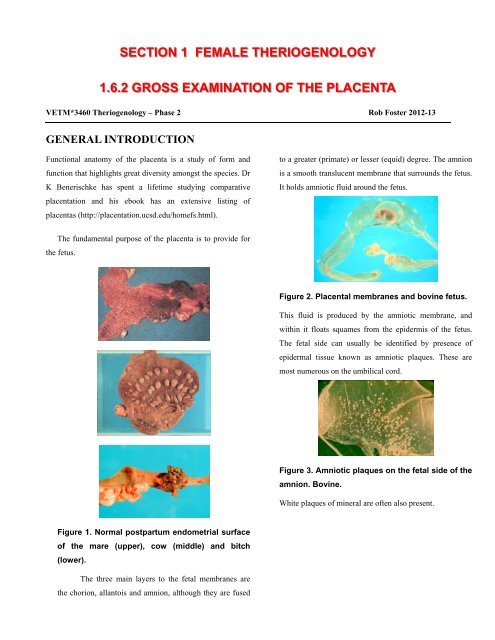
SECTION 1 FEMALE THERIOGENOLOGY1.6.2 GROSS EXAMINATION OF THE PLACENTAVETM*3460 Theriogenology – Phase 2 Rob Foster 2012-13GENERAL INTRODUCTIONFunctional anatomy of the placenta is a study of form andfunction that highlights great diversity amongst the species. DrK Benerischke has spent a lifetime studying comparativeplacentation and his ebook has an extensive listing ofplacentas (http://placentation.ucsd.edu/homefs.html).to a greater (primate) or lesser (equid) degree. The amnionis a smooth translucent membrane that surrounds the fetus.It holds amniotic fluid around the fetus.The fundamental purpose of the placenta is to provide forthe fetus.Figure 2. Placental membranes and bovine fetus.This fluid is produced by the amniotic membrane, andwithin it floats squames from the epidermis of the fetus.The fetal side can usually be identified by presence ofepidermal tissue known as amniotic plaques. These aremost numerous on the umbilical cord.Figure 3. Amniotic plaques on the fetal side of theamnion. Bovine.White plaques of mineral are often also present.Figure 1. <strong>Normal</strong> postpartum endometrial surfaceof the mare (upper), cow (middle) and bitch(lower).The three main layers to the fetal membranes arethe chorion, allantois and amnion, although they are fused
2membranes is seen and there are necrotic tips of whereadjoining <strong>placentae</strong> touch.Figure 4. <strong>Normal</strong> placental mineralization. Bovine.Amniotic fluid is swallowed by the fetus and postpartum sampling of amniotic fluid is possible by takingstomach content. Fetuses probably do not inhale amnioticfluid past the larynx – unless they are distressed. Likewise,meconium is not normally present in the amniotic fluidunless the fetus is distressed and defecates. Fetusescovered with meconium are said to have ‘foetal diarrhoea’.The chorion is the layer that contacts the mother –and in most species it is fused with the allantois. Thechorioallantois is thus formed.The allantoic cavity, where it exists, contains fetalurine, and other fluids that arise from the membrane itself.The final part of the placenta is the umbilical cord.This structure contains 2 umbilical arteries, an umbilicalvein and a urachal space, which empties into the allantois.The cord is often covered with amniotic plaques, and isoccasionally gently twisted.D.H. Schlafer, P.J. Fisher, C.J. Davies. (2000) Thebovine placenta before and after birth: placentaldevelopment and function in health and disease. AnimalReproduction Science 60-61: 2000. 145-160.Figure 5. Chorionic surface with mineralizationand chorionic cysts in a porcine placenta.THE HORSEIn the horse, the amnion and the chorioallantois arecompletely separate. The chorionic surface is diffuse andmicrocotyledonary and gives it a luxurious velvetyappearanceFigure 6. Chorionic surface of equine placentaAvillous regions are located in those areaswhere the trophoblasts do not contact theendometrium – over large vessels, at the uterotubaljunction, and at the cervix where the appearance ofthe avillous regions is star shaped to form the cervicalstar.THE PIGThe membranes are completely fused in the placenta ofpigs. The chorionic surface of the porcine placenta hassmall microscopic projections or villi. They are barelyrecognizable grossly. Chorionic cysts are found in theplacenta of pigs – these are where the secretion of uterineglands is trapped. Prominent mineralization of theFigure 7. An avillus region of equine placenta –the cervical star.
3The avillous chorioallantoic pouches are formedover the sites of the endometrial cups around the chorionnear the attachment site of umbilical cord.The umbilical cord of the equine fetus is normally 36 and83 cm long, and the insertion site should be at the junctionof the horn and body of uterus.THE COWThe ruminant has a cotyledonary placenta with cotyledonsand intercotyledonary regions.Figure 8. Endometrial cups (left) andcorresponding chorioallantoic pouches (right).Horse.Hippomanes and allantoic pouches are often foundin the horse allantois. Hippomanes are present in virtuallyall equine <strong>placentae</strong> and are proteinaceous soft calculi.They also occur in the <strong>placentae</strong> of cows, sheep, andlemurs! Some are found in the amniotic cavity too.Figure 9. Hippomane from allantoic cavity. Horse.Occasionally one finds pedunculated structures attached tothe chorioallantois on the allantoic side. These are calledallantoic pouches. They are incidental in most cases. Yolksac remnants are less frequently found, and when presentare attached to the cord at the junction with the allantois.They often are mineralized.Figure 10 Yolk sac remnant. Horse.Figure 11 Cotyledons of a bovine placenta withadventitial placentation (left lower).The exchange unit is the placentome made up of cotyledon(fetal) and caruncle (maternal) components. Thecotyledonary (chorionic) surface of the placentalmembranes often has additional regions or adventitialplacentation. This is assumed to be an attempt atcompensatory hyperplasia, however it is seen as an ageassociated change.The amnion of ruminants is fused with the allantoisover the dorsum of the fetus.The normal intercotyledonary placenta is clearenough to read a book through. The normal cotyledon isred and even. Autolysis makes the placenta appear palerthan normal, and autolysis affects the entire placenta, notjust one part.The placentomes develop in 4 rows, two dorsal andtwo ventral. There are 70-140 placentomes that have anexchange area of more than 18 sq m. Fetal growth iscorrelated with vascular development within theplacentome. Placentomes increase in size during pregnancyand the largest ones are found nearest the attachment of theumbilical vessels. A cotyledon larger than 15 cm diameterin the bovid is regarded as increased in size. After birth,the hypertrophic portion of the caruncles undergoes
4necrosis and is lost in the lochia, usually by day 12postpartum (PP). Reepithelialization occurs in about 21days PP. Because the outer part of the caruncle is lostanyway, it can be sampled without damage to the futurereproductive capacity of the cow. Such sampling isrecommended when there is no placenta for examination.Caruncles should never be twisted off, but the cotyledoncan be gently peeled off the caruncle.Placentomes have a capacity to compensate for loss,and they do so by hypertrophy, as functional area is lost;remaining placentomes become larger. There is also thefacility for additional or adventitial placentation to occur.THE DOG AND CATDomesticated carnivores have a zonary placenta.Interdigitation of fetal and maternal tissues occurs in thecenter of the zone and there are marginal hematomas at theedges. The amnion is separate from the allantois.Figure 12. Zonary placenta. Canine