

Eruptive Xanthoma - Medcom Limited
Eruptive Xanthoma - Medcom Limited
Eruptive Xanthoma - Medcom Limited

Create successful ePaper yourself
Turn your PDF publications into a flip-book with our unique Google optimized e-Paper software.
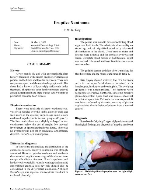
Case Reports<strong>Eruptive</strong> <strong>Xanthoma</strong>Dr. W. K. TangDate: 14 March, 2001Venue: Yaumatei Dermatology ClinicOrganizer: Social Hygiene Service, DH;Clinico-pathological SeminarCASE SUMMARYHistoryA two-month-old girl with unremarkable birthhistory presented with sudden onset of erythematouspapules on the limbs and face for one week. There wasno systemic upset, and she remained asymptomatic. Hersister had a history of hypertriglyceridaemia undertreatment. The patient's other family members enjoyedgood physical health and there was no family history ofpremature coronary heart disease.Physical examinationThere were multiple discrete erythematous,yellowish papules over the limbs, anterior trunk andface, more on the extensor surface, and some lesionscoalesced together to form small plaques (Figure 1).The liver and spleen were slightly enlarged at twocentimetres below the costal margin. No mucosalinvolvement or lipaemia retinalis was found. There wasno dysmorphism nor other congenital abnormalitydetected. Darier's sign was negative.InvestigationsThe patient was found to have raised fasting bloodsugar and lipid levels. The whole blood was milky onstanding, which signified markedly elevatedchylomicrons in the blood. Urine protein, sugar andketones were negative and the amylase level was notraised. Complete blood picture with differential countwas normal. The renal and liver functions were alsounremarkable.The patient's parents and elder sister were asked forblood screening and the results were stated in Table 1.Skin biopsy showed scattered foci of a few foamcells in the superficial dermis, admixed withlymphocytes, histiocytes and eosinophils. The overlyingepidermis was unremarkable. The features weresuggestive of eruptive xanthoma. Since the patient'splasma lipoprotein lipase level was normal, abnormalor deficient apoprotein C-II cofactor was suspected. Itwas later confirmed by dramatic lowering of plasmatriglycerides after infusion of plasma from a normalcontrol.DiagnosisBased on the "sky-high" hypertriglyceridaemia andhistological findings, the diagnosis of eruptive xanthomaDifferential diagnosisIn view of the morphology and distribution of thecutaneous lesions, eruptive xanthoma was stronglysuspected. However, papular xanthoma and xanthomadisseminatum might at some stage of the disease sharecomparable clinical features. Non-Langerhans' cellhistiocytosis especially juvenile xanthogranuloma andgeneralized eruptive histiocytosis should also beconsidered in the differential diagnosis. AlthoughDarier's sign was negative, mastocytosis could not beexcluded clinically.Figure 1: Multiple yellowish papules over the right thighextensor surface172Hong Kong Dermatology & Venereology Bulletin
Case Reportsinjury, oedema, altered lipoprotein structure orparaproteins in the plasma, can also lead to lipoproteinleakage into the dermis. Direct phagocytosis oflipoproteins by dermal histiocytes or a reactive processinvolving in-situ lipid synthesis in the histiocytes willthen evolve into foam cells. 9Differential diagnosisClinically eruptive xanthoma may sometimes beconfused with other xanthomatosis or non-Langerhanss'cell histiocytosis (Table 2). 10,11 Rarely the cutaneouslesions may mimic Sweet's syndrome. 12 Most of the timea skin biopsy can reliably differentiate eruptivexanthoma from other xanthomatoses and non-Langerhans' cell histiocytosis (Table 3).TreatmentTreatment of eruptive xanthoma is directed to theunderlying causes. Since eruptive xanthoma secondaryto hypertriglyceridaemia typically responds well todietary control, a dietician's advice should be soughtfirst. In general, a low carbohydrates and saturated fatdiet is the first treatment of choice. Anti-hyperlipidaemicagents should be considered when dietary control fails. 13PrognosisUnless the underlying causes can be corrected, thepatient should be put on life-long dietary control andregular follow-up is needed. However patients can bereassured that the cutaneous lesions and lipoproteinabnormalities can revert to normal, in terms of weeks,with appropriate treatment. 13Table 2. Clinical features of the common differential diagnosis of eruptive xanthoma<strong>Eruptive</strong> Papular Juvenile <strong>Xanthoma</strong> Generalized eruptivexanthoma xanthoma xanthogranuloma disseminatum histiocytosisAge Children, adult Mainly adult 80%
Case ReportsLearning points:Development of eruptive xanthoma is indicativeof an underlying hyper-triglyceridaemia.Appropriate dietary changes, correction ofsecondary factors along with antihyperlipidaemicagents, if needed, will lower plasmatriglycerides and allow the xanthomatous lesionsto resolve.References1. Ruggero C, Marcello M, Emilio B, Giovanni G. Normolipaemiceruptive cutaneous xanthomatosis. Arch Dermatol 1986;12:1294-7.2. Antonio M, Gotto J. Clinical diagnosis of hyperlipoproteinaemia.Am J Med 1983;74:5-9.3. Parker F. <strong>Xanthoma</strong>s and hyperlipidaemias. J Am AcadDermatol 1985;13:1-30.4. Dicken CH, Connolly SM. <strong>Eruptive</strong> xanthomas associatedwith isotretinoin (13-cis-retinoic acid). Arch Dermatol 1980;116:951-2.5. Parker F. Normocholesterolaemic xanthomatosis. ArchDermatol 1986;122:1253-6.6. Goldstein GD. The Koebner phenomenon with eruptivexanthomas. J Am Acad Dermatol 1984;10:1064-5.7. Eeckhout I, Vogelaers D, Gleerts ML, Naeyaert JM. <strong>Xanthoma</strong>sdue to generalized edema. Br J Dermatol 1997;136:601-3.8. Mahmoud SF Lawrence. Seip Syndrome: Report of a case fromEgypt. Cutis 1997;60:91-3.9. Ghislaine R, Marianne X, Jean D. <strong>Eruptive</strong> and tubero-eruptivexanthomas of the skin arising on sited of prior injury; two casereports. JAMA 1988;260:1282-3.10. Ferrando J, Campo-Voegeli A, Soler-Carrillo J, et al. Systemicxanthohistiocytoma: a variant of xanthoma disseminatum? BrJ Dermatol 1998;138:155-60.11. Zelger BW, Sidoroff A, Orchard G, Cerio R. Non-Langerhanscell histiocytoses. A new unifying concept. Am J Dermatopathol1996;18:490-504.12. Ahn SJ, Choi JH, Sung KJ, Moon KC, Koh JK. Sweet'ssyndrome presenting with lesions resembling eruptivexanthoma. Br J Dermatol 2000;143:449-50.13. Archer CB, Macdonald DM. <strong>Eruptive</strong> xanthoma in type Vhyperlipoprotaeinemia associated with diabetes mellitus. ClinExp Dermatol 1984;9:312-6.Vol.9 No.4, December 2001 175

