

Case studies in novel narial anatomy - Digital Morphology
Case studies in novel narial anatomy - Digital Morphology
Case studies in novel narial anatomy - Digital Morphology

Create successful ePaper yourself
Turn your PDF publications into a flip-book with our unique Google optimized e-Paper software.
J. Zool., Lond. (2004) 264, 217–230 C○ 2004 The Zoological Society of London Pr<strong>in</strong>ted <strong>in</strong> the United K<strong>in</strong>gdom DOI:10.1017/S0952836904005540<strong>Case</strong> <strong>studies</strong> <strong>in</strong> <strong>novel</strong> <strong>narial</strong> <strong>anatomy</strong>: 3. Structure and function ofthe nasal cavity of saiga (Artiodactyla: Bovidae: Saiga tatarica)Andrew B. Clifford 1,2 and Lawrence M. Witmer 3 *1 Department of Biological Sciences, Ohio University, Athens, OH 45701, U.S.A.2 Department of Ecology and Evolutionary Biology, Brown University, Providence, RI 02912, U.S.A.3 Department of Biomedical Sciences, College of Osteopathic Medic<strong>in</strong>e, Ohio University, Athens, OH 45701, U.S.A.(Accepted 10 February 2004)AbstractMuch of the <strong>narial</strong> <strong>anatomy</strong> of the enigmatic antelope Saiga tatarica has been described by previous workers.However, the <strong>anatomy</strong> of the nasal cavity and the causally associated osteological correlates of proboscis structurerequire closer attention, because these data are <strong>in</strong>tegral for both a more comprehensive understand<strong>in</strong>g of saigafunctional morphology and more robust reconstructions of proboscis structure <strong>in</strong> fossil taxa. Saiga and outgroupspecimens were subjected to X-ray computed tomographic (CT) imag<strong>in</strong>g, gross dissection and skeletonization.The nasal cavity of saiga is characterized by an enlarged nasal vestibule and basal conchal fold. Many structures(e.g. turb<strong>in</strong>ates, lateral cartilages, mucosal folds, nasolacrimal duct) are retracted caudally to a small area <strong>in</strong> thecaudodorsal part of the nasal cavity proper. The enlarged vestibule is associated laterally and ventrally with pairedsacs. The nasal septum is largely membranous and conta<strong>in</strong>s a large patch of cavernous tissue that serves as adynamic baffle modify<strong>in</strong>g the flow of <strong>in</strong>spired air. Bones compris<strong>in</strong>g the <strong>narial</strong> marg<strong>in</strong> have modified attachmentsites for bucc<strong>in</strong>ator group muscles and the reduced lateral cartilages. The premaxilla is greatly modified by theenlarged musculature associated with nasolabial fusion. Ma<strong>in</strong>tenance of the topological relationships of <strong>narial</strong>structures compared to bovid outgroups has resulted <strong>in</strong> a nasal cavity with much larger area for seromucous glandsof the vestibule as well as <strong>narial</strong> musculature capable of controll<strong>in</strong>g the aperture of the nasal cavity. Maxillolabialmuscles and the lateralis nasi act together both to compress the nasal cavity and to control the dilation of the nostrilssuch that air flow through the cavity is highly modified relative to bovid outgroups. The lateral vestibular recessis an outpocket<strong>in</strong>g of the nasal vestibule that produces supplementary seromucous secretions and seems to haveno homologue among outgroups. The enlarged nasal vestibule, lateral vestibular recess, repositioned basal fold,and septal cavernous mass are regarded as a coord<strong>in</strong>ated adaptation to dusty habitats, such that nasal air flow canbe dynamically regulated allow<strong>in</strong>g for collection of <strong>in</strong>spired particulates <strong>in</strong> the vestibule and thus cleans<strong>in</strong>g of airdest<strong>in</strong>ed for the lungs.Key words: Artiodactyla, Bovidae, Saiga, nasal vestibule, proboscis, nasal physiologyINTRODUCTIONSaiga Saiga tatarica are a relatively little studied but morphologicallydisparate group of antelopes (Fig. 1). Bovidae(e.g. antelopes, cattle) have undergone a dramatic radiation<strong>in</strong> the past 18 million years, now encompass<strong>in</strong>g 135species (Vrba & Schaller, 2000). Previously of vary<strong>in</strong>gaffiliation (e.g. Capr<strong>in</strong>ae <strong>in</strong> Nowak, 1999), saiga are nowregarded as members of Antilop<strong>in</strong>ae with<strong>in</strong> Bovidae(Vrba & Schaller, 2000). Saiga have been difficult to placephylogenetically because they are bizarrely apomorphic,*All correspondence to: L. M. Witmer.E-mail: witmer@exchange.oucom.ohiou.eduparticularly <strong>in</strong> the head and skull as a result of theirevolution of unique <strong>narial</strong> structures. Like many otherantelopes, only males possess horns, but their most dist<strong>in</strong>guish<strong>in</strong>gcharacteristic, a conspicuous proboscis, is possessedby both males and females. The <strong>in</strong>flated proboscisof saiga provides a case study <strong>in</strong> the evolution of <strong>novel</strong><strong>narial</strong> <strong>anatomy</strong> with<strong>in</strong> an otherwise morphologically andphylogenetically well-resolved clade.Saiga are a relatively young species, first occurr<strong>in</strong>g<strong>in</strong> middle Pleistocene deposits approximately 1 millionyears ago, although some authors (Heptner, Nasimovich &Bannikow, 1988) speculated that this genus may occur asfar back as the late Pliocene. Only one other species ofSaiga is currently recognized (S. prisca), although theMongolian population previously received species rank
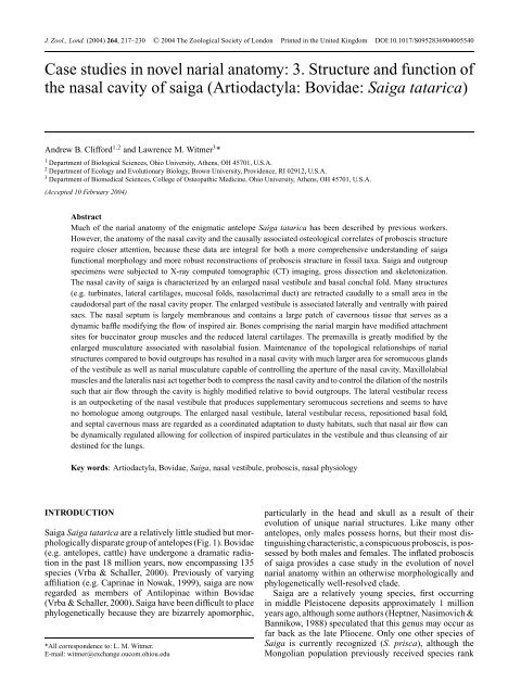
218 A. B. CLIFFORD AND L. M. WITMER(a)(b)(c)(Heptner et al., 1988). Generally, fossil saiga are virtuallyidentical to Recent forms, differ<strong>in</strong>g mostly <strong>in</strong> geographicalrange (Sokolov, 1974). Occurr<strong>in</strong>g now only <strong>in</strong> open,dusty, arid grasslands of central Asia, their range onceextended from the British Isles to eastern Alaska (Frick,1937).The <strong>in</strong>flated <strong>narial</strong> apparatus <strong>in</strong> saiga has been studiedto various extents by previous workers. Murie (1870)gave an extensive account of the <strong>anatomy</strong> of the wholeanimal, discuss<strong>in</strong>g much of the musculature and <strong>in</strong>nervationof the proboscis, as well as some aspects of thenasal cartilages and skull, attribut<strong>in</strong>g the peculiar noseto an <strong>in</strong>crease <strong>in</strong> tactile sensation (Murie, 1870). Boas &Paulli (1908) figured the skull but offered few otheranatomical details. Jacobi (1921) provided a diagram ofthe skull and nasal cartilages (seem<strong>in</strong>gly based largely onMurie, 1870) to describe the apparent convergence betweenmany phylogenetically disparate proboscis-bear<strong>in</strong>gmammals. Lodyshenskaya (1952) described the complex<strong>narial</strong> musculature and nasal cartilages, add<strong>in</strong>g developmentaland histological components. More recently,Frey & Hofmann (1995, 1997) conducted a series ofmorphological <strong>studies</strong> of the proboscis <strong>in</strong> saiga, focus<strong>in</strong>gon the skull, glands, musculature, and anatomical differentiationfrom another proboscis-bear<strong>in</strong>g bovid, Guenther’sdikdik Madoqua guentheri. All of these <strong>studies</strong> differ fromeach other <strong>in</strong> focus, completeness and term<strong>in</strong>ology.This study has two major aims. The first is to highlightfunctional aspects of the transformation of the nasalcavity of saiga, based on cross-sectional <strong>anatomy</strong> anddissection. The second is to detail the causally associatedbony modifications of the skull result<strong>in</strong>g from the evolutionof a proboscideal nose <strong>in</strong> saiga. The anatomicalconfiguration of <strong>in</strong>ternal <strong>narial</strong> structures rema<strong>in</strong>s largelyundescribed. Previous work has either omitted apomorphiesoccurr<strong>in</strong>g <strong>in</strong>side the nose or has only brieflydescribed certa<strong>in</strong> features. In addition, the osteologicalcorrelates of soft tissues compris<strong>in</strong>g the nose <strong>in</strong> saigarema<strong>in</strong> undescribed. The present study is part of a largereffort attempt<strong>in</strong>g to describe the functional <strong>anatomy</strong> ofapomorphic <strong>narial</strong> structures <strong>in</strong> extant amniotes (Witmer,Sampson & Solounias, 1999; Clifford & Witmer, 2001,2002a,b, 2004; Witmer, 2001a,b). The project as a wholeseeks to expla<strong>in</strong> how anatomical specialization results <strong>in</strong>causally associated bony features <strong>in</strong> proboscis-bear<strong>in</strong>gtaxa. A goal is to assess the presence or absence of <strong>novel</strong>soft-tissue structures <strong>in</strong> ext<strong>in</strong>ct taxa by appeal to osteologicalcorrelates of these soft tissues found <strong>in</strong> extanttaxa (Witmer, 1995). Previous work has concentratedlargely on muscular, cartilag<strong>in</strong>ous, nervous, and vascularspecializations <strong>in</strong> saiga without <strong>in</strong>tegrat<strong>in</strong>g many <strong>in</strong>ternalchanges <strong>in</strong> the nasal cavity <strong>in</strong>to potential causally associatedfeatures of the skull, and this study seeks to fillthat gap.Fig. 1. The head of Saiga tatarica <strong>in</strong> left lateral view, based on 3Dreconstructions of CT scans of AMNH 202492: (a) isosurface ofthe <strong>in</strong>tact head; (b) voxel reconstruction of <strong>in</strong>tact head to simulatea lateral radiograph; (c) skull isosurface. Scale bars = 5cm.MATERIALS AND METHODSA skull (AMNH 119649) and an <strong>in</strong>tact head (AMNH202492) were the primary source of data for this study.
Nasal cavity of saiga 219BovidaeAntilop<strong>in</strong>aeSaigaMadoquaCapraOvisBosBisonAlcesCamelusCystophoraCapr<strong>in</strong>aeFig. 2. Phylogenetic relationships of taxa and clades referred to <strong>in</strong>this study. Topology based on Hassan<strong>in</strong> & Douzery (2003).The <strong>in</strong>tact head was a zoo specimen later preserved asa fluid specimen. Before dissection, the <strong>in</strong>tact specimenwas subjected twice to X-ray computed tomography (CT)at O’Bleness Memorial Hospital <strong>in</strong> Athens, Ohio, us<strong>in</strong>ga GE HiSpeed Fx-i Helical CT Scanner. The first scanwas set at 140.0 kV, 170.0 mA, 5.0 mm slice thickness,us<strong>in</strong>g both standard and bone algorithms. The second scanwas set at 140.0 kV, 160.0 mA, 2.0 mm slice thickness,us<strong>in</strong>g a bone algorithm. The CT data were exported <strong>in</strong>DICOM format us<strong>in</strong>g eFilm (v. 1.5.3, Merge eFilm,Toronto). Analysis and postprocess<strong>in</strong>g used the softwarepackages eFilm (v. 1.8.3) and Amira (v. 3.0, TGS, Inc., SanDiego). Both sectional <strong>anatomy</strong> and 3D reconstructionsderived from the slice data were analysed. Study of thehead also <strong>in</strong>cluded gross dissection and sagittal section<strong>in</strong>gwith a band saw. Follow<strong>in</strong>g sagittal section<strong>in</strong>g, the rightside was CT scanned a third time (120.0 kV, 130.0 mA,1.0 mm slice thickness, bone algorithm). Dissectionswere recorded with digital photography. Figures 1, 3,5 & 6 were produced from reconstructed CT data us<strong>in</strong>gAmira.To determ<strong>in</strong>e the <strong>anatomy</strong> of bovid outgroups (Fig. 2),skulls of Madoqua saltiana (Salt’s dikdik; OUVC 9575),Ovis aries (domestic sheep; OUVC 9704), Bos taurus(domestic ox; OUVC 9473, 9474, 9475, 9476, 9477, 9478,9479, 9480, 9481, 9482, 9547, 9548, 9558), and Bisonbison (American bison; OUVC 9484, 9489, 9557), <strong>in</strong>addition to <strong>in</strong>tact heads of Capra hircus (domestic goat;OUVC 9744, 9746), were exam<strong>in</strong>ed before dissectionof the saiga. One Capra (OUVC 9744) was <strong>in</strong>jected<strong>in</strong> both carotid arteries with radio-opaque barium/latex(per Sedlmayr & Witmer, 2002), CT scanned (120.0 kV,100.0 mA, 1.0 mm slice thickness, bone algorithm), andanalysed as above. Additionally, previous research (seeabove), veter<strong>in</strong>ary texts (Nickel, Schummer, Seiferle &Sack, 1973; Getty, 1975; Nickel, Schummer, Seiferle,Frewe<strong>in</strong> et al., 1986; Schaller, 1992), and Nom<strong>in</strong>aAnatomica Veter<strong>in</strong>aria (NAV, 1994) were consulted tostandardize term<strong>in</strong>ology and homologize the <strong>narial</strong> structuresof saiga.RESULTSOverview of nasal cavityThe most remarkable attribute of the nasal cavity of saigais, relative to outgroups, the enormous nasal vestibule,the expansion of which has led to a major reorganizationof the entire nose. For example, the nasal passage canbe divided <strong>in</strong>to two dist<strong>in</strong>ct regions (Fig. 3). The firstregion is a wide-open chamber comprised of the expandednasal vestibule at the rostral end of the passage, thecentral space <strong>in</strong> the passage, and the nasopharyngeal duct(ductus nasopharyngeus; Figs 3a & 4i; dnp). The secondregion is the restricted, caudodorsal portion of the nasalcavity, which is tightly packed with conchae. At thecaudal end of the nasal vestibule, there is a bl<strong>in</strong>d recessor sac which opens rostrally and is l<strong>in</strong>ed with hair andvestibular mucosa (‘nasal sac’ of Murie, 1870). Thereis another recess whose dorsal wall forms the floor ofthe nasal vestibule (‘shelf’ of Murie, 1870). The mucosa<strong>in</strong> this ventral recess is much like the mucosa of thenasal vestibule, but it is not l<strong>in</strong>ed with hair. The nostrilsare the narrowest portion of the nasal passage. Thefollow<strong>in</strong>g description emphasizes the various parts of thenasal cavity and mucosal structure <strong>in</strong> saiga. Muscles andcartilages have been described elsewhere (Murie, 1870;Lodyshenskaya, 1952; Frey & Hofmann, 1995, 1997).Standard veter<strong>in</strong>ary nomenclature will be applied to anatomicalstructures.Nasal vestibuleVestibulum nasi (Figs 3a & 4b-d; vn). The nasal vestibuleextends immediately caudal to the nostrils. In saiga, thenostrils are separated only by a th<strong>in</strong> cont<strong>in</strong>uation of thenasal septum, rather than by a rh<strong>in</strong>arium (planum nasi),which is well-developed <strong>in</strong> other bovids but not <strong>in</strong> saiga(Murie, 1870). The nostrils are oval and set close to themouth. In the specimen described here, the nostrils didnot overhang the mouth, although other workers havedescribed a more pendulous proboscis (Murie, 1870;Sokolov, 1974; Heptner et al., 1988). The nostrils aretightly bound <strong>in</strong> position by musculature extend<strong>in</strong>g fromthe dorsolateral surface of the premaxilla (<strong>in</strong>cisive bone)and w<strong>in</strong>d<strong>in</strong>g around the nostril. The mucosa of the nasalvestibule is thick and fur-l<strong>in</strong>ed, although the hairs <strong>in</strong> thevestibule are much shorter and more sparsely distributedthan the hairs cover<strong>in</strong>g the head. From a gross perspective,small seromucous glands occur throughout the vestibuleon its dorsal, lateral and ventral walls. These glandsoccur <strong>in</strong> vestibular mucosa that has undergone significantfibrofatty elaboration compared to rum<strong>in</strong>ant outgroups.The vestibule extends caudally to the crescentic rostraledge of the basal fold (plica basalis) at the limen nasi. Atthe limen, the mucosa changes to respiratory mucosa, as istypical for mammals generally. Lateral to the limen is theostium of the lateral recess, and medial to it is the ostiumof the nasolacrimal duct.
220 A. B. CLIFFORD AND L. M. WITMER(a)pamnmcnm pr mndcnecndorvlpbvnrnvmnvcnvdnp(b)Fig. 3. Medial view of sagittally sectioned head of Saiga tatarica: (a) labelled draw<strong>in</strong>g of isosurface of AMNH 202492 based on 3Dreconstructions of CT scans; (b) stereopairs of specimen <strong>in</strong> (a). Scale bars = 5 cm. Abbreviations as <strong>in</strong> Table 1.Recessus vestibularis lateralis (Figs 3a & 4f; rvl). Thelateral recess of the nasal vestibule is roughly oval coronallyand semicircular <strong>in</strong> sagittal section. Its open<strong>in</strong>g <strong>in</strong>tothe ma<strong>in</strong> nasal vestibular chamber occurs <strong>in</strong> the middleof the rostrolateral edge of the basal fold about halfwayalong the rostrocaudal extent of the entire nasal passage.
Nasal cavity of saiga 221(a)vn(b)snpm(c)vn(d)papr(e)csndorvlnrnvsnccoppnomopprovnoppp(f)cnmoncnld (g) cndcnv(h)prcnlparvlcsvnlodbocipbgpocsnsmcnp(i)oelpcne(j) a b c d e f g h ibodnpFig. 4. Draw<strong>in</strong>gs of selected computerized tomographic slices of the head of Saiga tatarica (AMNH 202492) show<strong>in</strong>g <strong>narial</strong> structures<strong>in</strong> successive transverse sections (a–i); (j) skull <strong>in</strong> left lateral view to show the rostrocaudal levels of sections depicted <strong>in</strong> (a–i). Scalebars = 5 cm. Abbreviations as <strong>in</strong> Table 1.
222 A. B. CLIFFORD AND L. M. WITMERTable 1. List of abbreviations used <strong>in</strong> figures. ∗ , terms named <strong>in</strong>this study and not found <strong>in</strong> NAV (1994)bo Bulbus oculi (Fig. 4h & i)ci Canalis <strong>in</strong>fraorbitalis (Figs 4h & 6a)cnd Concha nasalis dorsalis (Figs 3a & 4g)cne Conchae nasalis ethmoidales (Figs 3a & 4i)cnl Canalis nasolacrimalis (Fig. 4f & g)cnld Cartilago nasi lateralis dorsalis (Fig. 4f)cnm Concha nasalis media (Figs 3a & 4g)cnp Cavum nasi proprium (Fig. 4h)cnv Concha nasalis ventralis (Figs 3a & 4g)csn Cartilago septi nasi (Fig. 4g)csnd Cartilago septi nasi, processus dorsalis (Fig. 4e)csnv Cartilago septi nasi, processus ventralis (Fig. 4e)dnp Ductus nasopharyngeus (Figs 3a & 4i)gpo Glandula preorbitalis (Fig. 4g)l L<strong>in</strong>gua (Fig. 4d & e)mnd Meatus nasi dorsalis (Fig. 3a)mnm Meatus nasi medius (Fig. 3a)mnv Meatus nasi ventralis (Fig. 3a)n Naris (Fig. 4a)ocnvOs conchae nasalis ventralis (maxilloturb<strong>in</strong>ate)(Figs 5a & 6a)od Os dentale (Mandibula) (Fig. 4a)oee Os ethmoidale, ethmoturb<strong>in</strong>ates (Fig. 6a)oelc Os ethmoidale, lam<strong>in</strong>a cribrosa (Fig. 6a)oelp Os ethmoidale, lam<strong>in</strong>a perpendicularis (Fig. 6a)oent Os ethmoidale, nasoturb<strong>in</strong>ate (Figs 5a & 6a)olpn Os lacrimale, processus nasolacrimalis (Fig. 5a)olt Os lacrimale, tuberculum (Fig. 5a)om Os maxillare (maxilla) (Fig. 6a)omcf Os maxillare, crista facialis (Fig. 5a)omfi Os maxillare, foramen <strong>in</strong>fraorbitale (Fig. 5a)omti Os maxillare, tuberculum <strong>in</strong>fraorbitale (Fig. 5a)on Os nasale (Figs 4f, 5a & 6a)onp Os nasale, processus (Fig. 5a)oppn Os premaxillare (os <strong>in</strong>cisivum), processus nasalis(Figs 4c, 5a & 6a)oppp Os premaxillare (os <strong>in</strong>cisivum), processus palat<strong>in</strong>us(Figs 4c & 6a)oppr Os premaxillare (os <strong>in</strong>cisivum), processus rostralis(Figs 4b, 5a & 6a)orvl Ostium recessus vestibularis lateralis(Figs 3a & 4e) ∗ovn Organum vomeronasale (Fig. 4c)pa Plica alaris (Figs 3a & 4d-f)pb Plica basalis (Figs 3a & 4f)pr Plica recta (Figs 3a, 4e & f)rnv Recessus nasi ventralis (Figs 3a, 4c & d) ∗rvl Recessus vestibularis lateralis (Fig. 4f) ∗sm S<strong>in</strong>us maxillaris (Figs 4h & 6a)sncc Septum nasi, corpus cavernosum (Fig. 4e) ∗snpm Septum nasi, pars membranacea (Fig. 4b)vn Vestibulum nasi (Figs 3a & 4b)The sac then extends caudolaterally, somewhat mediolaterallycompressed, over the edge of the nasomaxillary<strong>in</strong>cisure. Its caudalmost extent is lateral to the maxilla,underly<strong>in</strong>g musculature (e.g. m. levator labii superiorisand m. can<strong>in</strong>us) responsible for compress<strong>in</strong>g the proboscis(Murie, 1870) and situated just rostral to the preorbitalgland (Fig. 4g; gpo; Frey & Hofmann, 1997). The mucosal<strong>in</strong><strong>in</strong>g the <strong>in</strong>terior of the lateral recess is nearly identicalto that <strong>in</strong> other parts of the nasal vestibule, even to theextent that it conta<strong>in</strong>s small hairs and m<strong>in</strong>ute glandularostia. Its mucosa is thickened by fibrofatty elaboration, aselsewhere <strong>in</strong> the nasal vestibule. No muscles, cartilages,or bones could be found associated with the lateralrecess; it occurs simply as an outpocket<strong>in</strong>g of the nasalvestibule.Canalis nasolacrimalis (Fig. 4f & g; cnl). The nasolacrimalcanal is the primary connection between the nasalcavity and the orbit. Generally <strong>in</strong> bovids, this canal opensnear the limen nasi, at the juncture of the vestibular mucosaand the respiratory mucosa of the nasal cavity proper(Nickel, Schummer, Seiferle & Sack, 1973). In saiga, thisdel<strong>in</strong>eation can be clearly seen on the basal fold. As thenasolacrimal canal exits the lacrimal bone to enter thenasal vestibule, it next enters a space with<strong>in</strong> the basal foldbetween the ventral portion of the dorsal lateral cartilage(Fig. 4f; cnld) and the ventral lateral cartilage (‘sesamoidcartilage’ of Murie, 1870). The mucosal ostium of thenasolacrimal canal occurs relatively far dorsal with<strong>in</strong> thenasal vestibule, just medial to the rostral lip of the crescentof the basal fold.Ma<strong>in</strong> nasal cavityCavum nasi proprium. The nasal cavity proper extendsfrom the limen nasi caudally to the nasopharyngeal duct.The mucosa <strong>in</strong> the nasal cavity proper is differentiated<strong>in</strong>to a respiratory region (regio respiratoria) on the ventralconcha and rostral parts of the ethmoid conchae and apigmented olfactory region (regio olfactoria) cover<strong>in</strong>g thecaudal portion of the ethmoid conchae. The turb<strong>in</strong>atesupportednasal conchae and the meatuses between themare displaced caudodorsally <strong>in</strong> the nasal cavity. In outgroups,the nasal cavity proper takes up the vast majorityof the nasal passage (Nickel, Schummer, Seiferle & Sack,1973), whereas <strong>in</strong> saiga it is much smaller and is telescopedcaudally by the expanded vestibule. The conchaeare somewhat closed off by the basal fold, and the mucosalfolds extend<strong>in</strong>g rostrally from the conchae are generallyreduced.Plica basalis (Figs 3a & 4f; pb). The basal fold is theventralmost mucosal fold of the ma<strong>in</strong> nasal cavity. Thisfold <strong>in</strong> saiga extends from the rostralmost extension of theturb<strong>in</strong>ate-supported portion of the ventral concha ventrallyto the floor of the nasal vestibule. The thickened, fattymucosa of the basal fold is covered with m<strong>in</strong>ute folds.Its rostral marg<strong>in</strong> is crescentic. The space rostral to thebasal fold is the nasal vestibule, the major space of thenasal passage <strong>in</strong> saiga. The space caudal to the basalfold is much smaller, form<strong>in</strong>g the ventral meatus (meatusnasi ventralis; Fig. 3a; mnv). Just rostral to the crescentof the basal fold is the ostium of the lateral recess ofthe nasal vestibule. With<strong>in</strong> the fibrofatty basal fold, themajor lateral cartilages of saiga can be found. The dorsallateral nasal cartilage (cartilago nasalis lateralis dorsalis;‘lower lateral cartilage’ and ‘upper lateral cartilage’ ofMurie, 1870) is suspended with<strong>in</strong> the rostral crescent ofthe basal fold. A smaller flange of cartilage attach<strong>in</strong>g to theventralmost edge of the dorsal lateral cartilage (cartilago
Nasal cavity of saiga 223nasalis lateralis ventralis; ‘sesamoid cartilage’ of Murie,1870) extends caudoventrally to attach on the lacrimalbone. These two cartilag<strong>in</strong>ous structures frame the rostralostium of the nasolacrimal canal, which opens onto thebasal fold near the edge of the rostral crescent.The dorsal lateral nasal cartilage is figured both <strong>in</strong> Murie(1870) and <strong>in</strong> Lodyshenskaya (1952), and the two majorpaired lateral components of the lateral cartilages aregiven different anatomical names. Some mammals sharethis feature (hav<strong>in</strong>g two cartilag<strong>in</strong>ous processes extend<strong>in</strong>gventrolaterally along the lateral wall of the nasal cavity),and the two cartilag<strong>in</strong>ous lam<strong>in</strong>ae are referred as dorsallateral cartilages (Nickel, Schummer, Seiferle & Sack,1973). Thus, the two paired lam<strong>in</strong>ae of lateral cartilages <strong>in</strong>saiga are most likely homologous to, and properly named,dorsal lateral nasal cartilages. Attach<strong>in</strong>g to the rostralportion of the dorsal lateral nasal cartilage and ventralportion of the nasomaxillary <strong>in</strong>cisure, many mammalsalso possess a ventral lateral nasal cartilage. Murie (1870)identified a cartilag<strong>in</strong>ous process extend<strong>in</strong>g away from thecaudoventral portion of the rostral lam<strong>in</strong>a of the dorsallateral cartilage, nam<strong>in</strong>g it the sesamoid cartilage. Inall other mammals, the cartilage attach<strong>in</strong>g to the ventralportion of the nasomaxillary fissure along the lateral wallof the nasal cavity is termed ventral lateral nasal cartilage(Nickel, Schummer, Seiferle & Sack, 1973).Concha nasalis ventralis (Figs 3a & 4g; cnv). Theventral nasal concha is the largest of the conchae with<strong>in</strong>the nasal cavity. It is double-scrolled (characteristic ofartiodactyls) and supported by the bony maxilloturb<strong>in</strong>ate(os conchae nasalis ventralis; Figs 5a & 6a; ocnv). Thedorsal scroll makes at least three complete turns, whereasthe ventral scroll completes one and a half. Also characteristicof other artiodactyls, the ventral concha is widesthalfway along the maxilloturb<strong>in</strong>ate. The ventral concha iscovered exclusively by respiratory mucosa. The ventralconcha is strongly angled dorsally, becom<strong>in</strong>g almostvertical, which is <strong>in</strong> marked contrast to other artiodactyls,<strong>in</strong> which it is basically horizontal.Plica alaris (Figs 3a & 4d-f; pa). The alar fold <strong>in</strong> saigais relatively very short compared to most other rum<strong>in</strong>ants.This mucosal fold is the direct rostral cont<strong>in</strong>uation of theventral nasal concha, and it ends rostrally just beyondthe rostral edge of the basal fold. Caudally, the alar foldreta<strong>in</strong>s a partial scroll of the ventral concha which curlsfirst dorsally and then ventrolaterally, mak<strong>in</strong>g a nearlycomplete turn. As the fold courses laterally along thelateral wall of the nasal cavity, it becomes less scrolled.Near the rostral term<strong>in</strong>ation of the straight fold (plicarecta), the attachment of the alar fold migrates dorsallyalong the wall of the nasal cavity. This migration cont<strong>in</strong>uesdorsally and medially, and the alar fold ultimatelyterm<strong>in</strong>ates on the nasal septum just <strong>in</strong>side the caudal endof the nasal vestibule and dorsal to the ostia of the lateralrecess and nasolacrimal canal. The mucosa of the alar foldis thick and fatty, as it is <strong>in</strong> much of the vestibule, but asthe fold travels onto the nasal septum, there is a collectionof cavernous tissue deep to the mucosa.Concha nasalis dorsalis (Figs 3a & 4g; cnd). Dorsally,the dorsal nasal concha, supported by the nasoturb<strong>in</strong>ate(Figs 5a & 6a; oen; endoturb<strong>in</strong>al I of Paulli, 1908), coursesdirectly rostrally from the cribriform plate just ventral tothe roof of the nasal cavity. The mucosa of the dorsalconcha is respiratory rostrally and olfactory caudally. Thisconcha is not <strong>in</strong>cl<strong>in</strong>ed relative to outgroups, but its rostralextent is shortened <strong>in</strong> saiga. The dorsal concha makesa complete scroll before unw<strong>in</strong>d<strong>in</strong>g to cont<strong>in</strong>ue as thestraight fold.Plica recta (Figs 3a & 4e-f; pr). The straight fold isthe dorsalmost mucosal fold of the nasal cavity, and is thedirect rostral cont<strong>in</strong>uation of the dorsal nasal concha. Thestraight fold travels from the end of the dorsal conchato just caudal to the po<strong>in</strong>t where the alar fold beg<strong>in</strong>s itsrotation on the lateral wall of the nasal cavity. The foldis very small, and there seem to be no glandular orificesassociated with lateral nasal glands as <strong>in</strong> other rum<strong>in</strong>ants(Nickel, Schummer, Seiferle & Sack, 1973), and nosuch gland was discovered despite careful dissection.Immediately lateral to the straight fold are the flattenedlam<strong>in</strong>ae of the dorsal lateral nasal cartilage. Between thesetwo lam<strong>in</strong>ae lateral to the straight fold, there is a sheet ofdense connective (non-cartilag<strong>in</strong>ous) tissue commenc<strong>in</strong>gat the apex of and partially overly<strong>in</strong>g the dorsal lateralcartilage. This plate was described by Murie (1870), andits function rema<strong>in</strong>s unknown. The straight fold does notundergo any rotation ak<strong>in</strong> to that of the alar fold. Themucosa of the straight fold is covered <strong>in</strong> much th<strong>in</strong>nermucosa characteristic of the respiratory mucosa of thenasal cavity proper.Concha nasalis media (Figs 3a & 4g; cnm). The middlenasal concha extends rostrally from the cribriform platebetween the dorsal and ventral nasal conchae. The middleconcha is roughly triangular <strong>in</strong> shape, with its base atthe cribriform plate (Fig. 6a; oelc) and its apex at the levelof the end of the nasoturb<strong>in</strong>ate. There are no mucosalfolds associated with the middle concha. As <strong>in</strong> the dorsalconcha, the mucosa of the middle concha is olfactory nearits attachment to the cribriform plate. Surround<strong>in</strong>g themiddle concha dorsally, there are several smaller ethmoidconchae (Figs 3a & 4i; cne) that also are l<strong>in</strong>ed withrespiratory and olfactory mucosa. The conchae associatedwith the ethmoid bone do not undergo significant rotationlike that <strong>in</strong> the ventral concha.Nasal meatuses. The air spaces between the conchaeare also modified <strong>in</strong> saiga relative to their artiodactyl outgroups.The ventral nasal meatus is the largest space <strong>in</strong> thenasal cavity proper of saiga. Below the ventral concha, theventral meatus (meatus nasi ventralis) occupies almostthe entire dorsoventral extent of the nasal cavity proper,ow<strong>in</strong>g to the <strong>in</strong>cl<strong>in</strong>ation of the ventral concha. Its narrowestpo<strong>in</strong>t occurs at the term<strong>in</strong>ation of the ventralconcha, where the nasopharyngeal duct meets the nasalcavity proper. The air space dorsal to the ventral conchaand ventral to the middle concha is the middle meatus(meatus nasi medius; Fig. 3a; mnm). This space isrestricted rostrally and open caudally, result<strong>in</strong>g from the
224 A. B. CLIFFORD AND L. M. WITMERrotation of the ventral concha. Farthest rostrally, the dorsalnasal meatus (meatus nasi dorsalis; Fig. 3a; mnd) occupiesthe space between the dorsal wall of the nasal cavity andthe dorsal nasal concha. This is the smallest space <strong>in</strong> thenasal cavity. Unlike <strong>in</strong> other bovids, this space does notbroadly communicate rostrally with other air spaces <strong>in</strong>the cavity due to the rotation of the alar fold. Travers<strong>in</strong>gbetween the nasal septum and the conchae with<strong>in</strong> thenasal cavity proper, the common meatus (meatus nasicommunis) connects the dorsal, middle and ventralmeatuses. The common meatus rema<strong>in</strong>s laterally compressed,as structures extend<strong>in</strong>g out from the lateral wallof the nasal cavity approximate the nasal septum.Recessus nasi ventralis (Figs 3a & 4c-d; rnv). Theventral recess <strong>in</strong> the nasal cavity is the ventralmost space <strong>in</strong>the cavity. The ostium of the ventral recess opens caudally<strong>in</strong>to the ventral nasal meatus just caudal to the ventralterm<strong>in</strong>ation of the basal fold. Directly ventrally, theventral recess is bounded by the palat<strong>in</strong>e processes of thepremaxilla and maxilla. The recess is bounded laterallyby the maxilla and medially by the nasal septum. Alongits ventromedial marg<strong>in</strong>, the recess lies directly next tothe vomeronasal organ. The dorsal relations of the ventralrecess are mostly muscular. A series of strong musclefibers from m. <strong>in</strong>cisivus superior extends from the rostralsurface of the premaxilla to <strong>in</strong>sert on the nasal vestibulenear the ventral term<strong>in</strong>ation of the basal fold.Organum vomeronasale (Fig. 4c; ovn). The vomeronasalorgan of saiga courses along the ventral and lateralsurface of the nasal septum. Caudally, the organ isassociated with the open<strong>in</strong>g of the ventral recess of thenasal cavity and the ventral attachment of the basal fold.The vomer and the palat<strong>in</strong>e process of the premaxillaseparate the vomeronasal organ from the ventral processof the septal cartilage. The organ itself is conta<strong>in</strong>ed <strong>in</strong>an envelope of cartilage (cartilago vomeronasale) thatextends from about the rostral extent of the palat<strong>in</strong>e boneto the <strong>in</strong>cisive duct. The organ communicates with theoral cavity through the <strong>in</strong>cisive duct, ultimately open<strong>in</strong>gon either side of the <strong>in</strong>cisive papilla. The lumen of thevomeronasal organ rema<strong>in</strong>s patent for much of its length,clearly separated from the ventral recess of the cavity <strong>in</strong>cross-section (Fig. 4c).Nasal septumThe nasal septum (septum nasi) <strong>in</strong> saiga consists ofthree major parts, from caudal to rostral: (1) lam<strong>in</strong>aperpendicularis of the ethmoid (Fig. 4i; oelp); (2) cartilagosepti nasi (Fig. 4g; csn); (3) pars membranacea (Fig. 4b;snpm). The perpendicular plate of the ethmoid lies betweencontralateral ethmoturb<strong>in</strong>ates. It is much reduced<strong>in</strong> saiga, accompany<strong>in</strong>g the caudodorsal retraction of theethmoturb<strong>in</strong>ates. Rostral to the bony portion of the nasalseptum is the cartilag<strong>in</strong>ous portion. The middle portionof the septal cartilage <strong>in</strong> saiga is emarg<strong>in</strong>ated such thatits rostral edge is roughly crescentic. The ventral prong ofthe septal cartilage (Fig. 4e; csnv) divides the contralateralventral recesses. Near the term<strong>in</strong>ation of the ventral prongof the septal cartilage, the palat<strong>in</strong>e processes of the premaxillaewiden along their dorsal surfaces to accept awidened bulb of cartilage ly<strong>in</strong>g just caudal to the nostrils.The dorsal prong of the septal cartilage (Fig. 4e; csnd)travels rostrally to the rostral term<strong>in</strong>ation of the basal fold.Farther rostrally, this prong is cont<strong>in</strong>ued by a fibrous cord(Murie, 1870), although this could not be directly verified<strong>in</strong> the specimen described here.Between the dorsal and ventral processes of the septalcartilage, the septum is membranous. The membranousportion of the septum extends from the rostral extentof the cartilag<strong>in</strong>ous septum to the open<strong>in</strong>g of the fleshynostrils. The mucosa of the septum is considerably th<strong>in</strong>nerthan on the lateral walls of the vestibule, as it is not as<strong>in</strong>vested with fibrofatty tissue or seromucous glands. Themembranous portion of the septum is fairly uniformlyth<strong>in</strong>, with the exception of a collection of cavernoustissue located directly opposite the ostium of the lateralrecess (Fig. 4e sncc). This cavernous tissue mass is placednear the rostralmost extent of the cartilag<strong>in</strong>ous septumalmost exactly halfway dorsoventrally and rostrocaudally<strong>in</strong> the nasal cavity. The cavernous tissue is unique amongartiodactyls studied here <strong>in</strong> that it occurs entirely with<strong>in</strong>the membranous portion of the nasal septum. The massis more or less spherical and cont<strong>in</strong>uous between the twosides of the nasal vestibule.Osteological correlates of the proboscis <strong>in</strong> saigaOs nasale (Figs 4f, 5a & 6a; on). As <strong>in</strong> many otherproboscis-bear<strong>in</strong>g mammals, the nasal bones are caudallyretracted <strong>in</strong> saiga. The frontonasal suture is closed <strong>in</strong> adults(frontonasal bone; Frey & Hofmann, 1995, 1997). Thenasal cartilages modify the nasal bones by leav<strong>in</strong>g a rugosesurface where they attach. The rostral, triangular processof the nasal bone, together with its contralateral process,supports the dorsalmost portions of the septal cartilagesand the dorsal lateral nasal cartilage. Laterally, where thenasal bone contacts the lacrimal bone, there is a shallow<strong>in</strong>vag<strong>in</strong>ation of the bony <strong>narial</strong> marg<strong>in</strong> that is smootherthan the medial processes. Ventral to this marg<strong>in</strong>, the nasalbone sends a short process along the nasolacrimal suture(Fig. 5a; onp), and this process is aga<strong>in</strong> rugose. The dorsallateral nasal cartilage additionally attaches on this process.Retraction of the nasal cartilages has led to the presenceof attachment sites on the nasal bones.Os lacrimale. The lacrimal bone of saiga is uniqueamong ungulates <strong>in</strong> separat<strong>in</strong>g the nasal bone from themaxilla (Murie, 1870). Near the nasolacrimal suture, thelacrimal is roughened for attachment of dorsal lateralcartilages. Dorsal to the lacrimal fossa, there is a shallowbut wide tubercle (Murie, 1870) that serves as theattachment of malaris, a fan-like muscle extend<strong>in</strong>g alongthe preorbital region. Along the bony <strong>narial</strong> marg<strong>in</strong>, thelacrimal bone sends out two processes, one dorsal andone ventral to the bony nasal ostium of the nasolacrimal
Nasal cavity of saiga 225(a)onponoentocnvolpnoppnomfiomtiomcfoltoppr(b)Fig. 5. Left lateral view of skull of Saiga tatarica; (a) labelled draw<strong>in</strong>g of isosurface of AMNH 202492 based on 3D reconstructions ofCT scans; (b) stereopairs of specimen <strong>in</strong> (a). Scale bars = 5 cm. Abbreviations as <strong>in</strong> Table 1.canal. The dorsal process (Fig. 5a; olpn) does not attachto any muscular or cartilag<strong>in</strong>ous structures but ratherapparently serves as a dorsal support for the nasolacrimalcanal, as its rostral edge abuts the curved process of theventral lateral nasal cartilage. This process results fromthe caudal relocation of the nasolacrimal canal. Ventral
226 A. B. CLIFFORD AND L. M. WITMER(a)oeeoentonoelcocnvcismopppoppnomoppr(b)Fig. 6. Medial view of sagittally sectioned skull of Saiga tatarica; (a) labelled draw<strong>in</strong>g of isosurface of AMNH 202492 based on 3Dreconstructions of CT scans; (b) stereopairs of specimen <strong>in</strong> (a). Scale bars = 5 cm. Abbreviations as <strong>in</strong> Table 1.to the nasolacrimal canal, a second, triangular tubercle(Fig. 5a; olt) serves as the attachment of the ventral lateralcartilage form<strong>in</strong>g the ventral support for the nasolacrimalcanal.Os maxillare (Figs 4d-e & 6a; om). The maxilla formsthe majority of the bony <strong>narial</strong> aperture <strong>in</strong> saiga. Theshallow angle taken by the maxilla gradually <strong>in</strong>creasescaudally. On the lateral surface of the maxilla between the<strong>in</strong>fraorbital foramen (Fig. 5a; omfi) and the facial crest(Fig. 5a; omcf ), an angled tubercle with a sharp rostralmarg<strong>in</strong> (Fig. 5a; omti) just rostral to the tuber facialeserves as the skeletal attachment of the maxillolabialmuscles (i.e. the levator labii superioris, the can<strong>in</strong>us, andthe depressor labii superioris). These muscles fan outalong the lateral side of the proboscis, act<strong>in</strong>g to compress
Nasal cavity of saiga 227the nasal vestibule (Frey & Hofmann, 1995, 1997). Themedial wall of the maxilla is extremely th<strong>in</strong>, as the conchaethat this wall supports are reduced <strong>in</strong> saiga compared tooutgroups. The basal fold takes up much of the space <strong>in</strong> thenasal cavity and has no bony skeletal support associatedwith it. As a result, the medial wall of the maxilla has lessconchal mass to support, permitt<strong>in</strong>g replacement of whatis bone <strong>in</strong> outgroups with a th<strong>in</strong> sheet of mucosa.Os premaxillare. As described previously (Murie,1870), the premaxilla is very short and shallow with atruncated nasal process (Figs 4c, 5a & 6a; oppn). Perhapsthe most dist<strong>in</strong>guish<strong>in</strong>g feature of the premaxilla hasrema<strong>in</strong>ed undescribed: the rostral end of the premaxilla iscurved ventrolaterally and possesses a semicircular rugosemarg<strong>in</strong> (Figs 4b, 5a & 6a; oppr). This area serves as theskeletal attachment of the <strong>in</strong>cisivus superior, which itselfcourses rostrocaudally. The enlarged attachment site ofthe <strong>in</strong>cisivus superior reflects its greater development <strong>in</strong>saiga. However, because the muscle’s orig<strong>in</strong> is ventrallydisplaced and its <strong>in</strong>sertion is caudally displaced, the<strong>in</strong>cisivus superior makes an unusual bend around the bodyof the premaxilla. The <strong>in</strong>cisivus superior also produces asmall bony lip on the medial surface of the premaxillanear the palat<strong>in</strong>e process (Figs 4c & 6a; oppp). Thus,the larger attachment site is reflective of a larger muscle,but the <strong>in</strong>cisivus superior now must travel over the dorsalsurface of the body of the premaxilla before attach<strong>in</strong>gto the vestibule near the ventral extent of the basalfold.Near the shortened nasal process of the premaxilla,the lateralis nasi (transversalis nasi of Lodyshenskaya,1952) leaves a conspicuous attachment site. This muscleis generally poorly understood <strong>in</strong> many taxa, and itsenlargement probably reflects greater control of the apertureof the fleshy nostril.DISCUSSIONReorganization of the nasal cavityThe nasal cavity of saiga is highly divergent from thecondition <strong>in</strong> rum<strong>in</strong>ant outgroups. The nasal vestibuledom<strong>in</strong>ates the rostral half of the cavity rather than be<strong>in</strong>grestricted to the rostralmost portion immediately near thenostrils. The effects of this transformation have implicationsfor many of the structures compris<strong>in</strong>g the nose,particularly those <strong>in</strong>teract<strong>in</strong>g with the nostrils. The distalattachment sites for musculature, the ostium of the nasolacrimalduct, and the mucosal folds of the nasal cavityare all modified as a result of the caudal expansion of thevestibular portion of the nasal cavity.The proximal attachment sites (orig<strong>in</strong>s) for musculatureof the proboscis reta<strong>in</strong> the same pattern as <strong>in</strong> outgroups,yet the distal attachments (<strong>in</strong>sertions) serve externallyas evidence for the caudal displacement of the nasalvestibule. For example, the levator nasolabialis reta<strong>in</strong>s thesame orig<strong>in</strong> as <strong>in</strong> many other mammals, yet its <strong>in</strong>sertionhas been relocated (Frey & Hofmann, 1997). Typically,this muscle attaches on the upper lip caudal to thenostrils (Nickel, Schummer, Seiferle, Frewe<strong>in</strong> et al., 1986;Schaller, 1992), but its caudal retraction accompaniesthe caudal retraction of the vestibule. The levator nasolabialisreta<strong>in</strong>s an attachment approximat<strong>in</strong>g the extentof the nasal vestibule. However, when the vestibule isexpanded as <strong>in</strong> saiga, the distal attachment of the levatornasolabialis tracks this change. The result is that the levatornasolabialis retracts the proboscis and turns the nostrilsupward, creat<strong>in</strong>g transverse furrows <strong>in</strong> the proboscis(Frey & Hofmann, 1997). This muscle still acts upon thenasal vestibule, but its l<strong>in</strong>e of action has been altered.Similarly, the maxillolabial musculature tracks theexpansion of the nostril area correspond<strong>in</strong>g to the nasalvestibule. Primitively, these muscles (i.e. the levator labiisuperioris, the can<strong>in</strong>us, and the depressor labii superioris)attach around the dorsal, caudal, and ventral portionsof the nostril on the lateral wall of the nasal vestibule.These vestibular walls are retracted <strong>in</strong> saiga, and so themaxillolabial musculature attaches more dorsally than <strong>in</strong>rum<strong>in</strong>ant outgroups. This relocation alters the l<strong>in</strong>e ofaction <strong>in</strong> the maxillolabial muscles from caudal retraction,as <strong>in</strong> other rum<strong>in</strong>ants, to ventral compression (Frey &Hofmann, 1997). In other mammals that have welldevelopedmusculature for compress<strong>in</strong>g the nasal vestibule(e.g. hooded seals: Clifford & Witmer, 2001; moose:Clifford & Witmer, 2004), this action is carried out byM. nasalis. However, the dorsal lateral cartilage, whichis the distal attachment for the nasalis, has retracted farcaudally <strong>in</strong> saiga. Thus, compression of the nasal vestibulehas been taken over by the maxillolabial musculature, thedorsal angulation of which makes this action possible.The unusual morphology of the <strong>in</strong>cisivus superioraga<strong>in</strong> reflects caudal expansion of the nasal vestibule.Primitively, this muscle attaches to ventral structures <strong>in</strong>the nasal vestibule, such as the cartilag<strong>in</strong>ous septumand basal fold, and fans outward to the upper lip. Saigacontrol the fleshy nostril to a much larger extent than <strong>in</strong>rum<strong>in</strong>ant outgroups (Frey & Hofmann, 1997), and thus itsattachment to the premaxilla is much more conspicuous.The caudal relocation of the ventral attachment of the basalfold, however, has taken the musculature associated withit caudally. The <strong>in</strong>cisivus superior now lies <strong>in</strong> the floor ofthe nasal vestibule. Thus, the ventral recess open<strong>in</strong>g <strong>in</strong>tothe ventral nasal meatus is an epiphenomenon result<strong>in</strong>gfrom the caudal direction of the <strong>in</strong>cisivus superior.The nasolacrimal canal is similarly affected by thecaudal relocation of the basal fold. The ostium of this canalprimitively opens <strong>in</strong>to the nasal cavity <strong>in</strong> the basal fold nearthe limen nasi (Nickel, Schummer, Seiferle & Sack, 1973),and saiga reta<strong>in</strong> this condition. However, when the basalfold is retracted caudally and its dorsal term<strong>in</strong>ation on thealar fold is retracted dorsally and caudally, the ostium ofthe nasolacrimal duct accompanies this transformation.The <strong>in</strong>teraction of the nasolacrimal canal with the dorsallateral and ventral lateral nasal cartilages is most probablythe result of these structures be<strong>in</strong>g reorganized to adjacentcaudodorsal locations. Aga<strong>in</strong>, as a result of caudallyrelocat<strong>in</strong>g the nasal cartilages, their attachments have alsotravelled caudally to meet the lacrimal bone.
228 A. B. CLIFFORD AND L. M. WITMERAs bovids, saiga are constra<strong>in</strong>ed to hav<strong>in</strong>g a scrolledmaxilloturb<strong>in</strong>ate and a series of ethmoturb<strong>in</strong>ates. Ratherthan lose these structures, saiga modify them to accommodatethe vestibular expansion. Conchae that primitivelyextend far rostrally <strong>in</strong>to the nasal cavity are either reducedrostrocaudally (as is the straight fold) or rotated (as is theventral concha). The retraction of the basal fold forces theturb<strong>in</strong>ate-supported portion of the ventral concha <strong>in</strong>to analmost vertical position. The mucosal cont<strong>in</strong>uation, thealar fold, is rotated around onto the nasal septum. Thesechanges have resulted <strong>in</strong> removal of many of the conchalstructures from the ma<strong>in</strong> airflow, such that air flow<strong>in</strong>gthrough the nasal cavity will more likely pass over structures<strong>in</strong> the nasal vestibule and through the ventral meatus.Proboscis function <strong>in</strong> saigaThe apomorphic nose of saiga has been implicated <strong>in</strong> anumber of different functions. The mechanism of anyof these functions has not been expla<strong>in</strong>ed with referenceto specific anatomical <strong>novel</strong>ties, lead<strong>in</strong>g to some mis<strong>in</strong>terpretationof the functions of anatomical <strong>novel</strong>ty <strong>in</strong> saiga.Murie (1870) regarded the proboscis of saiga as animproved tactile organ, by virtue of <strong>in</strong>creased <strong>in</strong>nervationand vasculature to structures compris<strong>in</strong>g the proboscis.Anatomically, however, an <strong>in</strong>crease <strong>in</strong> <strong>in</strong>nervation orvascularity could not be verified. The <strong>in</strong>fraorbital foramendoes not seem to be enlarged <strong>in</strong> saiga, nor does the<strong>in</strong>fraorbital nerve seem larger than <strong>in</strong> outgroups. Furthermore,the pelage cover<strong>in</strong>g the nose is characterized moreas dense fur than as tactile vibrissae (Heptner et al., 1988).The mobility of the proboscis would presumably beenhanced if tactile <strong>in</strong>formation from the nose was essential.The musculature of the proboscis does not contributeto enhanced movement, but rather it relates to compressionof the vestibule and control of the aperture of the nostrils,limit<strong>in</strong>g mobility of the vestibule as a whole. The proboscisdoes not extend far rostrally beyond the mouth,further limit<strong>in</strong>g lateral or dorsal excursion.It is clear <strong>in</strong> the proboscis of saiga that air <strong>in</strong>haled<strong>in</strong>to the nasal cavity will <strong>in</strong>teract with the enlarged nasalvestibule at the expense of the conchal structures of thenasal cavity. Several workers (Sokolov, 1974; Heptneret al., 1988; Frey & Hofmann, 1997) attributed <strong>narial</strong>specializations <strong>in</strong> saiga to an <strong>in</strong>creased performance ofthe air-condition<strong>in</strong>g mechanisms of the nose. When air is<strong>in</strong>haled through the nasal cavity, it must be warmed andhumidified so as not to damage the sensitive exchangesurfaces of the lungs. Upon exhalation, air is passed overthe same surfaces to recover the heat and humidity passedto the air dur<strong>in</strong>g <strong>in</strong>halation. This process is dependentupon the surface area of the conchae project<strong>in</strong>g medially<strong>in</strong>to the nasal cavity (Schmidt-Nielsen, Ha<strong>in</strong>sworth &Murrish, 1970). Saiga, however, do not either <strong>in</strong>creasethe surface area available for counter-current exchange orbr<strong>in</strong>g those surfaces <strong>in</strong>-l<strong>in</strong>e with the ma<strong>in</strong> airflow. Thus, acounter-current exchange mechanism is clearly not be<strong>in</strong>genhanced <strong>in</strong> saiga, and, if anyth<strong>in</strong>g, is compromised byvestibular expansion.Frey & Hofmann (1997) support a second hypothesis,orig<strong>in</strong>ally proposed by Bannikov et al. (1961), which<strong>in</strong>tegrates behavioural observations of saiga with theirapomorphic <strong>narial</strong> apparatuses. Saiga live <strong>in</strong> dusty, aridhabitats and use a highly efficient mode of locomotion <strong>in</strong>which the head is held low so that cervical musculaturemay be recruited <strong>in</strong>to forelimb movement (Heptner et al.,1988). As a result, saiga are constantly <strong>in</strong>hal<strong>in</strong>g airladen with dust particles (Frey & Hofmann, 1997). Themucosa of the nasal vestibule is l<strong>in</strong>ed extensively withseromucous glands (Murie, 1870), provid<strong>in</strong>g moistureand surface area for adher<strong>in</strong>g suspended particulates. Themusculature of the proboscis is aligned to produce forcefulcompression of the vestibule, thus ridd<strong>in</strong>g it of dust thathas accumulated on the moist mucosa. Frey & Hofmann(1997) described the nasal ‘sneeze’ of the proboscis as amechanism to expel these collections of dust particlesfrom <strong>in</strong>haled air, recruit<strong>in</strong>g maxillolabial muscles, thelevator nasolabialis, and the lateralis nasi.Our data support this hypothesis. The mechanism bywhich particulate matter is adhered to the mucosa of thenasal vestibule can be expla<strong>in</strong>ed anatomically by virtue ofthe size and orientation of vestibular structures. Becausethe nostrils are the narrowest portion of the nasal cavity, asparticle-laden air enters the vestibule, its speed decreasesas it passes <strong>in</strong>to a space with a larger cross-sectional area(Poiseuille’s Law). The largest space with<strong>in</strong> the vestibuleis rostral to the basal fold and caudal to the nostrils, andhence air would be travell<strong>in</strong>g slowest with<strong>in</strong> the vestibule.As the air slows, the suspended particles would tend toprecipitate out of the air and adhere to the moist mucosaof the vestibule. The dynamics of air flow with<strong>in</strong> thenasal vestibule are further altered by the cavernous tissuemass on the membranous septum, the basal fold, and thelateral recess. The cavernous mass, which can change itsdimensions based on relative engorgement, may functionas a baffle, dynamically adjust<strong>in</strong>g the flow of air beyond themoist nasal vestibule, perhaps, when engorged, direct<strong>in</strong>g<strong>in</strong>haled air and the particles suspended with<strong>in</strong> it, laterallyto the side wall of the nasal vestibule around the ostiumof the lateral recess. The basal fold similarly would assistthis <strong>in</strong>terruption of direct passage of air travell<strong>in</strong>g fromthe vestibule to the pharynx. Fluid dra<strong>in</strong><strong>in</strong>g from thenasolacrimal canal would follow the crescentic rostraledge of the basal fold, further trapp<strong>in</strong>g dust suspendeddust particles immediately rostral to the ma<strong>in</strong> nasal cavity.Despite the arid environment <strong>in</strong> which they occur, saigaprobosces are better suited anatomically for cleans<strong>in</strong>g<strong>in</strong>spired air, perhaps even at the expense of the waterconservation functions that would normally be set up bythe conchae.Lateral recess homology and functionA functional hypothesis suggested here for the evolutionof an enlarged lateral recess <strong>in</strong> the vestibule is forthe production of extra seromucous secretions availablefor the collection of <strong>in</strong>haled particles. This recess may
Nasal cavity of saiga 229perform a similar function to that of lateral nasal glands<strong>in</strong> other mammals. The lateral nasal glands serve as asignificant source of fluid available for evaporative cool<strong>in</strong>g<strong>in</strong> the nasal passageways (Blatt, Taylor & Habal, 1972),and the ostium for these glands <strong>in</strong> other mammals isassociated with the straight fold (Schaller, 1992). Thestraight fold <strong>in</strong> saiga, however, is nearly obliterated andany secretions from glands here would lie well outsidethe ma<strong>in</strong> air passage, <strong>in</strong> the space dorsal to the rotatedventral concha. These secretions would, <strong>in</strong> saiga as <strong>in</strong>their rum<strong>in</strong>ant outgroups, dra<strong>in</strong> onto the dorsal scroll ofthe ventral nasal concha. This concha is modified <strong>in</strong>to adorsoventral orientation <strong>in</strong> saiga, and lateral nasal glandsecretions would be isolated from the airflow through thenasal cavity and would dra<strong>in</strong> directly ventrally <strong>in</strong>to thenasopharynx. The lateral recess does not seem to havemusculature capable of dilat<strong>in</strong>g or widen<strong>in</strong>g it, and sothis space would presumably rema<strong>in</strong> outside the airflow.Nevertheless, the mucosa of the recess is morphologically<strong>in</strong>dist<strong>in</strong>guishable from the mucosa l<strong>in</strong><strong>in</strong>g the rema<strong>in</strong>der ofthe nasal vestibule. The secretions produced throughoutthe vestibule are also produced <strong>in</strong> the lateral recess, yet thesecretion produced <strong>in</strong> the recess represents an additionalsource as the recess is not <strong>in</strong> the ma<strong>in</strong> airflow. Thus, saigahave evolved a mechanism for alternative supplementaryproduction of fluids available for collect<strong>in</strong>g suspendedparticles <strong>in</strong> <strong>in</strong>spired air.The homology of this recess is uncerta<strong>in</strong>, as no otherbovid develops a large recess <strong>in</strong> the nasal vestibule.One difficulty <strong>in</strong> assess<strong>in</strong>g potential homologues of thisstructure is that the majority of the nasal vestibule <strong>in</strong>saiga is homologous to a very small area just <strong>in</strong>sidethe nostril <strong>in</strong> other rum<strong>in</strong>ants. Bovids such as oxen andgoats have a basal fold, but the space ventral to the basalfold is very restricted (Nickel, Schummer, Seiferle &Sack, 1973; Schaller, 1992), so a homologous recess<strong>in</strong> these rum<strong>in</strong>ants does not have the space to form orhave significant functional consequences. In none of theseanimals is a glandular ostium described, nor was onefound <strong>in</strong> the specimens of Capra exam<strong>in</strong>ed here. Theostia found <strong>in</strong> most other artiodactyls are those associatedwith the nasolacrimal duct and the lateral nasal glands.The ostium of the nasolacrimal gland and the lateralrecess <strong>in</strong> saiga are on opposite sides of the basal fold.The ostium of the lateral nasal gland is always associatedwith the straight fold, a structure nearly obliterated <strong>in</strong>saiga. Thus, the lateral recess is most likely a neomorphof saiga, perhaps hav<strong>in</strong>g evolved as a direct adaptationto liv<strong>in</strong>g <strong>in</strong> dusty habitats. The only known potentiallyanalogous structure to the lateral recess <strong>in</strong> saiga is anasal sac <strong>in</strong> Camelus described by Arnautovic & Abdalla(1969). This sac is much longer than that <strong>in</strong> saiga, yet it iscapable of be<strong>in</strong>g compressed by musculature of the nose,specifically the maxillolabial musculature. As <strong>in</strong> saiga, thesac <strong>in</strong> camels is l<strong>in</strong>ed with vestibular mucosa, produc<strong>in</strong>gexcess seromucous secretions to keep the mucosa of thenasal cavity moist (Arnautovic & Abdalla, 1969). Saigamay have evolved a convergent structure not so much toconserve water but more to use vestibular secretions toadhere particles <strong>in</strong>haled <strong>in</strong> an arid environment.AcknowledgementsFor access to specimens, we thank R. D. MacPhee,N. Simmons, and B. J. Mader (American Museum ofNatural History, New York City). For assistance with CTscann<strong>in</strong>g, we thank H. D. Mayle (O’Bleness MemorialHospital, Athens, Ohio). For assistance with the figures,we thank R. C. Ridgely. For assistance with vascular <strong>in</strong>jection,we thank B. L. Beatty. For help <strong>in</strong> specimenpreparation, we thank T. L. Hieronymus, C. M. Hollidayand M. J. Papp. Special thanks go to S. M. Reilly, A. R.Biknevicius and the EvoMorph group at Ohio Universityfor lively discussion and advice. Fund<strong>in</strong>g was providedby a John Houk Memorial Research Grant to A. B. C.and by grants to L. M. W. from the Ohio UniversityCollege of Osteopathic Medic<strong>in</strong>e and the National ScienceFoundation (IBN-9601174).REFERENCESArnautovic, I. & Abdalla, O. (1969). Unusual bl<strong>in</strong>d sac on the faceof the one-humped camel. Acta Anat. 73: 272–277.Bannikov, A. G., Zhirnov, L. V., Lebedeva, L. S. & Fandeev, A. A.(1961). Biology of saiga. Moscow: SelskokchozyajstvennayaLiterature Publications.Blatt, C. M., Taylor, R. C. & Habal, M. B. (1972). Thermal pant<strong>in</strong>g<strong>in</strong> dogs: the lateral nasal gland, a source of water for evaporativecool<strong>in</strong>g. Science 177(4051): 804–805.Boas, J. E. V. & Paulli, S. (1908). The elephant’s head: <strong>studies</strong> <strong>in</strong>the comparative <strong>anatomy</strong> of the organs of the head of the Indianelephant and other mammals. Part I. Copenhagen: Folio, GustavFisher.Clifford, A. B. & Witmer, L. M. (2001). The <strong>narial</strong> <strong>anatomy</strong>of hooded seals (Cystophora cristata) with respect to otherCarnivora. Annual Meet<strong>in</strong>g of the Society of Integrativeand Comparative Biology, Chicago, Ill<strong>in</strong>ois. Am. Zool. 40(6):976.Clifford, A. B. & Witmer, L. M. (2002a). Proboscis evolution <strong>in</strong>Mammalia: prelim<strong>in</strong>ary <strong>studies</strong>. Annual Meet<strong>in</strong>g of the Societyof Integrative and Comparative Biology, Anaheim, California.Am. Zool. 41(6): 153.Clifford, A. B. & Witmer, L. M. (2002b). Not all noses are hoses: anappraisal of proboscis evolution <strong>in</strong> mammals. Annual Meet<strong>in</strong>gof the Society of Vertebrate Paleontology, Norman, Oklahoma.J. Vertebr. Paleontol. 22(Suppl. to 3): 66A.Clifford, A. B. & Witmer, L. M. (2004). <strong>Case</strong> <strong>studies</strong> <strong>in</strong> <strong>novel</strong><strong>anatomy</strong>. 2. The enigmatic nose of moose (Artiodactyla:Cervidae: Alces). J. Zool. (Lond.) 262: 339–360.Frey, R. & Hofmann, R. R. (1995). Der Kopf der Saiga-Antilope (Saiga tatarica tatarica L<strong>in</strong>naeus 1766, Mammalia:Bovidae) – Ausgewählte funktionsmorphologische Aspekte. 1.Die Speicheldrüsen, die Mandibula und die Zunge. Zool. Beitr.36(2): 169–198.Frey, R. & Hofmann, R. R. (1997). Skull, proboscis musculatureand preorbital gland <strong>in</strong> the saiga antelope and Guenther’s dikdik(Mammalia, Artiodactyla, Bovidae). Zool. Anz. 235: 183–199.Frick, C. (1937). Horned rum<strong>in</strong>ants of North America. Bull. Am.Mus. Nat. Hist. 69.Getty, R. (1975). Sisson and Grossman’s The Anatomy of theDomestic Animals. I: General, equ<strong>in</strong>e, rum<strong>in</strong>ant. 5th edn.Philadelphia: W. B. Saunders.Hassan<strong>in</strong>, A. & Douzery, E. J. P. (2003). Molecular and morphologicalphylogenies of Rum<strong>in</strong>antia and the alternative position ofthe Moschidae. Syst. Biol. 52(2): 206–228.
230 A. B. CLIFFORD AND L. M. WITMERHeptner, G. G., Nasimovich, A. A. & Bannikow, A. G. (1988). Mammalsof the Soviet Union. I: Artiodactyla and Perissodactyla.Wash<strong>in</strong>gton, DC: Smithsonian Institution Libraries and theNational Science Foundation.Jacobi, A. (1921). Die Rüsselbildung bei Säugetieren der Gegenwartund Vorzeit. Jen. Z. Naturwiss. 57: 199–218.Lodyshenskaya, W. I. (1952). <strong>Morphology</strong> of the upper respiratorypathways of the saiga antilope (Saiga tatarica). Uch. Zap. Karelo-F<strong>in</strong>sk. Univ. Petrazavodsk. 4: 17–41.Murie, J. (1870). On the saiga antelope, Saiga tatarica (Pall.). Proc.Zool. Soc. Lond. 451: 503.Nickel, R., Schummer, A., Seiferle, E., Frewe<strong>in</strong>, J., Wilkens, H. &Wille, K.-H. (1986). The <strong>anatomy</strong> of the domestic animals. I:The locomotor system of the domestic mammals. Siller, W. G. &Stokoe, W. M. (trans.). Berl<strong>in</strong>: Verlag Paul Parey.Nickel, R., Schummer, A., Seiferle & Sack, W. O. (1973). Theviscera of the domestic mammals. Siller, W. G. & Stokoe, W. M.(trans.). Berl<strong>in</strong>: Verlag Paul Parey.Nom<strong>in</strong>a Anatomica Veter<strong>in</strong>aria (1994). Ithaca: World Associationof Veter<strong>in</strong>ary Anatomy.Nowak, R. M. (1999). Walker’s mammals of the World. 6th edn.Baltimore: Johns Hopk<strong>in</strong>s University Press.Paulli, S. (1900). Über die Pneumaticität des Schädels beiden Säugerthieren. E<strong>in</strong>e morphologische Studie. II. Über dieMorphologie des Siebbe<strong>in</strong>s und die der Pneumaticität bei denUngulaten und Probosciden. Gegenbaurs Morphol. Jahrb. 28:179–251.Schaller, O. (1992). Illustrated veter<strong>in</strong>ary anatomical nomenclature.Stuttgart: Ferd<strong>in</strong>and Enke.Schmidt-Nielsen, K., Ha<strong>in</strong>sworth, F. R. & Murrish, D. E.(1970). Counter-current heat exchange <strong>in</strong> the respiratorypassages: effect on water and heat balance. Respir. Physiol. 9:263–276.Sokolov, V. E. (1974). Saiga tatarica. Mamm. Species 38: 1–4.Vrba, E. S. & Schaller, G. B. (2000). Phylogeny of Bovidae basedon behavior, glands, skulls, and postcrania. In Antelopes, deer,and relatives. Vrba, E. S. & Schaller, G. B. (Eds). New Haven,CT: Yale University Press.Witmer, L. M. (1995). The extant phylogenetic bracket and theimportance of reconstruct<strong>in</strong>g soft tissues <strong>in</strong> fossils. In Functionalmorphology <strong>in</strong> vertebrate paleontology: 19–33. Thomason, J. J.(Ed.). New York: Cambridge University Press.Witmer, L. M. (2001a). A nose for all reasons. Nat. Hist. 110:64–71.Witmer, L. M. (2001b). Nostril position <strong>in</strong> d<strong>in</strong>osaurs and othervertebrates and its significance for nasal function. Science 293:850–853.Witmer, L. M., Sampson, S. D. & Solounias, N. (1999). Theproboscis of tapirs (Mammalia: Perissodactyla): a case study<strong>in</strong> <strong>novel</strong> <strong>narial</strong> <strong>anatomy</strong>. J. Zool. (Lond.) 249: 249–267.