notice inviting expression of interest (eoi) for design, installation ...
notice inviting expression of interest (eoi) for design, installation ...
notice inviting expression of interest (eoi) for design, installation ...

You also want an ePaper? Increase the reach of your titles
YUMPU automatically turns print PDFs into web optimized ePapers that Google loves.
NOTICE INVITINGEXPRESSION OF INTEREST (EOI)FOR DESIGN, INSTALLATION, REFURBISHMENT, OPERATIONALIZATION AND MAINTENANCEOF IMAGING CENTRES IN 6 GOVERNMENT MEDICAL COLLEGE & HOSPITALS, 33 DISTRICTHOSPITALS AND LNJP ORTHO HOSPITAL RAJVANSHI NAGAR, PATNA IN BIHAR ON PPP MODEExpression <strong>of</strong> Interest (EOI) is invited from reputed agencies <strong>for</strong> the <strong>design</strong>, <strong>installation</strong>,refurbishment, operationalization and maintenance <strong>of</strong> Imaging Centre (CT & MRI) in PPP mode in 6Government Medical College & Hospitals, 33 District Hospitals and LNJP Ortho Hospital RajvanshiNagar, Patna. The <strong>installation</strong> <strong>of</strong> CT & MRI machines is proposed at PMCH Patna, NMCH Patna,SKMCH Muzaffarpur, DMCH Darbhanga, JLNMCH Bhagalpur, ANMMCH Gaya & LNJP Ortho Hospital,Rajvanshi Nagar, Patna. Apart from the above mentioned facilities CT Scan facility is proposed <strong>for</strong>District Hospitals in Araria, Arwal, Aurangabad, Banka, Begusarai, Bhojpur, Buxar, East Champaran,Gopalganj, Jamui, Jehanabad, Kaimur, Katihar, Khagaria, Kishanganj, Lakhisarai, Madhepura,Madhubani, Munger, Nalanda, Nawada, Purnia, Rohtas, Saharsa, Samastipur, Saran, Sheikhpura,Sheohar, Sitamarhi, Siwan, Supaul, Vaishali, & West Champaran.The detailed terms and conditions may be downloaded from website(http://www.statehealthsocietybihar.org). A pre-bid meeting is scheduled at 3:00 pm on15/12/2013 in the Conference Room <strong>of</strong> State Health Society Bihar building, Sheikhpura in Patna.Competent agencies are requested to submit the details <strong>of</strong> their proposal to the SHSB on or be<strong>for</strong>e30/12/2013 at 5.00 P.M. Based on the documents submitted, firms shall be short listed. The shortlisted firms shall have to submit their financial bid in sealed cover immediately thereafter.For any further clarifications, please contact Mr. Kumar Anuj, Senior Deputy Collector cumI/C Radiology on phone no: 9470003012 during working hours.Sd/-Secretary Health cum Executive DirectorState Health Society, Bihar1
EOI FOR DESIGN, INSTALLATION, REFURBISHMENT, OPERATIONALIZATION ANDMAINTENANCE OF IMAGING CENTRE IN 6 GOVERNMENT MEDICAL COLLEGE & HOSPITALS, 33DISTRICT HOSPITALS AND LNJP ORTHO HOSPITAL RAJVANSHI NAGAR, PATNA IN BIHAR ONPPP MODEI. INTRODUCTIONHealth sector in Bihar has witnessed notable developments in the last few years particularlyin fields <strong>of</strong> health infrastructure, institutional delivery & improvement in different healthparameters. The results <strong>of</strong> health initiatives in Bihar are positive and the state needs to sustain andstrengthen the momentum during the coming years. Radiology Diagnostics being one <strong>of</strong> the mostimportant components <strong>of</strong> health, second generation <strong>of</strong> re<strong>for</strong>ms and positive initiatives is warranted<strong>for</strong> establishment <strong>of</strong> high technology Radio imaging centres in the state.State Health Society Bihar (SHSB), thus, intends to establish Imaging Centre in 6Government Medical College Hospitals (MCH), 33 District Hospitals and at LNJP Ortho Hospital,Rajvanshi Nagar, Patna through Public Private Partnership (PPP). The key objective is to provideaccess to high quality services at competitive rate under PPP mode to the people <strong>of</strong> Bihar. For thementioned objectives, Expression <strong>of</strong> Interest is invited from competent agencies <strong>for</strong> <strong>design</strong>ing,installing, refurbishing, operationalizing and maintaining MRI & CT scan Machines in the followingcentres in the Public- Private Partnership mode: Proposed MRI Centres: (7 centres)1. PMCH Patna,2. NMCH Patna,3. SKMCH Muzaffarpur,4. DMCH Darbhanga,5. JLNMCH Bhagalpur,6. ANMMCH Gaya.7. LNJP Ortho Hospital, Rajvanshi Nagar.Proposed CT Scan Centres (40 centres)1. PMCH Patna,2. NMCH Patna,3. SKMCH Muzaffarpur,4. DMCH Darbhanga,5. JLNMCH Bhagalpur,6. ANMMCH Gaya.7. District Hospital Araria,8. District Hospital Arwal9. District Hospital Aurangabad10. District Hospital Banka,11. District Hospital Begusarai12. District Hospital Bhojpur2
13. District Hospital Buxar14. District Hospital East Champaran,15. District Hospital Gopalganj16. District Hospital Jamui17. District Hospital Jehanabad18. District Hospital Kaimur19. District Hospital Katihar20. District Hospital Khagaria21. District Hospital Kishanganj22. District Hospital Lakhisarai23. District Hospital Madhepura24. District Hospital Madhubani25. District Hospital Munger26. District Hospital Nalanda27. District Hospital Nawada28. District Hospital Purnia29. District Hospital Rohtas30. District Hospital Saharsa31. District Hospital Samastipur32. District Hospital Saran33. District Hospital Sheikhpura34. District Hospital Sheohar35. District Hospital Sitamarhi36. District Hospital Siwan37. District Hospital Supaul38. District Hospital Vaishali39. District Hospital West Champaran40. LNJP Ortho Hospital, Rajvanshi Nagar.II. SCOPE OF WORKThe selected agency will be allotted covered space in all the proposed government<strong>installation</strong>s. The allotted space would be around 1500 SFT <strong>for</strong> CT Scan facility and 2500 SFT <strong>for</strong> CT& MRI scanning facility. The selected agency will have to carry out complete civil & electrical work,employ technical manpower as well as carry out all the necessary work <strong>for</strong> the establishment <strong>of</strong> theImaging centres in the proposed <strong>installation</strong>s. The selected agency/agencies will have to submitdetailed <strong>design</strong> diagram <strong>of</strong> their proposed MRI/CT facility with the SHSB. The allotment <strong>of</strong> abovementioned facilities (<strong>for</strong> CT & MRI) would be done in clusters. The details <strong>of</strong> the clusters areenclosed in Annexure I. The financial bids would be allowed <strong>for</strong> the clusters and under nocircumstances would the competing agencies be allowed to bid within the clusters.III.MRI MACHINES FOR IMAGING CENTRES3
The agency will <strong>design</strong>, install, refurbish, operationalize and maintain one MRI machineeach at the above mentioned centres. The agency will provide complete technical support in terms<strong>of</strong> <strong>design</strong> and execution <strong>of</strong> the proposed MRI Centre. The agency will supply equipments & furnitureas required including <strong>installation</strong>, operationalization and maintenance <strong>of</strong> MRI Centre, also includingrecruitment and necessary training to the staffs <strong>for</strong> operating and managing the machines as well asgenerating reports. The Technical Details <strong>for</strong> the MRI Machines are in Annexure II. Theselected Agency will have to install latest product introduced globally within last 3 years.Be<strong>for</strong>e <strong>installation</strong> <strong>of</strong> the machines the agency will provide necessary document evidence(including an affidavit) <strong>of</strong> the machines being new with the SHSB.IV. CT SCAN MACHINES FOR THE IMAGING CENTRESThe agency will <strong>design</strong>, install, refurbish, operationalize and maintain one CT Scan machine eachat the above mentioned centres. The agency will provide complete technical support in terms <strong>of</strong><strong>design</strong>, <strong>installation</strong> and maintenance <strong>of</strong> the CT Scan machines, supply equipments & furniture asrequired <strong>for</strong> operationalization <strong>of</strong> the facility. The agency would hire and train required manpower<strong>for</strong> operation, maintenance as well as generating reports <strong>for</strong> the CT Scan facility. The TechnicalDetails <strong>for</strong> the CT Scan Machines are in Annexure III. The selected Agency will have to installlatest product introduced globally within last 3 years. Be<strong>for</strong>e <strong>installation</strong> <strong>of</strong> the machines theagency will provide necessary document evidence (including an affidavit) <strong>of</strong> the machinesbeing new with the SHSB.V. GENERAL TERMS & CONDITIONS1. The selected agency/agencies will have to install, operate and maintain the ImagingCentre <strong>for</strong> initial five years. The period <strong>of</strong> engagement may be extended beyond thestipulated 5 years (based on per<strong>for</strong>mance) by the SHSB.2. Rogi Kalyan Samiti at each proposed facility will provide vacant covered space includingwater and electricity connection to the selected agency (charges payable by the agency).3. Allotment to the competing agencies would done as per cluster/s indicated in Annexure I.Under no circumstances would competing agencies be allowed to bid within the clusters. Noagency would be allotted more than three (3) clusters.4. The competing agency will have to place competitive financial bid <strong>for</strong> each clusterseparately which would be opened only after the agency qualifies in the technical bid. Thefloor rate <strong>for</strong> bidding <strong>for</strong> each cluster would be announced on the date <strong>of</strong> pre-bid meeting on15.12.2013. Under no circumstances, bidding <strong>for</strong> facilities within the clusters will beallowed. After the qualification <strong>of</strong> any bidder, it will have to deposit the agreed amount withthe SHSB in two <strong>installation</strong>s (<strong>for</strong> the first 3 years & <strong>for</strong> the next two years). No agencywould be allowed to bid <strong>for</strong> more than three clusters.4
5. Selected agency will set up the equipments at the Hospitals identified under the projectunder consideration and bear all cost (cost <strong>of</strong> equipment, personnel, training facilities,repair & maintenance, security, etc.) <strong>for</strong> setting up and running <strong>of</strong> the unit.6. The selected agency will have to provide the Imaging Centres free <strong>of</strong> cost <strong>for</strong> educationaland training purposes to the Medical Colleges & Hospitals. To the extent mentioned above,the services <strong>of</strong> the Imaging Centres at the Medical Colleges & Hospital will be placed at thedisposal <strong>of</strong> the Principals/ Head <strong>of</strong> the Radiology Department <strong>of</strong> the person/s authorized bythem <strong>of</strong> the respective Medical Colleges & Hospitals.7. Selected agency will provide Imaging services to all patients at approved CGHS rates.The receipt <strong>for</strong> user charges will be issued by the Operator. The Centre Operator will utilizethe user charge <strong>for</strong> operation <strong>of</strong> imaging center including salary payments and otherbusiness expenses. The Centre Operator may retain any surplus. Centre Operators arealso to charge CGHS rates from patients referred to by private practitioners. Under nocircumstances would any rate other than the approved CGHS rate, be allowed.8. The selected agency would be required to develop a dynamic website <strong>for</strong> managing andcoordinating the services. Server maintenance and online security would be the sole legalresponsibility <strong>of</strong> the Agency. Each and every patient availing the scanning facility wouldhave to be registered at the website in real time. The website may be used by the agency (asper legal norms) <strong>for</strong> generating reports after the scan. The selected agency will have tonecessarily provide administrative password and link to the <strong>of</strong>ficial website <strong>of</strong> SHSB. Anyunexplained difference between the real time patient registration report and physicalinspection <strong>of</strong> scanning facility would be considered as a serious violation <strong>of</strong> the terms <strong>of</strong>contract which might draw financial fine from the SHSB after following due process.9. The selected party would have to maintain and provide <strong>for</strong> competent radiologists <strong>for</strong>generating reports. The Agency can maintain its own channel <strong>of</strong> internet based telemedicinereporting system within legal norms.10. Sub-contracting <strong>for</strong> any part or any setup would not be allowed. The selected agencywould have to report to the SHSB, the name, qualification, photograph and otherdetails <strong>of</strong> every personnel working in different capacity in different <strong>installation</strong>sunder the proposed set up through the proposed dynamic website.11. Selected agency will operate and maintain these centers as required by any law or board,such as the AERB norms and shall display the requisite licenses <strong>for</strong> operationalizing thesame. The Agency shall also display the ‘do’s and don’ts’ <strong>for</strong> the imaging centre to preventany hazard.5
12. The Agency shall take all responsibility <strong>for</strong> maintaining monitoring <strong>of</strong> the Thermoluminescent Dosimeter (TLD) badges <strong>for</strong> the employees as per rule to ensure employeesafety.13. In case <strong>of</strong> any technology breakthrough, the agency would present its case be<strong>for</strong>e the SHSBwhich after thorough consultation, may allow technological up gradation <strong>of</strong> the imagingfacility with terms and condition. The SHSB can also demand technological up gradation onthe basis <strong>of</strong> available technology.14. All services are to be provided as per standard guidelines prescribed by the Government.The SHSB reserves the right <strong>for</strong> third party inspection <strong>for</strong> assessment <strong>of</strong> physical andfinancial quality <strong>of</strong> services.15. Nature <strong>of</strong> Partnership:i. The State Health Society will empanel the shortlisted vendors <strong>for</strong> the next five years.ii. The Agreement will then be done between District Health Society in each district orRKS (<strong>for</strong> MCH) and the selected agency. The RKS/DHS will sign the Usage right <strong>for</strong>the agency <strong>for</strong> the space and facilities provided in every allotted hospital.iii. The Selected agency shall function under the overall supervision <strong>of</strong> Rogi KalyanSamiti <strong>of</strong> the respective hospital.iv. The SHSB would be overall in-charge <strong>of</strong> the timely operationalization, qualitycontrol, arbitration among others.16. Monitoring System:i. The RKS under whose jurisdiction the hospital falls will monitor the facility.ii. Each facility will follow the advice <strong>of</strong> superintendent/ MOIC in whose jurisdictionthe hospital falls <strong>for</strong> day-to-day activity. The agency will be free to have its ownadministrative system <strong>for</strong> management <strong>of</strong> its facility but it will have to take intoconsideration the advice and monitoring <strong>of</strong> operational activity from the competentauthority <strong>of</strong> the RKS.iii. At the end <strong>of</strong> each month, within 5 th day <strong>of</strong> the next month, the agency will berequired to submit monthly report in a prescribed <strong>for</strong>mat on the activities andresult areas <strong>for</strong> the month to the RKS, DHS and SHSB.iv. DHS will make one visit in three months to the facilities as part <strong>of</strong> monitoringactivity. The monitoring can be comprehensive in terms <strong>of</strong> quality control, usage <strong>of</strong>manpower, cleanliness <strong>of</strong> the premises among others.v. In the event (supported by necessary evidence) <strong>of</strong> any agency following anymalpractices including charging the patients higher than the prescribed rate, thematter would be reported immediately to the SHSB, which after following dueprocess may impose financial fine on the agency and may also order the termination<strong>of</strong> the contract. The decision <strong>of</strong> the SHSB in this regard would be final.6
vi.vii.The agency in consultation with SHSB will develop the quality assurance systems <strong>for</strong>ensuring quality <strong>of</strong> services. The agency will have to abide by these guidelines.The SHSB, as mentioned earlier, would monitor overall functioning, quality control,timely operationalization among others <strong>of</strong> the Imaging centres. SHSB would act asan arbitrator in case <strong>of</strong> any dispute or lack <strong>of</strong> co ordination between the agency andthe different RKS.17. The Agency shall provide the following and shall he responsible <strong>for</strong> the same:i. The centres should function 24 X7.ii. The reports must be <strong>of</strong> highest quality standard.iii. The agency shall be responsible <strong>for</strong> hiring qualified technical personal as perguidelines and Standard Operating Procedures (SOPs) and training them <strong>for</strong>running the centers. The Radiologist has to be also provided by the agency.iv. The agency shall maintain the premises and it shall be the responsibility <strong>of</strong> theagency to carry out disposal <strong>of</strong> waste <strong>of</strong> the center as per the Biomedical Waste(Management and Handling) Rules, 1998.v. The agency shall obtain all necessary provision and business such as laboratorylicenses, Trade license and comply with all statutory requirements <strong>for</strong> running theCentre and produce relevant documents during inspection by statutory authorities.The Agency shall be responsible <strong>for</strong> getting the Center registered/ authorizedfrom Atomic Energy Regulation Board.vi. Agency shall be responsible <strong>for</strong> setting up <strong>of</strong> their own operations in respect <strong>of</strong>inventory management, customers servicing, financing accounting, record keepingand MIS.vii. Agency shall coordinate with superintendent/MOIC concerning operationalactivities, patient servicing on day to day basis.viii. Agency shall make provision <strong>for</strong> a suggestion box to give feedback based on whichremedial action would be taken <strong>for</strong> patient/ customers satisfaction through aservices mechanism.18. Agency shall display the SHSB approved price list <strong>of</strong> essential tests at a prominent place <strong>for</strong>clients to see. The list would be in Hindi and English both. The Agency has to maintaintransparency in all financial transaction.19. Equipment/ system should be certified/accredited wherever applicable. It must behighlighted here that all the equipments installed in the facilities have to be new withannual maintenance contract, where ever applicable. The agency will have to submitdocumentary pro<strong>of</strong> with the SHSB about procurement <strong>of</strong> every instruments and equipmentinstalled in every facility. In case <strong>of</strong> any lacuna on this part, the SHSB may impose financialfine on the agency after following due process and may even terminate the contract.20. In case the agency fails to make the facility functional within the stipulated time frame, the<strong>of</strong>fer may be withdrawn after following the due process by the SHSB.7
21. All internet usage charges have to be met by the agency. The hardware and s<strong>of</strong>tware <strong>for</strong>internet usage has also to be provided by the agency. The agency will have to provide 24 hrspower back up.22. The Bidder shall be responsible <strong>for</strong> all <strong>of</strong> the cost associated with the preparation <strong>of</strong> the EOIand its participation in the pre-bid meeting. The SHSB will not be responsible <strong>for</strong> any cost,regardless <strong>of</strong> the conduct <strong>for</strong> outcome <strong>of</strong> the bidding process.23. Delivery and <strong>installation</strong>: The Imaging Centre shall be installed and commissioned within6 (six) months from the date <strong>of</strong> handing over <strong>of</strong> premises by the hospital to the agency.24. Payment Terms & Security deposit: The selected agency will have to deposit a bankguarantee as security money valid <strong>for</strong> 5 years with the SHSB. In the event <strong>of</strong>termination <strong>of</strong> contract be<strong>for</strong>e the stipulated time frame, the SHSB may <strong>for</strong>feit thesecurity deposit and blacklist the agency after following the due process. The amount<strong>of</strong> security deposit as bank guarantee (facility wise) would be finalized after pre-bidmeeting.25. Uptime guarantee: During the contract period, the agency shall maintain the equipmentwith 95% uptime. The Agency shall give a written commitment <strong>for</strong> 95% uptime <strong>of</strong> the unit,calculated on annual basis, with penalty equivalent to double the amount <strong>of</strong> daily averagerevenue <strong>of</strong> the unit <strong>for</strong> each day's delay in proper functioning <strong>of</strong> the unit beyond 5% downtime per annum.26. Penalty Clause: The agency will be bound to establish the unit within the stipulated periodas mentioned above, failing which the following penalty will be levied on the agency:i. For delayed setting up <strong>of</strong> Unit: A penalty <strong>of</strong> 0.5% <strong>of</strong> the security deposit will beimposed <strong>for</strong> each day delayed. Such matters are to be reported by the RKS to SHSBwhich would arbitrate on such matters after following due process.ii. For Non-setting up <strong>of</strong> Unit: Security Deposit <strong>of</strong> the firm shall be <strong>for</strong>feited.iii. In case <strong>of</strong> downtime <strong>of</strong> more than 24 hours: A penalty <strong>of</strong> 0.5% <strong>of</strong> the securitydeposit will be imposed <strong>for</strong> each day delayed. Such matters are to be reported by theRKS to SHSB which would arbitrate on such matters after following due process.27. Correctness and Completeness <strong>of</strong> Imaging Centre: The Imaging Centre should be correctand complete in every aspect with all the mounting fitting, fixtures, standard accessorieswhich are normally supplied even though not specifically listed out in the technicalspecification. The Agency should calculate costs considering all these aspects.28. Annual Maintenance Contract (Next 5 years)8
The Agency shall further commit to provide unconditional AMC <strong>for</strong> the next 60Months (five years) after completion <strong>of</strong> 5 (five) years <strong>of</strong> completion <strong>of</strong> Comprehensivewarranty/ guarantee to ensure satisfactory/ flawless functioning <strong>of</strong> the Imaging CentresUnit to give the desired result. The Agency shall bear only the costs <strong>of</strong> spares at theprescribed prices, in case required as necessary/ essential, to keep the above equipmentfunctional. The Agency shall submit a list <strong>of</strong> most commonly required components/ spareparts <strong>of</strong> the equipment along with their prevailing rates. The Agency will also furnish thelist <strong>of</strong> items not covered under warranty/ guarantee.29. The Agency will have to procure and install the equipment/ fixtures/ articles manufacturedonly by the reputed companies/ manufacturers. They will have to indicate the name <strong>of</strong>manufacturers in the bid document which will be duly approved by the TechnicalCommittee <strong>of</strong> the SHSB be<strong>for</strong>e executing the work. The refurbished gold seal units willnot be accepted.VI. ELIGIBILITY CRITERIA:Original Equipment Manufacturers (OEM) <strong>of</strong> Radiological Equipments as well as ServiceProviders can apply. The <strong>interest</strong>ed parties must have the following minimumcredentials to qualify <strong>for</strong> the proposed task:i. The minimum average annual turnover <strong>for</strong> OEMs in radiology equipmentsmanufacture required <strong>for</strong> agencies taking part in this EOI shall be more than Rs.150 crores in the last three years. Service Providers who are willing to participate inthis EOI must have a minimum average turnover <strong>of</strong> Rs. 20 crores from radiology <strong>for</strong>the past three years. In the event <strong>of</strong> any competing agency being both the ServiceProvider as well as OEM, it will have to fulfill either <strong>of</strong> the two conditions.ii. Minimum <strong>of</strong> 3 years <strong>of</strong> experience <strong>of</strong> working in setting up/ running <strong>of</strong> a MRI Unit/CT Scan machines.iii. The agency should at least have installed 5 MRI machines and/or 10 CT Scan in oneor multiple centres/ locations.iv. Must not be blacklisted by any government dept./institutionVII. SUBMISSION REQUIREMENTSInterested Agencies wishing to undertake the above task on behalf <strong>of</strong> State HealthSociety Bihar, may submit their application in a sealed envelope marked “Expression <strong>of</strong>Interest <strong>for</strong> Setting Up & Operating Imaging Centre in Government Hospitals in Biharunder PPP mode”. Agency is required to clearly indicate the relevant page number againsteach <strong>of</strong> the submission requirements mentioned below in your cover letter/applicationaccompanying the EOI.The EOI should include the following:i. Background pr<strong>of</strong>ile <strong>of</strong> the firm/organization (along with contact details viz. Name,Address, Phone No., E-mail Address <strong>of</strong> the party). Competing agencies must submitmemorandum <strong>of</strong> association, article <strong>of</strong> association/ partnership deed.9
ii. Capability Statement (List <strong>of</strong> major work completed/on-going assignments similarto present assignment).iii. Detailed Technical Proposal providing approach to the project along with <strong>of</strong>fer <strong>of</strong>services and the process <strong>of</strong> rollout <strong>of</strong> the service.iv. No-conviction certificate <strong>for</strong> the last three years submitting affidavit fromMagistrate that they are not blacklisted by any Govt. Dept. /Govt. organizationv. List <strong>of</strong> Hospitals set up and managed and copy <strong>of</strong> Installation Orders and certificates.vi. Audited Financial Statements <strong>for</strong> 2010-11, 2011-12 and 2012-13.VIII.WHO CAN APPLYThe applicant(s) may be an Original Equipment Manufacturer (OEM) or a ServicesProvider in the field <strong>of</strong> Radiology. The detailed criterions’ regarding eligibility ismentioned in the eligibility criteria (Section VI). The SHSB at its pre- bid meetingmay allow <strong>for</strong>mation <strong>of</strong> Consortium with one or more members. Under nocircumstances, however, sub- contracting <strong>of</strong> one or more components <strong>of</strong> theservices would be allowed.IX. OTHER CONDITIONSi) The SHSB may in its sole discretion place additional conditions on the ability <strong>of</strong>applicant(s) to affiliate or <strong>for</strong>m consortia at the time <strong>of</strong> submission <strong>of</strong> proposals.The SHSB, reserves the right to seek any additional clarification and/orin<strong>for</strong>mation from the applicant(s).ii) SHSB reserves the right to reject any application(s) if:(a) At any time, a material misrepresentation is made or uncovered, or(b) The applicant(s) does not provide, within the time specified by SHSB, thesupplemental in<strong>for</strong>mation sought by SHSB <strong>for</strong> evaluation <strong>of</strong> theapplication(s). Such misrepresentation/ improper response shall lead to thedisqualification <strong>of</strong> the applicant(s). If the applicant(s) is a Consortium, thenthe entire Consortium shall be disqualified / rejected.X. SELECTION PROCESSSteps involved in selection <strong>of</strong> PPP partner are as follows:-i. Invitation <strong>of</strong> Expression <strong>of</strong> Interestii. Pre-bid meetingiii. Submission <strong>of</strong> EOIiv. Tender Floatingv. Short listing as per technical criteriavi. Financial Bid Opening <strong>for</strong> technically qualified biddervii. Negotiation and award <strong>of</strong> contractXI. SUBMISSION DEADLINE AND ADDRESS:10
Completed Expression <strong>of</strong> Interest in English in a sealed envelope marked as“Expression <strong>of</strong> Interest <strong>for</strong> Setting Up & Operating Imaging Centre in GovernmentHospitals in Bihar under PPP mode” should reach the <strong>of</strong>fice <strong>of</strong> Executive Director, StateHealth Society Bihar, Sheikhpura, Patna - 800014 by 5:00 p.m. on 30/12/2013 throughregistered post/ courier/ speed post or dropped in the bid box at SHSB <strong>of</strong>fice located at thereception room at the following address.A pre-bid meeting is scheduled at 3:00 p.m. on 15/12/2013 in the Conference Room <strong>of</strong>the State Health Society Bihar building, Sheikhpura in Patna.The decision <strong>of</strong> SHSB to accept or reject any or all <strong>of</strong> the application(s) shall be final andbinding and shall not be subject to any review or revision by any judicial or quasi-judicialauthority. The State Health Society Bihar reserves all rights to reject any or all the EOI/tender without assigning any reason.For any further clarifications, please contact Mr. Kumar Anuj on Phone +91- 9470003012 duringworking hours.-sd-Secretary Health cum Executive DirectorState Health Society, Bihar11
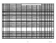
ANNEXURE – 1THE LIST OF CLUSTERS‣ CLUSTER 1.a) MRI Centres1) PMCH Patna2) LNJP Ortho Hospital Rajvanshi Nagarb) CT Scan Centres1) PMCH Patna2) LNJP Ortho Hospital Rajvanshi Nagar3) District Hospital Nalanda4) District Hospital Bhojpur5) District Hospital Buxar6) District Hospital Rohtas7) District Hospital Kaimur‣ CLUSTER 2a) MRI Centres1) ANMMCH Gaya2) NMCH Patnab) CT Scan Centres1) ANMMCH Gaya2) NMCH Patna3) District Hospital Jehanabad4) District Hospital Arwal5) District Hospital Nawada6) District Hospital Aurangabad‣ CLUSTER 3a) MRI Centres1) SKMCH Muzaffarpur12
) CT Scan Centres1) SKMCH Muzaffarpur2) District Hospital Saran3) District Hospital Siwan4) District Hospital Gopalganj5) District Hospital West Champaran6) District Hospital East Champaran7) District Hospital Sitamarhi8) District Hospital Sheohar9) District Hospital Vaishali‣ CLUSTER 4a) MRI Centres1) DMCH Darbhangab) CT Scan Centres1) DMCH Darbhanga2) District Hospital Madhubani3) District Hospital Samastipur4) District Hospital Begusarai5) District Hospital Munger6) District Hospital Sheikhpura7) District Hospital Lakhisarai8) District Hospital Jamui9) District Hospital Khagaria‣ CLUSTER 5a) MRI Centres1) JLNMCH Bhagalpurb) CT Scan Centres1) JLNMCH Bhagalpur2) District Hospital Banka3) District Hospital Saharsa13
4) District Hospital Supaul5) District Hospital Madhepura6) District Hospital Purnia7) District Hospital Kishanganj8) District Hospital Araria9) District Hospital Katihar14
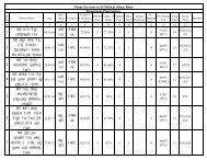
ANNEXURE II‣ Applications <strong>of</strong> MRI Machines at Medical College & Hospital MRI brain and spectroscopy MRI neuro vascular imaging with 12 channel or more coil and angio MRI dorsal/lumbar/cervical/sacrum MRI screening head neck spine imaging without Repositioning MRI upper abdomen 45cm fov in one scan in all axis MRI lower abdomen 45cm fov in one scan in all axis MRIKUB MRI joints with dedicated shoulder coil & knee foot coil MRI breast MR pelvic imaging MRI contrast and non contrast renal angio MRI MRCP MRI whole body 200cm diffusion without repositioning MRI dti and fibertrak imaging MR cartillage color mapping MR contrast kinematic imaging MR neuro perfusion MR blood oxygen level dependent (bold)imaging <strong>of</strong> brain MR sequence <strong>for</strong> triglyceride quantification in liver MR sequence and reconstruction package to get water only, fat only, in phase and out <strong>of</strong>phase in one scan. MR sequence scanning technique to acquire multiple echoes at different TE and generategreyscale and color images <strong>for</strong> assessment <strong>of</strong> iron. MR sequence and post processing <strong>for</strong> cardiac wall motion analysis, Cardiac perfusiondelayed enhancement and non contrast imaging <strong>of</strong> Coronary vasculature MR sequence which uses water in arterial blood as contrast Medium <strong>for</strong> tissue perfusionand quantitative assessment <strong>of</strong> Cerebral blood flow .(asl)Technical Specifications - Specifications <strong>of</strong> High End 1.5 Tesla MRITECHNICAL SPECIFICATIONS Medical Colleges & HospitalS.N1.5 TESLA MAGNETIC RESONANCE IMAGING SYSTEM,16 channelDescription15Competitive bids are invited <strong>for</strong> <strong>installation</strong> <strong>of</strong> 1.5T MRI System 16 channel with state-<strong>of</strong>the-artlatest features commercially available at the time <strong>of</strong> supply. The system should becost effective, with user friendly plat<strong>for</strong>m, reliable & capable <strong>of</strong> providing excellentper<strong>for</strong>mance <strong>for</strong> clinical imaging & research. The detailed specification that follows shallbe understood to be minimum requirement
1 Magneta Whole body 1.5 Tesla Magnetic Resonance Imaging System optimised <strong>for</strong> higherper<strong>for</strong>mance in whole body & vascular examinations with superconducting magnet, highper<strong>for</strong>mance gradients & digital radio frequency systemb 1.5T active shielded super conductive magnet should be short & non-claustrophobiccdefIt should have at least 60 cm patient bore with flared openingMagnet length should be less than 200 cmHomogenecity <strong>of</strong> magnet should be less than 2 ppm over 45 cm DSVThe magnet should be well ventilated & illuminated with built-in 2 way intercom <strong>for</strong>communication with patientg It should have a built-in cryo-cooler such that helium consumption does not exceed 0.05lit / hour2 Shim systema High per<strong>for</strong>mance, highly stable shim system with global & localised automatedshimmimg <strong>for</strong> high homogenicity magnetic field <strong>for</strong> imaging & spectroscopyb Auto shim should be available to shim the magnet with patient in position3 Gradient Systema Actively shielded gradient systemb the gradient should be actively shielded with each axis having independently a slewrate(120). The gradient should be capable <strong>of</strong> providing spatial and temporal resolutionimaging <strong>for</strong> radiology applications with a rise time less than 280 microsecondsc The system should have efficient & adequate eddy current compensationd Effective cooling system <strong>for</strong> gradient coil & power supply4 RF Systema A fully digital RF system with Optical Receive system <strong>for</strong> higher SNRb It should also have independent DIGITAL RF receiver channels with each havingbandwidth <strong>of</strong> 1 MHZ or more along with necessary hardware to support quadrature ICParray / matrix coils.c It should support parallel acquisition techniques with a factor <strong>of</strong> upto 4 in 2Dd Should allow remote selection <strong>of</strong> coils and / or coil elements5 Patient Tablea The table should be fully motorised, computer controlled table movements in vertical &bhorizontal there should directions. be a handheld Horizontal alarm travel <strong>for</strong> patients. should be 200 cm or more.6 Computer system / Image Processor / Operator Consolea The main host computer should have a 19 inches or more high resolution LCD TFT colormonitor with 1024 x 1024 matrix displayb The system should have image storage capacity <strong>of</strong> 100 GB <strong>for</strong> at least 2,00,000 imagesin 256 x 256 matrixc The reconstruction speed should be at least 1000 or more <strong>for</strong> full FOV 256 matrix16
defthe main console should have facility <strong>for</strong> music system <strong>for</strong> patient in the magnet room.The system should have DVD / CD / flash drive archiving facility.Two way intercom system <strong>for</strong> patient communicationMRI system should be DICOM ready in all parameters with no additional requirement <strong>of</strong>licenses <strong>for</strong> connectivity to any PACS / HIS / Radiotherapy treatment planning systemstations7 Measurement Systema Largest field <strong>of</strong> view should be at least 48 cm in all 3 axisb the measurement matrix should be from 128 x 128 to 1024 x1024c Minimum 2D slice thickness should be equal to or less than 0.5mmd minimum 3D slice thickness mm should be equal to or less than 0.1mm8 Coil Systema the main body coil integrated to the magnet must be quadrature/CP. In addition to thisfollowing coils should be quoted .b Multichannel head coils with at least 8 elements <strong>for</strong> high resolution brain imaging.c Neurovascular coil with 16 or more elements or head / neck coil combined, capable <strong>of</strong>high resolution neuro-vascular imagingd Spine array / matrix coils with 12 elements or more <strong>for</strong> thoracic & lumbar spine imaging .System capabale to image Head neck and spine by combining coils without repositioningpatient.e Single Body array / matrix coil with at least 48 cm z axis coverage <strong>for</strong> imaging <strong>of</strong>abdomen, angiograms & heart.fgdedicated Shoulder coil ( not General purpose Flex coil)Suitable coil <strong>for</strong> peripheral angiography application. If inbuilt body coil is used <strong>for</strong> thisplease specify the FOV available and whether multistep s<strong>of</strong>tware/whole body coverage is<strong>of</strong>feredh Bilateral breast coil with at least 4 channel . It should be capable <strong>of</strong> bilateral breastimaging in axial and sagittal planesi Dedicated Knee/ foot coilj Dedicated orbit coilk general purpose flex coil9 Application Sequencesa The system should have basic sequence package with spin echo, inversion recovery,turbo spin echo with high turbo factor <strong>of</strong> 200 or more, gradient echo, FLAIRbSingle slice, multiple single slice, multiple slice, multiple stacks, radial stacks & 3Dcdefg17acquisitions single & multishot <strong>for</strong> all EPI applications imaging techniques with ETL factor <strong>of</strong> 200 or moreFat suppression <strong>for</strong> high quality images both STIR & SPIRThe system should acquire motion artifact free images in T2 studies <strong>of</strong> brain in restlesspatients (propeller, multivane, blade etc)Dynamic study <strong>for</strong> pre & post contrast scans & time intensity studiesMR angio imaging : should have 20 / 30 TOF, 20 / 30 PC, MTS & TONE. ceMRA,facilities <strong>for</strong> accelerated time resolved vascular imaging with applications like treats /tracks / tricks sequences or equivalent
h Fat & water excitation package. Diffusion weighted imaging with at least b value <strong>of</strong> 5000or morei Bolus chasing with automatic & manual triggeringj Non contrast enhanced peripheral angiography <strong>for</strong> arterial flow with native / trance /klmninhance Whole body sequences screening or equivalent imaging studies <strong>for</strong> metastasisWhole body diffusion weighted imaging with background suppressionhigh resolution abdominal & liver imaging in breath hold & free breathing modes withrespirator triggered volume acquisitionsthe system should have basic & advanced MRCP packages including free breathing & 3Do techniques The system should have the hydrogen, single voxel spectroscopy, multi voxel, Thecomplete processing / post processing s<strong>of</strong>tware including color metabolite maps shouldbe available on main console.p Time resolved contrast kinetics like TRICKS/TRACKS/TWIST to be <strong>of</strong>feredq Advanced breast imaging package (bilateral breast imaging protocols)r Perfusion imaging <strong>of</strong> brain with s<strong>of</strong>tware <strong>for</strong> analysiss motion correction <strong>of</strong> imaging like Propeller/Blade/Multivane to be <strong>of</strong>feredt Multi direction DWI & DTI with minimum <strong>of</strong> 32 directions (complete package includingquantification & tractography s<strong>of</strong>tware).u Functional BOLD imaging & evaluation & spectroscopic imagingv system should be capable <strong>of</strong> fat quantification in liver by specfying an region <strong>of</strong> intrestareaw Sequence and application package <strong>for</strong> applying Multi B value diffusion in 3directions simultaneously and also with varying Nex to Improve SNRx Sequence and reconstruction package to get water only, fat only, in phase and out <strong>of</strong>phase in one scany Sequence and application package to utilize water in arterial blood as contrast medium<strong>for</strong> tissue perfusion and quantitative assessment <strong>of</strong> cerebral blood flow.z Scanning technique to acquire multiple echoes at different TE and generate greyscaleand color images <strong>for</strong> assessment <strong>of</strong> IRON.a1 System should have compatibility <strong>for</strong> addition <strong>of</strong> hardware , acquisistion andreconstruction to produce images giving relative stiffness <strong>of</strong> s<strong>of</strong>t tissue.b1 System should have s<strong>of</strong>t wares <strong>for</strong> cardiac wall motion analysis, cardiac perfusion delayedenhancement and non contrast imaging <strong>of</strong> coronary vasculaturec1 Sequence and post processing <strong>for</strong> non invasive T2 mapping <strong>of</strong> cartilage to be <strong>of</strong>feredd1e1aSequence and post processing <strong>for</strong> visualizing micro bleedsTractography facility should be availableWork StationsA workstation with preferably the same user interface as <strong>of</strong> main console is required withthe availability <strong>of</strong> all necessary s<strong>of</strong>tware including basic post-processing s<strong>of</strong>twareincluding MIP, MPR, surface reconstruction & volume rendering techniqueb18It should have at least 21 inch(Medical Grade) LCD TFT color monitor, with hard disk <strong>of</strong>at least 500GB <strong>for</strong> at least 2,50,000 image storage in 256 matrix & 4 GB RAM capacity ormore, with self playing DVD/CD archiving facility.
c The workstation should display cardiac cine images in movie mode .d The workstation should enable printing in laser film camera & color printers11 safety FeaturesThe system should have following safety features:a The magnet system should include an emergency ramp down unit (ERDU) <strong>for</strong> fastreduction <strong>of</strong> the magnetic field with ramp down time below 3 minutesb The magnet should have quench bands that contain the fringe fields to a specified valuein the event <strong>of</strong> a magnet quenchc real time SAR calculation should be per<strong>for</strong>med by s<strong>of</strong>tware to ensure that RF powerlevels comply with regulatory guidelines & are displayed on each imaged The system shall have manual override <strong>of</strong> the motor drive <strong>for</strong> quick removal <strong>of</strong> thepatients from the magnet boree Temperature sensor (built in) <strong>for</strong> magnet refrigeration efficiency must be provided12 Documentationa DICOM compatible dry chemistry laser camera with integrated processor <strong>for</strong> filming frommain console & workstationb Printing on films <strong>of</strong> 14" x 17", 11" x 14" & 10" x 8" sizes in a resolution <strong>of</strong> 500 or more dpi.It should be possible to connect other imaging modalities including CT & ultrasound to theprinter.13 UPSa The system should be provided with on-line UPS system <strong>for</strong> the complete system with atleast 1 hr backup.14 Suitable RF Enclosurea RF cabin: The system should be supplied with RF room shielding, RF door screen &interiors <strong>for</strong> the same should be carried out suitably15 Accessoriesa MR compatible pulse oximeter to be providedb Chiller <strong>for</strong> cold head gradientsc MRI compatible anaesthesia unit & ventilator - all <strong>of</strong> international manufacturer (Thevendor should quote model & make <strong>of</strong> the equipment)d 1 non-ferromagnetic patient transfer trolley <strong>of</strong> international make should beprovided(imported trolley)efgFire fighting system, detectors & 3 fire extinguishers and one MR compatible fireextinguisherhandheld metal detectorsphantoms <strong>for</strong> image quality audits19
1 The model with 'the best and latest technical features’ available with the vendor should bequoted in tender response with original printed vendor data sheets.2 All product catalogues in original.3 A s<strong>of</strong>t copy in word <strong>for</strong>mat in addition to a hard copy to be provided in a CD.4 When the vendor data sheet disagrees with the bid response, clarification shouldaccompany in the <strong>for</strong>m <strong>of</strong> letter/certificates from the principals in original.5 The System should be DICOM – 3MPPS & should be ready to integrate with any existingPACS/HIS System.6 Quoting vendor should have 2 or more working MRI systems in Bihar to confirmavailabiity <strong>of</strong> service.7 Up-time: 95% uptime as per standard government terms and conditions or downtimeconversion to extended warranty8 Silent D.G. set <strong>of</strong> required capacity9 The model quoted must be US FDA approved. Copy <strong>of</strong> the certificate must be submitted10 Installation site approval by AERB will be the responsibility <strong>of</strong> successful bidder .11 All civil/ electrical work at site will be done by successful bidder.20
ANNEXURE – IIISPECIFICATION FOR A WHOLE BODY MULTI-SLICE SPIRAL CT SCANNER 16 sliceWhole body spiral C.T. Scanner (Multi Slice Capabilities with 16 slice per rotations or more andsystem should be currently under production.The System should have following essential features1. X-RAY GENERATOR:High frequency generator on board gantry fitted.The KV range <strong>of</strong> at least 80-140 KVP with minimum 3 or more steps, 80 and 100 KVstation is must <strong>for</strong> pediatric scanning2. X-RAY TUBE:Mention X-Ray Tube with anode heat storage capacity -any scan techniques which canimprove the effective heat capacity if available to be included and quoted Peak Anode heat dissipation rate <strong>of</strong> at least 500KHU/min. e. Mention focal spotsize3. GANTRY AND SCANNING TABLE:Gantry Aperture <strong>of</strong> at least 65 cms.Gantry tilt <strong>of</strong> at least -30/+30 degree or equi valen t digital tilt w i t h same capabilityScan Field <strong>of</strong> View (FOV)<strong>of</strong> at least 40cm or above. At least two Scan Field <strong>of</strong> Viewshould be possible, mention themGantry should have a small foot print smaller the better. Scanning Table load <strong>of</strong> at least 200kg o r m o r e with position accuracy <strong>of</strong> 0.25mm f.Metal free Scanable range <strong>of</strong> at least100cm. Gantry should have v i s u a l instruction display <strong>for</strong> patient breadth hold IndicationDedicated Pediatrics Scan FOV should be available4. OPERATOR CONSOLE:The operators should have architecture to allow simultaneous scan, reconstruct, andarchive. With a RAM <strong>of</strong> at least 16GB or more and a hard disk capacity to store at least250000 images in 512 matrix. The console should allow simultaneous scan,reconstruct, and archive. The total hard disk capacity should be more than 500 GB <strong>for</strong>image storage, raw data storage and application s<strong>of</strong>tware. Custom <strong>design</strong>ed Key21
oard that includes all <strong>of</strong> the controls necessary to control scan, display and archiveincluding emergency stop and patient intercom.Laser Camera functions shall be initiated from operator’s console.Multi <strong>for</strong>mat filming with flexible filming <strong>for</strong>mat should be possible5. Detector16 Rows <strong>of</strong> Solid State detectors <strong>of</strong> rare earth.It should be free from repeated Calibrations.The number <strong>of</strong> detectors elements/row should be more than 750 physical elementsin a single row.Minimum Slice Thickness should be 0.625mm or lower.The Z axis detector width <strong>for</strong> sub mm slice should be equal or more than 10mm.6. HIGH CONTRAST SPATIAL RESOLUTION:It should be at least 15lp/cm at 0% MTF using clinical algorithm7. LOW CONTRAST DETECTABILITY:5mm or less @0.3% using 20cm CATPHAN on 10mm slice thickness. Mention the doseto achieve this IQ.8. SCAN TIME: Mention the minimum rotation time - should be less than or equal to 1s <strong>for</strong> 360degrees.Mention all scan times possible with the sytem in Axial and Spiral mode.9. SLICE THICKNESS:The system Should provide multiple selections <strong>of</strong> slice thicknesses. Minimum slice thicknessshould be 0.625mm or lower.10. SPIRAL MODE SPECIFICATIONS :Continuous data acquisition with overlapping slices.22
Maximum helical <strong>for</strong> single continuous spiral <strong>of</strong> at least 120 Seconds.Spiral mode must be extended spiral, back to back spiral and multi spiral as well as tiltedand should be available on all the slice thickness <strong>of</strong>fered.There should be algorithmic correction <strong>for</strong> cone beam artifact <strong>for</strong> highpitch spiral. Mentionthe maximum table speed/s with cone beam correction.Dedicated pediartic protocols with colour coded or BMI based protocol selection must beavailable.11. IMAGE PROCESSING SYSTEM:Main CPU unit <strong>of</strong> at least Random Accessory Memory(RAM) at least 4GB with 2.33 GHz dualor quad processorsImage reconstruction speed must be more than 5 images.12. IMAGE PROCESSING SYSTEM: Image Reconstruction matrix at least 512 x 512. Display matrix at least 1024x1024.High resolution colour LCD Monitors 19"…2nos(Medical Grade).13. IMAGE STORAGE:Image storage Capacity <strong>of</strong> at least 250000 images in 512 matrix.14. IMAGE ARCHIVING:Image archiving on DVD/ CD/MOD should be available. In case <strong>of</strong> MOD -10MODsrewritable may be supplied. In case <strong>of</strong> CD-1000CDs may be supplied along with.15. FILMING:Laser Camera DICOM Compatible, with latest model camera should be able to take allstandard make <strong>of</strong> films.16. WORKSTATION: The workstation should be <strong>of</strong> at latest version with Dual LCD Monitors(MedicalGrade) and with RAM <strong>of</strong> at least 12 GB & Hard disc capacity <strong>of</strong> minimum 200 GB .Alsomust have fastest reconstruction time available with the tenderer and should haveDICOM-3.0 compatibility. It should have parallel processing capabilities. Direct filmingfacility from the main console and workstation must be provided. 3D rendering methods - Following 3D rendering methods should be possible - Surface,MiP, MinIP, RaySum, Integral, SSD, VED Multi object merging, Joint disarticulation should be possible23
17. IMAGE TRANSFORMER/NETWORKING: The unit should have DICOM Interface <strong>for</strong> transmitting images and in<strong>for</strong>mation inDICOM standard and also to permit communication between devices <strong>of</strong> variousmanufactures. The unit should have provision <strong>for</strong> connectivity <strong>of</strong> the HospitalIn<strong>for</strong>mation System/Radiology In<strong>for</strong>mation System.18. STANDARD SOFTWARES:Routine s<strong>of</strong>tware <strong>for</strong> image evaluation and display with minimum three region <strong>of</strong><strong>interest</strong> (ROI), Angles and distance measurements. Histogram display pr<strong>of</strong>ile, symmetrycomparison, variable, multiple image display with independent window image, annotationand labelling, image addition and subtraction and volume artifact reduction capability,reversal <strong>of</strong> gray scale value and image filter functions reference scales topogram evaluationetc.The CT console should have 3D and 2D processing capabilities such as 3D surface, Volumerendering, multiobject merge, CT angio. MPR, MPVR, Virtual Scopy etcDirect Multiplannar reconstruction should be standard feature - should be able to create MPRimgaes in atleast in 3 orthogonal planes directly during scan.Dynamic CT Scanning, oblique MPR, MIP capabilities to be provided as standard s<strong>of</strong>tware.Real time re<strong>for</strong>ming <strong>of</strong> secondary views with reconstruction facilities in saggital, coronal,paraxial, oblique and irregular (curvilinear). Outline should be determined in the topogramor in sagital images.Zooming by reconstruction <strong>of</strong> the raw data. Image planning and zooming should bepossible. Real time monitoring <strong>of</strong> contrast bolus injection. All these s<strong>of</strong>tware’s are to beavailable on the main console.Should have Virtual endoscopy s<strong>of</strong>tware <strong>for</strong> visualization <strong>of</strong> vessels and air filled structuresand Colonography s<strong>of</strong>tware <strong>for</strong> virtual endoscopic, colon study. Should have the facility <strong>of</strong>Perfusion CT <strong>for</strong> study <strong>of</strong> dynamic data <strong>of</strong> brain following contrast and study <strong>of</strong> cerebralperfusion.19. ESSENTIAL ACCESSORIES TO BE INCLUDED WITH THE UNIT:UPS On- line UPS with MF batteries <strong>for</strong> the main computer system, digital imagingprocess and provision <strong>of</strong> light in console room and gantry room with backup time<strong>of</strong> 1hr.Voltage Stabilizer: Servo Voltage stabilizer <strong>for</strong> whole unit including accessories.The voltage vary at this Institute between 330-460V.24
Color Printer : Please <strong>of</strong>fer color printer to take color images plain paper only <strong>of</strong>VR / CTA/CTP images on plain paper.Lead Glass: 60cm X 120 cm or more with lead equivalent as per the safety norms.e. Pressure Injector: CT pressure injector with single head - remote console <strong>of</strong>Medrad or equivalent make latest model with inter phase s<strong>of</strong>tware along with200 disposable syringes with connectors.Silent D.G. set <strong>of</strong> required capacityLatest model should be installed.20. The model quoted must be US FDA approved. Copy <strong>of</strong> the certificate must be submitted with the<strong>of</strong>fer.21. Installation site approval by AERB will be the responsibility <strong>of</strong> successful bidder .22. All civil/ electrical work at site will be done by successful bidderSpecification <strong>of</strong> 128 Slice Spiral CT Scanner (MCH)Features :The 128 Multi slice Spiral CT Scanner <strong>design</strong>ed <strong>for</strong> the whole body and cardiac scanning The system consisting <strong>of</strong> Gantry, Couch, Operating console with 3D, re<strong>for</strong>mat and volumerendering processing capabilities, Separate Multi-Modality post processing workstation. Filmingwith DICOM compatible Dry laser Imager, Color laser printer, Dual Head power injector. An Online UPS <strong>for</strong> the complete system <strong>for</strong> backup power in case <strong>of</strong> mains failure and fluctuation Image storage in system HD and image transfer through CD/DVD Writer The system must have DICOM (latest version) on online data /image transfer capability Rotating anode type X-ray tube and Ceramic / Solid state detector Automatic injector synchronized with the system Voice link: Vocal instruction the patient.Specification :Gantry & Scan Parameter : Scan Regions : Whole body including Head Gantry Aperture : 60 cm or more Scan field : 50 cm or more Scan time : 0.40sec or faster Recon Slice thickness : adjustable from 0.625mm to 5 mm Recon time : Min 16-20 images /secX-Ray Generator : X-Ray Generator : 70 kw or higher X-Ray Generator type : High frequency Inverter type X-ray Exposure : Continuous 100s or aboveX-Ray Tube Computer controlled monitoring <strong>of</strong> anode temperature25
X-Ray Tube Voltage : Adjustable from 80 to 140 kVX-Ray Tube Current : 20 to 600mA or moreAnode heat storage capacity : 6 MHU or higherCooling rate : 800 KHU/min or moreX-Ray Detection Detector Material : Ceramic/ Solid state Detector Z-axis detector width : 40mm or more Number <strong>of</strong> slices /rotation : capable <strong>of</strong> acquiring 128 slice/rotation Number <strong>of</strong> detector rows : at least 64 or more Coverage per rotation in axial mode : 40 mm or more Numer <strong>of</strong> detector element s/row : 1200 or moreImage quality Specifications High Contrast detectability (using clinical high resolution algorithm) – helical mode X-Y planeshould be more than 17lp/cm and Z –plane should be more than 19lp/cm Low Contrast detectability state at 3 mm at 0.32% contrast difference, specify the dose, using 8inch catphan phantom. (lower dose system will be preferred) Latest Iterative Reconstruction method (such as Safire, ASiR, IMR, AIDR ) is to be quoted asstandard.Patient Table Remote control <strong>of</strong> the couch from console The table move vertically and the table top moves longitudinally Motor driven couch top movement Load capacity : 200 kg or more Scanable range: 1700 mm or more.Cardiac Acquisition Mode Prospective Gated Coronary acquisition The total time taken to cover 12cm <strong>of</strong> heart in Prospective gated acquisition mode Retrospective gating should be possible Minimal temporal resolution in multi sector mode must be less than or equal to 45mS Should be able to combine gated and non-gated acquisition in a single seriesPediatric Scan Pediatric Scan FOV should be available Dedicated BMI based/colour coded pediatric patientAcquisition consoleHigh per<strong>for</strong>mance computer <strong>for</strong> data acquisition, image reconstruction and routine post processing Processor : 2 x Xeon 2.6 or more GHz Dual Core Monitor : TFT/ LCD 21” (medical grade) or more with 1280 x 1024 or better resolution RAM : 18 GB or more HDD : Raw data 1TB & Image data 800GB or more Graphics Accelerator : For fast 3D processing DVD / CD writer : CD ( 700 MB or more ), DVD ( 4.7 GB or more ) DICOM VIEWER : Automatically included in every CD / DVD <strong>for</strong> viewing in viewer’s PC USB port<strong>for</strong> connecting USB storage devices like pen drive, external hard disk etc.26
S<strong>of</strong>tware: MPR ( Multi Planer Reconstruction ) 3D SSD ( Shaded Surface Display ) VRT ( Volume Rendering Technique ) <strong>for</strong> advanced 3D application Bolus CT Video Capture and Editing Tool Fly through Double – oblique image from standard scanning protocol Cardiac post processingMulti Modality Post processing work station – 1numberHigh per<strong>for</strong>mance computer with advanced multi modality post processing facilityThis should be from the same CT manufacturer to ensure full functionality Processor : 2 x Xeon 2.6 or more GHz Dual Core Monitor : TFT /LCD medical grad 21” or more, with 1280 x 1024 or better resolution RAM : 16 GB or more HDD : Preferably 1TB or more Graphics Accelerator : For fast 3D processing with additional 2 GB image memory DVD / CD writer : Minimum 16X DICOM Viewer : Automatically included in every CD / DVD <strong>for</strong> viewing in viewer’s PCSpecial Processing S<strong>of</strong>t wares <strong>for</strong> Multi- Modality cardiac workstation : Volume rendering with multi object merging Fly through (Virtual Endoscopy with Virtual bronchoscopy and CT Colonoscopy facilities) Bone Removal S<strong>of</strong>tware Automatic Quantification <strong>of</strong> stenosis and plaque analysis Automatic Vessels Measurement Automatic coronary vessel tree with auto labeling <strong>of</strong> vessels. Myocardial relative perfusion is must Perfusion <strong>for</strong> Neuro and body applications Calcium scoring s<strong>of</strong>tware Cardiac function with LV function analysisAdditional fully functional independent work station <strong>of</strong> similar capacity as satellite work station shouldbe installed in Radiology Department with registered Pulmonary and Cardiac S<strong>of</strong>tware.CT Fluoroscopy facility <strong>for</strong> CT Guided FNAC and other interventional procedures (to be quotedseparately)Dual Head CT pressure injector with compatible syringe -100 pairs <strong>of</strong> syringes with pressure tubing andother required material to be suppliedFlow rate: 0.1- 10 ml/sec in 0.1 ml incrementsProgrammable pressure limit <strong>for</strong> 200 ml syringe : 2241 kPaSyringe : 200 mlDisplay control unit with standDry Laser imager : Resolution : 500 pi or more Throughput : 70 films /h more Film size : 35 x 43 cm / 35 x 35 cm- one size online27
Color laser printer: Resolution : 600 x 600 dpi Throughput : 30 pages / min Media types : A UPS : True on line UPS with backup <strong>of</strong> one hour <strong>for</strong> the whole system in case <strong>of</strong> mains failure & Generator: Silent DG set should be installed separately <strong>for</strong> running the machines. Lead Aprons: 6 sets with thyroid protector and radiation protector glass spectacles. Lead glass : 100 cm x 80 cm lead glass between machine room and control room <strong>for</strong> radiationprotection. Quality Standard: Model to be quoted must be USFDA certified must be submitted with the <strong>of</strong>fer. AERB type approval / NOC is to be attached. Latest model should be preferred. Installation site approved by AERB will be responsibility <strong>of</strong> the successful bidder. All civil and electrical work should be done by successful bidder.Bidder are required to have silent D.G. set <strong>of</strong> required capacity at the center.28