

A rare case of Verruciform Xanthoma-pdf - Wonder Smile Clinic
A rare case of Verruciform Xanthoma-pdf - Wonder Smile Clinic
A rare case of Verruciform Xanthoma-pdf - Wonder Smile Clinic

You also want an ePaper? Increase the reach of your titles
YUMPU automatically turns print PDFs into web optimized ePapers that Google loves.
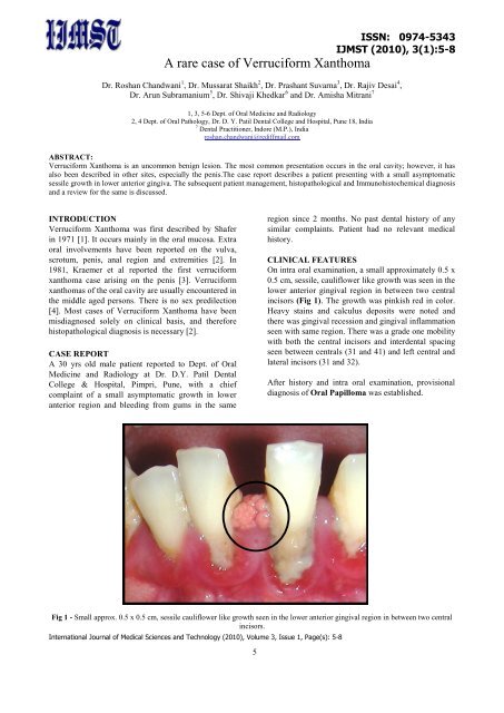
A <strong>rare</strong> <strong>case</strong> <strong>of</strong> <strong>Verruciform</strong> <strong>Xanthoma</strong>ISSN: 0974-5343IJMST (2010), 3(1):5-8Dr. Roshan Chandwani 1 , Dr. Mussarat Shaikh 2 , Dr. Prashant Suvarna 3 , Dr. Rajiv Desai 4 ,Dr. Arun Subramanium 5 , Dr. Shivaji Khedkar 6 and Dr. Amisha Mitrani 71, 3, 5-6 Dept. <strong>of</strong> Oral Medicine and Radiology2, 4 Dept. <strong>of</strong> Oral Pathology, Dr. D. Y. Patil Dental College and Hospital, Pune 18, India7 Dental Practitioner, Indore (M.P.), Indiaroshan.chandwani@rediffmail.comABSTRACT:<strong>Verruciform</strong> <strong>Xanthoma</strong> is an uncommon benign lesion. The most common presentation occurs in the oral cavity; however, it hasalso been described in other sites, especially the penis.The <strong>case</strong> report describes a patient presenting with a small asymptomaticsessile growth in lower anterior gingiva. The subsequent patient management, histopathological and Immunohistochemical diagnosisand a review for the same is discussed.INTRODUCTION<strong>Verruciform</strong> <strong>Xanthoma</strong> was first described by Shaferin 1971 [1]. It occurs mainly in the oral mucosa. Extraoral involvements have been reported on the vulva,scrotum, penis, anal region and extremities [2]. In1981, Kraemer et al reported the first verruciformxanthoma <strong>case</strong> arising on the penis [3]. <strong>Verruciform</strong>xanthomas <strong>of</strong> the oral cavity are usually encountered inthe middle aged persons. There is no sex predilection[4]. Most <strong>case</strong>s <strong>of</strong> <strong>Verruciform</strong> <strong>Xanthoma</strong> have beenmisdiagnosed solely on clinical basis, and thereforehistopathological diagnosis is necessary [2].CASE REPORTA 30 yrs old male patient reported to Dept. <strong>of</strong> OralMedicine and Radiology at Dr. D.Y. Patil DentalCollege & Hospital, Pimpri, Pune, with a chiefcomplaint <strong>of</strong> a small asymptomatic growth in loweranterior region and bleeding from gums in the sameregion since 2 months. No past dental history <strong>of</strong> anysimilar complaints. Patient had no relevant medicalhistory.CLINICAL FEATURESOn intra oral examination, a small approximately 0.5 x0.5 cm, sessile, cauliflower like growth was seen in thelower anterior gingival region in between two centralincisors (Fig 1). The growth was pinkish red in color.Heavy stains and calculus deposits were noted andthere was gingival recession and gingival inflammationseen with same region. There was a grade one mobilitywith both the central incisors and interdental spacingseen between centrals (31 and 41) and left central andlateral incisors (31 and 32).After history and intra oral examination, provisionaldiagnosis <strong>of</strong> Oral Papilloma was established.Fig 1 - Small approx. 0.5 x 0.5 cm, sessile cauliflower like growth seen in the lower anterior gingival region in between two centralincisors.International Journal <strong>of</strong> Medical Sciences and Technology (2010), Volume 3, Issue 1, Page(s): 5-85
ISSN: 0974-5343IJMST (2010), 3(1):5-8RADIOGRAPHIC FEATURESIntra Oral Periapical radiograph <strong>of</strong> lower anteriorregion reveals severe horizontal bone loss andradioopaque interproximal calculus (Fig2).DIFFERENTIAL DIAGNOSIS1. Pyogenic granuloma2. Peripheral Giant cell granuloma3. Common Wart4. Condyloma acuminatum5. Focal epithelial hyperplasia6. Peripheral ossifying fibroma7. Eosinophilic granulomaFig 2 – Intra Oral RadiographFINAL DIAGNOSISAfter complete oral prophylaxis, excision <strong>of</strong> the lesionwas performed under local anesthesia after sensitivitytest. Excision was performed in two pieces (Fig3) andthe sample was sent for histopathological examination.Histopathologic examination <strong>of</strong> hematoxylin and eosinstained slide showed, hyperparakeratotic stratifiedsquamous epithelium without dysplasia arranged in apapillary architecture. The underlying matureconnective tissue revealed presence <strong>of</strong> large foam cellsin the papillary area. The cytoplasm <strong>of</strong> foam cellsseemed to be granular in nature. The overallhistopathological features were suggestive <strong>of</strong><strong>Verruciform</strong> <strong>Xanthoma</strong> (Fig 4).Fig 3 - Excision <strong>of</strong> the lesion in two piecesImmunohistochemical staining using antimacrophageantibody – CD 68 showed the foam cells asmonocyte/macrophage lineage, confirming thediagnosis <strong>of</strong> a verruciform xanthoma (Fig 5 and Fig6).Foam cells are strongly positive for CD-68 seen inconnective tissue. Distribution <strong>of</strong> CD-68 forms adelicate network delineating small vessels.Follow up was done after 15 days (Fig 9) and thepatient was sent for further periodontal treatmentFig 4 – Histopathological diagnosis <strong>of</strong> <strong>Verruciform</strong><strong>Xanthoma</strong>International Journal <strong>of</strong> Medical Sciences and Technology (2010), Volume 3, Issue 1, Page(s): 5-86
ISSN: 0974-5343IJMST (2010), 3(1):5-8Fig 5 – Scanner viewFig 6 – High Power viewFig 7 - PAS without diastaseFig 8 - PAS with diastaseFig 9 - Follow up after 15 daysInternational Journal <strong>of</strong> Medical Sciences and Technology (2010), Volume 3, Issue 1, Page(s): 5-87
DISCUSSION<strong>Verruciform</strong> <strong>Xanthoma</strong> is an uncommon lesion thatusually occurs on the oral mucosa <strong>of</strong> middle-agedpersons or on the scrotum <strong>of</strong> middle-aged to elderlyJapanese men.The most common site for <strong>Verruciform</strong> <strong>Xanthoma</strong> isthe oral mucosa. Lesions that occur elsewhere usuallyarise on the perineum or on the skin with somepredisposing factor, such as lymphedema1 or anepidermal nevus [5]. It is also known as HistiocytosisY [4].The pathophysiology <strong>of</strong> <strong>Verruciform</strong> <strong>Xanthoma</strong> isunknown. Many authors consider it to be a reactiveprocess rather than a true neoplasm. Damage to thesquamous cells with increased epithelial cell turnover,leading to the deposition <strong>of</strong> epithelial cell debris that isengulfed by macrophages in the corium may lead tothe development <strong>of</strong> this lesion. Patients usuallypresent with a history <strong>of</strong> an asymptomatic or tenderlesion on the skin or mucosa [6].The clinical appearance <strong>of</strong> a <strong>Verruciform</strong> <strong>Xanthoma</strong> isnot diagnostic; the diagnosis is almost always made athistologic examination. Depending on the nature <strong>of</strong> theindividual lesion, <strong>Verruciform</strong> <strong>Xanthoma</strong> mayclinically resemble any Verrucous, papillary, orlichenoid oral lesion, particularly any such lesion thatis also hyperkeratotic. It is frequently misdiagnosed atclinical examination as a papilloma [6].It can occur at any site and is more frequently found onthe gingiva or alveolar ridge followed by buccalmucosa, palate, floor <strong>of</strong> mouth, lip and lowermuccobuccal fold, it has got normal or red in color butsometimes pale or hyperkeratotic with a rough pebblysurface and either a sessile or pedunculated base [4].Lesions have also been found in the oral cavity inassociation with lichen planus [7], pemphigus vulgaris[8], oral bullae, carcinoma in situ [9] or franksquamous cell carcinoma [10], the lesions were als<strong>of</strong>ound in a bone marrow transplant recipient [11].The most striking and characteristic histologic feature<strong>of</strong> the <strong>Verruciform</strong> <strong>Xanthoma</strong> is the presence <strong>of</strong> largefoam cells in the connective tissue papillae.ISSN: 0974-5343IJMST (2010), 3(1):5-8Ultrastructurally, most studies have concluded that thefoam cells in verruciform xanthoma are fat-ladenmacrophages. Immunohistochemical analysis revealedthese cells to be positive for CD68.They are also periodic-acid-Schiff (PAS) positive anddiastase resistant, indicating that the PAS-positivematerial is not glycogen [6].Local surgical excision is almost always curative. Theprognosis is excellent after local surgical excision.Recurrence is <strong>rare</strong> [6].REFERENCES[1] Shafer W.G. <strong>Verruciform</strong> xanthoma. Oral Surg Oral MedOral Pathol 1971; 31:784-9.[2] Tongli Xia, Guizhong Li, In Sun Jun1, Yanqun, NaYinglu Guo, Moon Kee Chung Korean J Urol 2004;45:297-298.[3] Kraemer BB, Schmidt WA, Foucar E, Rosen T.<strong>Verruciform</strong> xanthoma <strong>of</strong> the penis. Arch Dermatol 1981;117:516-8.[4] Shafer, Hine, Levy, a Textbook <strong>of</strong> Oral Pathology, 3 rdedition 153.[5] Grosshans E, Laplanche G. <strong>Verruciform</strong> xanthoma orxanthomatous transformation <strong>of</strong> inflammatory epidermalnevus? J Cutan Pathol. Oct 1981; 8(5):382-4.[6] Hong Li, W Clark Lambert, <strong>Verruciform</strong> <strong>Xanthoma</strong>,Article Last Updated: Nov 9, 2007, e-medicine.[7] Miyamoto Y, Nagayama M, Hayashi Y. <strong>Verruciform</strong>xanthoma occurring within oral lichen planus. J OralPathol Med. Apr 1996; 25(4):188-91.[8] Gehrig RD, Baughman RA, Collins JF. <strong>Verruciform</strong>xanthoma in a young male patient with a past history<strong>of</strong> pemphigus vulgaris. Oral Surg Oral Med OralPathol. Jan 1983; 55(1):58-61.[9] Drummond JF, White DK, Damm DD, CramerJR. <strong>Verruciform</strong> xanthoma within carcinoma insitu. J Oral Maxill<strong>of</strong>ac Surg. Apr 1989; 47(4):398-400.[10] Mannes KD, Dekle CL, Requena L, SanguezaOP. <strong>Verruciform</strong> xanthoma associated with squamouscell carcinoma. AmJDermatopathol. Feb 1999;21(1):66-9.[11] Allen CM, Kapoor N. <strong>Verruciform</strong> xanthoma in abone marrow transplant recipient. Oral Surg OralMed Oral Pathol. May 1993; 75(5):591-4.These cells characteristically fill the entire papilla butonly <strong>rare</strong>ly extend beyond the base <strong>of</strong> the papilla. Mostor all <strong>of</strong> the papillae are involved with these cells,which occasionally may also be seen in the epithelium(i.e., epidermis, mucosa) [6].International Journal <strong>of</strong> Medical Sciences and Technology (2010), Volume 3, Issue 1, Page(s): 5-88