indications and technique of the weil osteotomy - The Podiatry Institute
indications and technique of the weil osteotomy - The Podiatry Institute
indications and technique of the weil osteotomy - The Podiatry Institute
You also want an ePaper? Increase the reach of your titles
YUMPU automatically turns print PDFs into web optimized ePapers that Google loves.
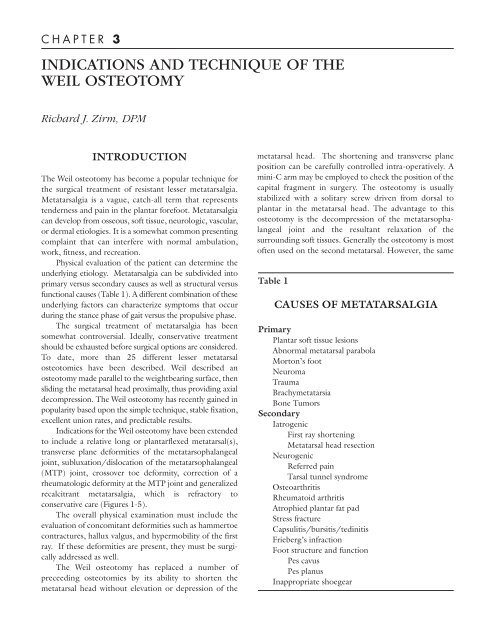
C H A P T E R 3INDICATIONS AND TECHNIQUE OF THEWEIL OSTEOTOMYRichard J. Zirm, DPMINTRODUCTION<strong>The</strong> Weil <strong>osteotomy</strong> has become a popular <strong>technique</strong> for<strong>the</strong> surgical treatment <strong>of</strong> resistant lesser metatarsalgia.Metatarsalgia is a vague, catch-all term that representstenderness <strong>and</strong> pain in <strong>the</strong> plantar forefoot. Metatarsalgiacan develop from osseous, s<strong>of</strong>t tissue, neurologic, vascular,or dermal etiologies. It is a somewhat common presentingcomplaint that can interfere with normal ambulation,work, fitness, <strong>and</strong> recreation.Physical evaluation <strong>of</strong> <strong>the</strong> patient can determine <strong>the</strong>underlying etiology. Metatarsalgia can be subdivided intoprimary versus secondary causes as well as structural versusfunctional causes (Table 1). A different combination <strong>of</strong> <strong>the</strong>seunderlying factors can characterize symptoms that occurduring <strong>the</strong> stance phase <strong>of</strong> gait versus <strong>the</strong> propulsive phase.<strong>The</strong> surgical treatment <strong>of</strong> metatarsalgia has beensomewhat controversial. Ideally, conservative treatmentshould be exhausted before surgical options are considered.To date, more than 25 different lesser metatarsalosteotomies have been described. Weil described an<strong>osteotomy</strong> made parallel to <strong>the</strong> weightbearing surface, <strong>the</strong>nsliding <strong>the</strong> metatarsal head proximally, thus providing axialdecompression. <strong>The</strong> Weil <strong>osteotomy</strong> has recently gained inpopularity based upon <strong>the</strong> simple <strong>technique</strong>, stable fixation,excellent union rates, <strong>and</strong> predictable results.Indications for <strong>the</strong> Weil <strong>osteotomy</strong> have been extendedto include a relative long or plantarflexed metatarsal(s),transverse plane deformities <strong>of</strong> <strong>the</strong> metatarsophalangealjoint, subluxation/dislocation <strong>of</strong> <strong>the</strong> metatarsophalangeal(MTP) joint, crossover toe deformity, correction <strong>of</strong> arheumatologic deformity at <strong>the</strong> MTP joint <strong>and</strong> generalizedrecalcitrant metatarsalgia, which is refractory toconservative care (Figures 1-5).<strong>The</strong> overall physical examination must include <strong>the</strong>evaluation <strong>of</strong> concomitant deformities such as hammertoecontractures, hallux valgus, <strong>and</strong> hypermobility <strong>of</strong> <strong>the</strong> firstray. If <strong>the</strong>se deformities are present, <strong>the</strong>y must be surgicallyaddressed as well.<strong>The</strong> Weil <strong>osteotomy</strong> has replaced a number <strong>of</strong>preceeding osteotomies by its ability to shorten <strong>the</strong>metatarsal head without elevation or depression <strong>of</strong> <strong>the</strong>metatarsal head. <strong>The</strong> shortening <strong>and</strong> transverse planeposition can be carefully controlled intra-operatively. Amini-C arm may be employed to check <strong>the</strong> position <strong>of</strong> <strong>the</strong>capital fragment in surgery. <strong>The</strong> <strong>osteotomy</strong> is usuallystabilized with a solitary screw driven from dorsal toplantar in <strong>the</strong> metatarsal head. <strong>The</strong> advantage to this<strong>osteotomy</strong> is <strong>the</strong> decompression <strong>of</strong> <strong>the</strong> metatarsophalangealjoint <strong>and</strong> <strong>the</strong> resultant relaxation <strong>of</strong> <strong>the</strong>surrounding s<strong>of</strong>t tissues. Generally <strong>the</strong> <strong>osteotomy</strong> is most<strong>of</strong>ten used on <strong>the</strong> second metatarsal. However, <strong>the</strong> sameTable 1CAUSES OF METATARSALGIAPrimaryPlantar s<strong>of</strong>t tissue lesionsAbnormal metatarsal parabolaMorton’s footNeuromaTraumaBrachymetatarsiaBone TumorsSecondaryIatrogenicFirst ray shorteningMetatarsal head resectionNeurogenicReferred painTarsal tunnel syndromeOsteoarthritisRheumatoid arthritisAtrophied plantar fat padStress fractureCapsulitis/bursitis/tedinitisFrieberg’s infractionFoot structure <strong>and</strong> functionPes cavusPes planusInappropriate shoegear
10CHAPTER 3considerations including length patterns, transverse planedeformities, subluxations, <strong>and</strong> intractable plantar skinlesions sometime merit <strong>the</strong> <strong>osteotomy</strong> to be preformed on<strong>the</strong> third <strong>and</strong> fourth metatarsal.Possible complications <strong>of</strong> <strong>the</strong> Weil <strong>osteotomy</strong> includea dorsiflexed, nonpurchasing toe, pain on MTP jointdorsiflexion, reduced MTP joint range <strong>of</strong> motion,proximal interphalangeal joint contracture <strong>and</strong> a floppytoe. <strong>The</strong> relaxed tension <strong>of</strong> <strong>the</strong> plantar fascial insertion at<strong>the</strong> base <strong>of</strong> <strong>the</strong> proximal phalangeal base may produce afloating toe. Long or prominent plantar fixation can alsocause postoperative pain <strong>and</strong> stiffness <strong>and</strong> needs to beremoved once <strong>the</strong> <strong>osteotomy</strong> is healed.Figure 1. Preoperative <strong>and</strong> postoperative view <strong>of</strong> along second metatarasal.Figure 2. Preoperative <strong>and</strong> postoperative view <strong>of</strong> ashortened first ray.Figure 3. Preoperative <strong>and</strong> postoperative view <strong>of</strong>hallux varus.Figure 4. Preoperative <strong>and</strong> postoperative view <strong>of</strong>hallux abducto valgus with second ray adductus.
CHAPTER 3 11Figure 5. Preoperative <strong>and</strong> postoperative view <strong>of</strong>digital adductus.TECHNIQUE TIPS AND PEARLSA 3.5- to 4.0-cm linear incision is made over <strong>the</strong> distalthird <strong>of</strong> <strong>the</strong> lesser metatarsal ending at <strong>the</strong> base <strong>of</strong> <strong>the</strong>proximal phalanx. This will reduce <strong>the</strong> chance <strong>of</strong> scar formationthat can contribute postoperative dorsal contracture.If digital work is done at <strong>the</strong> same time, a lazy-Sincision is made over <strong>the</strong> MTP joint as <strong>the</strong> incision endsdorsally over <strong>the</strong> middle phalanx.<strong>The</strong> extensor tendons are <strong>the</strong>n retracted laterally <strong>and</strong><strong>the</strong> dorsal periosteum <strong>and</strong> capsule are incised with <strong>the</strong> toedorsiflexed to protect <strong>the</strong> articular cartilage. <strong>The</strong> collateralligaments are partially incised to fully expose <strong>the</strong> metatarsalhead. <strong>The</strong>y do not need to be completely resected. Next,a McGlamry elevator may be used to release <strong>the</strong> plantarplate adhesions at <strong>the</strong> joint especially in <strong>the</strong> case <strong>of</strong> asignificant contracture. <strong>The</strong> McGlamry elevator strips <strong>the</strong>plantar synovial attachments (vincula), allowing <strong>the</strong>plantar plate to shift more proximally with <strong>the</strong> <strong>osteotomy</strong>.<strong>The</strong>re is rarely vascular compromise to <strong>the</strong> capital fragment<strong>and</strong> avascular necrosis <strong>of</strong> <strong>the</strong> head is extremely rare.<strong>The</strong> McGlamry elevator can double as a retractor for<strong>the</strong> ensuing <strong>osteotomy</strong>. <strong>The</strong> head should be fully visualizedbefore <strong>the</strong> <strong>osteotomy</strong> is made. <strong>The</strong> <strong>osteotomy</strong> is started2-mm inferior to <strong>the</strong> most dorsal aspect <strong>of</strong> <strong>the</strong> articularcartilage at an approximately 25° angle with respect to <strong>the</strong>metatarsal shaft. <strong>The</strong> <strong>osteotomy</strong> can be as long as 2.5- to3-cm in st<strong>and</strong>ard foot conditions.<strong>The</strong> plane <strong>of</strong> <strong>the</strong> <strong>osteotomy</strong> is made parallel to <strong>the</strong>ground as if <strong>the</strong> foot was bearing weight. <strong>The</strong> angle <strong>of</strong> <strong>the</strong><strong>osteotomy</strong> may vary according to <strong>the</strong> angle <strong>of</strong> <strong>the</strong>metatarsal declination. <strong>The</strong> angle <strong>of</strong> <strong>the</strong> <strong>osteotomy</strong> mustincrease toward <strong>the</strong> more lateral metatasals because <strong>the</strong>yare less plantarflexed than <strong>the</strong> second metatarsal.In severely plantarflexed metarsals, a second <strong>osteotomy</strong>is performed in effect to remove a slice <strong>of</strong> bone to fur<strong>the</strong>rdorsiflex <strong>the</strong> capital fragment. An easier, more stable tip isto use a double saw blade or a thicker saw blade to obtain <strong>the</strong>same goal <strong>of</strong> removing more bone to dorsally elevate <strong>the</strong>metatarsal head.When <strong>the</strong> <strong>osteotomy</strong> is completed, <strong>the</strong> metatarsal headwill suddenly shift to a more proximal position. <strong>The</strong>re is acertain neutral position that reflects <strong>the</strong> relaxation <strong>of</strong> <strong>the</strong>surrounding s<strong>of</strong>t tissues that feels just right. <strong>The</strong>n withmanual pressure, a solitary K-wire or bone clamp is used totemporarily fixate <strong>the</strong> <strong>osteotomy</strong>. <strong>The</strong> position can bechecked by fluoroscopy at this time. <strong>The</strong> metatarsal headposition can be changed from 2- to 10-mm with 3- to5-mm being <strong>the</strong> normal amount <strong>of</strong> shortening.<strong>The</strong> <strong>osteotomy</strong> can be slightly angulated in <strong>the</strong> frontalplane if more dorsal excursion is desired. <strong>The</strong> head can alsobe rotated <strong>and</strong> translocated somewhat in more difficulttransverse or crossover toe cases. However, too muchmanipulation <strong>of</strong> <strong>the</strong> <strong>osteotomy</strong> or displacement <strong>of</strong> capitalfragment can sacrifice <strong>the</strong> overall stability <strong>of</strong> <strong>the</strong> construct.Do not be too extreme. A st<strong>and</strong>ard parallel Weil <strong>osteotomy</strong>,without any geometric modifications has been found to besuccessful in treating most forefoot malalignments.A solitary screw driven from proximal dorsal to distalplantar into <strong>the</strong> metatarsal head is usually sufficient. <strong>The</strong>reare a multitude <strong>of</strong> different screws that can be used for thispurpose including <strong>the</strong> twist-<strong>of</strong>f screw by Depuy. I prefer <strong>the</strong>Syn<strong>the</strong>s modular h<strong>and</strong> set screws. <strong>The</strong>se are noncannulatedwith a smaller head that is not prominent. <strong>The</strong>y haveexcellent purchase due to <strong>the</strong> increased thread excursion,<strong>and</strong> <strong>the</strong>y can be minimally tapped as <strong>the</strong>y will self tap oncestarted into <strong>the</strong> capital fragment.<strong>The</strong> one consensus regarding screws is that a 12-mmscrew will fit in <strong>the</strong> vast majority <strong>of</strong> adult metatarsalswithout penetrating <strong>the</strong> plantar metatarsal head. Thisobviates <strong>the</strong> need to measure <strong>the</strong> pilot hole. I usually usea 2.0-mm screw in most adults for fixation <strong>of</strong> <strong>the</strong> Weil<strong>osteotomy</strong>. A 2.4-mm screw is useful in larger adults <strong>and</strong>a 1.6-mm can be used in a very slight individual. <strong>The</strong>resulting dorsal overhanging bone ledge is <strong>the</strong>n resectedwith a rongeur.Postoperatively, most patients are maintained partialweightbearing in an Orthowedge shoe using crutches or awalker to support <strong>the</strong>ir upper body weight. <strong>The</strong> patient canbe transferred into a st<strong>and</strong>ard surgical shoe at 4 weeks wi<strong>the</strong>arly radiographic signs <strong>of</strong> osseous healing. A severely
12CHAPTER 3dorsiflexed toe that is released may benefit from a 0.045K-wire across <strong>the</strong> MTP joint for 4 to 6 weeks. In mildercases, splinting or a Betadine splint is useful for <strong>the</strong> initialthree weeks. <strong>The</strong> patient should initiate rigorous sagittalplane exercises once early bone healing has occurred.<strong>The</strong> most common reported complication is <strong>the</strong>floating toe. One <strong>the</strong>ory suggests <strong>the</strong> Weil <strong>osteotomy</strong>changes <strong>the</strong> center <strong>of</strong> rotation <strong>of</strong> <strong>the</strong> MTP joint. <strong>The</strong>interosseous muscles <strong>the</strong>n act more as dorsiflexors thanplantarflexors contributing to a nonpurchasing toe. Iusually will perform a proximal interphalangeal jointarthrodesis in concert with a Weil <strong>osteotomy</strong> whenindicated. I strive to avoid excessively shortening <strong>the</strong> toewhile performing <strong>the</strong> arthrodesis to preserve <strong>the</strong> strongerplantarflexory lever arm associated with an arthrodesis.<strong>The</strong> arthrodesis should be mildly flexed by bending <strong>the</strong>K-wire after it is inserted into <strong>the</strong> toe. In addition, <strong>the</strong> longextensor tendon is leng<strong>the</strong>ned in a Z-plasty fashion as areall <strong>of</strong> <strong>the</strong> s<strong>of</strong>t tissues that are dorsally contracted with <strong>the</strong>sequential release. A K-wire can be carefully passed across<strong>the</strong> reconstructed metatarsophalangeal joint to ei<strong>the</strong>r side<strong>of</strong> <strong>the</strong> screw when necessary. <strong>The</strong>se measures greatlydecrease <strong>the</strong> incidence <strong>of</strong> a nonpurchasing toe.Significantly unstable, dorsiflexed toes will require aconcomitant flexor tendon transfer.<strong>The</strong> Weil <strong>osteotomy</strong> is an effective <strong>and</strong> safe procedurefor <strong>the</strong> treatment <strong>of</strong> plantar, central metatarsal symptomscaused by a relatively long metatarsal. Complications can becontrolled with a judicious amount <strong>of</strong> shortening <strong>of</strong><strong>the</strong> metatarsal while avoiding plantar depression <strong>of</strong> <strong>the</strong>capital fragment. Ancillary procedures directed at correctingcoexistent pathology must be considered in compounddeformities.


