


Create successful ePaper yourself
Turn your PDF publications into a flip-book with our unique Google optimized e-Paper software.
<strong>Cauda</strong> <strong>Equina</strong> <strong>Syndrome</strong>Learning outcomes;At the end of this course you should be able to :1. Describe the anatomy of the cauda equina;2. Discuss the relationship of the spinal nerves and dermatomal patters, andtheir application in clinical practice;3. Describe the clinical signs and symptoms of cauda equina syndrome andconus medullaris syndrome, and be able to differentiate between thetwo;4. Understand the implications of these syndromes on patient lifestyle.<strong>Cauda</strong> equina syndrome refers to a characteristic pattern of neuromuscular andurogenital symptoms resulting from the simultaneous compression of multiplelumbosacral nerve roots below the level of the conus medullaris. These symptomsinclude low back pain, sciatica (unilateral or, usually, bilateral), saddle sensorydisturbances, bladder and bowel dysfunction, and variable lower extremity motorand sensory loss. Conus medullaris syndrome is a related problem due to the veryclose proximity of the cauda equina and conus medullaris, and therefore it isuseful to consider the two syndromes when looking at clinical presentation.<strong>Cauda</strong> equina syndrome is caused by significant narrowing of the spinal canal thatcompresses the nerve roots below the level of the spinal cord. Numerous causes ofcauda equina syndrome have been reported, including traumatic injury, diskherniation, spinal stenosis, spinal tumours (neoplasms), such as metastatictumours, meningiomas, schwannomas, and inflammatory conditions, infectiousconditions, and accidental causes by medical intervention (iatrogenic causes).<strong>Cauda</strong> equina syndrome is considered a surgical emergency because if leftuntreated it can lead to permanent loss of bowel and bladder control and paralysisof the legs.
AnatomyThe spinal cord extends from the brain down through the spinal canal inside thevertebral column. Spinal nerves leave the spinal cord and exit the vertebralcolumn via the intervertebral foramen, although they level at which they exit thespinal cord is not always the same as the level that they leave the vertebralcolumn. During development, the vertebral column grows more rapidly than thespinal cord, so spinal nerves exit the vertebral column at progressively moreoblique angles because of the increasing distance between the spinal cordsegments and the corresponding vertebrae. Lumbar and sacral nerves travelvertically down the spinal canal before reaching their exiting foramen.The spinal cord ends between the first and second lumbar vertebrae, forming athickening known as the conus medullaris, and its tapering end continues as thefilum terminale. From this point onwards the nerve roots are separate from eachother, but continue to run together through the vertebral canal, and arecollectively known as the cauda equina (horse tail).The nerve roots are the connection between the central nervous system and theperipheral nervous system. They are bathed in cerebrospinal fluid (CSF) in thesubarachnoid space with the dural sac within the vertebral canal, ending at thelevel of second sacral vertebra.The nerve roots in the cauda equina region carry sensations from the lowerextremities, perineal dermatomes, and outgoing motor fibers to the lowerextremity myotomes. The conus medullaris obtains its blood supply predominantlyfrom three spinal arteries, and to a lesser extent from radicular arterial branchesfrom the aorta, lateral sacral arteries, and the fifth lumbar, iliolumbar, andmiddle sacral arteries. The latter contribute more to the vascular supply of thecauda equina.
Compression or inflammation of the spinal nerves at any level can cause symptomsof pain, altered reflexes, decreased strength, and decreased sensation. This canoccur either within the vertebral canal, at the point of exit from the vertebralcolumn, or at any point subsequent to this. An understanding of dermatomaldistribution of the spinal nerves is essential for an appreciation of the potentialpattern of resulting clinical signs and symptoms.
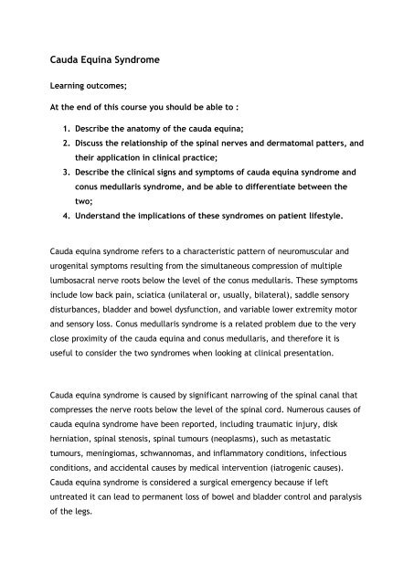
Dermatomal and peripheral nerve distributions – anterior and posterior
AetiologyThere are a number of events which can lead to the development of cauda equinasyndrome (and also conus medullaris syndrome).1. Trauma - Events leading to fracture or subluxation of the lumbar vertebrae mayresult in compression of the cauda equina. Additionally, if fluid collects in this areas a result of bleeding or inflammation this may also lead to compressions.2. Herniated IV disc - The majority of these will spontaneously resolve, but in upto 15% of cases cauda equine syndrome may result. Seventy percent of cases ofherniated disks leading to cauda equina syndrome occur in people with a history ofchronic low back pain, and 30% develop cauda equina syndrome as the firstsymptom of lumbar disc herniation. Most cases of cauda equina syndrome causedby disc herniation occur in men between the ages of 30 and 40 years. They involvelarge particles of disc material that have completely separated from the normaldisc and compress the spinal nerves.3. Spinal Stenosis - Spinal stenosis is any narrowing of the normal anterior /posterior dimensions of the spinal canal. This can be caused by a developmentalabnormality or degenerative process. Occasionally, one vertebral body can moveforward in relation to those either side (spondylolisthesis), resulting in a narrowingof the spinal canal and leading to cauda equina syndrome.4. Neoplasms - Isolated tumours (primary neoplasms) or metastatic spinalneoplasms may also cause cauda equine syndrome. Metastatic spine tumours aremost commonly from the prostate or lung in males, and from the lung and breastin females. The most common initial symptom of people with neoplasm-relatedcauda equina syndrome is severe lower back and leg pain, followed by lowerextremity weakness, sensory loss in the legs, and loss of bowel or bladder control.4. Inflammatory Conditions - Chronic inflammatory conditions of the spine,including Paget's disease and ankylosing spondylitis, can cause a narrowing of thespinal canal, leading to cauda equina syndrome.
5. Infectious Conditions – An abscess can lead to a narrowing of the spinal canal,or deformity of the nerve roots. Symptoms generally include severe back pain andrapidly worsening muscle weakness. Common causes are tuberculosis, meningitis,herpes simplex virus, meningovascular syphilis, and cytomegalovirus.6. Developmental defects – Conditions such as Spina bifida and tethered cordsyndromes.7. Causes related to medical intervention (iatrogenic) - Poorly positionedfixation devices placed in the spine can compress and injure nerves and causecauda equina syndrome. Continuous spinal anaesthesia has also been linked tocases of cauda equina syndrome. Lumbar puncture can cause a collection of bloodin the spinal canal (spontaneous spinal epidural hematoma) in patients receivinganticoagulation therapy, and accumulation of blood can compress the nerves andcause cauda equina syndrome.Much rarer causes include the following:Intravascular lymphomatosisMultiple sclerosisSpinal arteriovenous malformationsNeurosarcoidosisDeep venous thrombosis of the spinal veins (propagated)Inferior vena cava thrombosisSigns & SymptomsPatients can present with symptoms of isolated cauda equina syndrome, isolatedconus medullaris syndrome, or a combination of both. The symptoms and signs ofcauda equina syndrome tend to be mostly lower motor neuron (LMN) in nature,while those of conus medullaris syndrome are a combination of LMN and uppermotor neuron (UMN) effects. The history of onset, the duration of symptoms, andthe presence of other features or symptoms could point to the possible causes, andare summarised below.
Conus Medullaris <strong>Syndrome</strong> <strong>Cauda</strong> <strong>Equina</strong> <strong>Syndrome</strong>Presentation Sudden and bilateral Gradual and unilateralReflexesKnee jerks preserved but anklejerks affectedBoth ankle and knee jerksaffectedRadicular pain Less severe More severeLow back pain More LessSensorysymptoms andsignsNumbness tends to be morelocalized to perianal area;symmetrical and bilateral;sensory dissociation occursNumbness tends to bemore localized to saddlearea; asymmetrical, maybe unilateral; no sensorydissociation; loss ofsensation in specificdermatomes in lowerextremities withnumbness andparaesthesia; possiblenumbness in pubic areaMotor strength Typically symmetric, hyperreflexicdistal paresis of lowerlimbs that is less marked;fasciculations may be presentAsymmetric a-reflexicparaplegia that is moremarked; fasciculationsrare; atrophy morecommonImpotence Frequent Less frequent; erectiledysfunction, lack ofsensation in pubic area,and inability to ejaculateSphincterdysfunctionUrinary retention and atonic analsphincter cause overflow urinaryincontinence and faecalincontinence; tend to presentearly in course of diseaseUrinary retention; tendsto present late in courseof disease
The general symptoms of cauda equina syndrome include the following:Low back painPain in one leg (unilateral) or both legs (bilateral) that starts in thebuttocks and travels down the back of the thighs and legs (sciatica)Numbness in the groin or area of contact if sitting on a saddle (perineal orsaddle paraesthesia)Bowel and bladder disturbancesLower extremity muscle weakness and loss of sensationsReduced or absent lower extremity reflexesLow back pain can be divided into local and radicular pain. Local pain is generallya deep, aching pain resulting from soft tissue and vertebral body irritation,whereas radicular pain is generally a sharp, stabbing pain resulting fromcompression of the nerve roots. Radicular pain projects along the specific areas ofthe dermatomal distribution. Lower back pain in cauda equina syndrome may havesome characteristic that suggests something different from the far more commonlumbar strain. There may be a trigger, such as turning the head, which seemsunusual. Severe pain is an early finding in 96% of patients with cauda equinasyndrome secondary to spinal neoplasm. Later findings include lower extremityweakness due to involvement of the ventral roots. Patients generally develophypotonia and hyporeflexia. Sensory loss and sphincter dysfunction are alsocommon.DiagnosisThis is based on findings from the history, symptoms, and physical examination.Examination involves testing muscle strength of the lower extremities, evaluatingsensation to touch and pain, especially around the groin (perineum), checking thelower extremity reflexes, and evaluating rectal tone, sensation, and reflex. It isimportant to remember that back pain and/or leg pain, and changes in bowel orbladder function, are not necessarily due to cauda equina syndrome. More commoncauses of bladder changes are urinary tract infections, which can be identified by a
Gluteus medius Superior gluteal L4, 5, S1Gluteus maximus Inferior gluteal L5, S1, 2Posterior tibial Tibial L5, S1Flexor digitorum longus Tibial L5, S1Abductor hallucis brevis Tibial (medial plantar) L5, S1, 2Abductor digiti minimi Tibial (lateral plantar) S1, 2Gastrocnemius Tibial L5, S1, 2Soleus Tibial S1, 2Muscle strength of the following muscles should be tested to determine the levelof lesion:L2 - Hip flexors (iliopsoas)L3 - Knee extensors (quadriceps)L4 - Ankle dorsiflexors (tibialis anterior)L5 - Big toe extensors (extensor hallucis longus)S1 - Ankle plantar flexors (gastrocnemius/soleus)
Nerve roots and their distribution / deficit effect.Root Site of Pain Sensory Deficit Motor Deficit Reflex DeficitL2AnteriorUpper thighSlight quadricepsSlightly diminishedmedial thighweakness; hipsuprapatellarflexion; thighadductionL3AnteriorLower thighQuadricepsPatellar orlateral thighweakness; kneesuprapatellarextension; thighadductionL4Posterolateral Medial legKnee and footPatellarthigh,extensionanterior tibiaL5Dorsum ofDorsum of footDorsiflexion ofHamstringsfootfoot and toesS1-2 Lateral foot Lateral foot Plantar flexion offoot and toesAchillesS3-5 Perineum Saddle Sphincters Bulbocavernosus;anal
<strong>Cauda</strong> equina syndrome and conus medullaris can occur simultaneously simplybecause of the physical proximity of the two structures, therefore a single lesioncan result in an over-lap syndrome. The features of these two syndromes are listedbelow:Features <strong>Cauda</strong> <strong>Equina</strong> <strong>Syndrome</strong> Conus MedullarisVertebrallevelL2-sacrumL1-L2Spinal levelInjury to the lumbosacral nerverootsInjury of the sacral cord segment(conus and epiconus) and rootsSeverity ofsymptomsand signsUsually severeUsually not severeSymmetry ofsymptomsand signsUsually asymmetricUsually symmetricPainProminent, asymmetric, andradicularUsually bilateral and in theperineal areaMotor Weakness to flaccid paralysis Normal motor function to mild ormoderate weaknessSensorySaddle anaesthesia, may beasymmetricSymmetric saddle distribution,sensory loss of pin prick, andtemperature sensations (Tactilesensation is spared.)ReflexesA-reflexic lower extremities;bulbocavernosus reflex is absentin low CE (sacral) lesionsA-reflexic lower extremities
Sphincter and Usually late and of lessersexual magnitude; lower sacral rootsfunction involvement can cause bladder,bowel, and sexual dysfunctionEarly and severe bowel, bladder,and sexual dysfunction thatresults in a reflexic bowel andbladder with impaired erection inmalesEMGMultiple root level involvement;sphincters may also be involvedMostly normal lower extremitywith external anal sphincterinvolvementOutcomeMay be favourable compared withconus medullaris syndromeThe outcome may be lessfavourable than in patients withCESMore specifically, in cauda equina syndrome muscle strength in the lowerextremities is diminished. Sensation is decreased to pinprick and light touch in adermatomal pattern corresponding to the affected nerve roots. This includessaddle anesthesia and decreased sensation in the lower extremities in thedistribution of lumbar and sacral nerves. Vibration sense may also be affected.Reflexes may be absent or diminished in the corresponding nerve roots. Theplantar reflex is diminished or absent. Muscle tone in the lower extremities isdecreased, which is consistent with an LMN lesion.There are some differences with conus medullaris syndrome in that patients mayexhibit hypertonicity. Other signs are almost identical to those of the cauda equinasyndrome, except that in conus medullaris syndrome signs are more likely to bebilateral, the muscle stretch reflex may be hyper-reflexic, there may be a Babinskiresponse, and muscle tone might be increased to the point of spasticity.ManagementThe aim of management for patients is to act as soon as possible to reduce the riskof permanent damage to the affected nerves. In acute compression of the conusmedullaris or cauda equina, immediate surgical decompression is essential to
educe the pressure and increase the space in the vertebral canal. Traditionally,cauda equina syndrome has been considered a surgical emergency, with surgicaldecompression considered necessary within 48 hours after the onset of symptoms,and preferably performed within 6 h of injury.In a more chronic presentation with less severe symptoms, decompression could beperformed when medically feasible and should be delayed to optimise the patient'smedical condition; with this precaution, decompression is less likely to lead toirreversible neurological damage.Pain should be treated appropriately based on its origin; treatment may includenarcotics in the acute setting and tricyclic antidepressants later. Nerve rootischemia is partially responsible for the pain and decreased motor strengthassociated with cauda equina syndrome. As a result, vasodilatory treatment can beuseful in some patients. Mean arterial blood pressure should be maintained above90 mm Hg to maximise blood flow to the spinal cord and nerve roots.Treatment with lipoprostaglandin E1 and its derivatives has been reported to beeffective in increasing blood flow to the cauda equina region and reducingsymptoms of pain and motor weakness. This treatment option should be reservedfor patients with modest spinal stenosis with neurogenic claudication. No benefithas been reported in patients with more severe symptoms or patients withradicular symptoms.Corticosteroid therapy may be beneficial in suppressing an inflammatory responsebut treatment must be started within eight hours of injury. If treatment beginsafter this time there appears to be no benefit and may even have detrimentaleffects.Use of orthoses is advised to prevent contractures. Use of antispasticitymedications also is encouraged. Other medications include dantrolene, diazepam,clonidine, and tizanidine. These agents are thought to work centrally bysuppressing conduction at the spinal level.
Nerve blocks also could be done to relieve spasticity; appropriate agents includephenol, botulinum toxin, or local anesthetics. Botulinum toxin A binds to receptorsites on motor nerve terminals and inhibits the release of acetylcholine, which inturn inhibits transmission of impulses in neuromuscular tissue. It is most useful fortreating spasticity in the gastrocnemius and soleus muscles but is less effective inlarger muscles such as quadriceps.KEY LEARNING POINTS.1. <strong>Cauda</strong> equina syndrome (CES) is a pattern orneuromuscular and urogenital symptoms resultingfrom compression of multiple spinal nerves.2. Common symptoms are lower back pain, sciatica,saddle sensory disturbance, bladder & boweldysfunction.3. Narrowing of the spinal canal is responsible for thecompression on the nerve roots.4. Causes of CES include trauma, disc herniation,infection, neoplasm, spinal stenosis, and medicalintervention.5. Conus medullaris syndrome (CMS) is a closelyrelatedphenomenon to CES, although there aredifferences in clinical presentation.6. Signs of CES are mostly those of a lower motorneurone problem, where CMS is a combination ofupper and lower motor neurone signs.7. Diagnosis is based on good clinical history andinvolves examination of muscle strength, tone, andtendon reflexes.8. In many cases management of CES is a clinicalemergency in order to prevent permanent damageto the nerve roots. Surgery may be required toprovide decompression.
