Early pregnancy diagnosis by transrectal ultrasonography
Early pregnancy diagnosis by transrectal ultrasonography
Early pregnancy diagnosis by transrectal ultrasonography

You also want an ePaper? Increase the reach of your titles
YUMPU automatically turns print PDFs into web optimized ePapers that Google loves.
Original ArticleBuffalo Bulletin (June 2013) Vol.32 No.2EARLY PREGNANCY DIAGNOSIS BY TRANSRECTAL ULTRASONOGRAPHY INMEHSANA BUFFALOES (Bubalus bubalis)Mehrajuddin Naikoo 1 , D.M. Patel 2 and H.J. Derashri 3ABSTRACTThe aim of this study was to detect early<strong>pregnancy</strong> and early embryonic mortality using<strong>transrectal</strong> <strong>ultrasonography</strong> in Mehsana buffaloes.The study was carried out on 18 postpartum anestrusMehsana buffaloes of a dairy farm during breedingseason, using different estrus synchronizationprotocols, followed <strong>by</strong> fixed time AI. Ultrasoundexaminations were performed using a real timeB-mode ultrasound scanner (ALOKA SSD - 500)equipped with 5 MHz linear array transducer onday 26 and 40 post AI. Ultrasound scanning onday 26 post AI revealed visualization of embryonicvesicle around the conceptus. The embryo properwas observed in nine out of 13 animals (69.23%)on day 26 and all 12 conceived animals (100%)on day 40 post AI. The amniotic and allantoicvesicles were observed as non-echogenic cavitiesclosely surrounding the embryo proper on day 40post AI. Ovarian corpus luteum and foetal heartbeats were observed in conceived buffaloes onboth 26 and 40 days post AI. <strong>Early</strong> embryonicdeath occurred in one buffalo each between days26 and 40 and between day 40 and 60 post AI. Theoverall diagnostic accuracy of <strong>ultrasonography</strong>for early <strong>pregnancy</strong> was recorded as 94.44% and100% on day 26 and day 40, respectively. Plasmaprogesterone concentrations ranged from 3.00-6.02 ng/ml with an average of 4.42±0.52 ng/ml inpregnant animals and from 0.97-1.91 ng/ml withan average of 1.22±0.06 ng/ml in nonpregnantanimals on day 20 post AI. It was concluded thatultrasound scanning is a useful tool in detection ofearly <strong>pregnancy</strong>, early non-<strong>pregnancy</strong> and earlyembryonic mortality in buffaloes.Keywords: <strong>ultrasonography</strong>, early <strong>pregnancy</strong><strong>diagnosis</strong>, Mehsana buffaloesINTRODUCTIONLivestock production in general and milkproduction in particular play an important role inour national economy and thus dairying is oneof the most important agricultural activities inIndia. Buffaloes are the major contributor to milkproduction in our country, especially in Gujaratstate. Conditions like early embryonic mortality,early fetal death and unobserved abortions dueto the tropical environment lead to an increased1Department of Animal Reproduction, Gynaecology and Obstetrics, College of Veterinary Science andAnimal Husbandry, Anand Agricultural University, Anand - 388 001, India, E-mail: drmehraj@gmail.com2Teaching Veterinary Clinical Services Complex, Anand Agricultural University, Anand - 388 001, India3Department of Animal Reproduction, Gynaecology and Obstetrics, Navsari Agricultural University, Navsari,Gujarat - 396 450, India120
Buffalo Bulletin (June 2013) Vol.32 No.2anestrus period and there<strong>by</strong> increase the costlycalving interval. To overcome such problems,reproductive <strong>ultrasonography</strong> may serve as a bettertool.The use of <strong>ultrasonography</strong> has beenincreasing as an imaging modality in bovinereproduction. Its use can provide solutions toa number of unanswered questions in dealingwith the bovine reproductive cycle and itsconcurrent disorders, including early <strong>pregnancy</strong><strong>diagnosis</strong>. Ultrasonographic observation of thepostpartum changes in female reproductivetract, early <strong>pregnancy</strong> <strong>diagnosis</strong> <strong>by</strong> reproductive<strong>ultrasonography</strong> and detection of early embryonicmortality are some of the efficient means ofimproving domestic animal reproduction andproduction and help in fulfilling the basicnecessities of man in the day-to-day world.Ultrasound <strong>pregnancy</strong> <strong>diagnosis</strong> is a reliablemethod of determining the presence of a conceptusand viability of an embryo, which is essential toincrease the profitability of the animal (Tiwari et al.,2002). The present study was conducted to detectearly <strong>pregnancy</strong> and early embryonic mortality<strong>by</strong> <strong>transrectal</strong> <strong>ultrasonography</strong> in oestrus inducedpostpartum anestrus Mehsana buffaloes.MATERIALS AND METHODSA total of 18 postpartum anestrus Mehsanabuffaloes of a private dairy farm at Vadodara, Gujaratwere subjected to different estrus synchronizationprotocols and fixed time AI with good qualityfrozen semen. The buffaloes not returning to estruswere subjected to ultrasonographic scanning forearly <strong>pregnancy</strong> <strong>diagnosis</strong> on days 26 and 40 postAI and finally <strong>pregnancy</strong> was confirmed <strong>by</strong> rectalexamination on day 60 post AI.The ultrasound examinations wereperformed using a <strong>transrectal</strong> B-mode ultrasoundscanner (ALOKA SSD - 500, SN - M10408. Japan)equipped with a 5 MHz linear array transducerdesigned for intra-rectal placement. The scanningof uterine horns was carried out on their dorsaland lateral surfaces. Ovaries were also scanned forthe presence of a corpus luteum and/or any otherpalpable structure. The images displayed werethoroughly examined and frozen on the screenand the hard copies (sonograms) were taken usinga video graphic thermal printer (Sony, UP-895MDW, Japan).Blood samples were collected twice fromall the buffaloes for estimation of plasma levelsof progesterone, i.e. on the day of AI and on day20 post AI. Plasma progesterone concentrationswere estimated <strong>by</strong> employing the standard radioimmuno-assay(RIA) technique of Kubasic et al.(1984). Labeled antigen (I 125 ), antibody coated tubesand standards were procured from Immunotech -SAS, Marseille Cedex, 9, France. The sensitivityof assay was 0.1 ng/ml. Intra-assay coefficient ofvariation was 5.4 percent and inter-assay variationwas 9.1 percent. Cross reactivity of the antibodywith progesterone, 17α-dihydroprogesterone and20α-hydroxyprogesterone was 100, 0.13 and 0.96percent, respectively. The sensitivity, specificity,positive predictive value, negative predictive valueand accuracy were calculated based on findingsof ultrasound scanning on different days (day 26and 40 post AI) and those of plasma progesteroneconcentrations on day 20 post AI in relation tofindings of <strong>pregnancy</strong> <strong>diagnosis</strong> <strong>by</strong> rectal palpationon day 60 post AI.121
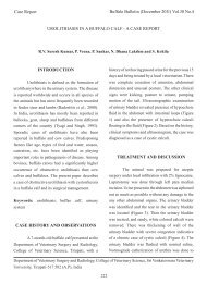
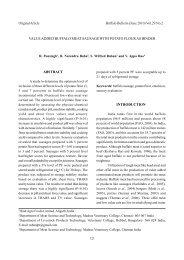
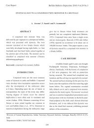
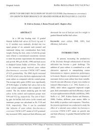
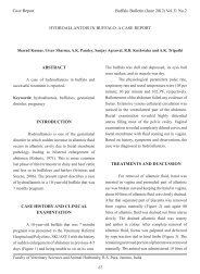
Buffalo Bulletin (June 2013) Vol.32 No.2RESULTS AND DISCUSSIONThe results of ultrasound scanning andprogesterone assay were correlated with thefindings of palpation per rectum on day 60 postAI with respect to accuracy and reliability of thesetwo tests. Although the sensitivity on days 26 and40 and the specificity on day 40 of ultrasoundscanning was 100 percent, the specificity was loweron day 26 being 80 percent. On day 26 one animalwas incorrectly diagnosed pregnant. However,the progesterone assay revealed two animalshaving been diagnosed incorrectly pregnant. Thesensitivity, specificity, positive predictive value,negative predictive value and overall diagnosticaccuracy of progesterone assay were comparativelylower as compared to ultrasound scanning resultson days 26 and 40 (Table 1).Developmental profile of embryonic vesicle andembryoThe scanning of recently bred buffaloesthrough B-mode <strong>transrectal</strong> <strong>ultrasonography</strong> onday 26 allowed the visualization of non-echogenicembryonic vesicle around the conceptus (Figure 1).The embryo proper was observed on day 26 postAI, however, it was observed in only nine out of 13(69.23%) buffaloes. Thereafter, the embryo properwas observed in all 12 conceiving animals (100%)on day 40 post AI (Figure 2). The amniotic andallantoic vesicles were observed as non-echogeniccavities closely surrounding the embryo proper onday 40 post AI. These cavities were surrounded<strong>by</strong> large anechoic area i.e. embryonic fluid. Thescanning of ovaries revealed the presence of a<strong>pregnancy</strong> corpus luteum in conceiving buffaloeson both the days i.e. day 26 and 40 post AI. Theheartbeats were observed as a pulsatile flickeringTable 1. Accuracy of ultrasonographic and P 4assay for early <strong>pregnancy</strong> <strong>diagnosis</strong> in Mehsana buffaloes ondifferent days after insemination.Plasma P4 assayDays of ultrasound scanningDiagnostic results/Predictive values<strong>by</strong> RIA †26 40Diagnosis pregnant correct (a) 13 12 11Diagnosis pregnant incorrect (b) 1 0 2Diagnosis nonpregnant correct (c) 4 6 4Diagnosis nonpregnant incorrect (d) 0 0 1Sensitivity (%) 100 x a /(a + d) 100% 100% 91.66 %Specificity (%) 100 x c /(c + b) 80% 100% 66.66 %Positive predictive value (%)100 x a /(a + b)92.85% 100% 84.61 %Negative predictive value (%)100 x c /(c + d)100% 100% 80.00%Overall diagnostic accuracy (%)100 x (a + c)/(a + b + c + d)94.44% 100% 83.33%(†) RIA performed at day 20 post-breeding.122
Buffalo Bulletin (June 2013) Vol.32 No.2Figure 1. Sonogram on day 26 post-insemination.a) Conceptus andb) Fluid filled embryonic vesicleFigure 2. Sonogram on day 40 post-insemination.a) Embryo proper andb) Fluid filled embryonic vesicle123
Buffalo Bulletin (June 2013) Vol.32 No.2at a faster rate in the centre of the embryo properwithin the embryonic vesicle on day 26 and 40 postAI. However, heartbeats were observed in only 10out of 13 animals on day 26 post AI and in all 12animals on day 40 post AI. The mean heart ratewas 174.60±0.81 beats per minute on day 26 postAI, which decreased to 165.28±1.4 on day 40 postAI. These findings were in accordance with thefindings of many workers (Rosiles et al., 2005 andAgarwal et al., 2007).In the present study it was possible to clearlyvisualize the embryonic vesicle in all animals andembryo proper in nine out of 13 animals on day 26post AI. Moreover, both the embryonic vesicle andthe embryo proper were detected in all pregnantanimals on day 40 post AI. Thus, <strong>ultrasonography</strong>facilitates <strong>diagnosis</strong> of all nonpregnant animals atan early stage (day 26 post AI) and is really moreadvantageous than <strong>pregnancy</strong> <strong>diagnosis</strong> <strong>by</strong> rectalpalpation method, wherein 100 percent reliableresults cannot be obtained at such an early stage of<strong>pregnancy</strong>. Agarwal et al. (2007); Ali and Fahmy(2008) reported similar findings in buffaloes. Theresults of the present study showed that <strong>transrectal</strong>ultrasound scanning of buffaloes on day 26 post AIwas less accurate than on day 40 post AI.The sensitivity of ultrasound scanningwas 100 percent on days 26 and 40 post AI (Table1). The specificity of ultrasound scanning was80.00 and 100 percent on days 26 and 40 post AI,respectively. The positive predictive value (PPV)of the ultrasound scanning was 92.85 and 100percent on day 26 and 40 post AI, respectively. Thenegative predictive value (NPV) of the ultrasoundscanning was 100 percent on both the days i.e. day26 and 40 post AI. The overall diagnostic accuracyin the present study was recorded as 94.44 and 100percent on days 26 and 40, respectively. Similarfindings were recorded <strong>by</strong> Bhosreker and Hangarge(2000); Rosiles et al. (2005). They observed 97.90and 100 percent accuracy in detecting <strong>pregnancy</strong>through <strong>ultrasonography</strong>, respectively. The possibleexplanation for higher overall diagnostic accuracyin the present study might be because ultrasoundscanning was carried out on days 26 and 40 post AIwith proper restraining of the animals.<strong>Early</strong> embryonic mortality and/or early fetaldeathIn one animal, ultrasound scanning revealedthe presence of a conceptus inside the fluid filledembryonic vesicle on day 26 post AI. However,<strong>ultrasonography</strong> on day 40 post AI revealed thesame animal as nonpregnant as evident <strong>by</strong> theabsence of a conceptus and resorption of embryonicfluid; suggestive of early embryonic death betweendays 26 and 40 post AI. In one buffalo, ultrasoundscanning revealed presence of a conceptus and anembryonic vesicle on both the occasions, i.e. days26 and 40 post AI. Later on, palpation per rectum atday 60 post AI revealed the animal as nonpregnant,suggestive of early embryonic or fetal deathbetween days 40 and 60 post AI. Similar findingswere recorded <strong>by</strong> Campanile et al. (2005); Vecchioet al. (2007).Hormone profileIn the present study, plasma progesteroneconcentrations ranged from 3.00-6.02 ng/ml withan average of 4.42±0.52 ng/ml in pregnant animalson day 20 post AI. In nonpregnant animals plasmaprogesterone concentrations ranged from 0.97-1.91ng/ml with an average of 1.22±0.06 ng/ml on day20 post AI. These findings were in close accordancewith the findings of Muhammad et al. (2000).The findings of the present study indicatethat ultrasound scanning of freshly bred animalsis helpful in detection of early <strong>pregnancy</strong>, early124
Buffalo Bulletin (June 2013) Vol.32 No.2non<strong>pregnancy</strong> and early embryonic deaths (EED)in buffaloes. The detection of early <strong>pregnancy</strong> inbuffaloes may be accomplished on or after day 26post AI through ultrasound scanning with positivepredictive value and accuracy over 92 percent andthe resul ts are instantly available.ACKNOWLEDGEMENTSThe authors would like to thank Mr. RaviShetty, owner of Geeta Dairy Farm, Vadodara forproviding buffaloes for this research purpose andfor his kind cooperation.REFERENCESAgarwal, S.K., S.K. Singh, T. Sarath, R. Rajkumar,S. Mehrotra, M. Hoque and Uma Shankar.2007. Ovarian and uterine changes duringpregnany in buffalo using real time B mode<strong>ultrasonography</strong>, p. 331. In Proceedings of23 rd Annual Convention of Indian Societyfor the Study of Animal Reproduction(ISSAR) and National Symposium, OUAT,Bhubaneswar, India.Ali, A. and S. Fahmy. 2008. Ultrasonographicfetometry and determination of fetal sex inbuffaloes (Bubalus bubalis). Anim. Reprod.Sci., 106: 90-99.Bhosreker, M.R. and I.M. Hangarge. 2000.Ultrasonography for early <strong>pregnancy</strong><strong>diagnosis</strong> in buffaloes. Indian J. Anim.Reprod., 21: 143-144.Campanile, G., G. Neglia, B. Gasparrini, G. Galiero,A. Prandi, R. Di Palo, M.J. D’Occhio andL. Zicarelli. 2005. Embryonic mortality inbuffaloes synchronized and mated <strong>by</strong> AIduring the seasonal decline in reproductivefunction. Theriogenology, 63: 2334-2340.Kubasic, N.P., G.D. Hallauer and R.G. Brodows.1984. Evaluation of direct solid phase RIAfor progesterone, useful for monitoringluteal function. Clin. Chem., 30: 284-286.Muhammad, F., A. Sarwar, C.S. Hayat andM.I. Anwar. 2000. Peripheral plasmaprogesterone concentration during early<strong>pregnancy</strong> in Holstein Friesian cows. Pak.Vet. J., 20: 166-168.Rosiles, V.A., C.S. Galina, M. Maquivar, R. Molinaand S. Estrada. 2005. Ultrasonographicscreening of embryo development in cattle(Bos indicus) between days 20 and 40 of<strong>pregnancy</strong>. Anim. Reprod. Sci., 90: 31-37.Tiwari, R.P., M.K. Awasthi and R.P. Gautam.2002. Principles, methods and clinical useof <strong>ultrasonography</strong> in bovine reproduction.Intas Polivet, 3: 78-85.Vecchio, D., R. Di Palo, L. Zicarelli, C. Grassi,A. Cammarano, M.J. D’Occhio and G.Campanile. 2007. Embryonic mortality inbuffaloes naturally mated. Italian J. Anim.Sci., 6: 677-679.125