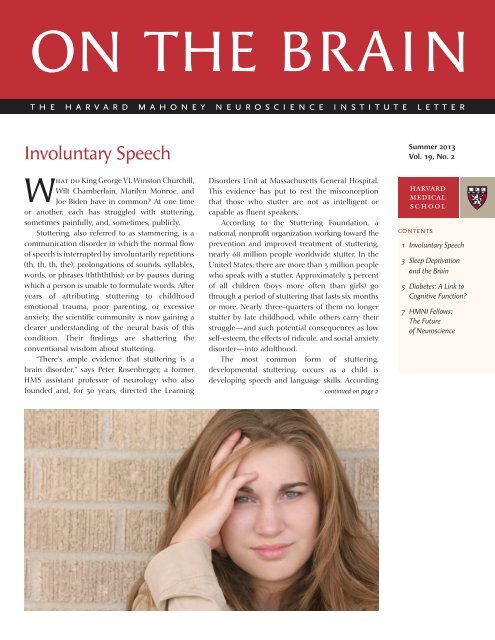
Involuntary Speech - Harvard Medical School - Harvard University
Involuntary Speech - Harvard Medical School - Harvard University
Involuntary Speech - Harvard Medical School - Harvard University

Create successful ePaper yourself
Turn your PDF publications into a flip-book with our unique Google optimized e-Paper software.
<strong>Involuntary</strong> <strong>Speech</strong>continued from page 1to some scientists, the stuttering is triggeredbecause those skills have not developed enoughto meet the child’s verbal needs. This form ofstuttering tends to run in families. In 2010, scientistsat the National Institute on Deafness and OtherCommunication Disorders (NIDCD) discoveredthree genes implicated in developmental stuttering.Another form, neurogenic stuttering, often followsan injury to the brain such as stroke, trauma, ortumor. Psychogenic stuttering, another, albeit rare,form of the disorder, is identified most often inpeople suffering some form of mental illness.Left side, right sideWhile much of stuttering remains a mystery toscientists, they do know, based on imaging studies,that the brains of people who stutter are structurallydifferent for those of people who do not stutter;these differences could affect behavior.Positron emission tomography studies of peoplewho stutter show decreased activity in corticalareas associated with language processing, such asBroca’s area, which controls motor functions linkedwith speech production. People with damage toBroca’s area can understand language but cannotsuccessfully form words or produce speech. Thesesame studies show hyperactivity in the motorareas of the brain, including the basal ganglia, astructure deep in the brain’s gray matter that helpscontrol muscle and nerve function.Positron emission tomography studies of people who stuttershow decreased activity in cortical areas associated with languageprocessing, such as Broca’s area, which controls motorfunctions linked with speech production.Previously, scientists had found evidence ofrewiring in the brains of people who beganstuttering as children. Fluent speakers use the leftside of their brain to integrate what they hear withword formation. People who stutter, however,transfer this workload to the right hemisphere; adefect in the left hemisphere prevents the motorcortex, which controls movement, from generatingthe instructions that coordinate the muscles of thethroat and mouth to aid in speech production. Thebrain compensates by shifting speech-related tasksto the right hemisphere. Thus, even though anindividual who stutters knows what words to say,those words are produced with a struggle and ashalting, stammering speech.Slow and deliberate does itSome 80 percent of children who stutter recoverspontaneously. For other children and adults whostutter, behavioral therapies to teach specific skillscan lead to improved oral communication. Forvery young children, early intervention andtreatment may help prevent stuttering frombecoming a lifelong problem. There is, however, nocure for the condition.According to NIDCD, many of the currentbehavioral therapies used to treat stuttering teachthe person how to minimize the expression of thedisorder by speaking slowly, regulating breathing,or gradually progressing from single-syllableresponses to longer words and more complexsentences. One technique, called delayed auditoryfeedback, takes an artificially modified version ofthe original acoustic speech signal and feeds itback to the individual through headphones. Thistechnique and other behavioral therapies, saysRosenberger, have been effective in helping stuttersbecome fluent speakers.In the 1970s, Rosenberger and others investigatedusing haloperidol, a powerful anti-dopaminergicdrug, to treat stuttering. Although they found thedrug could effectively help those who stutter tobecome more fluent, the drug’s side effectsoverpowered its benefits so its use was discontinued.The Food and Drug Administration has yet toapprove any drugs for stuttering, but some drugsapproved to treat other problems are now beingtested for the condition. Pagoclone, an anti-anxietymedication, and asenapine, an atypical antipsychoticdrug, have shown some promise. The formerenhances the activity of the neurotransmitter GABA,which may be disrupted in people who stutter, whilethe latter blocks dopamine receptors in the brain.Both potential treatments require further study.Two years ago, the obstacles facing those whostutter received popular attention when the movieThe King’s <strong>Speech</strong> won the Academy Award for BestPicture. The film depicted how England’s KingGeorge VI struggled with and overcame stutteringwith the help of a speech therapist.“The movie is quite good,” opines Rosenberger.“It provides an intelligent look at stuttering andsobers people’s attitudes toward people whostruggle with stuttering.”on the brain
This article is partof a series on theinternal and externalforces that affectthe brain.Sleep Deprivation and the BrainSleep is one of the body’s most importantfunctions. It is essential for cellular renewal andtissue maintenance, for the removal of cellulardebris, and for neural processing and archiving,including the firm establishment of memories. Inaddition, deep, restorative sleep stimulates brainregions used in learning.Unfortunately, though, many people in thiscountry average less than the seven to nine hoursof sleep per night recommended by the NationalSleep Foundation—and by many physicians andsleep researchers. This shortfall has consequences:Millions are sleep deprived and thousands sufferthe unexpected effects of sleep loss.Sleep deprivation is simply the condition ofnot getting enough sleep and can be either acute(a single night of poor sleep) or chronic (a string ofnights with insufficient sleep). Most of us know theeffects of a bad night’s sleep: We’re irritable,clumsy, and our cognition and recall are just a bitoff. Even one night of sleep deprivation can lead todifficulty concentrating, impaired judgment, anddiminished cognitive function. Over time, repeatedperiods of sleep loss can lead to such deleteriousmental effects as hallucinations or mania.“Every function of the brain that has been testedis affected by sleep loss—mood, learning, reactiontime,” says Elizabeth Klerman, an HMS associateprofessor of medicine and a sleep researcher atBrigham and Women’s Hospital.How sleep worksOur understanding of sleep has developed overdecades as researchers, including those in <strong>Harvard</strong><strong>Medical</strong> <strong>School</strong>’s Division of Sleep Medicine, havedeciphered its biological basis. The human sleep–wake cycle is controlled by two complementarymechanisms. Our circadian clock, which regulatesthe body’s internal processes and alertness levels, islocated in the hypothalamus, a region of the brainthat also produces hormones that control suchprocesses as sleep, mood, and hunger. Governedby light–dark cycles, this clock is key to controllingthe timing of our sleep. Circadian rhythms alone,however, are not enough to regulate our sleep.Sleep–wake homeostasis, or the accumulation ofsleep-inducing substances in the brain, essentiallyreminds our body that it needs to sleep after acertain period of being awake.continued on page 4on the brain
Sleep Deprivation and the Braincontinued from page 3Neurotransmitters in the brain also play animportant role in helping us fall asleep, remainasleep, and wake up. One of these chemicalmessengers is the neurotransmitter melatonin.During the day, the pineal gland, a small gland,located just above the middle of the brain, thatproduces melatonin, is inactive. Daytime levels ofmelatonin, in fact, are barely detectable. When thesun sets, or our exposure to light is lost, the pinealgland releases melatonin into the bloodstream,making us feel sleepy. Melatonin levels stayelevated throughout the night, but decrease as thelevels of light increase with the new day.The researchers found that areas of the prefrontal cortex,a center for working memory and reasoning, are moreactive in people who are sleep deprived. This indicatesthat, for a given task, the prefrontal cortex has to workharder in sleep-deprived people than in those who’ve hadsufficient sleep.During the hours we are awake, theneurotransmitter adenosine attaches to receptorsin the brain. The longer we are awake, the greaterthe amount of adenosine that attaches to thesereceptors and accumulates in the brain. When wesleep, adenosine is broken down and its levels inthe brain fall. If we don’t get enough sleep,adenosine remains in the brain, causing grogginess.SleepyheadA number of studies show how sleep deprivationaffects brain function. A <strong>University</strong> of California,San Diego, study used functional MRI to monitoractivity in sleep-deprived brains. The researchersfound that areas of the prefrontal cortex, a center forworking memory and reasoning, are more activein people who are sleep deprived. This indicatesthat, for a given task, the prefrontal cortex has towork harder in sleep-deprived people than inthose who’ve had sufficient sleep. In 2011,researchers at the <strong>University</strong> of Wisconsin foundthat the brains of rats kept awake for extendedperiods began to shut down, neuron by neuron,even though the rats were still awake. They foundthat the most-used neurons were the first to shutoff. Such results may help explain why we functionless well the longer we stay awake or when wehave inadequate sleep. Other researchers haveobserved genetic changes in the sleep-deprivedbrain that could lead to health problems such asheart failure, stroke, and depression and mooddisorders.Shifting schedulesSleep deprivation, says Klerman, is often selfimposed,a function of our hectic work schedulesand the presence of electronic devices and 24-hourentertainment sources that keep us occupied atall hours.Without adequate sleep, some people’sperformance suffers. “After a couple of days withoutadequate sleep, many of these folks say they arenot tired, but their performance worsens,” saysKlerman, who has conducted performance studieson the sleep deprived. “The brain is not accuratelyconveying alertness and tiredness and this affectsself-assessment of performance.”By some estimates, excessive sleepiness contributesto a greater-than-twofold risk of sustaining anoccupational injury. The National Highway TrafficSafety Administration estimates that drowsy driving,often a reflection of the lack of sleep, is responsiblefor at least 100,000 automobile crashes (about1 every 6 minutes), 71,000 injuries, and 1,550 deathseach year. Much of this mayhem and loss could beeradicated if drivers got adequate amounts of sleep.As part of her work for the U.S. space program,Klerman led a National Space Biomedical ResearchInstitute (NSBRI) team that developed computersoftware that uses sophisticated mathematicalmodeling to help astronauts and ground personneladjust to shifting work–sleep schedules. Thesoftware uses a complex mathematical formula topredict how a person will react to specificconditions and to allow users to interactivelydesign better work schedules. The software alsofeatures a “shifter,” a component that prescribeswhen to use light to reset, or shift, a person’scircadian clock to improve performance at criticaltimes, such as during a space mission.Klerman says the software can be adapted forpeople who work night shifts, including medicalpersonnel and first responders.“Our lives, including our safety,” she said in anNSBRI press release announcing the software, “areimpacted by people who have jobs requiring shiftwork or extremely long hours and who may beat increased risk of accidents affecting themselvesor others.”To diminish that risk, we should all say, “Sweetdreams.”on the brain
Diabetes: A Link to Cognitive Function?The complications of uncontrolled diabetesare well recognized: nerve damage, kidneydisease, blindness, and circulation problems thataffect the extremities. The disease’s impact on thebrain, however, is often overlooked. This oversightcould spell trouble for millions of Americanswho face the daily challenge of controlling theirblood sugar.According to the American Diabetes Association,an estimated 26 million Americans have diabetes.Another 79 million have prediabetes, a condition inwhich blood sugar levels are higher than normalbut not high enough for a diabetes diagnosis.A growing body of evidence suggests that thecognitive health of millions with the disease is asmuch at risk as are other body systems from theeffects of out-of-control blood sugar. “Unlike forcertain other diseases, scientists originally didn’tknow where to look in the brain for the effects ofdiabetes,” says Gail Musen, an HMS assistantprofessor of psychiatry and assistant investigatorin the Section on Clinical, Behavioral, andOutcomes Research at Joslin Diabetes Center. “Weknew, theoretically, that because it affects so muchelse in the body, it also could affect the brain.”Since Musen’s first study of diabetes and brainMore recently, Gail Musen and her colleagues discovered reduced white matter integrityand cortical thickness in patients with long-standing type 1 diabetes. “It’s not clear,” she says,“whether such changes to the brain will have a more profound effect as a patient ages.”function nearly a decade ago, the scientificcommunity has gained a greater understanding ofhow diabetes—primarily type 1 diabetes—affectsbrain function.Shrinking brainMusen’s 2006 study, reported in the journalDiabetes, was the first comprehensive study ofdensity changes in the brain’s gray matter as aresult of type 1 diabetes. Its findings suggested thatpersistent hyperglycemia, or high blood sugar, andacute severe hypoglycemia, or low blood sugar,have an effect on brain structure. The gray matterreductions were small and did not necessarilyshow any clinically significant cognitiveimpairment, but the brain regions involvedincluded the memory, attention, and languageprocessing centers. More recently, Musen and hercolleagues discovered reduced white matterintegrity and cortical thickness in patients withlong-standing type 1 diabetes. “It’s not clear,” shesays, “whether such changes to the brain will havea more profound effect as a patient ages.”Currently, Musen is using functional MRI,which measures the brain in action, to determinewhether regions of the brain with gray matterdensity loss show impaired function. Even thoughpeople with diabetes may show normalperformance in terms of accuracy or processingspeed on cognitive tasks, their brain activitymay differ from that of patients without diabetes.Such changes, she said, may precede clinicallyrelevant cognitive issues, such as memory loss andcontinued on page 6on the brain
HMNI Fellows: The Future of NeuroscienceTo encourage young scientists to pursue neuroscience research, the <strong>Harvard</strong> Mahoney NeuroscienceInstitute supports fellowships at <strong>Harvard</strong> <strong>Medical</strong> <strong>School</strong>. For more than two decades, partial or completefellowships have been provided to more than 60 HMNI fellows, supporting a variety of neuroscienceinvestigations, including research into the molecular biology of circadian rhythms, visual processing inprimates, the development of olfactory systems in mammals, and gene expression and memory.Three HMNI fellows are currently working in laboratories in the Department of Neurobiology at HMS.Gabriella BoultingNeuroscientists know thatfor neurons to functionproperly, they must beable to “see” and “hear”signals in the brain. Asthe brain develops, cellsin the brain physicallychange how they arewired and how they integrate the signals theyreceive. “This is what makes us cognitive beings,”says Gabriella Boulting, an HMNI fellow in thelaboratory of Michael Greenberg, the NathanMarsh Pusey Professor of Neurobiology and chairof the Department of Neurobiology at HMS.In Greenberg’s laboratory, Boulting is studyingenhancer sequences, the regulatory elements thatcontrol gene expression. Enhancers are poorlyunderstood; they have been difficult to studybecause they can be located far away fromthe genes they influence. Boulting is hoping todevelop methods that will ease the study of thesesequences; she’s adapting technologies that usespecially tailored proteins to target and repressenhancer function.Boulting is engineering these proteins to recognizebrain-derived neurotrophic factor (BDNF) genes inmice. These genes play a crucial role in regulatingneuronal synapse function, thus allowing neuronsto adapt their responses to stimuli and to guidebehavior accordingly. “By focusing on wellcharacterizedgenes, such as those for BDNF,” shesays, “I can examine the regulatory role thatenhancer sequences play in the basic neuronfunctions that underlie memory and learning.”Prior to her fellowship, Boulting earned adoctorate in molecular and cell biology at <strong>Harvard</strong><strong>University</strong> and worked in the laboratory of KevinEggan, an HMS professor of stem cell andregenerative biology, studying mouse models ofmotor neurons to better understand cognitive andneurodevelopmental disorders in humans.Noah DruckenbrodThe ability to detect,interpret, and respond tocomplex sounds in theenvironment depends onthe precise functioning ofneural circuits in theinner ear. Spiral ganglia,the primary neuronsinvolved in hearing, receive input from sensoryhair cells and transmit this information rapidlyand accurately to auditory processing centers inthe central nervous system. In addition, the cochlea,the portion of each inner ear that is shaped like asnail’s shell, are innervated by a small populationof motor neurons that provide reverse feedback;that is, signals that run from the brain to the ear.In the laboratory of Lisa Goodrich, an HMSassociate professor of neurobiology, HMNI fellowNoah Druckenbrod is studying both thedevelopment of the neural circuits that connectthe inner ear to the brain and the neurons in thespiral ganglion. “Our work,” he says, “aims tounderstand how these two populations of neuronsinteract to create the circuits that underlie theperception of sound.”Druckenbrod uses novel imaging and moleculartechniques to distinguish the critical cellular andmolecular events that “influence the final wiringdecision to form a functional cochlea.” He says thatunderstanding how the wiring of the inner earoccurs will give scientists a better understanding ofneurodevelopment and diseases of the nervoussystem that disrupt proper wiring, such asamyotrophic lateral sclerosis (Lou Gehrig’s disease)and progressive muscular atrophy, as well as theobstacles involved in motor neuron regeneration.Before he joined HMNI as a fellow, Druckenbrodearned both his bachelor’s degree and his doctoratein neuroscience at the <strong>University</strong> of Wisconsin—Madison.continued on page 8on the brain
HMNI Fellows: The Future of Neurosciencecontinued from page 7Zhihua LiuEach night as we sleep,extraordinary changesoccur in our brains: ourmemories reorganize andour mind rejuvenates.The process of sleep, andparticularly how and whythe brain switches from awaking to a sleeping state and back again, has longintrigued scientists. In the laboratory of DraganaRogulja, an HMS assistant professor of neurobiology,HMNI fellow Zhihua Liu is studying these questions.And he’s looking into why we need to sleep at all.Using Drosophila melanogaster, the common fruitfly, as a model system, Liu is characterizing genesidentified through large-scale genetic screening toreveal the molecular pathways they use to shut thebrain down during a good night’s sleep. Amongthe genes Rogulja identified in a previous study iscyclin A, a regulator of the cell cycle process. Shediscovered that cyclin A functions in neurons topromote sleep in Drosophila, and found thatreducing the amount of cyclin A in the flies’neurons delayed the sleep–wake transition andcaused multiple arousals from sleep.Liu is examining discrete populations of neuronsthat are active sleep centers. Fruit flies tend to sleepall night, as do humans, so finding sleepmechanisms in Drosophila may have implicationsfor the study of human sleep. “What’s interesting,”Liu says, “is that cyclin A is expressed in only a fewcells in the Drosophila brain. This small numbergreatly facilitates the mapping of sleep-regulatingneuronal circuits in which cyclin A acts.”Liu and Rogulja also want to understand howcyclin A regulates sleep on a molecular basis. Theysay many clues are provided by the thoroughcharacterization of the gene in the context of cellcycle regulation.Prior to his HMNI fellowship, Liu earned hisdoctorate at the Chinese Academy of Sciences’Institute of Genetics and Developmental Biology inBeijing.on the brainthe harvard mahoneyneuroscience institute letterHARVARD MAHONEYNEUROSCIENCE INSTITUTECouncil Members:Hildegarde E. Mahoney, ChairmanSteven E. Hyman, MDCaroline Kennedy SchlossbergAnn McLaughlin KorologosJoseph B. Martin, MD, PhDEdward F. RoverON THE BRAINOn The Brain is published electronically three timesa year through the Office of Communications andExternal Relations at <strong>Harvard</strong> <strong>Medical</strong> <strong>School</strong>, Gina Vild,Associate Dean and Chief Communications Officer.Editor: Ann Marie MentingFreelance Writer: Scott EdwardsDesign: Gilbert Design Associates, Inc.In collaboration with: Michael E. Greenberg,Nathan Pusey Professor of Neurobiologyand Chair, Department of Neurobiology<strong>Harvard</strong> Mahoney Neuroscience Institute<strong>Harvard</strong> <strong>Medical</strong> <strong>School</strong>107 Avenue Louis PasteurSuite 111Boston, MA 02115www.hms.harvard.edu/hmnihmni@hms.harvard.educorrespondence/circulation<strong>Harvard</strong> <strong>Medical</strong> <strong>School</strong>Gordon Hall25 Shattuck Street, Room 001Boston, MA 02115