Microtensile bond strength of a filled vs unfilled adhesive to dentin ...
Microtensile bond strength of a filled vs unfilled adhesive to dentin ...
Microtensile bond strength of a filled vs unfilled adhesive to dentin ...

Create successful ePaper yourself
Turn your PDF publications into a flip-book with our unique Google optimized e-Paper software.
Journal <strong>of</strong> Dentistry (2006) 34, 283–291<br />
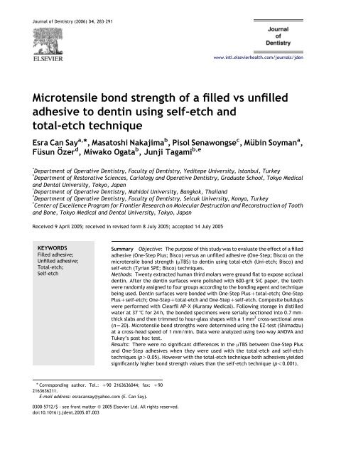
<strong>Microtensile</strong> <strong>bond</strong> <strong>strength</strong> <strong>of</strong> a <strong>filled</strong> <strong>vs</strong> un<strong>filled</strong><br />
<strong>adhesive</strong> <strong>to</strong> <strong>dentin</strong> using self-etch and<br />
<strong>to</strong>tal-etch technique<br />
Esra Can Say a, *, Masa<strong>to</strong>shi Nakajima b , Pisol Senawongse c ,Mübin Soyman a ,<br />
Füsun Özer d , Miwako Ogata b , Junji Tagami b,e<br />
a<br />
Department <strong>of</strong> Operative Dentistry, Faculty <strong>of</strong> Dentistry, Yeditepe University, Istanbul, Turkey<br />
b<br />
Department <strong>of</strong> Res<strong>to</strong>rative Sciences, Cariology and Operative Dentistry, Graduate School, Tokyo Medical<br />
and Dental University, Tokyo, Japan<br />
c<br />
Department <strong>of</strong> Operative Dentistry, Mahidol University, Bangkok, Thailand<br />
d<br />
Department <strong>of</strong> Operative Dentistry, Faculty <strong>of</strong> Dentistry, Selcuk University, Konya, Turkey<br />
e<br />
Center <strong>of</strong> Excellence Program for Frontier Research on Molecular Destruction and Reconstruction <strong>of</strong> Tooth<br />
and Bone, Tokyo Medical and Dental University, Tokyo, Japan<br />
Received 9 April 2005; received in revised form 8 July 2005; accepted 14 July 2005<br />
KEYWORDS<br />
Filled <strong>adhesive</strong>;<br />
Un<strong>filled</strong> <strong>adhesive</strong>;<br />
Total-etch;<br />
Self-etch<br />
* Corresponding author. Tel.: C90 2163636044; fax: C90<br />
2163636211.<br />
E-mail address: esracansay@yahoo.com (E. Can Say).<br />
0300-5712/$ - see front matter Q 2005 Elsevier Ltd. All rights reserved.<br />
doi:10.1016/j.jdent.2005.07.003<br />
www.intl.elsevierhealth.com/journals/jden<br />
Summary Objective: The purpose <strong>of</strong> this study was <strong>to</strong> evaluate the effect <strong>of</strong> a <strong>filled</strong><br />
<strong>adhesive</strong> (One-Step Plus; Bisco) versus an un<strong>filled</strong> <strong>adhesive</strong> (One-Step; Bisco) on the<br />
microtensile <strong>bond</strong> <strong>strength</strong> (mTBS) <strong>to</strong> <strong>dentin</strong> using <strong>to</strong>tal-etch (Uni-etch; Bisco) and<br />
self-etch (Tyrian SPE; Bisco) techniques.<br />
Methods: Twenty extracted human third molars were ground flat <strong>to</strong> expose occlusal<br />
<strong>dentin</strong>. After the <strong>dentin</strong> surfaces were polished with 600-grit SiC paper, the teeth<br />
were randomly assigned <strong>to</strong> four groups according <strong>to</strong> the <strong>bond</strong>ing agent and technique<br />
being used. Dentin surfaces were <strong>bond</strong>ed with One-Step PlusC<strong>to</strong>tal-etch; One-Step<br />
PlusCself-etch; One-StepC<strong>to</strong>tal-etch and One-StepCself-etch. Composite buildups<br />
were performed with Clearfil AP-X (Kuraray Medical). Following s<strong>to</strong>rage in distilled<br />
water at 37 8C for 24 h, the <strong>bond</strong>ed specimens were serially sectioned in<strong>to</strong> 0.7 mmthick<br />
slabs and then trimmed <strong>to</strong> hour-glass shapes with a 1 mm 2 cross-sectional area<br />
(nZ20). <strong>Microtensile</strong> <strong>bond</strong> <strong>strength</strong>s were determined using the EZ-test (Shimadzu)<br />
at a cross-head speed <strong>of</strong> 1 mm/min. Data were analyzed using two-way ANOVA and<br />
Tukey’s post hoc test.<br />
Results: There were no significant differences in the mTBS between One-Step Plus<br />
and One-Step <strong>adhesive</strong>s when they were used with the <strong>to</strong>tal-etch and self-etch<br />
techniques (pO0.05). However with the <strong>to</strong>tal-etch technique both <strong>adhesive</strong>s yielded<br />
significantly higher <strong>bond</strong> <strong>strength</strong> values than the self-etch technique (p!0.001).
284<br />
Introduction<br />
Resin composites with <strong>adhesive</strong> materials have<br />
been available on the dental market for about<br />
four decades and are widely used for both anterior<br />
and posterior res<strong>to</strong>rations due <strong>to</strong> the esthetic<br />
demands <strong>of</strong> patients, however they still have<br />
some undesirable properties. One is the polymerization<br />
shrinkage which produces contraction stresses<br />
generally concentrate at the <strong>bond</strong>ing<br />
interface. 1 If these stresses exceed the <strong>bond</strong><br />
<strong>strength</strong> <strong>to</strong> <strong>dentin</strong>, an interfacial gap will be<br />
formed, leading <strong>to</strong> bacterial infiltration, sensitivity,<br />
secondary caries and possible pulpal damage. 2<br />
Thicker <strong>adhesive</strong> layers or liners may act as an<br />
elastic intermediate layer (elastic cavity wall)<br />
between the cavity walls and the adjacent composite.<br />
That is, they could resist the polymerization<br />
shrinkage stress <strong>of</strong> the resin composites 1 and absorb<br />
the shock produced by occlusal loads and thermal<br />
cycling. 3 Using un<strong>filled</strong> <strong>adhesive</strong>s, thicker layers are<br />
not recommended because these materials have<br />
lower mechanical properties and usually provide no<br />
radioopacity which could mislead clinicians <strong>to</strong><br />
interprete the <strong>adhesive</strong> radiotransparency as gap<br />
formation or recurrent caries at the margin <strong>of</strong> the<br />
res<strong>to</strong>ration. 4 Based on this idea, <strong>filled</strong> <strong>adhesive</strong>s<br />
have been introduced, 5–7 which have included<br />
various types <strong>of</strong> fillers; such as conventional glass,<br />
ion leachable glass, silica and nanometer-sized<br />
aerosil silica fillers. 8–10 They have been reported<br />
<strong>to</strong> improve marginal and internal seal <strong>of</strong> composite<br />
res<strong>to</strong>rations 6,11–13 and have sufficient radioopacity<br />
<strong>to</strong> be discernible on dental X-ray films. 7<br />
Current <strong>adhesive</strong> systems employ two simplified<br />
application procedures <strong>to</strong> achieve the goal <strong>of</strong><br />
micromechanical retention between resin and<br />
<strong>dentin</strong>. The first method attempts <strong>to</strong> remove the<br />
smear layer completely via acid etching and rinsing,<br />
followed by the application <strong>of</strong> an <strong>adhesive</strong> agent on<br />
a wet <strong>dentin</strong> surface; the <strong>to</strong>tal-etch technique. 14<br />
The second category is the self-etch technique,<br />
which simultaneously demineralizes <strong>dentin</strong> and<br />
infiltrates it with <strong>adhesive</strong> monomers. 15 The strong<br />
versions <strong>of</strong> self-etch <strong>adhesive</strong>s can completely<br />
dissolve or disperse smear layers, forming thick<br />
hybrid layers in intact <strong>dentin</strong> that approach those<br />
Conclusion: The <strong>filled</strong> <strong>adhesive</strong> One-Step Plus did not show any beneficial effect than<br />
the un<strong>filled</strong> <strong>adhesive</strong> One-Step on the mTBS <strong>to</strong> <strong>dentin</strong> with <strong>to</strong>tal-etch and self-etch<br />
techniques. Irrespective from the <strong>adhesive</strong> type, self-etch technique revealed lower<br />
<strong>bond</strong> <strong>strength</strong>s than the <strong>to</strong>tal-etch technique.<br />
Q 2005 Elsevier Ltd. All rights reserved.<br />
achieved with conventional <strong>to</strong>tal-etch technique.<br />
Conversely, intermediate strong and mild versions<br />
incorporate smear layers as part <strong>of</strong> the <strong>bond</strong>ed<br />
interface, forming only thin hybrid layers. 16 Both<br />
<strong>to</strong>tal and self-etch approaches rely on the impregnation<br />
and polymerization <strong>of</strong> the monomers in<strong>to</strong><br />
the exposed collagen <strong>of</strong> the demineralized <strong>dentin</strong><br />
surfaces, creating a hybrid layer and the stabilization<br />
<strong>of</strong> the hybrid layer was established by the<br />
<strong>adhesive</strong>. 17 Recently, in comparison <strong>to</strong> the <strong>to</strong>taletch<br />
<strong>adhesive</strong>s two-step self-etch <strong>adhesive</strong>s are<br />
becoming increasingly popular, because <strong>of</strong> the<br />
reduced post-operative 18 and technique sensitivity.<br />
16 In addition <strong>to</strong> these, they are less likely<br />
<strong>to</strong> result in a discrepancy between the depth <strong>of</strong><br />
demineralization and the depth <strong>of</strong> resin infiltration<br />
19 since both processes occur simultaneously. 15<br />
Using <strong>to</strong>tal-etch systems, fillers incorporated in<br />
<strong>adhesive</strong> resins may increase the <strong>adhesive</strong> viscosity,<br />
resulting in a reduction in <strong>adhesive</strong> penetration<br />
in<strong>to</strong> the demineralized <strong>dentin</strong> and <strong>bond</strong>ing <strong>to</strong><br />
<strong>dentin</strong>. 20,21 There is however, less information on<br />
the effect <strong>of</strong> the <strong>filled</strong> <strong>adhesive</strong>s on the <strong>bond</strong>ing <strong>of</strong><br />
two-step self-etch systems <strong>to</strong> <strong>dentin</strong>.<br />
The purpose <strong>of</strong> this study was <strong>to</strong> evaluate the<br />
effect <strong>of</strong> a <strong>filled</strong> <strong>adhesive</strong> (One-Step Plus; Bisco)<br />
versus an un<strong>filled</strong> <strong>adhesive</strong> (One-Step; Bisco) on the<br />
microtensile <strong>bond</strong> <strong>strength</strong> (mTBS) <strong>to</strong> <strong>dentin</strong> using<br />
<strong>to</strong>tal-etch (Uni-etch; Bisco) and self-etch (Tyrian<br />
SPE; Bisco) techniques.<br />
Material and methods<br />
E. Can Say et al.<br />
The materials and their compositions used in this<br />
study are listed in Table 1. Twenty non-carious<br />
extracted human third molars, s<strong>to</strong>red in iso<strong>to</strong>nic<br />
saline with thymol crystals at 4 8C, were ground<br />
flat using 180-grit silicon carbide (SiC) abrasive<br />
paper under running water <strong>to</strong> expose occlusal<br />
<strong>dentin</strong>. After the superficial <strong>dentin</strong> surfaces were<br />
polished with 600-grit SiC <strong>to</strong> standardize the<br />
smear layer, the teeth were randomly assigned<br />
<strong>to</strong> four groups according <strong>to</strong> the type <strong>of</strong> <strong>adhesive</strong><br />
(One-Step Plus; One-Step) and technique (<strong>to</strong>taletch;<br />
self-etch) being used. Bonding procedures<br />
were performed according <strong>to</strong> the manufacturer’s
<strong>Microtensile</strong> <strong>bond</strong> <strong>strength</strong> <strong>of</strong> <strong>adhesive</strong> <strong>to</strong> <strong>dentin</strong> using self-etch and <strong>to</strong>tal-etch technique 285<br />
Table 1 Materials used in the study.<br />
Materials Composition Manufacturer<br />
One-Step Plus Bis-GMA, BPDM, HEMA, CQ, p-dimethylaminobenzoic acid<br />
(co-initia<strong>to</strong>r), ace<strong>to</strong>ne, 8.5% glass fillers<br />
Bisco Inc, USA<br />
One-Step Bis-GMA, BPDM, HEMA, ace<strong>to</strong>ne Bisco Inc, USA<br />
Tyrian SPE Primer A: thymol blue, ethanol, water<br />
Primer B: AMPS, BisMEP, TPO, ethanol<br />
Bisco Inc, USA<br />
Uni-etch 32% Phosphoric acid, BAC Bisco Inc, USA<br />
Clearfil AP-X Bis-GMA, TEGDMA, filler (Barium, SiO2) Kuraray Co, Japan<br />
BPDM, Biphenyl-dimethacrylate; HEMA, 2-hydroxyethyl-methacrylate; Bis-GMA, Bisphenyl-glysidyl-methacrylate; AMPS, 2-<br />
Acrylamido-2-methylpropanesulfonic acid; BisMEP, Bis(2-(methacryloyloxy)ethyl)phosphate; TEGDMA, Triethylene glycol-dimethacrylate<br />
TPO:2,4,6 (trimethylbenzoyldiphenylphosphine) oxide; BAC, Benzalkonium chloride.<br />
instructions and summarized in Table 2. Then a<br />
resin composite (Clearfil AP-X; Kuraray Medical<br />
Co, Ltd, Tokyo, Japan) was built up incrementally.<br />
Each <strong>of</strong> the three resin composite increments<br />
was light cured for 20 s using a light-curing<br />
unit (XL 3000, 3MESPE, St Paul MN, USA) with the<br />
intensity at 600 mW/cm 2 . Following s<strong>to</strong>rage in<br />
distilled water at 37 8C for 24 h, the <strong>bond</strong>ed<br />
specimens were serially sectioned in<strong>to</strong> 0.7 mmthick<br />
slabs using a low-speed diamond saw<br />
(Isomet; Buehler Ltd, Lake Bluff, IL 60044) and<br />
then trimmed with a superfine diamond bur <strong>to</strong><br />
form hour-glass shapes with approximately 1 mm 2<br />
cross-sectional areas (nZ20) using a digital<br />
micrometer.<br />
The specimens were attached <strong>to</strong> a table-<strong>to</strong>p<br />
material tester (EZ-test, Shimadzu Co, Kyo<strong>to</strong>,<br />
Japan) with a cyanoacrylate glue (Zapit, DVA,<br />
Anaheim, CA, USA) and subjected <strong>to</strong> microtensile<br />
testing at a crosshead speed <strong>of</strong> 1 mm/min until they<br />
fractured. All <strong>of</strong> the <strong>bond</strong>ed specimens were<br />
capable <strong>of</strong> being tested. In order <strong>to</strong> observe the<br />
failure modes, the de<strong>bond</strong>ed specimens were fixed<br />
for at least 8 h in 10% neutral buffered formalin,<br />
placed on stubs followed by room desiccation, gold<br />
sputter-coated and observed with a scanning<br />
electron microscopy (JXA-5400, JEOL, Tokyo,<br />
Japan). Failure modes were classified in<strong>to</strong> one <strong>of</strong><br />
four categories: <strong>adhesive</strong> if de<strong>bond</strong>ing occured<br />
between resin and <strong>dentin</strong>, mixed if it exhibited<br />
partially <strong>adhesive</strong>, partially cohesive failure in<br />
<strong>bond</strong>ing resin or in hybrid layer, or cohesive in<br />
resin or cohesive in <strong>dentin</strong>.<br />
Two-way ANOVA was performed <strong>to</strong> evaluate the<br />
effect <strong>of</strong> two experimental fac<strong>to</strong>rs: <strong>adhesive</strong> type<br />
and application technique, and the interaction <strong>of</strong><br />
these two fac<strong>to</strong>rs on mTBS. Multiple comparisons<br />
were performed by Tukey’s post hoc test at a<br />
significance level <strong>of</strong> 0.05 using a computer s<strong>of</strong>tware<br />
(SPSS 11, SPSS Inc Chicago IL). Failure modes <strong>of</strong> the<br />
specimens were analyzed using chi-square test.<br />
Scanning electron microscopy<br />
Four non-carious third molars (one per group)<br />
were used for SEM examination <strong>of</strong> the resin-<strong>dentin</strong><br />
interfaces after argon ion etching. The teeth were<br />
prepared in the same manner as the <strong>bond</strong>ing<br />
procedure and then sectioned longitudinally <strong>to</strong> the<br />
<strong>bond</strong>ing surface under running water using a lowspeed<br />
diamond saw (Isomet, Buehler Ltd, Lake<br />
Bluff IL, USA) and s<strong>to</strong>red for 24 h in neutral<br />
formalin. The specimens were then embedded in<br />
epoxy resin (Epon 815, NISSIN EM Co Ltd, Tokyo,<br />
Table 2 Bonding procedures.<br />
Groups Application procedures<br />
One-Step Plus <strong>to</strong>tal-etch Apply etchant for 15 s, rinse etchant, slightly dry, leaving moist, apply <strong>adhesive</strong><br />
in two coats, air dry <strong>adhesive</strong>, light cure <strong>adhesive</strong> for 10 s<br />
One-Step Plus self-etch Air dry <strong>dentin</strong> for 5 s, apply Tyrian SPE in two coats, air dry primer, apply <strong>adhesive</strong><br />
in two coats, air dry <strong>adhesive</strong>, light cure <strong>adhesive</strong> for 10 s<br />
One-Step <strong>to</strong>tal-etch Apply etchant for 15 s, rinse etchant, slightly dry, leaving moist, apply <strong>adhesive</strong><br />
in two coats, air dry <strong>adhesive</strong>, light cure <strong>adhesive</strong> for 10 s<br />
One-Step self-etch Air dry <strong>dentin</strong> for 5 s, apply Tyrian SPE in two coats, air dry primer, apply <strong>adhesive</strong><br />
in two coats, air dry <strong>adhesive</strong>, light cure <strong>adhesive</strong> for 10 s
286<br />
Table 3 Mean microtensile <strong>bond</strong> <strong>strength</strong> <strong>to</strong> <strong>dentin</strong> using One-Step Plus and One-Step <strong>adhesive</strong>s with <strong>to</strong>tal and<br />
self-etch technique.<br />
Type <strong>of</strong> <strong>adhesive</strong> Technique MeanGsd<br />
Filled (One-Step Plus) Total-etch 38.8G12.2 a (nZ20)<br />
Filled (One-Step Plus) Self-etch 22.4G4 b (nZ20)<br />
Un<strong>filled</strong> (One-Step) Total-etch 33.9G8.5 a (nZ20)<br />
Un<strong>filled</strong> (One-Step) Self-etch 26.4G7.5 b (nZ20)<br />
Different superscript letters indicate significant differences by two-way ANOVA and post hoc Tukey’s test (p!0.05).<br />
Japan) and polished using wet silicon carbide<br />
abrasive papers and diamond pastes <strong>of</strong> decreasing<br />
abrasiveness <strong>to</strong> 0,25 mm (DP-Paste, P, Streuers<br />
A/S, Copenhagen, Denmark). They were then<br />
subjected <strong>to</strong> argon ion etching (EIS-1E, Elionix<br />
Ltd, Tokyo, Japan) for 5 min with a constant<br />
voltage <strong>of</strong> 1 kV and ion current density <strong>of</strong><br />
0.2 mA/cm 2 , with the ion beam directed 908 <strong>to</strong><br />
the specimen surface, 22 gold-sputter coated and<br />
observed using a scanning electron microscope<br />
(JSM 5400; JEOL Ltd, Tokyo, Japan).<br />
Four additional non-carious third molars (one<br />
per group) were used for SEM examination <strong>of</strong> the<br />
resin-<strong>dentin</strong> interfaces <strong>to</strong> serial acid-base treatment<br />
resistance. The specimens were prepared as<br />
in the above mentioned manner and polished.<br />
They were then subjected <strong>to</strong> 10% phosphoric acid<br />
treatment for 3–5 s 23 followed by 5% sodium<br />
hypochloride immersion for 5 min. 24 After being<br />
extensively rinsed, the specimens were dried<br />
gold-sputter coated and observed using a scanning<br />
electron microscope (JSM 6335F; JEOL Ltd,<br />
Tokyo, Japan).<br />
Results<br />
Two-way ANOVA showed that the mTBS results<br />
(Table 3) were significantly influenced by the<br />
technique (pZ0.000) but not by the type <strong>of</strong><br />
<strong>adhesive</strong> (pZ0.836). The interaction <strong>of</strong> these two<br />
fac<strong>to</strong>rs was significant (pZ0.025), indicating that<br />
the differences that existed between two techniques<br />
(<strong>to</strong>tal-etch and self-etch) were not dependent<br />
on the type <strong>of</strong> <strong>adhesive</strong>.<br />
Multiple comparisons (post hoc Tukey’s test)<br />
revealed that <strong>to</strong>tal-etch technique exhibited significantly<br />
higher <strong>bond</strong> <strong>strength</strong> values than the selfetch<br />
technique with both <strong>filled</strong> (pZ0.000) and<br />
un<strong>filled</strong> <strong>adhesive</strong>s (pZ0.031). The mTBS <strong>of</strong> the <strong>filled</strong><br />
<strong>adhesive</strong> One-Step Plus (38.8G12.2 MPa) and the<br />
un<strong>filled</strong> <strong>adhesive</strong> One-Step (33.9G8.5 MPa) were<br />
not significantly different when they were used with<br />
the <strong>to</strong>tal etch (pZ0.298), and with the self-etch<br />
E. Can Say et al.<br />
technique (22.4G4.0 and 26.4G7.5 MPa, respectively)<br />
(pZ0.459).<br />
SEM observations <strong>of</strong> the resin-<strong>dentin</strong> interfaces<br />
revealed that the thickness <strong>of</strong> the hybrid layers<br />
Figure 1 (A) Scanning electron micrograph <strong>of</strong> the resin–<br />
<strong>dentin</strong> interface <strong>bond</strong>ed with One-Step Plus with <strong>to</strong>taletch<br />
technique after argon ion etching. Hybrid layer was<br />
3 mm thick. Fillers were uniformly dispersed in the<br />
<strong>adhesive</strong> layer and were evident around the tubuler<br />
orifices and in the tubules (arrow). c, composite; fa, <strong>filled</strong><br />
<strong>adhesive</strong>; h, hybrid layer; d, <strong>dentin</strong>. (B) Scanning electron<br />
micrograph <strong>of</strong> the resin–<strong>dentin</strong> interface <strong>bond</strong>ed with<br />
One-Step Plus with <strong>to</strong>tal-etch technique subjected <strong>to</strong><br />
sequential acid–base treatment. Hybrid layer was<br />
approximately 3 mm thick and resin-tags (arrow) penetrating<br />
in<strong>to</strong> <strong>dentin</strong>al tubules were clearly observed.<br />
Adhesive layer was 17 mm thick. c, composite; fa, <strong>filled</strong><br />
<strong>adhesive</strong>; h, hybrid layer; d, <strong>dentin</strong>.
<strong>Microtensile</strong> <strong>bond</strong> <strong>strength</strong> <strong>of</strong> <strong>adhesive</strong> <strong>to</strong> <strong>dentin</strong> using self-etch and <strong>to</strong>tal-etch technique 287<br />
Figure 2 (A) Scanning electron micrograph <strong>of</strong> the resin–<br />
<strong>dentin</strong> interface <strong>bond</strong>ed with One-Step Plus with selfetch<br />
technique after argon ion etching. Thickness <strong>of</strong> the<br />
hybrid layer (3.5 mm) was similar <strong>to</strong> the One-Step Plus<br />
<strong>to</strong>tal-etch group (Fig. 1(A) and (B)) and the <strong>adhesive</strong><br />
layer was 10 mm thick. c, composite; fa, <strong>filled</strong> <strong>adhesive</strong>;<br />
h, hybrid layer; d, <strong>dentin</strong>. (B) Scanning electron<br />
micrograph <strong>of</strong> the resin–<strong>dentin</strong> interface <strong>bond</strong>ed with<br />
One-Step Plus with self-etch technique subjected <strong>to</strong><br />
sequential acid–base treatment. Thickness <strong>of</strong> the hybrid<br />
layer (3.5 mm) and the <strong>adhesive</strong> layer (17 mm) were<br />
similar <strong>to</strong> the One-Step Plus <strong>to</strong>tal-etch group (Fig. 1(B)).<br />
Resin penetration in<strong>to</strong> <strong>dentin</strong>al tubules were observed<br />
(arrow) c, composite; fa, <strong>filled</strong> <strong>adhesive</strong>; h, hybrid layer;<br />
d, <strong>dentin</strong>; g, gap induced between hybrid layer and<br />
<strong>adhesive</strong>.<br />
created with the <strong>filled</strong> One-Step Plus and the<br />
un<strong>filled</strong> One-Step <strong>adhesive</strong>s using the <strong>to</strong>tal-etch<br />
technique were approximately 3 mm (Figs. 1(A) and<br />
(B), 3(A) and (B)), and when using self-etch<br />
technique, the hybrid layers <strong>of</strong> both <strong>adhesive</strong>s<br />
were also a similar thickness <strong>of</strong> 3.5 mm (Figs. 2(A)<br />
and (B), 4(A) and (B)). The <strong>adhesive</strong> layers achieved<br />
with the <strong>filled</strong> <strong>adhesive</strong> One-Step Plus both with<br />
<strong>to</strong>tal-etch and self-etch techniques were thicker<br />
(12–17 and 10–17 mm, respectively; Figs. 1(A) and<br />
(B), 2(A) and (B)) than the un<strong>filled</strong> <strong>adhesive</strong> One-<br />
Step (2 mm) (Figs. 3(A) and (B), 4(A)). Fillers were<br />
Figure 3 (A) Scanning electron micrograph <strong>of</strong> the resin–<br />
<strong>dentin</strong> interface <strong>bond</strong>ed with One-Step with <strong>to</strong>tal-etch<br />
technique after argon ion etching. The hybrid layer was<br />
3 mm thick and the <strong>adhesive</strong> layer was 2 mm thick.<br />
Adhesive layer (a) created with the un<strong>filled</strong> <strong>adhesive</strong><br />
was very thin (compare Fig. 1(A) and (B)). c, composite;<br />
h, hybrid layer; d, <strong>dentin</strong>. (B) Scanning electron<br />
micrograph <strong>of</strong> the resin–<strong>dentin</strong> interface <strong>bond</strong>ed with<br />
One-Step with <strong>to</strong>tal-etch technique subjected <strong>to</strong> sequential<br />
acid–base treatment. The hybrid layer was 3 mm thick<br />
and the <strong>adhesive</strong> layer was only 2 mm thick. Resin-tags<br />
(arrow) were penetrated deeply in<strong>to</strong> <strong>dentin</strong>al tubules.<br />
Adhesive layer (a) created with the un<strong>filled</strong> <strong>adhesive</strong><br />
was very thin (compare Fig. 1(A) and (B)). c, composite,<br />
h, hybrid layer, d, <strong>dentin</strong>.<br />
uniformly dispersed in the <strong>adhesive</strong> layers (Figs.<br />
1(A) and (B), 2(A)) and were evident around the<br />
tubular orifices and in<strong>to</strong> some <strong>dentin</strong> tubules<br />
(Fig. 1(A)) with the <strong>to</strong>tal-etch technique.<br />
Failure modes <strong>of</strong> the specimens in various<br />
groups are shown in Table 4. SEM <strong>of</strong> the<br />
representative de<strong>bond</strong>ed specimens are shown<br />
in Figs. 5–8. Chi-squared analysis revealed that<br />
One-Step Plus self-etch group, which provided<br />
the lowest mTBS among the all groups, was<br />
significantly different from the other groups<br />
(p!0.05) and 85% <strong>of</strong> the specimens showed
288<br />
Figure 4 (A) Scanning electron micrograph <strong>of</strong> the resin–<br />
<strong>dentin</strong> interface <strong>bond</strong>ed with One-Step with self-etch<br />
technique after argon ion etching. Thickness <strong>of</strong> the hybrid<br />
layer (3.5 mm) was similar <strong>to</strong> the One-Step <strong>to</strong>tal-etch<br />
group (Fig. 3(A) and (B)). The un<strong>filled</strong> <strong>adhesive</strong> layer was<br />
only 2 mm thick (compare Fig. 2(A) and (B)). c, composite;<br />
a, <strong>adhesive</strong>; h, hybrid layer; d, <strong>dentin</strong>. (B) Scanning<br />
electron micrograph <strong>of</strong> the resin–<strong>dentin</strong> interface <strong>bond</strong>ed<br />
with One-Step with self-etch technique subjected <strong>to</strong><br />
sequential acid–base treatment. Thickness <strong>of</strong> the hybrid<br />
layer (3.5 mm) was similar <strong>to</strong> the One-Step <strong>to</strong>tal-etch<br />
group (Fig. 3(A) and (B)). c, composite; a, <strong>adhesive</strong>; h,<br />
hybrid layer; d, <strong>dentin</strong>, g, gap induced in the <strong>adhesive</strong><br />
layer and between hybrid layer and <strong>adhesive</strong> layer.<br />
<strong>adhesive</strong> failures (Fig. 6). Using <strong>to</strong>tal-etch technique,<br />
the <strong>filled</strong> <strong>adhesive</strong> One-Step Plus (Fig. 5)<br />
and un<strong>filled</strong> <strong>adhesive</strong> One-Step (Fig. 7) showed<br />
mostly mixed failures.<br />
Discussion<br />
E. Can Say et al.<br />
Filled <strong>adhesive</strong>s were expected <strong>to</strong> act as an<br />
intermediate shock-absorbing elastic layer<br />
between composite resin and <strong>dentin</strong>, thus increasing<br />
the <strong>bond</strong> <strong>strength</strong> <strong>to</strong> <strong>dentin</strong>. 1,3,20 Many studies<br />
evaluated comparisons between commercially<br />
available <strong>filled</strong> and un<strong>filled</strong> <strong>adhesive</strong>s however,<br />
the advantages <strong>of</strong> these <strong>adhesive</strong>s as stress buffers<br />
remain unpredictable 7 and have not been substantiated<br />
with in vitro <strong>bond</strong> tests 25–29 and a clinical<br />
trial. 30 Filler type, size, shape, its surface characteristics,<br />
their interaction with the resin matrix and<br />
various solvents in the <strong>adhesive</strong>s may affect the<br />
<strong>bond</strong> <strong>strength</strong>. 20,21,31–33 The un<strong>filled</strong> <strong>adhesive</strong> One-<br />
Step and <strong>filled</strong> <strong>adhesive</strong> One-Step Plus, used in this<br />
study, utilizes ace<strong>to</strong>ne as a solvent. Besides, One-<br />
Step Plus consists <strong>of</strong> 8.5% Glass fillers with an<br />
average particle size <strong>of</strong> 1 mm. SEM observations <strong>of</strong><br />
the resin-<strong>dentin</strong> interfaces revealed that the<br />
thickness <strong>of</strong> the hybrid layer created with One-<br />
Step Plus was almost similar <strong>to</strong> that achieved with<br />
One-Step both with <strong>to</strong>tal-etch (approximately<br />
3 mm; Figs. 1(B) and 3(B), respectively) and selfetch<br />
(approximately 3.5 mm; Figs. 2(B) and 4(B))<br />
techniques. Conversely, the <strong>adhesive</strong> layer <strong>of</strong> the<br />
<strong>filled</strong> <strong>adhesive</strong> was thicker (Figs. 1(A) and (B), 2(A)<br />
and (B)) than the un<strong>filled</strong> <strong>adhesive</strong> (Fig. 3(A) and<br />
(B), 4(A)). Filler particles were found around the<br />
tubular orifices and in some <strong>dentin</strong> tubules<br />
(Fig. 1(A)) with the <strong>to</strong>tal-etch technique, however<br />
they were not capable <strong>of</strong> penetrating in<strong>to</strong> the<br />
spaces between collagen fibers because the width<br />
<strong>of</strong> interfibrillar spaces is about 20 nm. 9 The results<br />
<strong>of</strong> the microtensile <strong>bond</strong> test showed that <strong>bond</strong><br />
<strong>strength</strong>s <strong>to</strong> <strong>dentin</strong> between <strong>filled</strong> <strong>adhesive</strong> One-<br />
Step Plus and the un<strong>filled</strong> <strong>adhesive</strong> One-Step were<br />
not significantly different when they were used with<br />
the <strong>to</strong>tal etch (38.8G12.2 and 33.9G8.5 MPa,<br />
respectively) and with the self-etch technique<br />
(22.4G4.0 and 26.4G7.5 MPa, respectively).<br />
These results might indicate that the penetration<br />
ability <strong>of</strong> the resin monomer and the evaporation<br />
rate <strong>of</strong> water/solvent from <strong>dentin</strong> subsurface are<br />
not so different between <strong>filled</strong> and un<strong>filled</strong><br />
Table 4 Failure modes <strong>of</strong> the specimens after microtensile <strong>bond</strong> test.<br />
Groups Adhesive Mixed Cohesive in <strong>dentin</strong> Cohesive in composite<br />
One-Step Plus <strong>to</strong>tal-etch 3 15 2 –<br />
One-Step Plus self-etch 17 3 – –<br />
One-Step <strong>to</strong>tal-etch 5 15 – –<br />
One-Step self-etch 14 6 – –<br />
Adhesive, between resin and <strong>dentin</strong>; Mixed, partially <strong>adhesive</strong>, partially cohesive failure in <strong>bond</strong>ing resin or hybrid layer; Cohesive in<br />
<strong>dentin</strong>; Cohesive in resin.
<strong>Microtensile</strong> <strong>bond</strong> <strong>strength</strong> <strong>of</strong> <strong>adhesive</strong> <strong>to</strong> <strong>dentin</strong> using self-etch and <strong>to</strong>tal-etch technique 289<br />
Figure 5 Scanning electron micrograph <strong>of</strong> the fractured<br />
surface <strong>of</strong> One-Step Plus <strong>to</strong>tal-etch group showing<br />
mixed failure. d: cohesive in <strong>dentin</strong>, b: cohesive in<br />
<strong>bond</strong>ing resin, a: <strong>adhesive</strong> failure.<br />
<strong>adhesive</strong>s, although they have different viscosities<br />
because <strong>of</strong> the 8.5% filler load. Moreover, similar<br />
<strong>bond</strong> <strong>strength</strong>s <strong>to</strong> <strong>dentin</strong> may be due <strong>to</strong> the<br />
application <strong>of</strong> resin composites <strong>to</strong> a flat <strong>dentin</strong><br />
surface because the polymerization shrinkage <strong>of</strong><br />
the composite resin would minimize stress at the<br />
<strong>bond</strong>ed interface. 26<br />
Stress distribution during tensile testing<br />
between <strong>filled</strong> and un<strong>filled</strong> <strong>adhesive</strong>s has been<br />
expected <strong>to</strong> be different because <strong>adhesive</strong>s containing<br />
fillers showed higher elastic modulus than<br />
un<strong>filled</strong> resins. 25 However, the failure modes <strong>of</strong> the<br />
<strong>filled</strong> and un<strong>filled</strong> <strong>adhesive</strong>s with <strong>to</strong>tal-etch technique<br />
(Figs. 5 and 7, respectively) were similar<br />
Figure 6 Scanning electron micrograph <strong>of</strong> the fractured<br />
surface <strong>of</strong> One-Step Plus self-etch group showing<br />
<strong>adhesive</strong> failure.<br />
Figure 7 Scanning electron micrograph <strong>of</strong> the fractured<br />
surface <strong>of</strong> One-Step <strong>to</strong>tal-etch group showing<br />
mixed failure. a: <strong>adhesive</strong> failure, b: cohesive in <strong>bond</strong>ing<br />
resin.<br />
showing predominantly mixed failure (Figs. 5<br />
and 7, respectively) and also similar with the<br />
self-etch technique showing <strong>adhesive</strong> failure<br />
(Figs. 6 and 8, respectively). This may be due <strong>to</strong><br />
the fact that the application <strong>of</strong> a thin layer <strong>of</strong> a<br />
more flexible <strong>adhesive</strong> (lower elastic modulus)<br />
(Figs. 3(A) and 4(A)) led <strong>to</strong> the same stress relief as<br />
thick layers <strong>of</strong> a less flexible <strong>adhesive</strong> (higher<br />
elastic modulus) 34 (Figs. 1(A) and 2(A)).<br />
Self-etch <strong>adhesive</strong>s have been classified on their<br />
ability <strong>to</strong> penetrate smear layers and their depth <strong>of</strong><br />
demineralization in<strong>to</strong> the subsurface <strong>dentin</strong> as<br />
mild, intermediary strong and strong. 16,35 The<br />
self-etching primer Tyrian SPE used in this study<br />
Figure 8 Scanning electron micrograph <strong>of</strong> the fractured<br />
surface <strong>of</strong> One-Step self-etch group showing<br />
<strong>adhesive</strong> failure.
290<br />
can be considered as a strong self-etching primer<br />
because <strong>of</strong> its low pH, 1.0. 36 This high acidity<br />
results in deeper demineralization and the hybrid<br />
layer thickness using the self-etching primer Tyrian<br />
SPE resembled those <strong>of</strong> the <strong>to</strong>tal-etch technique<br />
both with <strong>filled</strong> and un<strong>filled</strong> <strong>adhesive</strong>s, whose<br />
thickness is different from the other self-etch<br />
primer/<strong>adhesive</strong> systems (i.e.: Clearfil SE Bond). 37<br />
However as previously reported hybrid layer thickness<br />
does not correlate with <strong>bond</strong> <strong>strength</strong> values 38<br />
and microtensile <strong>bond</strong> <strong>strength</strong>s <strong>to</strong> <strong>dentin</strong> in<br />
comparison <strong>to</strong> the <strong>to</strong>tal-etch technique was compromised<br />
when the self-etching primer Tyrian SPE<br />
was used (Table 3). In addition, the de<strong>bond</strong>ed<br />
specimens <strong>of</strong> the Tyrian SPE groups showed mainly<br />
<strong>adhesive</strong> failures (Figs. 6 and 8), whereas the <strong>to</strong>taletch<br />
specimens showed mostly mixed failures<br />
(Figs. 5 and 7). These lower <strong>bond</strong> <strong>strength</strong>s using<br />
Tyrian SPE both with <strong>filled</strong> and un<strong>filled</strong> <strong>adhesive</strong>s<br />
may be attributed <strong>to</strong> these fac<strong>to</strong>rs: Tyrian SPE<br />
contains a high concentration <strong>of</strong> ethanol and water.<br />
Although it would be desirable <strong>to</strong> remove all water<br />
and solvent at the end <strong>of</strong> the etching time, acidic<br />
monomers, dissolved calcium and phosphate ions<br />
may lower their vapor pressure. Residual water in<br />
the <strong>dentin</strong> subsurface may interfere with polymerization<br />
<strong>of</strong> the mixture <strong>of</strong> the self-etching primer<br />
and the <strong>adhesive</strong> resin, thereby lowering the quality<br />
<strong>of</strong> the hybrid layer. 39<br />
In addition <strong>to</strong> this, the problem may be further<br />
aggravated by the addition <strong>of</strong> One-Step Plus and<br />
One-Step, which also contains a high concentration<br />
<strong>of</strong> ace<strong>to</strong>ne. It has been shown that incomplete<br />
removal <strong>of</strong> ace<strong>to</strong>ne from the <strong>adhesive</strong> layer <strong>of</strong> twostep<br />
<strong>to</strong>tal-etch <strong>adhesive</strong>s resulted in poor polymerization<br />
<strong>of</strong> resin and crack formation in the<br />
<strong>adhesive</strong> layer or between <strong>adhesive</strong> and hybrid<br />
layer, leading <strong>to</strong> premature <strong>bond</strong> failure. 40–42 The<br />
observed gaps in One-Step Plus (Fig. 2(B)) and One-<br />
Step (Fig. 4(B)) self-etch groups may have been<br />
originated from or may have been increased by air<br />
drying and desiccating the specimens for SEM<br />
observation. However since such gaps or cracks<br />
were not evident in <strong>to</strong>tal-etch groups, and since all<br />
specimens were treated in the same manner, they<br />
may have been attributed <strong>to</strong> poorly polymerized<br />
hybrid/<strong>adhesive</strong> layers.<br />
In conclusion, the 8.5% glass-<strong>filled</strong> <strong>adhesive</strong> One-<br />
Step Plus did not show any beneficial effect than the<br />
un<strong>filled</strong> <strong>adhesive</strong> One-Step on the mTBS <strong>to</strong> <strong>dentin</strong><br />
with <strong>to</strong>tal-etch and self-etch techniques. Irrespective<br />
from the <strong>adhesive</strong> type, self-etch technique<br />
revealed lower <strong>bond</strong> <strong>strength</strong>s than the <strong>to</strong>tal-etch<br />
technique. Further research is necessary <strong>to</strong> compare<br />
the effects <strong>of</strong> fillers in <strong>adhesive</strong>s with the same resin<br />
composition and solvent on <strong>bond</strong>ing <strong>to</strong> <strong>dentin</strong> within<br />
various cavity shapes and on the durability <strong>of</strong> the<br />
resin-<strong>dentin</strong> <strong>bond</strong>s made with these <strong>adhesive</strong>s using<br />
<strong>to</strong>tal-etch and self-etch techniques.<br />
References<br />
E. Can Say et al.<br />
1. Kemp-Scholte CM, Davidson CL. Complete marginal seal <strong>of</strong><br />
Class V resin composite res<strong>to</strong>rations effected by increased<br />
flexibility. J Dent Res 1990;69:1240–3.<br />
2. Brannström M. Communication between the oral cavity and<br />
the dental pulp associated with res<strong>to</strong>rative treatment.<br />
Operative Dent 1984;9:57–68.<br />
3. Van Meerbeek B, Willems G, Celis JP, Roos JR, Braem M,<br />
Lambrechts P, et al. Assessment by nano-indentation <strong>of</strong> the<br />
hardness and elasticity <strong>of</strong> the resin-<strong>dentin</strong> <strong>bond</strong>ing area.<br />
J Dent Res 1993;72:1434–42.<br />
4. Opdam NJ, Roeters FJ, Feilzer AJ, Smale I. A radiographic<br />
and scanning electron microscopic study <strong>of</strong> approximal<br />
margins <strong>of</strong> Class II resin composite res<strong>to</strong>rations placed<br />
in vivo. J Dent 1998;26:319–27.<br />
5. Perdigão J, Lambrechts P, Van Meerbeek B, Braem M,<br />
Yıldız E, Yücel T, et al. The interaction <strong>of</strong> <strong>adhesive</strong> systems<br />
with human <strong>dentin</strong>. Am J Dent 1996;9:167–73.<br />
6. Swift Jr EJ, Triolo Jr P, Barkmeier WW, Bird JL, Bounds SJ.<br />
Effect <strong>of</strong> low viscosity resins on the performance <strong>of</strong> dental<br />
<strong>adhesive</strong>s. Am J Dent 1996;9:100–4.<br />
7. Labella R, Lambrechts P, Van Meerbeek B, Vanherle G.<br />
Polymerization shrinkage and elasticity <strong>of</strong> flowable composites<br />
and <strong>filled</strong> <strong>adhesive</strong>s. Dent Mater 1999;15:128–37.<br />
8. Van Meerbeek B, Yoshida Y, Lambrechts P, Vanherle G,<br />
Duke ES, Eick JD, et al. TEM study <strong>of</strong> two water based<br />
<strong>adhesive</strong> systems <strong>bond</strong>ed <strong>to</strong> dry and wet <strong>dentin</strong>. J Dent Res<br />
1998;77:50–9.<br />
9. Tay FR, Moulding KM, Pashley DH. Distribution <strong>of</strong> nan<strong>of</strong>illers<br />
from a simplified-step <strong>adhesive</strong> in acid-conditioned <strong>dentin</strong>.<br />
J Adhes Dent 1999;1:103–17.<br />
10. Tay FR, Sano H, Tagami J, Hashima<strong>to</strong> M, Moulding KM, Yiu C,<br />
et al. Ultrastructural study <strong>of</strong> a glass ionomer-based, all-inone<br />
<strong>adhesive</strong>. J Dent 2001;29:489–98.<br />
11. Choi KK, Condon JR, Ferracane JL. The effects <strong>of</strong> <strong>adhesive</strong><br />
thickness on polymerization contraction stress <strong>of</strong> composite.<br />
J Dent Res 2000;79:812–7.<br />
12. da Cunha Mello FS, Feilzer AJ, de Gee AJ, Davidson CL.<br />
Sealing ability <strong>of</strong> eight resin <strong>bond</strong>ing systems in a Class II<br />
res<strong>to</strong>ration after mechanical fatiguing. Dent Mater 1997;13:<br />
372–6.<br />
13. Deliperi S, Bardwell DN, Papathanasiou A, Perry R. Microleakage<br />
<strong>of</strong> resin based liner materials and condensable<br />
composites using <strong>filled</strong> and un<strong>filled</strong> <strong>adhesive</strong>s. Am J Dent<br />
2003;16:351–5.<br />
14. Kanca J. Resin <strong>bond</strong>ing <strong>to</strong> wet substrate, I. <strong>bond</strong>ing <strong>to</strong><br />
<strong>dentin</strong>. Quintessence Int 1992;23:39–41.<br />
15. Watanabe I, Nakabayashi N, Pashley DH. Bonding <strong>to</strong> ground<br />
<strong>dentin</strong> by a phenyl-P self etching primer. J Dent Res 1994;73:<br />
1212–20.<br />
16. Van Meerbeek B, De Munck J, Yoshida Y, Inoue S, Vargas M,<br />
Vijay P, et al. Adhesion <strong>to</strong> enamel and <strong>dentin</strong>: current status<br />
and future challenges. Operative Dent 2003;28:215–35.<br />
17. Nakabayashi N, Kojima K, Masahura E. The promotion <strong>of</strong><br />
adhesion by the infiltration <strong>of</strong> monomers in<strong>to</strong> <strong>to</strong>oth<br />
substrates. J Biomed Mater Res 1992;16:265–73.<br />
18. Türkün SL. Clinical evaluation <strong>of</strong> a self-etching and a one<br />
bottle <strong>adhesive</strong> system at two years. J Dent 2003;31:527–34.
<strong>Microtensile</strong> <strong>bond</strong> <strong>strength</strong> <strong>of</strong> <strong>adhesive</strong> <strong>to</strong> <strong>dentin</strong> using self-etch and <strong>to</strong>tal-etch technique 291<br />
19. Spencer P, Wang Y, Walker MP, Wieliczka DM, Swafford JR.<br />
Interfacial chemistry <strong>of</strong> the <strong>dentin</strong>/<strong>adhesive</strong> <strong>bond</strong>. J Dent<br />
Res 2000;79:1458–63.<br />
20. Nunes MF, Swift Jr EJ, Perdigao J. Effects <strong>of</strong> <strong>adhesive</strong><br />
composition on microtensile <strong>bond</strong> <strong>strength</strong> <strong>to</strong> human <strong>dentin</strong>.<br />
Am J Dent 2001;14:340–3.<br />
21. Kim JS, Cho BH, Lee IB, Um CM, Lim BS, Oh MH, et al. Effect<br />
<strong>of</strong> the hydrophilic nan<strong>of</strong>iller loading on the mechanical<br />
properties and the microtensile <strong>bond</strong> <strong>strength</strong> <strong>of</strong> an ethanolbased<br />
one-bottle <strong>dentin</strong> <strong>adhesive</strong>. J Biomed Mater Res Part B<br />
2005;72B:284–91.<br />
22. Inokoshi S, Hosoda H, Harnirattisai C, Shimada Y. Interfacial<br />
structure between <strong>dentin</strong> and seven <strong>dentin</strong> <strong>bond</strong>ing systems<br />
revealed using argon beam etching. Operative Dent 1993;<br />
19:8–16.<br />
23. Gwinnett AJ, Kanca J. Interfacial morphology <strong>of</strong> resin<br />
composite and shiny erosion lesions. Am J Dent 1992;5:315–7.<br />
24. Wang T, Nakabayashi N. Effect <strong>of</strong> 2-(methacryloxy)ethylphenyl<br />
hydrogen phosphate on adhesion <strong>to</strong> <strong>dentin</strong>. J Dent<br />
Res 1991;70:59–66.<br />
25. Takahashi A, Sa<strong>to</strong> Y, Uno S, Pereira PN, Sano H. Effects <strong>of</strong><br />
mechanical properties <strong>of</strong> <strong>adhesive</strong> resins on <strong>bond</strong> <strong>strength</strong> <strong>to</strong><br />
<strong>dentin</strong>. Dent Mater 2002;18:263–8.<br />
26. Braga RR, Cesar PF, Gonzaga CC. Tensile <strong>bond</strong> <strong>strength</strong> <strong>of</strong><strong>filled</strong><br />
and un<strong>filled</strong> <strong>adhesive</strong>s <strong>to</strong> <strong>dentin</strong>. Am J Dent 2000;13:73–6.<br />
27. Gallo JR, Comeaux R, Haines B, Xu X, Burgess JO. Shear <strong>bond</strong><br />
<strong>strength</strong> <strong>of</strong> four <strong>filled</strong> <strong>dentin</strong> <strong>bond</strong>ing systems. Operative<br />
Dent 2001;26:44–7.<br />
28. Montes MAJR, de Goes MF, da Cunha MRB, Soares AB. A<br />
morphological and tensile <strong>bond</strong> <strong>strength</strong> evaluation <strong>of</strong> an<br />
un<strong>filled</strong> <strong>adhesive</strong> with low-viscosity composites and a<br />
<strong>filled</strong> <strong>adhesive</strong> in one and two coats. J Dent 2001;29:<br />
435–41.<br />
29. Nikolaenko SA, Lohbauer U, Roggendorf M, Petschelt A,<br />
Dasch W, Frankenberger R. Influence <strong>of</strong> c-fac<strong>to</strong>r and layering<br />
technique on microtensile <strong>bond</strong> <strong>strength</strong> <strong>to</strong> <strong>dentin</strong>. Dent<br />
Mater 2004;20:579–85.<br />
30. Swift Jr EJ, Perdigao J, Wilder AD, Heymann Jr HO,<br />
Sturdevant JR, Bayne SC. Clinical evaluation <strong>of</strong> two one<br />
bottle <strong>dentin</strong> <strong>adhesive</strong>s at three years. J Am Dent Assoc<br />
2001;132:1117–23.<br />
31. Li Y, Swarts ML, Phillips RW, Moore BK, Roerts TA. Effect <strong>of</strong><br />
filler content and size on properties <strong>of</strong> composites. J Dent<br />
Res 1985;64:1396–401.<br />
32. Miyazaki M, Ando S, Hinoura K, Onose H, Moore BK. Influence<br />
<strong>of</strong> filler addition <strong>to</strong> <strong>bond</strong>ing agents on shear <strong>bond</strong> <strong>strength</strong> <strong>to</strong><br />
bovine <strong>dentin</strong>. Dent Mater 1995;11:234–8.<br />
33. Armstrong SR, Keller JC, Boyer DB. The influence <strong>of</strong><br />
water s<strong>to</strong>rage and C-fac<strong>to</strong>r on the <strong>dentin</strong>-resin composite<br />
microtensile <strong>bond</strong> <strong>strength</strong> and de<strong>bond</strong> pathway utilizing<br />
a <strong>filled</strong> and un<strong>filled</strong> <strong>adhesive</strong> resin. Dent Mater 2001;17:<br />
268–76.<br />
34. Ausiello P, Apicella A, Davidson CL. Effect <strong>of</strong> <strong>adhesive</strong> layer<br />
properties on stress distrubution in composite res<strong>to</strong>rations-a<br />
3D finite element analysis. Dent Mater 2002;18:295–303.<br />
35. Tay FR, Pashley DH. Aggressiveness <strong>of</strong> contemporary selfetching<br />
systems, I: depth <strong>of</strong> penetration beyond <strong>dentin</strong><br />
smear layers. Dent Mater 2001;17:296–308.<br />
36. Lopes GC, Marson FC, Vieira LCC, de Andrada MAC,<br />
Baratieri LN. Composite <strong>bond</strong> <strong>strength</strong> <strong>to</strong> enamel with selfetching<br />
primers. Operative Dent 2004;29:424–9.<br />
37. Tay FR, Carvalho R, Sano H, Pashley DH. Effect <strong>of</strong> smear<br />
layers <strong>of</strong> the <strong>bond</strong>ing <strong>of</strong> a self-etching primer <strong>to</strong> <strong>dentin</strong>.<br />
J Adhes Dent 2000;2:99–116.<br />
38. Prati C, Chersoni S, Mongiorgi R, Pashley DH. Resininfiltrated<br />
<strong>dentin</strong> layer formation <strong>of</strong> new <strong>bond</strong>ing systems.<br />
Operative Dent 1998;23:185–94.<br />
39. Pashley EL, Zhang Y, Lockwood PE, Rueggeberg FA,<br />
Pashley DH. Effects <strong>of</strong> HEMA on water evaporation from<br />
water-HEMA mixtures. Dent Mater 1998;14:6–10.<br />
40. Zheng L, Pereira PN, Nakajima M, Sano H, Tagami J.<br />
Relationship between <strong>adhesive</strong> thickness and microtensile<br />
<strong>bond</strong> <strong>strength</strong>. Operative Dent 2001;26:97–104.<br />
41. Cho BH, Dickens SH. Effects <strong>of</strong> the ace<strong>to</strong>ne content <strong>of</strong> single<br />
solution <strong>dentin</strong> <strong>bond</strong>ing agents on the <strong>adhesive</strong> layer<br />
thickness and the microtensile <strong>bond</strong> <strong>strength</strong>. Dent Mater<br />
2004;20:107–15.<br />
42. Dickens SH, Cho BH. Interpretation <strong>of</strong> <strong>bond</strong> failure through<br />
conversion and residual solvent measurements and Weibull<br />
analyses <strong>of</strong> flexural and microtensile <strong>bond</strong> <strong>strength</strong>s <strong>of</strong><br />
<strong>bond</strong>ing agents. Dent Mater 2005;21:354–64.