Skull Deformity: ! Radiographic Diagnosis of ! "Sticky Suture"in ...
Skull Deformity: ! Radiographic Diagnosis of ! "Sticky Suture"in ...
Skull Deformity: ! Radiographic Diagnosis of ! "Sticky Suture"in ...

Create successful ePaper yourself
Turn your PDF publications into a flip-book with our unique Google optimized e-Paper software.
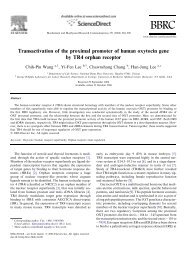
<strong>Skull</strong> <strong>Deformity</strong>: !<strong>Radiographic</strong> <strong>Diagnosis</strong> <strong>of</strong> !"<strong>Sticky</strong> Suture"<strong>in</strong> !Occipital Plagiocephaly"J. E. Losee, 1 P.-L. Westesson, 2 M. Ketkar, 2 D. S<strong>in</strong>gh, 3 !R. E. Kirschner, 3 and S. P. Bartlett 3 !"1The Childrens Hospital <strong>of</strong> Pittsburgh, PA, USA, !2University <strong>of</strong> Rochester School <strong>of</strong> Medic<strong>in</strong>e & Dentistry, Rochester, NY, USA,3Childrens Hospital <strong>of</strong> Philadelphia, PA, USA"Presentation material is for education purposes only. All rights reserved. ©2004 URMC Radiology Page 1 <strong>of</strong> 21
Back-to-Sleep"S<strong>in</strong>ce 1992 when the AAP suggested sup<strong>in</strong>e sleep !position, the <strong>in</strong>cidence <strong>of</strong> Occipital Plagiocephaly !has dramatically risen."Presentation material is for education purposes only. All rights reserved. ©2004 URMC Radiology Page 2 <strong>of</strong> 21
Occipital Plagiocephaly: OC"OC may result from either:"!!Non-synostotic occipitalplagiocephaly (NSOP)"-! positional mold<strong>in</strong>g"-! deformational plagiocephaly"!!Lambdoid craniosynostosis (LC)"-! posterior synostoticplagiocephaly"Presentation material is for education purposes only. All rights reserved. ©2004 URMC Radiology Page 3 <strong>of</strong> 21
Occipital Plagiocephaly:<strong>Diagnosis</strong>"!!Historically controversial"!!LC thought to be unique:characteristic radiographic f<strong>in</strong>d<strong>in</strong>gsnot necessary for diagnosis"!!Lambdoid suture described asfunctionally fused or stickysuture"Presentation material is for education purposes only. All rights reserved. ©2004 URMC Radiology Page 4 <strong>of</strong> 21
Occipital Plagiocephaly"!!Recent cl<strong>in</strong>ical criteria for diagnosisNSOP and LC have been del<strong>in</strong>eated"!!However radiographicdifferentiation is obscure"Presentation material is for education purposes only. All rights reserved. ©2004 URMC Radiology Page 5 <strong>of</strong> 21
NSOP: Cl<strong>in</strong>ical Exam"Cl<strong>in</strong>ically NSOP presents with a parallelogram shaped vertex cranial morphologyand a symmetric mastoid skull base."""""""""""As seen <strong>in</strong> this cl<strong>in</strong>ical case, with an abnormal, however patent and non-fusedlambdoid suture."Presentation material is for education purposes only. All rights reserved. ©2004 URMC Radiology Page 6 <strong>of</strong> 21
LC: Cl<strong>in</strong>ical Exam"Cl<strong>in</strong>ically LC presents with a trapezoid shaped vertex cranial morphology withipsilateral mastoid skull base boss<strong>in</strong>g."""""""""""As illustrated <strong>in</strong> this cl<strong>in</strong>ical case <strong>of</strong> right sided LC."Presentation material is for education purposes only. All rights reserved. ©2004 URMC Radiology Page 7 <strong>of</strong> 21
Aim <strong>of</strong> the study"•! To characterize changes <strong>of</strong> lambdoid suture<strong>in</strong> NSOP"•! To establish radiographic criteria for NSOP"•! To compare affected sutures <strong>in</strong> NSOP andLC"Presentation material is for education purposes only. All rights reserved. ©2004 URMC Radiology Page 8 <strong>of</strong> 21
Methods"•! CT scans children cl<strong>in</strong>ically diagnosed withNSOP and LC were evaluated by bothNeuroradiologist and Crani<strong>of</strong>acial Surgeonto compare: "–! lambdoid suture "–! cranial morphology"–! ear position"–! endocranial base angles"•! Statistical analysis was performed"Presentation material is for education purposes only. All rights reserved. ©2004 URMC Radiology Page 9 <strong>of</strong> 21
Methods"•! CT scans <strong>of</strong> 26 children with NSOP"–! 18 male, 8 female"–! 12 right side, 8 left side, 6 bilateral"•! 7 children diagnosed with LC"–! 5 male, 2 female"–! 4 left side, 3 right side"•! 32 sutures <strong>of</strong> NSOP and 7 sutures LC werecompared "Presentation material is for education purposes only. All rights reserved. ©2004 URMC Radiology Page 10 <strong>of</strong> 21
Sutures <strong>of</strong> NSOP evaluated for"•! focal fusion"•! endocranial heap<strong>in</strong>g/ridg<strong>in</strong>g"•! narrow<strong>in</strong>g"•! perisutural th<strong>in</strong><strong>in</strong>g"•! sclerosis"•! change <strong>in</strong> orientation: overlapp<strong>in</strong>g to endto-endand were "Compared to sutures <strong>of</strong> LC (p values)"Presentation material is for education purposes only. All rights reserved. ©2004 URMC Radiology Page 11 <strong>of</strong> 21
NSOP !Focal-Skip Fusion: !25% (p=0.308)"NSOP sutures demonstratedareas <strong>of</strong> skip fusion 25% <strong>of</strong>the time."NSOP"Endocranial Heap<strong>in</strong>g: !78% (p=0.313)"Ectocranial Heap<strong>in</strong>g: !0% (p=
NSOP Suture Narrow<strong>in</strong>g: !63% (p=0.008)"NSOP sutures demonstratedsutural narrow<strong>in</strong>g <strong>in</strong> 63% <strong>of</strong>cases."NSOP Sclerosis: !16% (p=0.319)"Sutural sclerosis was noted<strong>in</strong> 16% cases."Presentation material is for education purposes only. All rights reserved. ©2004 URMC Radiology Page 13 <strong>of</strong> 21
NSOP Change <strong>in</strong> Suture Orientation: !63% (p=0.001)"Sutures <strong>of</strong> NSOP demonstrated a change <strong>in</strong> sutureorientation from overlapp<strong>in</strong>g to end-to-endmorphology."Presentation material is for education purposes only. All rights reserved. ©2004 URMC Radiology Page 14 <strong>of</strong> 21
Suture Morphology: LC"!!Near complete obliteration:!100% (p=
Cranial Morphology: NSOP"!! Ipsilateral occipital flatten<strong>in</strong>g <strong>in</strong> allcases: 100%"!! Ipsilateral frontal boss<strong>in</strong>g: !85% (p=0.012)"!! Contralateral occipital boss<strong>in</strong>g: 95%(p=0.003)"Suture Morphology: NSOP "!! Compar<strong>in</strong>g affected to contra-lateralnon-affected control suture"!! Significant difference (p
NSOP PerisuturalTh<strong>in</strong>n<strong>in</strong>g:!78% (p=0.313)"Subarachnoid Spac<strong>in</strong>g:!47%"Ipsilateral <strong>in</strong>crease <strong>in</strong> subarachnoidspac<strong>in</strong>g was noted<strong>in</strong> 47% <strong>of</strong> NSOP and <strong>in</strong> nocases <strong>of</strong> OP. "Presentation material is for education purposes only. All rights reserved. ©2004 URMC Radiology Page 17 <strong>of</strong> 21
!Midl<strong>in</strong>e Cranial BaseDeviation Angle"!! Significant difference was found"!! LC: Angles were greater and represented alarger deviation from"!! mid sagittal cranial base axis"-! average 10.3 0 " (range 0-15 0 )"!! NSOP"-! average 4.1 0 "(range 0-9 0 )"!! p=0.02"Petrus Ridge Angle"!! Significant difference between affected and nonaffected side also between the affected sides <strong>in</strong> NSOPand LC"!! NSOP"-! affected: av. 121.8 0 "(range 117-127 0 ) "-! non-affected: av. 125.8 0 "(range 117-134 0 )"-! p=0.0016"!! LC"-! affected: av. 115.7 0 "(range 112-120 0 )"-! non-affected: av. 132 0 "(range 128-140 0 )"-! p=0.0156"!! NSOP vs. LC"-! p=0.0039"Presentation material is for education purposes only. All rights reserved. ©2004 URMC Radiology Page 18 <strong>of</strong> 21
Ear Position"!!Vertex view"!!LC"-!anterior "14%"-!symmetric "86%"!!NSOP"-!anterior "85%"-!symmetric "15%"Presentation material is for education purposes only. All rights reserved. ©2004 URMC Radiology Page 19 <strong>of</strong> 21
Conclusions"•! Cranial sutures"–! Open - <strong>in</strong>fants"–! Closed - adults"–! Obliterated - craniosynostosis (not prematurely fused)"–! Deformed or sticky - non-synostotic plagiocephaly"Presentation material is for education purposes only. All rights reserved. ©2004 URMC Radiology Page 20 <strong>of</strong> 21
Conclusion: !<strong>Radiographic</strong> <strong>Diagnosis</strong>"!! Changes <strong>in</strong> lamboid suture previously consideredto be LC:"-! endocranial heap<strong>in</strong>g"-! focal fusions"-! sutural narrow<strong>in</strong>g"-! perisutural th<strong>in</strong>n<strong>in</strong>g"-! Sclerosis"!! LC not unique among craniosynostosis:"-! suture obliteration"-! compensatory boss<strong>in</strong>g"Presentation material is for education purposes only. All rights reserved. ©2004 URMC Radiology Page 21 <strong>of</strong> 21